Myofascial Pain Treatment Guide with Trigger Point Injections
Find out how trigger point injections can help manage myofascial pain and improve your quality of life effectively.
Abstract
Welcome to our educational journey into the complex world of chronic musculoskeletal pain. I am Dr. Alex Jimenez, and I am honored to guide you through this exploration from the perspective of our multidisciplinary team here in El Paso, Texas. In this post, we will dissect the physiological underpinnings of muscle spasms, the formation of trigger points, and the subsequent development of chronic pain cycles. We will explore the latest evidence-based approaches for managing these conditions, with a particular focus on trigger point injection (TPI) therapy. We’ll discuss the rationale behind using specific agents like lidocaine and natural anti-inflammatories, the mechanics of the injection technique itself—often referred to as a “star pattern”—and the crucial role of anatomical knowledge in ensuring safety and efficacy.
Furthermore, we will illuminate how our unique collaborative model at Injury Medical Clinic PA integrates the expertise of chiropractic care, advanced practice nursing, and internal medicine. I will share insights from my dual roles as a Doctor of Chiropractic and a Family Nurse Practitioner, and explain how we work alongside our esteemed Medical Director, Dr. Maria Guadalupe Cardenas, MD, a board-certified internist with over four decades of experience. This integrated approach allows us to provide a comprehensive spectrum of care, from initial diagnosis and medical oversight to hands-on chiropractic adjustments, functional medicine, and personalized rehabilitation. Our goal is not just to alleviate symptoms but to uncover and address the root causes of pain, empowering our patients to reclaim their health and vitality. Join us as we unravel the science behind pain and the art of integrative healing.
Our Collaborative Care Philosophy: The Synergy of Medicine and Chiropractic
My name is Dr. Alex Jimenez, and I hold several professional titles that reflect my passion for a comprehensive and holistic approach to health: DC, APRN, FNP-BC, CFMP, IFMCP, ATN, and CCST. For years, my primary mission has been to understand the intricate web of human physiology and find the most effective ways to restore function and alleviate suffering. Here at our practice in El Paso, Texas, Injury Medical Clinic PA (also known as Mission Plaza Injury Medical Clinic), we have built a unique environment dedicated to this mission.
A cornerstone of our practice is the powerful synergy we’ve cultivated between different medical disciplines. We are not just a chiropractic office or a standard medical clinic; we are a fully integrated, multidisciplinary team. Our Medical Director and Collaborative Physician champions this structure: Dr. Maria Guadalupe Cardenas, MD. Dr. Cardenas is a highly respected, board-certified internist with over 40 years of experience. Her NPI is #1164426749, and she holds Texas MD License #J2933. Her role is absolutely pivotal. She provides essential medical oversight, diagnostic acumen, and an internal medicine perspective that elevate our patient care to the highest standard. This collaborative model, where a Medical Doctor provides directorship and works in tandem with a Doctor of Chiropractic, is common in forward-thinking integrative and injury care clinics, and it is the bedrock of our success.
This partnership allows us to offer a seamless continuum of services. When a patient walks through our doors, they have access to:
- Medical Oversight from Dr. Cardenas: She ensures that all treatments are medically appropriate, oversees complex cases, and manages any underlying health conditions that could be contributing to a patient’s pain, such as inflammatory disorders, metabolic issues, or nutritional deficiencies.
- Chiropractic and Functional Neurology from Myself (Dr. Jimenez): I focus on the biomechanical and neurological aspects of a patient’s condition. This includes performing precise chiropractic adjustments to restore spinal alignment and nerve function, as well as applying principles of functional neurology to rehabilitate the nervous system.
- Advanced Practice Nursing: In my capacity as a Family Nurse Practitioner (FNP-BC), I can perform advanced assessments, order and interpret diagnostic tests, and administer treatments like the trigger point injections we will be discussing in depth. This dual qualification allows me to bridge the gap between the chiropractic and medical models seamlessly.
- A Unified Team Approach: Our team also includes specialists in functional medicine, personal injury care, physical rehabilitation, and nutrition. We hold regular case conferences where Dr. Cardenas, I, and the rest of our team review patient progress and strategize the next steps in their care plan. This ensures every patient benefits from our collective expertise.
This integrated system is designed to break down the silos that too often exist in healthcare. Instead of a patient being bounced between a chiropractor, a primary care physician, and a pain specialist—with each provider having only a partial view of their condition—we bring all that expertise under one roof. This allows us to create a truly personalized and cohesive treatment plan that addresses the patient from every angle: structurally, neurologically, biochemically, and medically. It is within this collaborative framework that we approach complex issues like chronic myofascial pain and utilize advanced therapies like trigger point injections.
The Anatomy of a Knot: Unraveling Muscle Spasms and Trigger Points
Before we can effectively treat chronic muscle pain, we must first understand its origins. The conversation often begins with patients describing a “knot” in their muscle—a tender, hard lump that aches, burns, and sometimes sends pain to other parts of their body. Clinically, we refer to this as a myofascial trigger point. But what exactly is it, and how does it form? The process is a fascinating and often vicious cycle rooted in our body’s response to stress and injury.
The Initial Injury: Microscopic Tears and the Energy Crisis
Everything starts at the microscopic level within the muscle fibers. Imagine your muscles are composed of millions of tiny, overlapping filaments called actin and myosin. When a muscle contracts, these filaments slide past each other, a process powered by adenosine triphosphate (ATP), the energy currency of our cells. For the muscle to relax, it needs another surge of ATP to actively pump calcium ions out of the muscle cell, allowing the filaments to detach and lengthen.
Now, consider what happens when a muscle is subjected to stress. This stress can be:
- Acute: A sudden trauma, like a car accident (whiplash), a fall, or lifting an object that is too heavy.
- Repetitive: Chronic overuse from poor posture (e.g., “tech neck” from staring at a screen), repetitive occupational movements, or an imbalanced exercise routine.
- Biochemical: Nutritional deficiencies (especially magnesium, calcium, and B vitamins), hormonal imbalances, or systemic inflammation.
Any of these stressors can cause microscopic tearing and damage to the muscle fibers and, critically, to the sarcoplasmic reticulum—the intricate network within the muscle cell that stores and releases calcium. When the sarcoplasmic reticulum is damaged, it can lead to an uncontrolled, sustained leakage of calcium into the muscle fiber. This excessive calcium forces the actin and myosin filaments in that localized area to remain in a state of maximum contraction. They are locked together.
This creates an energy crisis. The sustained contraction dramatically increases the metabolic demand of that small segment of muscle fiber, requiring huge amounts of ATP. At the same time, this intense, localized contraction physically compresses the tiny blood vessels (capillaries) that supply the area. This compression chokes off the supply of oxygen and nutrients needed to produce ATP. The result is a vicious cycle:
- Sustained Contraction: Leaking calcium keeps muscle fibers locked.
- Increased Energy Demand: The locked fibers burn through their local ATP supply.
- Reduced Blood Flow (Ischemia): The contraction squeezes capillaries, preventing fresh oxygen and nutrients from arriving.
- Energy Production Failure: Without oxygen, the cell cannot produce enough ATP to pump the calcium out and allow the muscle to relax.
This small, localized segment of muscle fiber is now metabolically exhausted and physically trapped in a contracted state. This is the physiological birth of a trigger point taut band.
From Microscopic Crisis to Palpable Pain
Over time, the body’s inflammatory response kicks in. The area of ischemia and metabolic waste buildup (including substances like bradykinin, substance P, and prostaglandins) irritates nearby nerve endings. These irritated nerves send a barrage of pain signals to the spinal cord and brain. Furthermore, the body attempts to “wall off” this dysfunctional area by laying down fibrous connective tissue, or scar tissue, around the contracted muscle fibers.
This is the “knot” you can actually feel. It’s no longer just a microscopic problem; it has become a palpable, hard nodule of dysfunctional tissue. This scar tissue further restricts movement, exacerbates the lack of blood flow, and perpetuates the pain cycle.
From my clinical experience as a chiropractor, I see this process manifest in predictable patterns. A patient with forward head posture will develop trigger points in their upper trapezius and levator scapulae muscles. An office worker who sits all day will have them in their quadratus lumborum and gluteus medius. These patterns are not random; they are a direct consequence of biomechanical load and muscular endurance limits. Understanding this underlying physiology is absolutely critical because it dictates our treatment strategy. We aren’t just “rubbing a sore spot”; we are intervening in a complex neuro-biochemical cascade. Our goal is to break this cycle at multiple points: to mechanically disrupt the scar tissue, restore blood flow, flush out inflammatory chemicals, and reset the neurological signaling that is perpetuating the spasm.
The “Star Pattern” Technique: A Mechanical and Neurological Reset

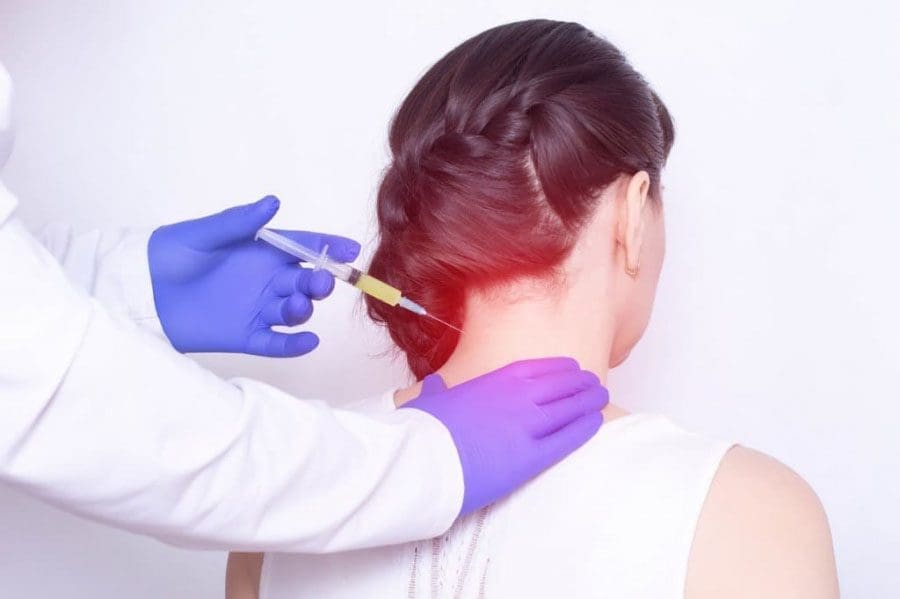
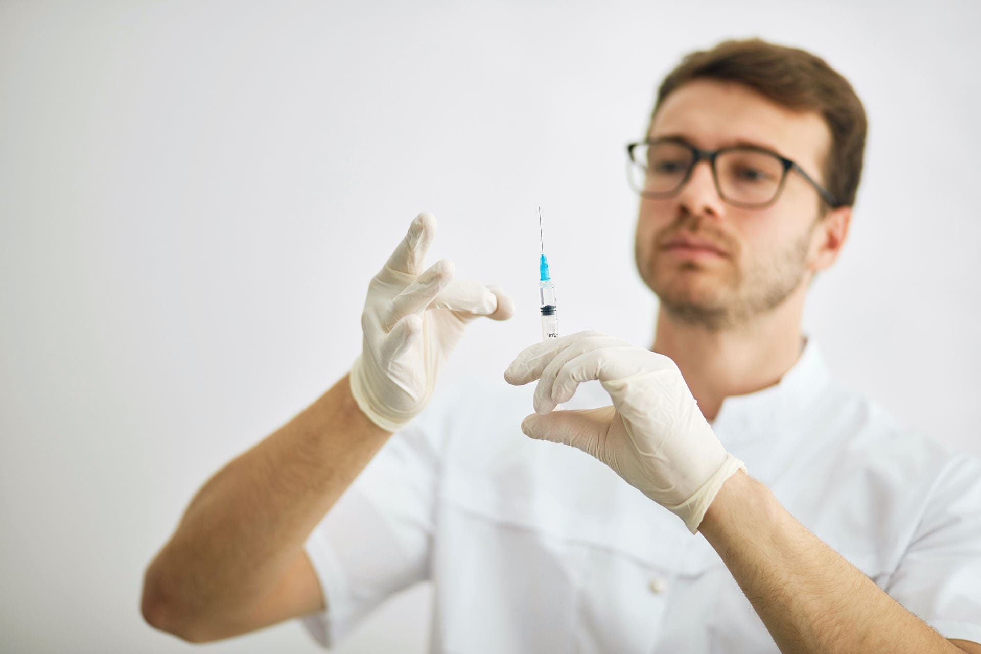
One of the most effective tools we have for directly addressing these stubborn trigger points is Trigger Point Injection (TPI). The technique itself is just as important as the substances we inject. A common and highly effective method, which was discussed in our training, involves what is colloquially known as the “star pattern” or fanning technique. This approach is an evolution of an older technique called dry needling.
The Evolution from Dry Needling to Injections
In traditional dry needling, a thin filiform needle (similar to an acupuncture needle) is inserted directly into the trigger point. The practitioner then manipulates the needle—moving it up and down and angling it in multiple directions within the knot—without injecting any substance. The primary goal is mechanical. By repeatedly piercing the taut band and surrounding scar tissue, we achieve several things:
- Mechanical Disruption: The needle physically breaks up the fibrous adhesions and scar tissue that have formed around the contracted muscle fibers. This helps release the entrapped tissues.
- Eliciting a Local Twitch Response (LTR): When the needle accurately contacts the most irritable spot within the trigger point, it often causes a brief, involuntary contraction or “twitch” of the muscle band. This LTR is considered a crucial therapeutic sign. It indicates that the dysfunctional segment of the muscle has been stimulated, leading to a subsequent reflex relaxation and a reset of the local nerve supply. Research by Shah and Gilliams (2008) suggests the LTR is associated with reduced inflammatory chemicals and improved pain outcomes.
- Stimulating a Healing Response: The minor trauma caused by the needle prompts a localized inflammatory and healing response. The body increases blood flow to the area to repair the micro-trauma, and in doing so, it also flushes out the accumulated metabolic waste and pain-sensitizing chemicals from the original energy crisis.
From my own observations, dry needling can be remarkably effective. I have seen many patients experience significant relief from this technique alone. However, we’ve found that we can enhance and accelerate the healing process by introducing therapeutic substances directly into the tissue. This is where TPI comes in.
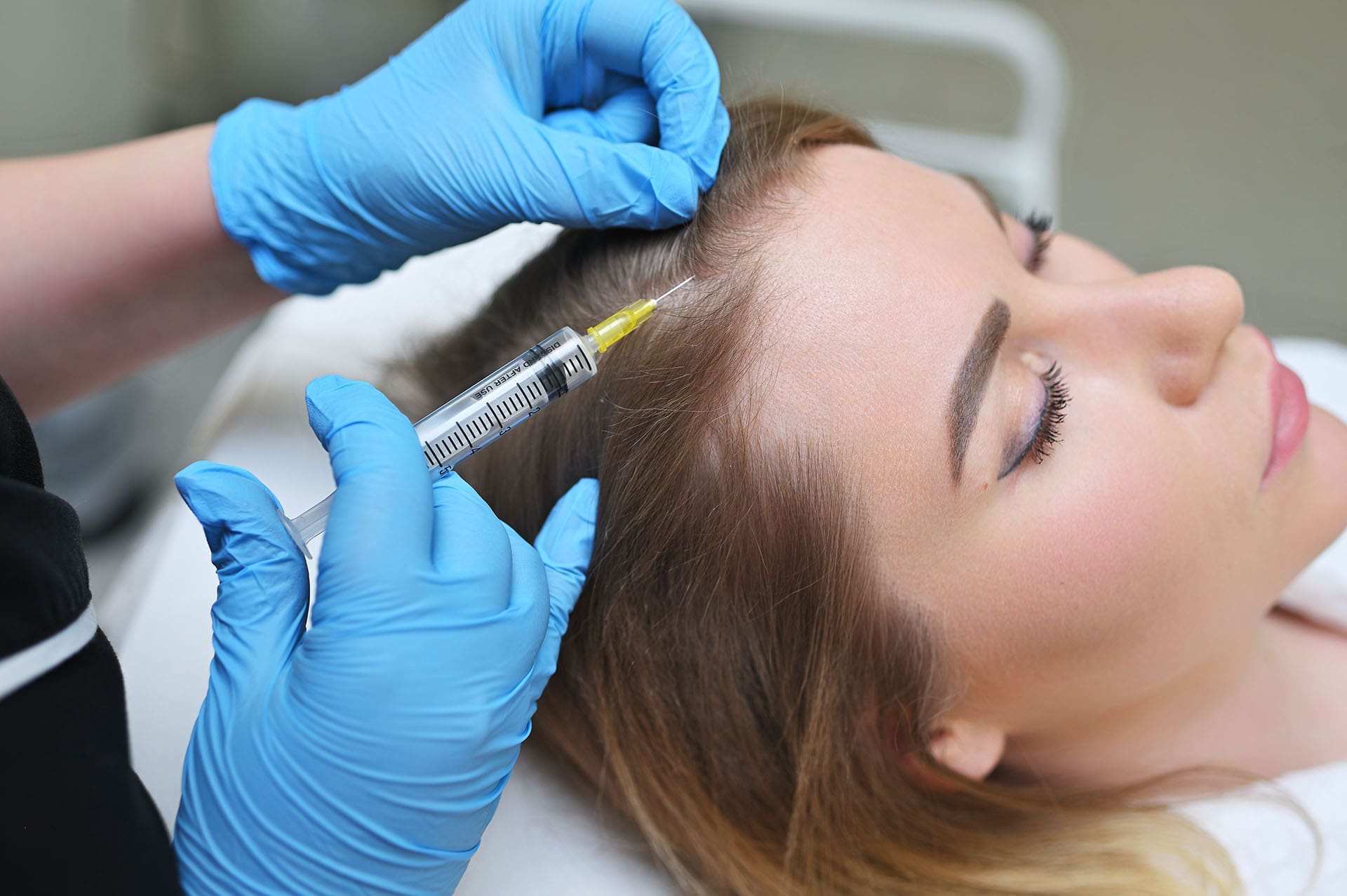
The Mechanics of the “Star Pattern” Injection
With TPI, we use the same foundational principle of mechanical disruption but add the benefit of targeted medication. The “star pattern” describes the way the needle is maneuvered after the initial insertion. Here’s a step-by-step breakdown from my perspective as the practitioner:
- Identification: First, I meticulously palpate the muscle to locate the epicenter of the trigger point. I am searching for that exquisitely tender, dense nodule within a taut band of muscle. Often, pressing on this spot will reproduce the patient’s familiar pain, sometimes even referring pain to a distant area (e.g., pressing a trigger point in the shoulder that sends a sensation down the arm). This confirms I’ve found the right target.
- Insertion: After sterilizing the skin, I insert the needle directly into the heart of the trigger point. The goal is to feel that subtle change in tissue resistance as the needle enters the dense, fibrotic knot.
- The “Star” Maneuver: This is the crucial part. Without withdrawing the needle from the skin, I begin the fanning motion. I will slightly pull the needle back (while keeping the tip within the muscle) and then re-angle it to probe a different quadrant of the trigger point. I repeat this process, moving the needle in and out and changing the angle, creating a pattern that resembles the points of a star or the spokes of a wheel.
- Injection: With each pass or in each new direction, I inject a tiny amount of the therapeutic solution. This ensures the fluid is distributed throughout the entire volume of the trigger point, not just in a single pocket.
Why is this “star pattern” so important? A trigger point is not a simple sphere; it’s a three-dimensional complex of knotted fibers and scar tissue. A single injection into the center might miss the full extent of the dysfunctional tissue. By fanning the needle out in multiple directions, we ensure we both mechanically break up adhesions and chemically treat the entire affected area. We are turning a single injection into a multi-pronged therapeutic assault on the trigger point. This method combines the mechanical benefits of dry needling with the chemical benefits of the injected solution, leading to a more comprehensive and lasting result. It helps the muscle release from its spastic state, which in turn alleviates the pain and restores normal function.
The Therapeutic Cocktail: Why We Choose Lidocaine and Sarapin
The effectiveness of trigger point injections is not just about the technique; it’s profoundly influenced by what we inject. In our practice, after careful consideration and review of the evidence, we have settled on a combination that we find provides superior results: a one-to-one mixture of lidocaine and a natural anti-inflammatory agent called Sarapin. Some practitioners might use Marcaine, another local anesthetic, but the principles remain similar. Let’s break down why this specific combination is our choice.
Lidocaine: More Than Just a Numbing Agent
Most people think of lidocaine (a 1% solution is common for this procedure) simply as a local anesthetic—something that numbs the area. And it certainly does that. The immediate numbing effect provides rapid pain relief for the patient, which can be psychologically and physically beneficial. It breaks the immediate pain-spasm-pain cycle and makes the procedure itself much more tolerable.
However, the role of lidocaine goes far beyond simple numbing. From a physiological standpoint, lidocaine is a sodium channel blocker. Here’s why that’s so important in the context of a trigger point:
- Interrupting Pain Signals: Pain signals are transmitted along nerve fibers as electrical impulses, which depend on the rapid opening and closing of sodium channels in the nerve cell membrane. By blocking these sodium channels, lidocaine physically prevents pain signals from irritated nerve endings in the trigger point from traveling to the spinal cord and brain. We are literally cutting off the “pain hotline.”
- Breaking the Neurological Loop: Chronic pain is not just a local tissue problem; it becomes ingrained in the nervous system. The constant barrage of pain signals from a trigger point can lead to a state called central sensitization, where the central nervous system becomes hypersensitive and amplifies pain signals. By silencing the peripheral nerve with lidocaine, we give the central nervous system a break from this constant input, helping to downregulate this hypersensitivity.
- Muscle Relaxation Properties: While not its primary mechanism, blocking nerve signals to the muscle can also contribute to muscle relaxation, further helping to release the taut band.
A common concern patients have is allergies. A small fraction of the population can indeed have an allergic reaction to lidocaine or other “-caine” anesthetics. We’ve seen estimates suggesting this occurs in about one percent of people. This is why a thorough medical history, overseen by Dr. Cardenas, is non-negotiable before any procedure. If a patient has a known allergy, we do not use it. In these cases, we can still perform dry needling. The mechanical disruption alone still provides significant benefit, though the immediate pain relief from the anesthetic will be absent.
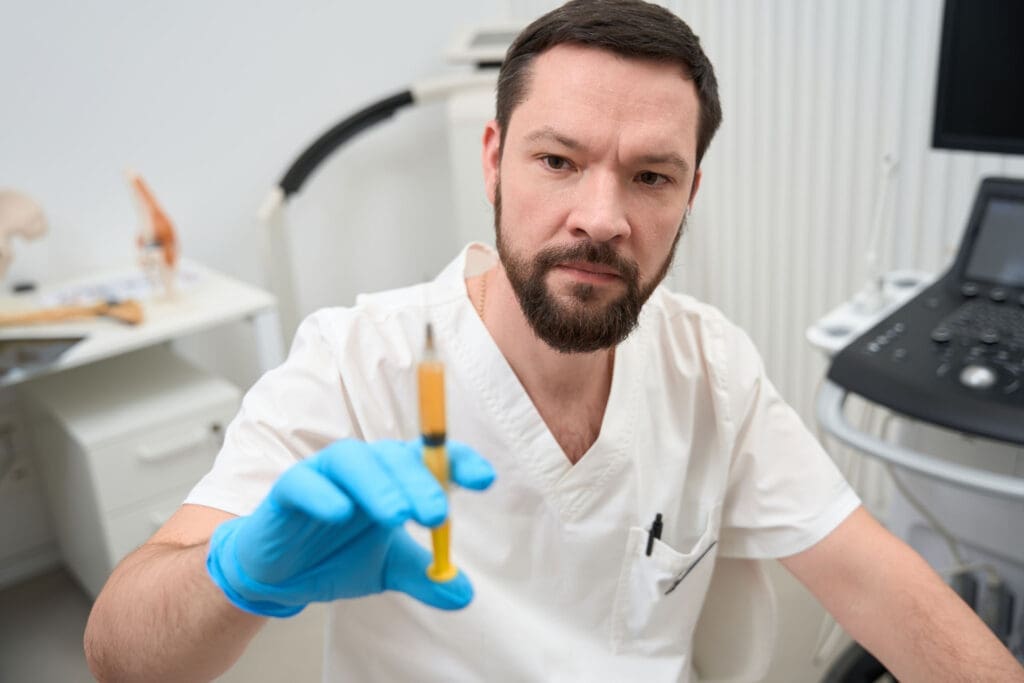
Sarapin: The Power of a Natural Anti-Inflammatory
This is where our approach diverges from many conventional pain management practices. While some practitioners might use corticosteroids (steroids) as the anti-inflammatory agent in their injections, I have a strong preference for a natural, plant-based alternative called Sarapin.
Sarapin is a biological medicine derived from the pitcher plant (Sarracenia purpurea). It has been used as a safe and effective analgesic and anti-inflammatory for decades. Its mechanism of action is unique and, in my opinion, superior to steroids for this particular application.
- How Sarapin Works: Unlike steroids, which act by suppressing the entire immune response in a broad-spectrum way, Sarapin works primarily as a neurolytic agent on sensory C-fibers. These are the small, unmyelinated nerve fibers responsible for transmitting dull, aching, chronic pain signals—the exact type of pain associated with trigger points. Sarapin appears to inhibit the function of these specific nerve fibers without affecting motor nerves or other sensory nerves (like those for touch or pressure). This means it targets the pain signals without causing muscle weakness or widespread numbness.
- Why I Prefer Sarapin Over Steroids: My decision to use Sarapin is based on several key factors:
- Safety Profile: Steroids, especially with repeated use, carry potential risks. They can weaken local tissues like tendons and ligaments, cause skin depigmentation, and have systemic side effects. Sarapin, being a natural biological product, is exceptionally safe. It does not damage local tissues and has no known systemic side effects. This allows us to perform injections as needed without the long-term concerns associated with corticosteroids.
- Targeted Action: As mentioned, Sarapin specifically targets the pain-transmitting sensory nerves. This is a more elegant and focused approach than the “shotgun” immunosuppression of steroids. We are addressing the pain signal, not just broadly suppressing inflammation.
- Avoiding Tissue Degradation: A core principle in regenerative and integrative medicine is to “first, do no harm.” My concern with repeated steroid injections into muscle and fascia is the potential for catabolic effects—the breakdown of tissue. Since our ultimate goal is to heal and regenerate the tissue, using an agent that could potentially weaken it seems counterintuitive. Sarapin supports the healing process without this risk.
By combining lidocaine and Sarapin in a one-to-one ratio, we create a powerful synergistic cocktail. The lidocaine provides immediate, profound pain relief by blocking all local nerve signals. This makes the patient comfortable and breaks the acute pain cycle. Meanwhile, the Sarapin provides a longer-lasting analgesic and anti-inflammatory effect by specifically targeting the chronic pain fibers. The result is a much better, more durable outcome than using either agent alone. I find that my patients experience more complete relief, and the effects last longer, reducing the need for frequent repeat injections. It’s a perfect example of how combining modern pharmacology with natural biological medicine can yield superior clinical results.
Procedural Safety: The Critical Importance of Anatomical Precision
Performing trigger point injections is a skill that blends tactile sensitivity with a deep, unwavering respect for human anatomy. While the procedure is generally very safe when performed by a trained professional, there are inherent risks that must be mitigated through knowledge and careful technique. One of the most significant concerns, particularly when working in the thoracic region, is the risk of causing a pneumothorax, or a punctured lung.
Navigating the Thoracic Cage: Needle Depth and Safety Margins
The question of “How deep is too deep?” is one that every practitioner must have a definitive answer for. The lungs are located within the thoracic cavity, protected by the rib cage. The space between each rib, known as the intercostal space, is filled with intercostal muscles, and it is through these muscles that a needle could potentially pass to reach the lung.
Here are the general guidelines and anatomical considerations we adhere to:
- Needle Length is Key: For injections in the cervical (neck) and upper thoracic (upper back) regions, we typically use a 30-gauge, 1-inch needle. The small gauge makes the injection more comfortable, and the 1-inch length provides a built-in safety measure. In the vast majority of adults, a 1-inch needle, even if inserted to its full depth in an intercostal space, will not be long enough to traverse the chest wall and puncture the parietal pleura, the outer lining of the lung.
- The 1.5-Inch Threshold: As a rule of thumb, a needle length of 1.5 inches is where the risk begins to increase, especially in individuals who are very thin, frail, or elderly. In these populations, the muscle and fat layers are thinner, meaning a 1.5-inch needle has a greater potential to reach the pleural space. Therefore, extreme caution and precise depth control are required if using a longer needle in the thoracic area. I often advise practitioners to avoid it altogether in these regions unless they have advanced training (e.g., ultrasound guidance).
- Palpating the Ribs: Before any injection near the rib cage, I always use my palpating hand to identify the ribs. The goal is to inject into the muscle belly overlying a rib or in the thick paraspinal muscles adjacent to the spine, not directly into the unprotected intercostal space. This simple tactile feedback is a crucial safety check.
- Angle of Insertion: The angle of the needle also matters. By angling the needle obliquely (a technique known as “tenting” the skin and muscle), rather than perpendicular to the skin, we can increase the distance the needle must travel through the tissue before it could ever reach the lung, further enhancing safety.
- Patient Body Habitus: Anatomy is not one-size-fits-all. A heavily muscled bodybuilder has a much thicker chest wall than a petite, older woman. I must constantly assess the patient in front of me and mentally adjust my “safe depth” calculation. In my clinical experience, even in a “little skinny old lady,” a 1-inch needle used with proper technique is exceptionally safe. You are not going to puncture a lung. The danger arises when practitioners become complacent or use inappropriately long needles for the patient’s anatomy.
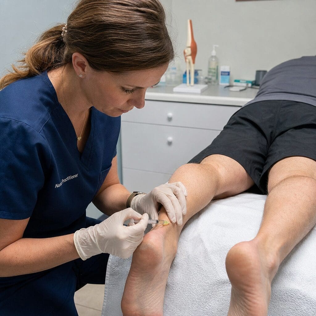
For the lower thoracic and lumbar regions, the anatomy is different. The risk of pneumothorax is gone, and the muscles, like the quadratus lumborum and the erector spinae, are much thicker and deeper. In these areas, it is safe and often necessary to use a longer needle (e.g., 1.5 to 2 inches) and inject a larger volume (up to 2 mLs per site) to reach and effectively treat the target muscles.
The Foundation of All Practice: Know Your Anatomy
This discussion underscores a point I cannot emphasize enough to any student or colleague in manual or injection-based medicine: you must constantly review and master your anatomy. When I was in school, I’ll admit, anatomy was a struggle. It’s a monumental amount of information, and for many of us, it’s a challenge. But in professional practice, anatomy is not an academic exercise; it is the roadmap that keeps your patients safe and makes your treatments effective.
Whether you are performing a chiropractic adjustment, designing an orthotic, or administering a trigger point injection, you are interacting with a complex, three-dimensional system of bones, nerves, muscles, and organs. You need to have a mental MRI of what lies beneath your fingertips.
- Where is the brachial plexus in relation to the scalene muscles?
- What is the course of the sciatic nerve as it passes through the gluteal region?
- How thick are the paraspinal muscles at L4 versus T4?
I urge everyone in this field to make anatomical review a regular habit. Go back to the textbooks, use 3D anatomy apps, attend dissection labs if you can. The more you immerse yourself in the elegant architecture of the human body, the more confident, precise, and—most importantly—safe your hands will become. This is the ultimate foundation of patient trust and clinical excellence.
The Root Causes of Pain- Video
The Patient’s Journey: The Psychology of Pain and Treatment
Treating the physical body is only half the battle. As clinicians, we must also be keenly aware of the patient’s psychological and emotional experience, especially when it comes to pain and needles. The fear of pain and the anxiety surrounding a procedure can significantly impact the outcome. A core part of my practice philosophy is to guide the patient through the treatment journey in a way that builds trust and minimizes distress.
One simple but profound strategy I’ve learned over years of practice is to save the worst for last.
When a patient comes in with multiple trigger points, they are not all created equal. There is almost always one “main offender”—that one knot that is the most sensitive, the most irritable, and the most likely to make the patient jump when it’s treated. From a purely mechanical perspective, one might think to tackle that worst one first. Get the biggest problem out of the way. However, from a psychological perspective, this is often the wrong approach.
Imagine the patient is already anxious. If the very first injection is the most painful one, their nervous system immediately goes on high alert. Their muscles tense up, their fear is validated, and they spend the rest of the procedure dreading the next needle. I have had experiences early in my career where I took this “worst-first” approach, and the patient would literally say, “No, I’m not doing any more.” I would then find myself in a position of having to coax or argue with them to complete a treatment that I knew would help them. This creates an adversarial dynamic, which is the complete opposite of the therapeutic alliance we want to build.
Instead, I’ve found it far more effective to start with the less sensitive trigger points. I will strategically choose the first few injection sites in areas that are sore but not excruciating. The patient experiences the procedure, realizes it’s manageable, and begins to feel the numbing effect of the lidocaine. Their anxiety starts to subside, and trust is established. They think, “Okay, I can handle this.”
By the time we get to that “son of a…” spot—the one that’s going to be the most intense—the patient is already partially numb, more relaxed, and confident in me and the process. The intense sensation from that final injection is then just a brief moment in an otherwise manageable experience, rather than the traumatic opening act.
This approach achieves several important goals:
- Builds Trust and Rapport: It shows the patient that I am mindful of their comfort and am strategically guiding them through the process.
- Reduces Overall Muscle Guarding: A relaxed patient is easier to treat. Their muscles are not defensively tensed up, allowing me to palpate more accurately and the needle to enter the tissue more easily.
- Improves Patient Compliance: Patients who have a positive (or at least tolerable) experience are far more likely to return to complete their course of care and adhere to the treatment plan.
- Leverages the Anesthetic: By the time we reach the worst spot, the lidocaine from the surrounding injections may have already started to diffuse slightly, providing a small degree of pre-numbing to the area.
It may seem like a small detail, but managing the psychological journey of the treatment is paramount. We are not just treating trigger points; we are treating a person who is in pain and likely fearful. Empathy, communication, and a thoughtful, patient-centered approach can make all the difference between a successful treatment and a traumatic one.
Clarifying the Terminology: Prolotherapy, PRP, and Trigger Point Injections
The world of regenerative and pain medicine is filled with an ever-expanding list of therapies, and the terminology can often be confusing for patients and even practitioners. It’s important to understand the differences between treatments like Trigger Point Injections (TPI), Prolotherapy, and Platelet-Rich Plasma (PRP), as they are designed for different purposes and work through different mechanisms.
Trigger Point Injections (TPI): A Focus on Muscle
As we have discussed at length, TPI is a procedure specifically designed to treat myofascial trigger points—those hyperirritable knots within a muscle’s taut band.
- Target Tissue: Muscle and fascia.
- Primary Goal: To release muscle spasms, break up fibrous adhesions, and resolve the localized energy crisis within the muscle fiber.
- Injectate: Typically a local anesthetic (like lidocaine) and an anti-inflammatory agent (like Sarapin or, less commonly, a corticosteroid).
- Mechanism: A combination of mechanical disruption (from the needle) and chemical action (from the injectate) to reset the neuromuscular junction and resolve the spasm.
TPI is the frontline treatment for direct muscular pain and dysfunction originating from these specific points of irritation.
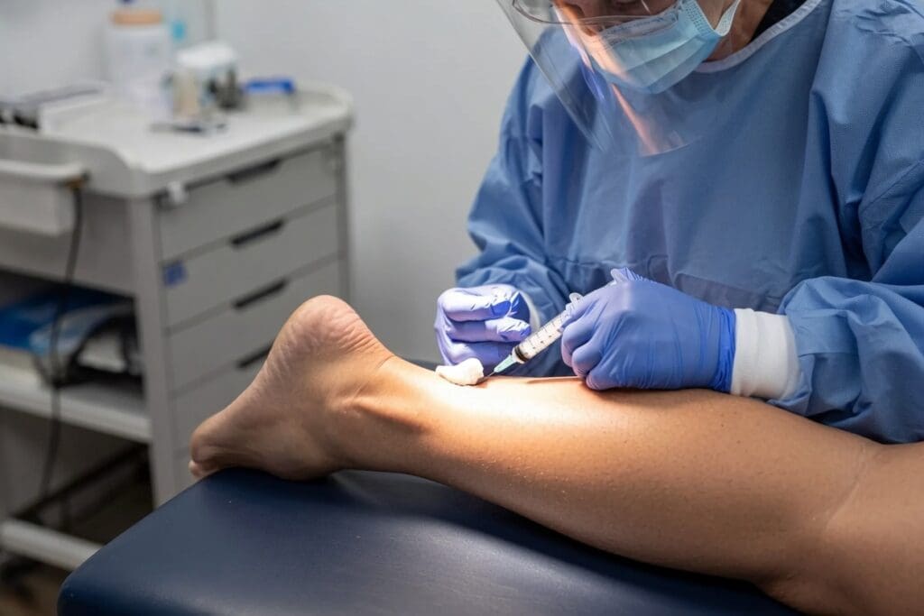
Prolotherapy: A Focus on Ligament and Tendon Laxity
Prolotherapy, short for “proliferative therapy,” is a fundamentally different treatment. It is not designed to treat muscle spasms but rather to address chronic pain originating from ligament and tendon laxity or instability.
- Target Tissue: Ligaments (which connect bone to bone) and tendons (which connect muscle to bone), specifically at their insertion points onto the bone (the “enthesis”).
- Primary Goal: To strengthen weak and damaged connective tissues and stabilize joints.
- Injectate: A “proliferant” solution, which is a mild irritant designed to stimulate a healing response. The classic solution is a dextrose (sugar) solution, often mixed with an anesthetic like lidocaine. Some practitioners incorporate other substances, including ozone.
- Mechanism: The proliferant solution intentionally creates a mild, controlled inflammatory response in the targeted ligament or tendon. This localized inflammation signals the body to initiate its natural healing cascade. The body sends growth factors, fibroblasts (cells that produce collagen), and other healing cells to the area. Over a series of treatments, this process leads to the deposition of new, strong collagen tissue, effectively tightening and strengthening the lax ligaments and tendons.
You would use prolotherapy for conditions like chronic low back pain due to sacroiliac ligament instability, chronic shoulder pain from a loose joint capsule, or tennis elbow (lateral epicondylitis) where the tendon is degenerated. It addresses joint instability, which is a common underlying cause of chronic musculoskeletal pain that TPI does not directly treat.
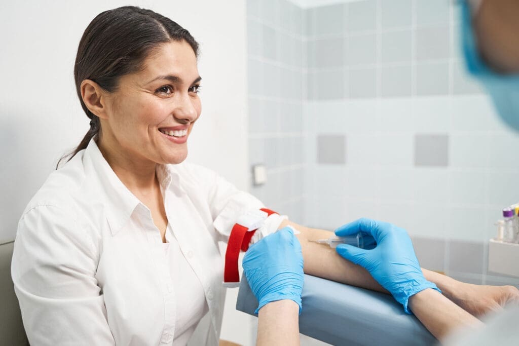
Platelet-Rich Plasma (PRP): A Focus on Cellular Regeneration
Platelet-Rich Plasma (PRP) therapy is a more advanced form of regenerative medicine that harnesses the body’s own healing power in a concentrated form.
- Target Tissue: Can be used on a wide variety of tissues, including tendons, ligaments, muscles, and joints (intra-articular).
- Primary Goal: To provide a high concentration of natural growth factors directly to an area of injury or degeneration to accelerate and enhance cellular repair and regeneration.
- Injectate: The patient’s own blood, which has been drawn and processed in a centrifuge to separate and concentrate the platelets. The resulting “platelet-rich plasma” is then injected back into the patient at the site of injury.
- Mechanism: Platelets are cell fragments in our blood that are critical for clotting, but they are also tiny storehouses of powerful growth factors. These are proteins that act as signaling molecules, telling local cells to proliferate, differentiate, and form new tissue. Key growth factors include Platelet-Derived Growth Factor (PDGF), Transforming Growth Factor-Beta (TGF-β), and Vascular Endothelial Growth Factor (VEGF). By injecting a concentrated dose of these growth factors directly into damaged tissue, we are essentially telling the body, “Heal this area, and do it now!”
PRP is used for more significant injuries and degenerative conditions, such as moderate osteoarthritis, chronic tendinopathies (like Achilles tendinosis), and muscle tears.
Is PRP in a Trigger Point Overkill?
This brings us to an important question: can you use PRP for a simple trigger point? My answer is generally yes, you can, but it’s often overkill. A standard myofascial trigger point, as we’ve defined it, is primarily a problem of neuromuscular dysfunction and localized ischemia—a spasm. It is not typically a tear or a significant degenerative condition of the muscle tissue itself.
The combination of mechanical needling and a solution of lidocaine/Sarapin is usually perfectly sufficient to break this cycle. It’s a highly effective, safe, and relatively inexpensive treatment.
Using PRP for a standard trigger point would be like using a sledgehammer to hang a picture frame. While PRP does have anti-inflammatory properties and would likely help, the primary issue in the trigger point (the spasm) is not what PRP is best designed to fix. PRP’s strength lies in regenerating tissue architecture, which is not the main problem in a trigger point. Furthermore, PRP is a more complex and expensive procedure.
Therefore, we reserve PRP for cases where there is evidence of actual tissue damage—a partial muscle tear, significant tendinosis adjacent to the trigger point, or a degenerative joint condition that is causing the muscle to go into a protective spasm. In those cases, treating the underlying degenerative condition with PRP makes perfect sense. But for the trigger point itself, TPI remains the more appropriate and cost-effective tool. Understanding these distinctions is key to creating a precise, effective, and responsible treatment plan for our patients.
The Integrated Approach in Action: A Patient Case Study
To bring all these concepts together, let me walk you through how a typical, yet complex, patient case would be managed at our clinic. This illustrates the true power of our integrated model, where chiropractic, internal medicine, and advanced injection therapies work in concert.
Imagine a 45-year-old female patient, “Jane,” comes to our clinic. Her primary complaint is chronic, nagging right-sided neck and shoulder pain with headaches that have persisted for over two years. She works an office job, spending 8-10 hours a day at a computer. She has seen multiple practitioners, tried physical therapy, and gets temporary relief from massage, but the pain always returns.
Phase 1: The Comprehensive, Multidisciplinary Assessment
Jane’s journey does not start with a treatment; it starts with a deep and comprehensive evaluation involving our entire team.
- Initial Consultation with Me (Dr. Jimenez): I perform a detailed history and a functional examination from my dual DC/FNP perspective.
- Chiropractic/Biomechanical Exam: I immediately notice a significant forward head posture and a high right shoulder. Her cervical range of motion is restricted, particularly in left rotation and lateral flexion. Palpation reveals exquisitely tender trigger points in her right upper trapezius, levator scapulae, and suboccipital muscles. Pressing on the trapezius trigger point reproduces her familiar headache pattern—a classic sign of a cervicogenic headache originating from myofascial dysfunction. A structural analysis shows a C5/C6 spinal misalignment (subluxation).
- Nurse Practitioner Assessment: I take a broader medical history. Jane reports feeling fatigued, having trouble sleeping due to the pain, and experiencing “brain fog.” She mentions a high-stress job and reliance on coffee and sugary snacks to get through the day. These are red flags for underlying systemic issues.
- Medical Consultation with Dr. Cardenas: Based on my initial findings, Jane is scheduled for a consultation with Dr. Cardenas. As our Medical Director, she reviews Jane’s history and my findings and conducts her own medical evaluation.
- Internal Medicine Perspective: Dr. Cardenas is concerned about the fatigue and brain fog. She suspects there may be more to the story than just poor posture. She orders a comprehensive blood panel to investigate potential contributing factors. The panel might include a complete blood count, a metabolic panel, thyroid hormones (TSH, free T3, free T4), inflammatory markers (hs-CRP, ESR), Vitamin D levels, and a full iron panel.
- Diagnostic Findings: The lab results come back and reveal several key issues: Jane is pre-diabetic (indicated by an elevated HbA1c), has subclinical hypothyroidism, and is severely deficient in Vitamin D.
Phase 2: The Integrated Treatment Plan
Now we have a complete picture. Jane’s pain is not just a mechanical problem. It’s a multifactorial issue where poor biomechanics, chronic muscle strain, systemic inflammation from her metabolic state, and low energy production from her thyroid and vitamin deficiencies are all feeding into each other. A purely chiropractic or purely medical approach would fail. Our integrated plan, designed in a case conference with Dr. Cardenas and me, attacks the problem from all angles simultaneously.
- Medical and Functional Medicine Interventions (Overseen by Dr. Cardenas and me as FNP/CFMP):
- Nutritional Counseling: We immediately implement a nutrition plan to address her pre-diabetes and inflammation. This involves removing processed sugars and refined carbohydrates and focusing on a whole-foods, anti-inflammatory diet rich in vegetables, healthy fats, and quality protein.
- Supplementation: We prescribe high-dose Vitamin D to correct her deficiency, along with magnesium (which is crucial for muscle relaxation and often depleted in pre-diabetic states) and a B-complex vitamin to support cellular energy production. Dr. Cardenas will manage and monitor her thyroid function, considering a low-dose thyroid support if necessary.
- Chiropractic Care (Dr. Jimenez):
- Spinal Adjustments: I begin a course of specific chiropractic adjustments to the C5/C6 segment and upper thoracic spine. The goal is to restore proper joint mechanics, reduce nerve interference, and take mechanical stress off the overworked muscles. Restoring proper spinal curves is fundamental to long-term success.
- Myofascial and Regenerative Therapies (Dr. Jimenez as FNP):
- Trigger Point Injections: To provide immediate relief and break the chronic pain cycle, I perform a series of trigger point injections into Jane’s right trapezius and levator scapulae using our one-to-one lidocaine/Sarapin mixture. The “star pattern” technique ensures the entire knotted area is treated. This immediately calms the hyperactive muscles, reduces her headaches, and improves her range of motion.
- Rehabilitation: We don’t just inject and adjust. We teach Jane specific therapeutic exercises to strengthen her weakened deep neck flexors and scapular stabilizers. We also provide ergonomic coaching for her workstation to correct the postural habits that caused the problem in the first place.
Phase 3: Progress and Long-Term Management
Over the next several weeks, Jane experiences a dramatic transformation.
- The TPIs provide rapid pain relief, allowing her to sleep better and engage more effectively in her rehabilitation exercises.
- The chiropractic adjustments improve her posture and mobility, taking the chronic strain off her muscles.
- The nutritional changes and supplements, overseen by Dr. Cardenas, begin to improve her energy levels, reduce her brain fog, and stabilize her blood sugar. The systemic inflammation in her body decreases.
After a course of care, Jane is not just pain-free; she is healthier. Her headaches are gone, her posture is improved, and she has the energy and knowledge to maintain her health. Her follow-up bloodwork shows her HbA1c is back in the normal range, and her Vitamin D levels are optimal.
This case perfectly demonstrates why our integrated model is so powerful. Had she only seen a chiropractor, the underlying metabolic issues would have been missed, and her muscles would have remained inflamed and prone to spasm. Had she only seen an internist, her biomechanical dysfunctions and trigger points would have persisted. It was the synergy of both approaches, under one roof, that allowed for a complete and lasting resolution. This is the future of effective, patient-centered healthcare.
References
- Shah, J. P., & Gilliams, E. A. (2008). Uncovering the biochemical milieu of myofascial trigger points using in vivo microdialysis: An application of muscle pain concepts to post-traumatic headache. Journal of Musculoskeletal Pain, 16(1-2), 1-13. https://doi.org/10.1080/10582450801979183
SEO Tags: Trigger Point Injections, Dr. Alex Jimenez, El Paso Chiropractor, Integrative Medicine, Chronic Pain, Myofascial Pain Syndrome, Dr. Maria Cardenas, Lidocaine Injection, Sarapin, Dry Needling, Chiropractic Care, Functional Medicine, Pneumothorax Risk, Star Pattern Injection, Muscle Spasm, Prolotherapy, PRP Therapy, Integrated Healthcare, Injury Medical Clinic PA, Pain Management El Paso