Give us a Call
+1 (915) 412-6680
Send us a Message
[email protected]
Opening Hours
Mon-Thu: 7 AM - 7 PM
Fri - Sun: Closed
How PRP Concentration Impacts Tissue Healing

How PRP Concentration Impacts Tissue Healing

How PRP Concentration Impacts Tissue Healing

Abstract

In the ever-evolving landscape of regenerative medicine, platelet-rich plasma (PRP) has emerged as a cornerstone therapy for a multitude of musculoskeletal conditions, most notably osteoarthritis. However, the clinical conversation is shifting from a simple “yes or no” regarding its use to a more sophisticated understanding of “how” and “why” it works. This post will take you on an educational journey into the nuanced world of PRP therapy. We will explore the critical concepts of platelet concentration, the evolving debate over leukocyte ratios (leukocyte-rich vs. leukocyte-poor PRP), and the latest evidence-based findings reshaping our treatment protocols. Drawing upon modern research and my clinical observations, we will demystify the physiological mechanisms at play, explaining how we can optimize treatments by focusing on absolute platelet dosage and understanding the synergistic roles of different cell types. Furthermore, we will connect these advanced regenerative techniques to the foundational principles of integrative chiropractic care, illustrating how a comprehensive approach that addresses biomechanical integrity and systemic health is paramount for achieving lasting patient outcomes.


The Evolution of PRP: Beyond Leukocyte Ratios to Precise Dosing

For years, the regenerative medicine community has engaged in a robust discussion about the ideal formulation of PRP. A central point of this debate was the white blood cell (leukocyte) ratio in the PRP preparation. This led to the common classification of PRP into leukocyte-rich (LR-PRP) and leukocyte-poor (LP-PRP) subtypes. The prevailing thought was that one type might be superior for specific conditions—for instance, that the pro-inflammatory nature of leukocytes in LR-PRP could be detrimental for an already inflamed arthritic joint.

This classification system, born around 2011-2012, was a significant step forward. It gave us a framework to begin conceptualizing and comparing different PRP preparations. It was a way for clinicians like myself to ask, “What is our patient actually receiving?” However, as science progresses, so must our understanding.

A Paradigm Shift in Understanding

Recent research has begun to challenge this dichotomous view. In a fascinating turn, some of the very same researchers who first proposed the importance of leukocyte ratios published a pivotal paper around 2022. Their updated findings, specifically regarding joint arthritis, suggested that, in the long run, the distinction between leukocyte-rich and leukocyte-poor PRP may not be as critical as we once believed (Le et al., 2022).

This finding aligns with a growing body of evidence that points to a different, perhaps more crucial, variable: the absolute platelet dose. Instead of focusing solely on the cell ratio, the focus is shifting to the total number of platelets delivered to the target tissue. The question is evolving from “Is it rich or poor in leukocytes?” to “How many billion platelets are we administering?” This represents a significant paradigm shift, moving us toward a more precise, dose-dependent approach to regenerative therapy.

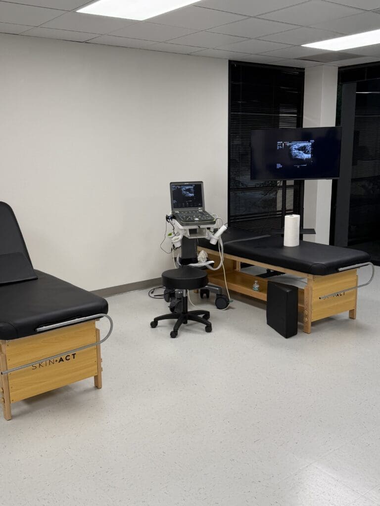
In my own practice, I’ve observed this principle in action. A recent case involved a patient for whom we prepared PRP with a concentration factor of approximately 7.5 times their baseline platelet count. While this number can vary from patient to patient due to individual physiology, our advanced processing systems consistently yield concentrations in the 6x-10x range. The key was not just the concentration but the processing method, which allowed us to capture a high platelet yield, ensuring we delivered a therapeutically significant dose.

The Intricate Cellular Symphony Within PRP

To truly appreciate the power of PRP, we must look at the intricate components of the preparation and how they work together. Using modern separation systems, we can meticulously isolate different fractions of the blood.

Understanding the Buffy Coat and Its Components

When blood is centrifuged, it separates into three main layers:

  • Red Blood Cells (Erythrocytes): The dense bottom layer.
  • Platelet-Poor Plasma (PPP): The clear, liquid top layer.
  • The Buffy Coat: A thin, whitish layer sandwiched between the other two.

The buffy coat is the treasure trove of regenerative medicine. It is densely packed with platelets and most leukocytes. The historical fear was the inclusion of the reddish layer just below the buffy coat, as it was thought to contain pro-inflammatory cells that could worsen conditions like arthritis.

However, our understanding of these cells has become much more refined. Advanced analysis reveals that this reddish zone, while containing some red blood cells, is also rich in specific leukocyte types, namely lymphocytes and monocytes. Far from being purely detrimental, these cells play a vital, beneficial role in the healing cascade.

  • Monocytes are particularly fascinating. When introduced to an injury site, they can differentiate into macrophages, which are essential for clearing cellular debris. More importantly, they help orchestrate the subsequent regenerative phases. The presence of lymphocytes helps guide these monocytes toward a pro-regenerative (M2) phenotype rather than a pro-inflammatory (M1) one.

This means that a PRP preparation that strategically includes these cell populations can create a more robust and sophisticated healing signal. The lymphocytes and monocytes don’t just add to the inflammation; they help manage it and then initiate a structured, beneficial healing response. This is why the conversation is moving away from simply labeling PRP as “leukocyte-rich.” It’s about understanding which leukocytes are present and their specific functions. The granulocytes (like neutrophils), which are more associated with acute inflammation, are largely separated out, while the beneficial monocytes and lymphocytes are retained.

This new perspective helps explain a retrospective observation: systems that produced “leukocyte-rich” PRP often happened to capture more platelets. The superior outcomes seen in some studies using LR-PRP for tendon injuries, for example, may have been less about the leukocytes and more about the higher absolute platelet dose being delivered (Filardo et al., 2018).

The Crucial Role of Integrative Chiropractic Care

Advanced regenerative treatments like PRP are powerful tools, but they do not exist in a vacuum. To achieve the best possible outcomes, we must address the entire patient, including the underlying biomechanical and structural issues that contributed to the injury or degeneration in the first place. This is where integrative chiropractic care becomes an indispensable partner to regenerative medicine.

Imagine injecting a highly potent, regenerative PRP preparation into a knee joint that is suffering from osteoarthritis. If that knee remains misaligned, with improper patellar tracking and imbalanced forces from dysfunctional muscles in the hip and ankle, the regenerative therapy is fighting an uphill battle. The very same pathological forces that wore down the cartilage remain, poised to degrade the newly formed tissue.

Creating an Optimal Healing Environment

As a chiropractor and functional medicine practitioner, my approach is to create an optimal environment for these regenerative cells to do their work. This involves a multi-faceted strategy:

  • Biomechanical Correction: Through precise chiropractic adjustments, we restore proper joint alignment not just in the affected joint but along the entire kinetic chain. For a knee issue, this means assessing and correcting imbalances in the spine, pelvis, hips, and ankles. This ensures that forces are distributed evenly, reducing pathological stress on the healing tissues.
  • Myofascial Release and Rehabilitation: We use advanced soft-tissue techniques to release adhesions, correct muscle imbalances, and restore proper function. This might involve active release techniques, instrument-assisted soft tissue mobilization, and targeted therapeutic exercises. This step is crucial for ensuring the joint is supported by a strong, balanced, and functional muscular system.
  • Nutritional and Metabolic Support: Healing is a metabolically demanding process. Through a functional medicine lens, we assess and optimize the patient’s nutritional status. This includes ensuring adequate levels of key vitamins and minerals (like Vitamin C, Zinc, and Magnesium) and managing systemic inflammation through diet and targeted supplementation (e.g., omega-3 fatty acids, curcumin). A systemically inflamed body will have a blunted response to any localized regenerative therapy.

By integrating these approaches, we are not just treating the site of pain; we are re-establishing the foundation for health. The chiropractic adjustments and physical rehabilitation prepare the “soil” by correcting the biomechanical environment, while the PRP injection acts as the “seed,” providing the cellular machinery for growth and repair. This comprehensive model significantly enhances the potential for long-term success and is a core tenet of my clinical philosophy at Injury Medical & Chiropractic Clinic.

A New Frontier in Regenerative Orthopedics

We stand at an exciting new frontier in the treatment of musculoskeletal conditions. The science of PRP is moving beyond simplistic classifications and toward a more sophisticated, evidence-based approach centered on precise dosing and a deeper understanding of cellular interactions. The latest research from leading experts is guiding us to optimize our methods, not by eliminating certain cells, but by understanding how to harness their synergistic potential to orchestrate a powerful healing response.

For patients suffering from conditions like joint arthritis, this means more effective and reliable outcomes. By combining these cutting-edge regenerative therapies with the foundational principles of integrative chiropractic care, we can address both the symptoms and the root cause of their condition. This holistic approach ensures that we are not only repairing damaged tissue but also restoring function, improving biomechanics, and empowering the body’s innate capacity to heal itself, resulting in lasting relief and improved quality of life.


References

Filardo, G., Di Matteo, B., Kon, E., Merli, G., & Marcacci, M. (2018). Platelet-rich plasma in tendon-related disorders: results and indications. Knee Surgery, Sports Traumatology, Arthroscopy, 26(7), 1984–1999. https://doi.org/10.1007/s00167-016-4261-4

Le, A. D. K., Enweze, L., DeBaun, M. R., & Dragoo, J. L. (2022). Current clinical recommendations for use of platelet-rich plasma. Current Reviews in Musculoskeletal Medicine, 15(6), 442–453. https://doi.org/10.1007/s12178-022-09787-z

PRP Therapy for Pain Relief, Healing, and Recovery

PRP Therapy for Pain Relief, Healing, and Recovery

PRP Therapy for Pain Relief, Healing, and Recovery

Abstract

Welcome to an in-depth exploration of Platelet-Rich Plasma (PRP), a cornerstone of modern regenerative medicine. We will delve into the very essence of platelets, exploring the powerful growth factors, cytokines, and other bioactive molecules they release. Drawing from the latest findings of leading researchers, we’ll examine how these components orchestrate the body’s natural healing and anti-inflammatory processes. I will explain the critical concept of PRP dosing, the importance of different platelet types, and how specific growth factors such as PDGF, TGF-β, and FGF contribute to tissue repair and regeneration. Finally, we’ll connect these principles to clinical practice, showing how integrative chiropractic care can be synergistically combined with PRP therapy to optimize patient outcomes, reduce pain, and restore function by addressing both the biochemical and biomechanical aspects of healing.


The Cellular Orchestra: Understanding the Power Within Your Blood

For over a decade, my clinical practice has been deeply rooted in the principles of functional and regenerative medicine. A recurring theme in helping patients overcome chronic pain and injury is the quest to harness the body’s innate ability to heal itself. One of the most powerful tools we have in this endeavor is Platelet-Rich Plasma (PRP). While many have heard the term, the true depth of what’s happening at a cellular level is often misunderstood. Today, I want to take you on a journey into the microscopic world of PRP, moving beyond the buzzwords to appreciate the sophisticated biological symphony it conducts.

When we talk about PRP, we’re focusing on concentrating a specific component of your blood: the platelets, also known as thrombocytes. While red blood cells and white blood cells play their own roles, it’s the platelets that act as the master conductors of tissue repair. The therapeutic magic of PRP lies in the wealth of bioactive components housed within these tiny cell fragments. These include:

  • Growth Factors: Proteins that signal cells to grow, proliferate, and differentiate.
  • Cytokines: Small proteins that are crucial in controlling the growth and activity of other immune system cells and blood cells.
  • Anti-inflammatory Molecules: Compounds that help modulate and resolve inflammation, which is a key barrier to healing.

The fundamental idea is that by concentrating these platelets, we can deliver a supraphysiological dose of these healing molecules directly to an injured area, amplifying the body’s natural repair signals and creating an optimal environment for regeneration.

Inside the Platelet: A Treasure Trove of Healing Granules

To truly grasp how PRP works, we need to look inside the platelet itself. Think of a platelet as a microscopic delivery vehicle packed with different types of cargo containers, or granules. The main therapeutic benefit we seek comes from the contents of these granules, which are released upon activation at the site of injury.

The three primary types of granules are:

  • Alpha Granules: These are the most abundant and arguably the most important for regeneration. Each platelet contains about 50 to 80 alpha granules, which are filled with a vast array of powerful growth factors. When a platelet is activated—for instance, by contact with exposed collagen in damaged tissue—it undergoes a process called degranulation, releasing growth factors into the surrounding environment. This is the primary event that initiates the healing cascade.
  • Dense Granules (or Delta Granules): These granules contain small molecules, including ADP, ATP, serotonin, and calcium. Their role is to amplify the healing response. They enhance platelet aggregation (helping form a stable scaffold) and vasoconstriction (controlling bleeding), and they also modulate the local immune response.
  • Lysosomes: These act as the cleanup crew. They release enzymes that help break down and remove damaged tissue and cellular debris from the injury site. This process, known as enzymatic debridement, clears the way for new, healthy tissue to form and also contributes to antimicrobial defense.

Recent research, including studies on platelet biology, highlights that over 280 distinct proteins are involved in this process, underscoring the incredible complexity of this “orchestra” of molecules working in concert (Golebiewska & Poole, 2015).

The Significance of Platelet Quality and “Dosing”

A critical concept that has emerged from evidence-based research is that not all PRP is created equal. The effectiveness of a treatment depends heavily on both the concentration and quality of the platelets. This is where the idea of PRP dosing becomes paramount. Simply put, the more functional platelets we can deliver to a target tissue, the greater the concentration of bioactive molecules we release, and potentially, the more robust the healing response.

Furthermore, we are learning about the importance of reticulated platelets. These are essentially “younger,” more robust platelets, recently released from the bone marrow (typically within the last 24-72 hours). They are denser and contain more alpha granules than their older counterparts. This means they are packed with more growth factors and have a greater regenerative potential. In our clinical processing, we use advanced techniques to preferentially harvest these denser, more potent reticulated platelets, ensuring that the PRP we inject is of the highest possible quality and biological activity.

The Key Players: A Closer Look at Essential Growth Factors

While hundreds of proteins are released, a few key growth factors are the primary drivers of the regenerative effects seen with PRP. Understanding their specific roles helps us appreciate why this therapy is so effective for a range of musculoskeletal conditions.

Platelet-Derived Growth Factor (PDGF)

As its name implies, PDGF was first discovered in platelets, but it’s also produced by other cells involved in healing. Think of PDGF as the “first responder” or the primary chemoattractant. It sends out a powerful signal that calls other crucial healing cells to the site of injury. Most importantly, it recruits Mesenchymal Stem Cells (MSCs)—the body’s own master repair cells—to the area. While PRP itself does not contain stem cells, it potently signals the body’s resident stem cells to migrate to the site, proliferate (make copies of themselves), and differentiate into the specific cell types needed for repair (e.g., cartilage, tendon, or bone cells). The PDGF-BB isoform is recognized as the most biologically active and is a major focus of current research for its potent role in initiating this cascade.

Transforming Growth Factor-Beta (TGF-β)

TGF-β is a master regulator of tissue regeneration. Its primary functions include:

  • Stimulating Collagen Synthesis: It powerfully enhances the production of type I collagen, the fundamental building block of tendons, ligaments, and the matrix of our bones and cartilage. This is essential for rebuilding the structural integrity of injured tissue.
  • Promoting Angiogenesis: the formation of new blood vessels. A healthy blood supply is critical for delivering oxygen and nutrients to the healing tissue and removing waste products.
  • Coordinating with Other Growth Factors: TGF-β works synergistically with PDGF to enhance endothelial cell proliferation and capillary sprouting, leading to a robust network of new blood vessels (neovascularization) that supports long-term tissue health.

The effect of PRP on angiogenesis is dose-dependent. Studies, such as those published in the Journal of Orthopedic Research, suggest that a platelet concentration of approximately 1.5 billion platelets per milliliter is required to achieve a significant pro-angiogenic effect (Mazzocca et al., 2012). This underscores the importance of precise processing and quantification to achieve optimal clinical results.

Fibroblast Growth Factor (FGF)

FGF is one of the most potent mitogens released by platelets, meaning it is exceptionally effective at stimulating cell division. It acts on a wide variety of cell types, including MSCs, chondrocytes (cartilage cells), and osteoblasts (bone-forming cells). By promoting the rapid proliferation of these essential repair cells, FGF accelerates tissue regeneration.

The Anti-Inflammatory Power of PRP

Chronic, unresolved inflammation is a major culprit behind persistent pain and tissue degradation, especially in conditions like osteoarthritis. While PRP initiates an acute, controlled inflammatory response to jumpstart healing, one of its most profound long-term benefits is its ability to modulate and resolve chronic inflammation.

This is achieved through several mechanisms:

  1. Leukocyte Interaction: The concentrated platelets in PRP interact with white blood cells (leukocytes) at the injury site. This interaction can shift the behavior of these immune cells, prompting them to release anti-inflammatory cytokines like IL-10 and IL-4, which actively suppress chronic inflammation.
  2. Macrophage Polarization: PRP can influence macrophage behavior, a type of white blood cell. It promotes a shift from the pro-inflammatory (M1) phenotype to an anti-inflammatory and pro-reparative (M2) phenotype. M2 macrophages are critical for cleaning up debris, resolving inflammation, and secreting factors that promote tissue remodeling and regeneration.
  3. Chemokine Secretion: Platelets release chemokines that not only recruit healing cells but also act as survival factors for monocytes, preventing their premature death and promoting their differentiation into beneficial M2 macrophages.

In essence, PRP acts as a biological “reset button,” transforming a chronically inflamed, degenerative environment into one that is actively anti-inflammatory and pro-regenerative.

Integrating Chiropractic Care for a Holistic Healing Approach

As a Doctor of Chiropractic, I view the body through both biomechanical and biochemical lenses. A successful outcome depends on addressing both the “parts” and the “system.” This is where the synergy between PRP therapy and integrative chiropractic care becomes so powerful.

Imagine a patient with chronic knee osteoarthritis. The PRP injection will address the biochemical problem inside the joint—reducing inflammation, signaling cartilage repair, and improving the quality of the synovial fluid. However, if the patient’s knee pain is also caused or exacerbated by poor biomechanics—such as a misaligned pelvis, muscle imbalances in the leg, or improper gait—the joint will remain under abnormal stress. This persistent mechanical strain can hinder the regenerative process initiated by the PRP and lead to a recurrence of symptoms.

This is why our integrative approach includes:

  • Chiropractic Adjustments: We perform precise adjustments to the spine and extremities to restore proper alignment and mobility of the joints. Correcting pelvic alignment, for example, can ensure that forces are distributed evenly through the knees, reducing abnormal wear and tear.
  • Myofascial Release and Soft Tissue Work: We address muscle imbalances, trigger points, and fascial restrictions that contribute to faulty movement patterns. This ensures that the muscles supporting the joint are functioning optimally.
  • Customized Rehabilitation: We design targeted exercise programs to strengthen weak muscles, stretch tight ones, and retrain proper movement patterns (neuromuscular re-education). This stabilizes the joint and protects it from future injury.

By combining the powerful biochemical signaling of PRP with the essential biomechanical corrections of chiropractic care, we create a comprehensive healing environment. The PRP rebuilds tissue from the inside out, while chiropractic care ensures the entire musculoskeletal system functions correctly to support and protect the newly regenerated tissue. In my clinical experience, this holistic strategy is the key to achieving durable, long-term pain relief and true restoration of function for my patients.


References

What You Should Know About PRP Therapy for Knee Osteoarthritis

Learn the benefits of PRP therapy for osteoarthritis for better mobility and reduced discomfort in your knee joints.

Abstract

In this educational post, I present a comprehensive, first-person overview of how I integrate platelet-rich plasma (PRP) therapy into care plans for patients with symptomatic knee osteoarthritis (OA). Drawing on modern, evidence-based research and my clinical observations as Dr. Alexander Jimenez, DC, APRN, FNP-BC, CFMP, IFMCP, ATN, CCST, I explain why PRP often outperforms corticosteroid and hyaluronic acid injections in pain, function, and potentially in cartilage preservation. I outline how PRP interacts with joint biology, the cellular signaling that underpins its therapeutic effects, and the integrative chiropractic strategies that optimize outcomes—covering biomechanics, neuromuscular control, functional nutrition, and lifestyle medicine. This piece synthesizes findings from leading researchers and aligns them with an integrated clinical protocol focused on measurable outcomes, safety, and long-term joint health.

Why I Use Platelet-Rich Plasma for Symptomatic Knee Osteoarthritis

As a clinician working daily with musculoskeletal conditions, I have witnessed how knee osteoarthritis impacts quality of life—pain, stiffness, loss of mobility, and reduced confidence in movement. Over years of clinical practice and continual review of the literature, platelet-rich plasma (PRP) has become a central part of my integrative treatment approach when patients present with symptomatic knee OA.
Here is why PRP often earns a place in my treatment plans:

  • Evidence favors PRP over corticosteroids and hyaluronic acid for pain and functional outcomes in many patient populations with knee OA.
  • Mechanistic plausibility: PRP delivers concentrated growth factors that can modulate inflammation, encourage matrix synthesis, and potentially support chondrocyte resilience.
  • Safety profile: Autologous biologic with relatively low risk when processed and delivered under rigorous protocols.
  • Synergy with integrative chiropractic care: Biologic modulation is most effective when combined with biomechanical alignment, neuromuscular training, and lifestyle interventions that reduce joint load and support metabolic health.

In practice, I find PRP excels for patients who are struggling with persistent symptoms despite conservative care, who are hesitant to rely on corticosteroid cycles, or who want to explore joint-preserving options alongside manual therapy and functional rehab.

Understanding Platelet-Rich Plasma: What It Is and How It Works

PRP is an autologous blood product derived from the patient’s own venous sample. After centrifugation, we isolate a plasma fraction with an elevated platelet concentration, typically ranging from 3x to 7x baseline, depending on the preparation. The platelets carry alpha granules packed with bioactive proteins that can influence joint microenvironments.
Key bioactive mediators in PRP include:

  • Platelet-derived growth factor (PDGF): Stimulates cell proliferation, chemotaxis, and angiogenesis.
  • Transforming growth factor-beta (TGF-β): Modulates matrix synthesis and can influence chondrocyte phenotype.
  • Insulin-like growth factor-1 (IGF-1): Supports cartilage matrix synthesis and cellular survival.
  • Vascular endothelial growth factor (VEGF): Angiogenic signaling with context-dependent roles in tissue repair.
  • Epidermal growth factor (EGF) and fibroblast growth factors (FGFs): Promote cellular activity relevant to tissue recovery.

When introduced into the osteoarthritic knee, these mediators can:

  • Modulate inflammation: Downregulate pro-inflammatory cytokines such as IL-1β and TNF-α while promoting anti-inflammatory signaling.
  • Support extracellular matrix homeostasis: Encourage anabolic pathways (e.g., collagen II, aggrecan synthesis) and potentially reduce catabolic enzyme activity (MMPs, ADAMTS).
  • Influence chondrocyte behavior: Support survival, reduce apoptosis in hostile joint environments, and promote a more regenerative phenotype.
  • Impact synovial environment: Improve synovial fluid characteristics and reduce synovitis, thereby decreasing nociceptive drive.

This physiologic rationale aligns with reported clinical outcomes—reduced pain, improved function, and, in some studies, signals that PRP may exert chondroprotective effects.

Comparing PRP With Corticosteroids and Hyaluronic Acid: What the Data Shows

Corticosteroids and hyaluronic acid (HA) have established roles in knee OA, but their mechanisms and durability differ from PRP:

  • Corticosteroids: Potent anti-inflammatory effects via genomic and non-genomic pathways that reduce prostaglandins and cytokines. Benefits tend to be short-lived, and repeated injections may have deleterious effects on cartilage metabolism. I reserve corticosteroids for acute synovitis flare-ups when rapid control is essential (e.g., severe swelling impeding rehab).
  • Hyaluronic acid (viscosupplementation): Aims to restore viscoelastic properties of synovial fluid and interact with CD44 receptors to modulate inflammation and joint mechanics. Responses vary, and HA alone may not match PRP’s performance in many trials.
  • PRP: Offers biologic signaling that can shift the joint toward repair and better matrix dynamics. Clinical benefits can last longer than those of steroids in many cohorts and often outperform HA alone on functional indices.

The literature has increasingly focused on head-to-head comparisons:

  • Multiple meta-analyses and randomized trials report greater pain reduction and functional improvement with PRP than with placebo, corticosteroids, or HA at mid-term follow-up, especially in mild-to-moderate OA phenotypes (e.g., Kellgren-Lawrence II–III) (see references below).
  • Dose and product characteristics matter: Leukocyte-poor PRP generally demonstrates better tolerability in the joint with lower post-injection flare rates than leukocyte-rich formulations for OA applications. Platelet concentration in a moderate range tends to be optimal; excessively high concentrations may increase catabolic signaling.
  • Repeat dosing strategies (e.g., series of 2–3 injections spaced 2–4 weeks apart) often yield more durable outcomes than single injections, likely reflecting cumulative biologic conditioning of the synovial and cartilage microenvironments.

These lines of evidence help me set patient expectations and tailor protocols to the OA phenotype they present with.

Chondroprotection: Can PRP Help Preserve Cartilage?

One of the most exciting themes in the recent literature is chondroprotection—the idea that PRP might help maintain cartilage integrity or slow degenerative processes. Mechanistically, PRP may:

  • Reduce catabolic enzyme activity: Suppress matrix metalloproteinases (MMP-1, MMP-3, MMP-13) and aggrecanases (ADAMTS-4/5).
  • Enhance anabolic signaling: Promote the synthesis of type II collagen and aggrecan by modulating IGF-1 and TGF-β.
  • Improve subchondral crosstalk: Alter inflammatory signaling between cartilage, synovium, and subchondral bone marrow lesions.
  • Dampen oxidative stress: Increase antioxidant capacity, which supports chondrocyte survival and reduces apoptosis.

While ongoing studies continue to refine estimates of the magnitude and consistency of these effects, the convergence of mechanistic and clinical data supports the potential of PRP to be chondroprotective in appropriately selected patients. This has meaningful implications for delaying progression, prolonging function, and deferring invasive interventions.

How Integrative Chiropractic Care Enhances PRP Outcomes

PRP works best inside a systems approach. As a chiropractor and advanced practice provider, I integrate manual therapy, neuromuscular re-education, biomechanical correction, functional nutrition, and lifestyle medicine to amplify PRP’s benefits.
Key pillars of my integrative care:

  • Spinal and lower-extremity alignment: Targeting pelvic tilt, sacroiliac mechanics, and tibiofemoral/ patellofemoral tracking reduces aberrant joint loading. Precise chiropractic adjustments, mobilizations, and soft-tissue releases help normalize kinetic-chain function.
  • Neuromuscular control: Focused training for gluteus medius/maximus, vastus medialis obliquus (VMO), hamstrings, and calf complex improves dynamic valgus control and reduces medial compartment stress. We use progressive closed-chain drills that restore proprioception and joint stability.
  • Gait and movement retraining: Patients learn load-management strategies—such as cadence optimization, stride mechanics, and foot placement—to reduce peak joint forces. We frequently pair this with orthotic support or footwear interventions.
  • Functional nutrition: Anti-inflammatory dietary patterns (Mediterranean-style, low-glycemic-load), adequate protein intake, omega-3 fatty acids, and micronutrients (vitamin D, magnesium) support cartilage metabolism and systemic recovery.
  • Weight management: Even modest reductions in body mass can translate to significant decreases in knee joint compressive forces per step. We implement structured plans for sustainable change.
  • Sleep and stress modulation: Optimizing sleep architecture and stress resilience (via HRV-guided practices and mindfulness) supports hormonal balance and reduces systemic inflammation, which influences joint pain perception.

This integrated framework enables PRP’s biologic signals to operate in an environment with optimized mechanics and metabolism, often resulting in faster symptom relief and more durable function.

Patient Selection and Personalized Protocols

Not every knee OA patient is an ideal candidate for PRP. Success starts with phenotyping and individualized planning.
I consider:

  • OA severity: PRP tends to perform best in mild-to-moderate OA. In advanced (Kellgren-Lawrence IV) cases, expectations may shift toward pain control and function rather than structural change.
  • Inflammatory burden: Synovitis on ultrasound or exam can guide timing and adjuvant therapies. Short-course anti-inflammatories or a single corticosteroid injection may be considered to quiet a severe flare before PRP.
  • Metabolic health: Insulin resistance, vitamin D deficiency, and obesity hinder repair. Addressing these factors improves outcomes.
  • Biomechanics: Valgus/varus alignment, foot mechanics, and hip control inform specific chiropractic and rehab priorities.
  • Activity goals: Tailoring protocols to sports, occupation, or daily living tasks ensures the plan is relevant and sustainable.

Typical PRP protocol elements:

  • Preparation: Hydration, avoidance of NSAIDs for a specified window (often 5–7 days) to minimize interference with platelet function.
  • Product selection: I prefer leukocyte-poor PRP for intra-articular knee OA due to better tolerability and reduced post-injection inflammatory flares.
  • Dosing strategy: A series of 2–3 injections spaced 2–4 weeks apart is common. Some cases respond well to a single injection; we decide based on response and phenotype.
  • Guidance: Ultrasound-guided intra-articular injection improves accuracy and consistency.
  • Post-procedure: Relative rest for 24–48 hours, then graded return to activity with a structured rehab emphasizing alignment and neuromuscular control. Heat or cold as indicated; avoid NSAIDs in the early window.
  • Follow-up metrics: We use validated outcome measures (e.g., WOMAC, KOOS) and functional tests to track progress.

Physiologic Rationale: From Synovial Biology to Pain Relief

Pain in knee OA is multifactorial:

  • Synovitis: Inflamed synovial tissue releases cytokines that sensitize nociceptors.
  • Cartilage degeneration: Loss of smooth articulation increases mechanical stress and generates pro-algesic debris.
  • Subchondral bone changes: Bone marrow lesions and altered microarchitecture amplify pain signaling.
  • Peripheral and central sensitization: Chronic input leads to heightened pain response.

PRP can address several of these contributors:

  • Anti-inflammatory modulation: Growth factors and cytokine interplay reduce drivers of synovial pain.
  • Cartilage matrix support: Enhanced anabolic signals promote smoother gliding surfaces, reducing mechanical irritation.
  • Improved joint homeostasis: A better synovial fluid environment reduces friction and improves lubrication.
  • Neuroimmune effects: Biologic shifts in cytokine milieu may reduce peripheral sensitization.

When paired with chiropractic alignment and neuromuscular strategies, we also reduce aberrant load transfer, which diminishes local tissue stress and pain generation.

How I Integrate PRP With Manual Therapy and Rehab

The timing and sequencing of care are crucial. My approach:

  • Pre-injection priming:
    • Educate the patient on expectations and activity pacing.
    • Begin gentle mobility work to reduce capsular stiffness and improve movement quality.
    • Initiate low-grade isometrics to maintain neuromuscular tone without provoking pain.
  • Injection day and immediate post-care:
    • Ultrasound-guided PRP injection.
    • Relative rest for 24–48 hours; avoid NSAIDs; use acetaminophen if needed.
  • Week 1–2:
    • Gradual reintroduction of manual therapy: targeted mobilizations for tibiofemoral and patellofemoral joints, soft-tissue release of quadriceps, hamstrings, IT band.
    • Begin closed-chain stability drills: mini-squats to a pain-free range, step-downs, hip abduction work, and balance drills.
  • Week 3–6:
    • Progress to eccentric strengthening for quads and glutes, dynamic valgus control drills, and controlled plyometrics where appropriate.
    • Gait retraining with cadence adjustments (often a 5–10% increase reduces peak joint load).
  • Beyond 6 weeks:
    • Higher-level functional training aligned with patient goals (hiking, recreational sports).
    • Maintenance care: periodic manual therapy tune-ups and home program updates.

This cohesive plan ensures the biologic effects of PRP translate into functional gains.

My Clinical Observations in Integrative PRP Care

From daily practice at my clinic and years of collaborative care, certain patterns consistently emerge:

  • Patients who combine PRP with precise chiropractic adjustments and neuromuscular re-education show faster improvements in gait symmetry and stair negotiation than those receiving injections alone.
  • Leukocyte-poor PRP produces fewer short-term flares; when flares occur, they typically resolve within 48–72 hours with conservative measures.
  • Metabolic optimization (vitamin D sufficiency, omega-3 intake, glycemic control) correlates with better mid-term outcomes.
  • Commitment to movement retraining—especially hip-centric control—reduces medial compartment loading and improves pain scores.
  • A series approach (2–3 injections) yields more stable 6–12 month outcomes than single injections in moderate OA phenotypes.

You can learn more about my approach and patient outcomes at:

Knee Injury Rehabilitation Story- Video

Practical Tips for Patients Considering PRP

When I counsel patients on PRP for knee OA, I emphasize:

  • Set goals clearly: Pain reduction, functional improvement, and joint preservation are achievable, but individual responses vary.
  • Commit to the full program: PRP works best with integrative care—alignment, strength, gait, and nutrition.
  • Expect some post-injection soreness: This is usually self-limited and part of the healing arc.
  • Track progress: Use standardized tools and functional milestones (stairs, walking distance, sit-to-stand ease).
  • Maintenance plan: Keep gains through ongoing exercise, weight management, and periodic tune-ups.

Safety, Contraindications, and Quality Control

PRP is generally safe, but appropriate screening and technique are essential:

  • Contraindications: Active infection, severe uncontrolled systemic disease, bleeding disorders, anticoagulation that cannot be paused, platelet dysfunction syndromes.
  • Relative considerations: Poorly controlled diabetes, significant anemia, severe vitamin D deficiency, or systemic inflammatory states—optimize first.
  • Quality control: Sterile technique, validated kits, clearly defined platelet concentration targets, and ultrasound guidance reduce variability.
  • Adverse events: Transient pain or swelling; rare complications include infection or hemarthrosis. We provide clear aftercare instructions and prompt follow-up.

The Future of Orthobiologics in Knee OA

The orthobiologic field is evolving rapidly:

  • Standardization: Outcome reporting and PRP characterization (platelet count, leukocyte status) are improving, enabling better meta-analytic clarity.
  • Combination therapies: PRP with hyaluronic acid or microfragmented adipose tissue are under investigation to achieve synergistic effects for certain phenotypes.
  • Imaging biomarkers: Advanced MRI and ultrasound can help track cartilage and synovial changes, refining patient selection and timing.
  • Personalized medicine: Matching biologic profiles and biomechanical signatures to specific orthobiologic strategies will enhance precision care.

As data quality increases, PRP’s role in knee OA management is likely to expand—particularly in integrated settings that marry biologic, mechanical, and behavioral strategies.

Why PRP Fits Naturally Into Integrative Chiropractic Care

Integrative chiropractic care is built on restoring function, alignment, and resilience. PRP complements this philosophy by offering biologic support that enhances the tissue environment while we correct load, movement, and systemic contributors. In my practice, this synergy helps patients:

  • Reduce pain more quickly and sustain improvements.
  • Restore confidence in movement and return to meaningful activities.
  • Delay or avoid more invasive interventions.
  • Take a proactive, evidence-based approach to joint health.

This is why, for many of my patients with symptomatic knee OA, PRP has become a high-value option integrated into a comprehensive, individualized plan.

Key Takeaways

  • PRP provides concentrated biologic signals that modulate inflammation, support matrix synthesis, and may offer chondroprotective benefits in knee OA.
  • Evidence often shows PRP outperforms corticosteroids and hyaluronic acid for pain and function, particularly in mild-to-moderate OA.
  • Leukocyte-poor PRP with series dosing and ultrasound guidance optimizes outcomes.
  • Integrative chiropractic care—alignment, neuromuscular control, gait retraining, nutrition, and lifestyle—amplifies PRP benefits and durability.
  • Careful patient selection and personalized protocols drive success and safety.
  • Continuous research and standardized reporting are strengthening PRP’s place in modern, evidence-based musculoskeletal medicine.

References

SEO tags: platelet rich plasma, PRP for knee osteoarthritis, integrative chiropractic care, chondroprotection, orthobiologics, knee pain treatment, leukocyte-poor PRP, functional rehabilitation, biomechanics, neuromuscular training, ultrasound-guided injection, evidence-based musculoskeletal medicine