Give us a Call
+1 (915) 412-6680
Send us a Message
[email protected]
Opening Hours
Mon-Thu: 7 AM - 7 PM
Fri - Sun: Closed

Regenerative Medicine Best Practices for Patient Optimization

Understand the importance of patient optimization in regenerative medicine and its role in modern healthcare advancements.

Abstract

Hello, I’m Dr. Alex Jimenez. With my extensive background in integrative and functional medicine, holding titles including DC, APRN, FNP-BC, CFMP, IFMCP, ATN, and CCST, I’ve dedicated my career to optimizing patient health from every angle. In this educational post, we will journey through the critical yet often overlooked phase of preparing the body for orthobiologic and regenerative therapies. I believe that optimizing the patient’s internal environment—their personal “pharmacy”—is just as crucial as the biologic treatment itself. We will explore the six pillars of lifestyle medicine: diet, exercise, sleep, stress mitigation, social connectedness, and the avoidance of risky substances. Drawing on the latest evidence-based research, we’ll discuss how conditions such as obesity, chronic low-grade inflammation, sarcopenia, and gut dysbiosis can significantly affect the success of regenerative procedures. I will provide a comprehensive framework for assessing and enhancing a patient’s metabolic health, including specific dietary recommendations, exercise protocols, and screening tools. We will also delve into how integrative chiropractic care complements this process by addressing the biomechanical and neurological factors that influence healing, ensuring a truly holistic approach to recovery and long-term wellness.

The Foundation of Healing: Why Patient Optimization is Non-Negotiable

As a practitioner deeply invested in both chiropractic and functional medicine, my perspective is uniquely shaped by a diverse background that includes public health and a passion for holistic wellness. When a patient comes to me for an orthobiologic procedure, my focus isn’t just on the treatment itself. It extends to a fundamental question: Is this patient’s body prepared to heal? I am passionate about making sure every patient is as metabolically optimized as possible before we proceed.

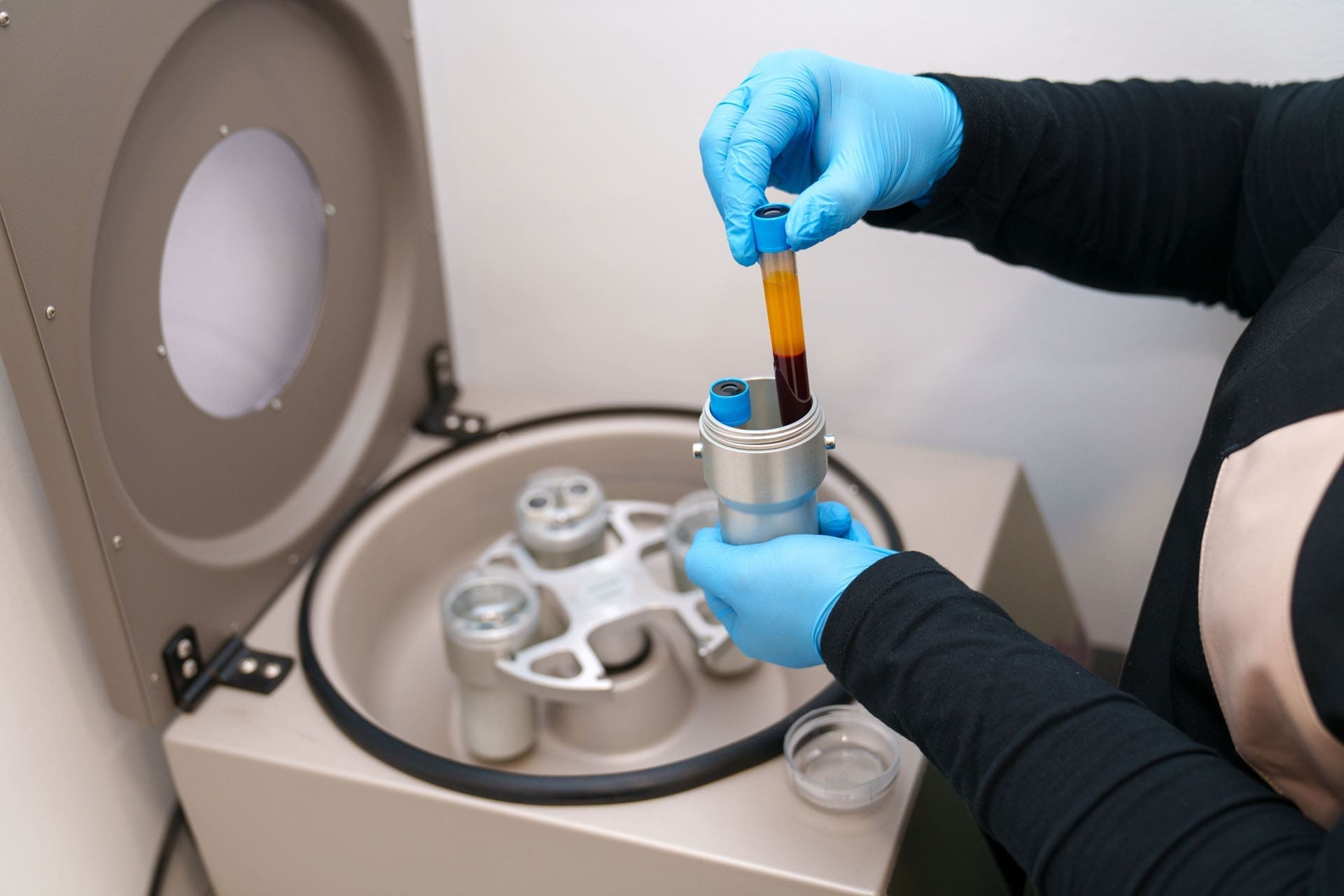
Think of it this way: the biologics we use, whether platelet-rich plasma (PRP) or cellular therapies, are catalysts. But the real work of healing happens within the patient’s own body. We are, in essence, optimizing their internal pharmacy. By improving their metabolic health, we are ensuring that the “raw materials” for regeneration are abundant and that the environment is conducive to repair rather than breakdown. This concept is the cornerstone of my practice and is supported by a growing body of research highlighting the profound connection between lifestyle and regenerative potential.

The Six Pillars of Lifestyle Medicine in Regenerative Care

To structure this optimization process, I use the framework of lifestyle medicine. This evidence-based approach focuses on six key areas that collectively determine our overall health. When we improve these pillars, we achieve maximum metabolic optimization, creating the ideal conditions for regenerative treatments to succeed. While we have few, if any, large-scale randomized controlled trials (RCTs) directly linking these pillars to biologic outcomes, a wealth of data from other fields allows us to extrapolate and apply these principles with confidence. We know, for instance, that dietary interventions can improve platelet function and that exercise can enhance cellular activity (Paolucci et al., 2023).

Here are the six pillars we focus on:

  • Diet and Nutrition: Fueling the body for repair.
  • Physical Activity: Moving to enhance cellular function.
  • Restorative Sleep: The non-negotiable recovery phase.
  • Stress Management: Taming the silent saboteur of healing.
  • Social Connection: The powerful influence of community on health.
  • Avoidance of Risky Substances: Eliminating toxins that hinder regeneration.

Identifying and Addressing Key Barriers to Healing

A pivotal review article has identified six specific aspects of health that can significantly influence the outcomes of our procedures (Centeno et al., 2023). Our goal is to ensure that patients walk away from our care not just with temporary relief but with a profound, lasting improvement in their quality of life. By addressing these factors, we build a reputation for excellence.

These six critical aspects are:

  1. Obesity: Excess weight doesn’t just add mechanical stress to joints. For every pound of weight lost around the midsection, there’s a roughly four-fold decrease in the load on the knee joint. But beyond the mechanics, obesity is a state of metabolic dysregulation, characterized by adipokines—inflammatory molecules secreted by fat cells—that create a hostile environment for healing.
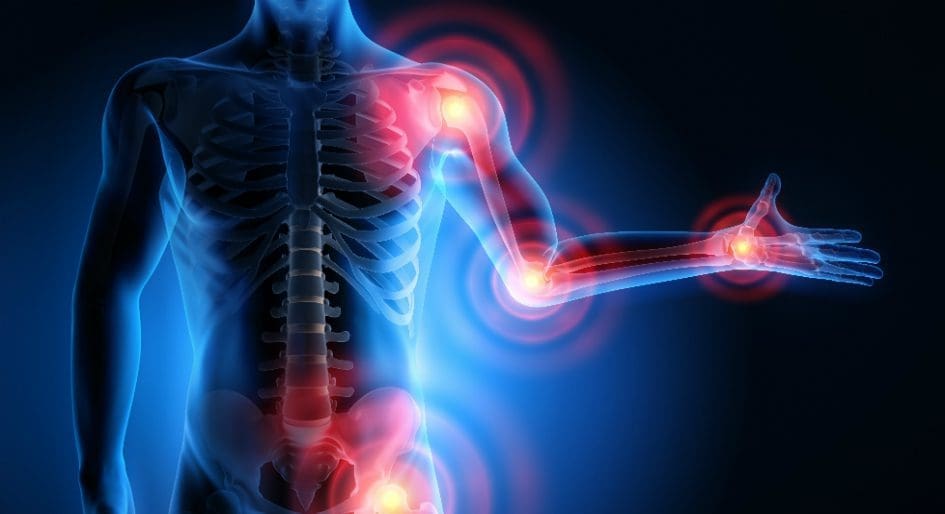
  2. Chronic Low-Grade Inflammation: This isn’t the acute, beneficial inflammation that initiates healing after an injury. This is a persistent, systemic inflammation that slowly degrades tissues and impairs regenerative processes.
  3. Sarcopenia: the age-related loss of muscle mass and function. Muscle is a metabolic powerhouse, and its decline compromises the body’s ability to heal and maintain stability.
  4. Gut Dysbiosis: This term describes an imbalance in your gut microbiota—the trillions of microorganisms living in your digestive tract. When these communities are out of balance, they can produce toxic byproducts that leak into the bloodstream, driving systemic inflammation and disrupting immune function.
  5. Sleep Deprivation: Anyone with a child understands how crucial sleep is. Lack of sleep dysregulates key hormones like cortisol, impairs immune function, and heightens pain sensitivity.
  6. Unhealthy Lifestyle Behaviors: A patient might be a marathon runner, but if they are also smoking heavily, the toxic exposure will negate many of the benefits. We must look at the patient’s life in its entirety.

Fueling Regeneration: The Power of an Anti-Inflammatory Diet

Your diet is one of the most powerful tools for influencing your body’s internal environment. We know that conditions like obesity and insulin resistance are detrimental, leading to impaired cellular function and a reduced capacity for healing. This is because high blood sugar and insulin levels create a pro-inflammatory state hostile to the very regeneration we aim to stimulate.

The gut-body connection is another critical piece of the puzzle. An imbalanced gut microbiome, or gut dysbiosis, can directly contribute to systemic inflammation, sabotaging our efforts. I believe we are only scratching the surface of how the gut impacts musculoskeletal health, and future research will undoubtedly reinforce this link.

Proposed Dietary Approach

My recommendation for patients is to adopt an anti-inflammatory diet. This isn’t a fad; it’s a dietary pattern that has been used successfully for years in rheumatology to manage inflammatory arthritis. The principles are simple and effective:

  • Increase Fiber and Leafy Greens: These feed beneficial gut bacteria and are rich in phytonutrients that help combat inflammation.
  • Boost Omega-3 Fatty Acids: Found in fatty fish, flaxseeds, and walnuts, these fats are precursors to powerful anti-inflammatory molecules.
  • Focus on Low-Glycemic-Index Foods: Choose whole grains, legumes, and non-starchy vegetables to help stabilize blood sugar and insulin levels.
  • Ensure Adequate Protein Intake: Protein provides the essential amino acids needed for tissue repair and collagen synthesis.
  • Avoid Processed Foods and Refined Sugars: These are primary drivers of inflammation and metabolic dysfunction.

Do We Need Supplements?

For a patient eating a varied, whole-foods diet, supplementation is often unnecessary. However, if there are concerns about nutritional gaps, certain nutrients can be particularly helpful:

  • Vitamin C: Essential for collagen synthesis, the primary protein in our connective tissues.
  • Vitamin D & Magnesium: Crucial for muscle function, bone health, and immune regulation.
  • Zinc & Copper: These minerals are cofactors in numerous enzymatic reactions vital for tissue repair.
  • Probiotics: Can help restore balance to the gut microbiome, potentially reducing systemic inflammation.

It’s important to note that the evidence regarding supplementation in relation to orthobiologic procedures is mixed. Some studies may suggest discontinuing certain supplements before a procedure, so it is always best to work with a knowledgeable provider to create a personalized plan.

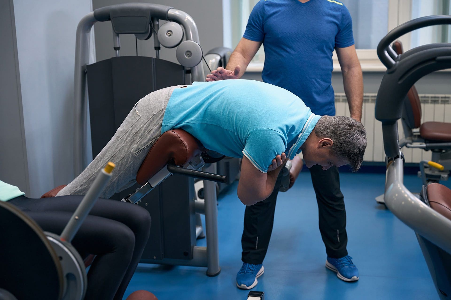
Movement as Medicine: Exercise Protocols for Enhanced Healing

Exercise is a potent medicine that positively impacts every barrier to healing we’ve discussed. It combats obesity and insulin resistance, lowers chronic inflammation, improves sleep quality, and reverses sarcopenia.

From an orthobiologic perspective, the benefits are even more direct:

  • Optimize Cellular Quality: Exercise can increase platelet counts and even enhance growth factor concentrations within platelets.
  • Improve Tissue Responsiveness: Regular physical activity makes your tissues more receptive to the growth signals initiated by regenerative treatments.
  • Limit Cellular Senescence: Exercise helps clear out old, dysfunctional “zombie” cells and improves the function, replication, and differentiation capacity of your own mesenchymal stem cells (MSCs).

Exercise Recommendations

As part of my intake, I use what’s called an “exercise vital sign,” a quick two-question screen to gauge a patient’s activity level. The goal is to meet or exceed the following recommendations:

  • Aerobic Exercise: At least 150 minutes per week of moderate-intensity activity (e.g., brisk walking, cycling). I often tell my patients that while this is the ideal, any movement toward this goal is a step in the right direction.
  • High-Intensity Interval Training (HIIT): Incorporating short bursts of intense effort followed by recovery periods is particularly effective at improving endothelial function—the health of your blood vessel lining—which is critical for delivering nutrients and healing factors to tissues.
  • Resistance Training: Aim for at least two sessions per week. Building and maintaining muscle is metabolically protective and provides crucial support for our joints.
  • Pre-Procedure Exercise: Intriguing research suggests that an acute bout of high-intensity exercise immediately before a PRP blood draw can temporarily increase circulating platelet levels (Liao et al., 2021). This is a simple strategy I often incorporate, having patients perform a short workout at a nearby gym just before their appointment.

The Healing Diet: Combat Inflammation, Embrace Wellness- Video

The Critical Role of Restorative Sleep

Sleep is when the body’s most important repair processes occur. The standard recommendation of seven to nine hours per night is not arbitrary; it’s a biological necessity. Inadequate sleep disrupts the delicate balance of our endocrine system, particularly affecting cortisol regulation. While often vilified, cortisol plays a vital role in managing inflammation, but chronically elevated levels due to poor sleep suppress the very pro-inflammatory signals needed to kickstart healing.

Furthermore, poor sleep significantly impacts central pain modulation. A sleep-deprived patient will perceive more pain from the procedure and during recovery. In my clinical observations at our clinics, patients who prioritize sleep hygiene consistently report better pain control and smoother recoveries. Special attention should be paid to conditions like obstructive sleep apnea (OSA), which can cause endothelial dysfunction due to intermittent hypoxia (low oxygen levels). I routinely screen for OSA, and it’s not uncommon for this screening to lead to a new diagnosis and life-changing treatment for a patient.

Eliminating Toxic Burdens: Tobacco and Alcohol

I am very direct with my patients about this: tobacco and alcohol are Group 1 carcinogens, meaning they are definitively known to cause cancer. Their negative impact on healing is just as definitive.

  • Tobacco: Nicotine is directly cytotoxic to MSCs (your stem cells) and causes abnormal platelet aggregation. It constricts blood vessels, starving tissues of the oxygen and nutrients they desperately need to heal.
  • Alcohol: Extensive surgical data shows that alcohol consumption increases the risk of post-procedure infection and impairs wound healing. It also directly damages MSCs and depletes key nutrients.

My approach is to counsel patients frankly about these risks and connect them with resources such as quit lines, pharmacotherapy, or a referral back to their primary care provider to develop a cessation strategy. A regenerative procedure is a significant investment of time, money, and hope—it makes no sense to undermine it with toxic exposures.

The Biopsychosocial Model: Stress, Social Connection, and Pain

The mind-body connection is not a new-age concept; it is a biological reality. Chronic stress leads to elevated cortisol levels, which, as we’ve discussed, impair tissue healing, suppress beneficial inflammation, and restrict the proliferation and differentiation of MSCs.

Conversely, strong social connections and effective stress management techniques are powerful buffers. Much of the data in this area revolves around pain mitigation. Patients who feel supported, understood, and emotionally resilient experience less pain and have better functional outcomes. This is why I advocate for a biopsychosocial evaluation, where we assess for stress, anxiety, and depression. It is crucial, however, that if you screen for these conditions, you must have the resources in place to provide or refer for appropriate support, such as behavioral health counseling. You can’t just ask the question and leave the patient hanging.

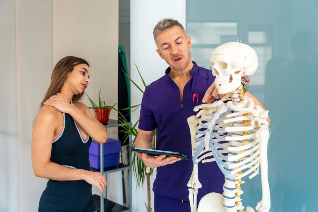
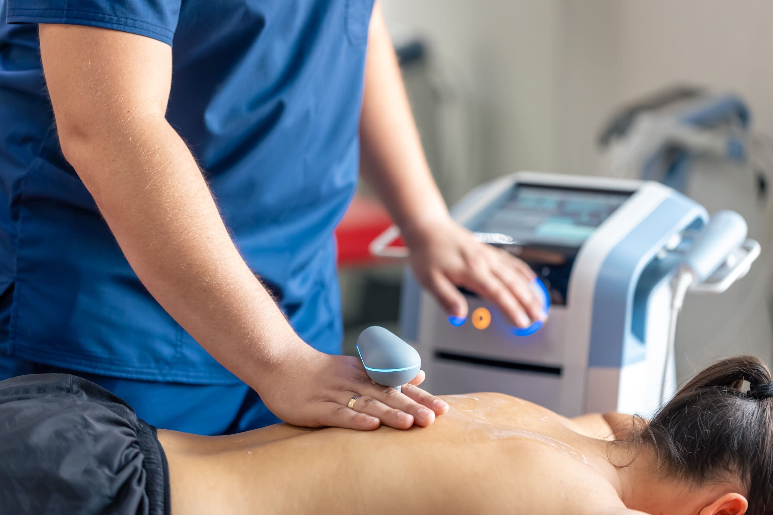
The Role of Integrative Chiropractic Care

This is where the principles of integrative chiropractic care fit seamlessly into the patient optimization plan. While functional medicine addresses the body’s biochemistry, chiropractic care focuses on its biomechanics and neurology. The two are inextricably linked.

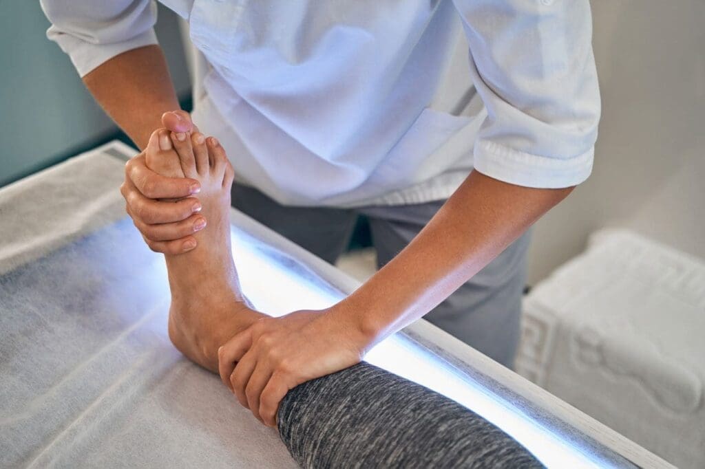
  • Restoring Biomechanical Function: A misaligned joint or dysfunctional movement pattern places abnormal stress on tissues. This chronic mechanical strain can perpetuate inflammation and create a “stuck” point that resists healing, even with biologics. Through precise spinal and extremity adjustments, we restore proper joint mechanics, unload compromised tissues, and create a better environment for regenerative cells to work.
  • Improving Neurological Input: Chiropractic adjustments have a profound effect on the nervous system. By stimulating mechanoreceptors in the joints and soft tissues, we can downregulate pain signals (nociception) and improve proprioception (the body’s sense of its position in space). This helps break the chronic pain cycles that often accompany degenerative conditions and can improve a patient’s tolerance for rehabilitative exercise.
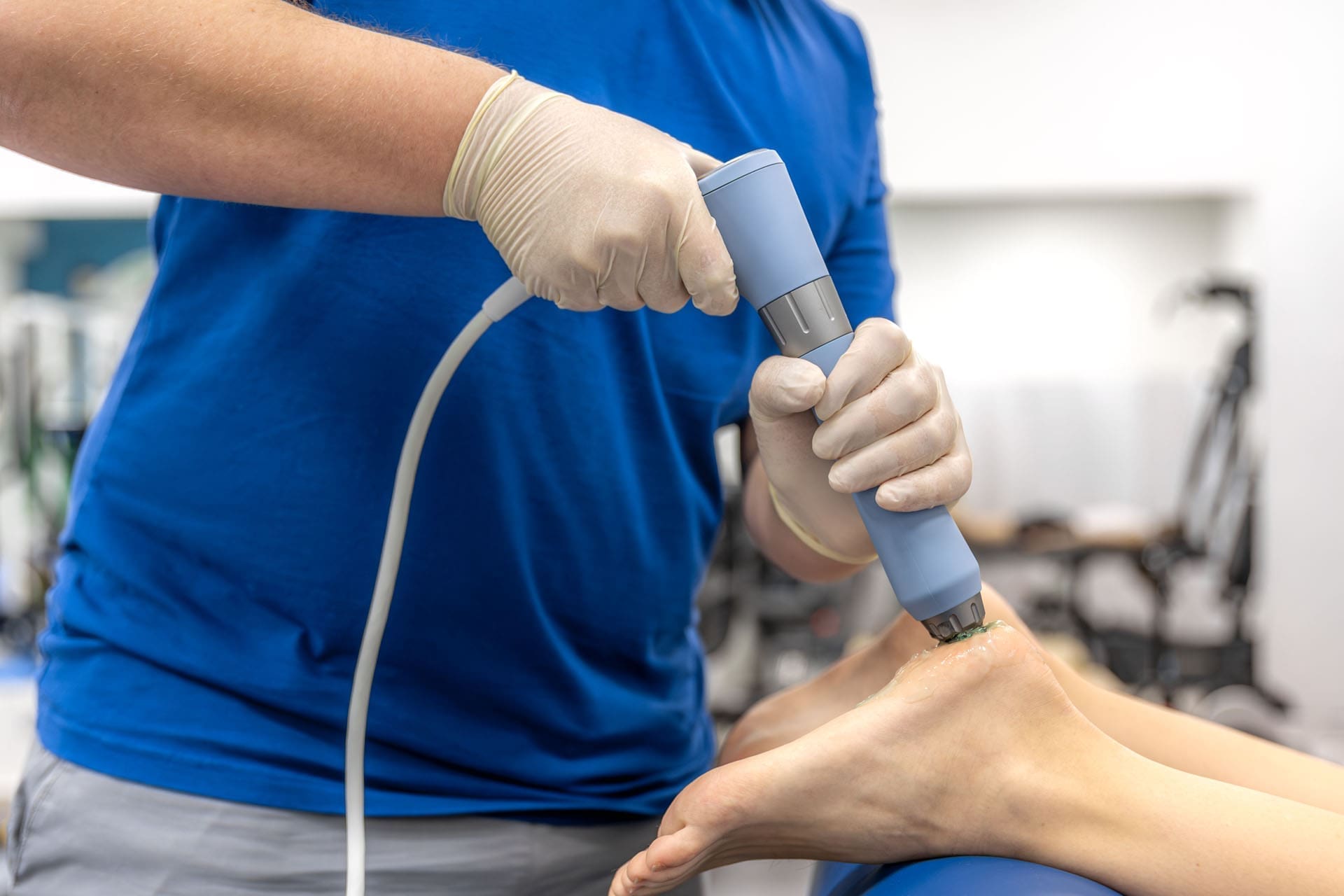
  • Enhancing Blood Flow and Fluid Dynamics: Proper movement is essential for pumping blood and lymphatic fluid, which deliver nutrients and remove waste products. Chiropractic care, combined with soft-tissue modalities, helps release restrictions and improve circulation in the target area, ensuring that regenerative therapies are delivered where they are needed most.

By integrating chiropractic adjustments, we are not just treating the site of injury; we are optimizing the entire kinetic chain and the neurological signaling that governs it. This ensures the patient’s body is mechanically and neurologically receptive to healing.

A Practical Approach to Pre-Procedure Assessment

So, how do we put all this into practice? It starts with a thorough assessment. If a patient’s recent medical records (within the last six months) are available and appear to be in good condition, extensive new testing may not be needed. However, I typically start with some simple point-of-care measurements.

Initial Screening:

  • Vitals: Height, weight, blood pressure, and waist circumference (a key indicator for metabolic syndrome).
  • Point-of-Care Labs: A fasting glucose and a lipid panel can quickly identify or rule out metabolic syndrome. Key markers are triglycerides and HDL cholesterol.
  • Further Labs (if indicated): Based on the initial screen and patient history, I might order a Hemoglobin A1c (to assess long-term blood sugar control), C-Reactive Protein (CRP, a marker of inflammation), or a renal function panel.

Screening Questionnaires:

Validated questionnaires are an efficient way to gather crucial information:

  • Lifestyle: Simple screens for tobacco/alcohol use, exercise, and diet.
  • Sleep: Questionnaires like the STOP-BANG can screen for sleep apnea risk.
  • Mental Health: Tools to assess for stress, anxiety, and depression (e.g., PHQ-9, GAD-7).

Creating an Optimization Plan

Based on this comprehensive assessment, I classify a patient’s metabolic risk as low, moderate, or high.

  • Low Risk: A patient with no signs of metabolic syndrome.
  • High Risk: A patient presenting with, for example, a Hemoglobin A1c of 11% and uncontrolled hypertension.

For a patient with moderate-to-high metabolic risk who is otherwise a good candidate for a biologic procedure (e.g., rotator cuff tendinopathy), this is the perfect opportunity to intervene. I present them with the information, we establish baseline markers, and we collaboratively set a timeline—often 8 to 12 weeks—to focus on optimization. We then repeat the key markers to track progress. This process of shared decision-making empowers the patient and dramatically increases their chances of a successful outcome.

The optimization “prescription” might include:

  • Specific Exercise Goals: “You need to achieve 150 minutes of brisk walking per week.”
  • Dietary Counseling: Providing clear guidelines or referring to a registered dietitian.
  • Sleep Hygiene Strategies.
  • Stress Mitigation Techniques: Recommending mindfulness apps, deep breathing exercises, or a referral for counseling.
  • Cessation Support: For tobacco and alcohol use.

This pre-habilitation period is an investment that pays dividends long after the procedure, fostering lifestyle changes that promote lifelong health. Thank you for joining me on this exploration of patient optimization.


References


SEO Tags: regenerative medicine, orthobiologics, patient optimization, lifestyle medicine, integrative chiropractic, platelet-rich plasma, PRP, stem cell therapy, metabolic health, anti-inflammatory diet, gut dysbiosis, sarcopenia, chronic inflammation, exercise physiology, sleep hygiene, stress management, functional medicine, Dr. Alex Jimenez, chiropractic care, holistic healing

Platelet-Rich Plasma PRP Therapy Guide for Recovery

Platelet-Rich Plasma PRP Therapy Guide for Recovery

Platelet-Rich Plasma PRP Therapy Guide for Recovery
Integrative Chiropractic Improves Movement and Health

Abstract

Welcome to this in-depth exploration of Platelet-Rich Plasma (PRP) therapy. My name is Dr. Alexander Jimenez, and in this educational post, we will journey together through the intricate world of regenerative medicine. We will unravel the complexities of PRP, moving beyond the surface-level understanding to explore the crucial details that determine its success. I will guide you through the latest findings from leading researchers, breaking down concepts like platelet dosing, the composition of the biologic product, and why not all PRP is created equal. We will discuss the physiological underpinnings of PRP, from the cellular level to its effects on tissues such as tendons and joints. A significant focus will be on the importance of achieving a specific therapeutic dose to elicit a healing response, particularly in conditions like osteoarthritis (OA) and soft tissue injuries. We will also examine how factors like patient age and the specific preparation system used can dramatically influence outcomes. Furthermore, I will explain how integrative chiropractic care plays a vital supportive role in this process, enhancing recovery and optimizing the body’s response to treatment. This post is designed to provide you with a comprehensive, evidence-based understanding of PRP therapy, empowering you to make informed decisions about your health.


As a clinician with a diverse background spanning chiropractic (DC), advanced practice nursing (APRN, FNP-BC), and functional medicine (CFMP, IFMCP), my goal is to bridge gaps across healthcare fields to provide a truly holistic and effective treatment model. My clinical experience, available at chiromed.com and detailed on my LinkedIn profile, has consistently shown me the power of combining advanced biologic treatments with foundational care. Let’s begin our journey into the science of PRP.

What Is a Platelet and Why Does It Matter?

To truly grasp the power of PRP, we have to go back to a fundamental concept from our early science education: what is a platelet? Many of us remember them as tiny components of our blood that help with clotting. But they are so much more than that.

Platelets are small, anucleated (meaning they lack a nucleus) cell fragments that are essentially little packets filled with a treasure trove of proteins. These proteins include powerful growth factors and cytokines, which are signaling molecules that orchestrate the body’s natural healing and repair processes.

  • Key Characteristics of Platelets:
    • They have a lifespan of about 7 to 10 days. This is a critical piece of information. When I advise patients to avoid anti-inflammatory medications like NSAIDs before a PRP procedure, it’s because these drugs can inhibit platelet function, and we need their full healing potential for the therapy to be effective.
    • A normal platelet count in the blood ranges from about 150,000 to 400,000 per microliter.
    • The FDA’s definition of PRP is simply a platelet concentration that is “above baseline.” This vague definition is partly why there is so much variability in the PRP products available today.

The core principle of PRP therapy is to concentrate these powerful healing cells and their associated growth factors and then deliver them with precision to an area of injury or degeneration. The goal is to amplify the body’s natural healing cascade, transforming a chronic, non-healing state into an active, acute healing phase.

The Problem of Variability in PRP Preparations

A significant challenge in the field of regenerative medicine is the immense variability among different PRP systems. This is a critical point that both patients and practitioners must understand. The idea that “PRP is PRP” is a dangerous oversimplification.

A compelling study by Jaewoo Pak and his colleagues highlighted this issue perfectly. They analyzed five different commercial PRP systems and found dramatic differences in both the final platelet concentration and the white blood cell (WBC) count in the final product (Pak et al., 2017).

I often show my patients a slide from a presentation by Dr. Gerben van de Meijden that drives this point home. It shows the blood of a single patient processed through four different systems. The resulting PRP products are all different colors—from light yellow to deep red—each representing a unique cellular makeup. This isn’t just an aesthetic difference; it signifies a profound variability in the biologic drug we are creating. The “dose” and “formulation” are completely different, which inevitably leads to different clinical outcomes.

The Evidence for PRP: A Growing Body of Research

Despite the variability, the evidence supporting PRP therapy, particularly for certain conditions, is robust and growing. When colleagues or patients ask about the evidence, I point out a fascinating fact: there are now more patients enrolled in high-quality clinical trials for PRP in knee osteoarthritis (OA) than for hyaluronic acid injections, a long-standing and widely accepted treatment.

This wealth of data, as highlighted in a meta-analysis by Meheux et al. (2016), generally shows that PRP therapy tends to outperform hyaluronic acid, especially for medium- to long-term pain relief and functional improvement. This suggests that PRP is not just a temporary fix but may have a more lasting biological effect.

How We Create Your Personalized PRP Treatment in Our Clinic

So, how do we go from a simple blood draw to a powerful healing injectate? Let me walk you through the process we use in our clinic, which is designed for precision and quality.

  1. Blood Draw: We begin by drawing a specific volume of your blood. This is not a one-size-fits-all step. The amount of blood we draw is a strategic decision based on the target dose we need to achieve. A larger blood volume allows us to harvest a greater total number of platelets.
  2. First Centrifugation: The blood is placed into a sterile, closed-system kit. This kit is then placed in a centrifuge, a machine that spins at high speeds. This first “hard spin” uses centrifugal force to separate the blood into its different components based on their density. The heavier red blood cells are forced to the bottom, the lighter plasma rises to the top, and a thin, precious layer forms in the middle. This is the “buffy coat.”
  3. Isolating the Buffy Coat: The buffy coat is where the magic is. It’s incredibly rich in platelets and white blood cells. The plasma above it, known as platelet-poor plasma (PPP), is carefully removed.
  4. Second Centrifugation & Concentration: We are then left with the buffy coat and a small amount of plasma. In some systems, a second, slower spin is used to further concentrate the platelets. The key is understanding exactly where the platelets reside within the tube. In the system I often use, about 85% of the platelets are concentrated within a tiny 2-millimeter layer. This allows us to create a high concentration of platelets in a very small, precise volume.

Understanding the specific mechanics of the system you use is paramount. It’s the only way to reliably create a therapeutic product and move away from guesswork.

The Critical Concept of PRP Dosing

I encourage my patients and colleagues to think of PRP not as a generic “procedure” but as a biologic drug. And like any drug, it has a dose-response relationship. There is a minimum dose—a therapeutic threshold—that must be reached to trigger a significant biological effect. If the dose is too low (subtherapeutic), the treatment is likely to fail.

So, what is the right clinical dose of PRP? This is the million-dollar question, and the answer is slowly being pieced together by dedicated researchers. The optimal dose likely varies by the type of tissue being treated (e.g., tendon vs. cartilage) and the specific pathology.

Dosing for Tendons and Soft Tissues

Early research in cell cultures provided the first clues. Studies have shown that a specific platelet concentration stimulates the proliferation of tenocytes (tendon cells). However, if the concentration became too high, it had an inhibitory effect, slowing cell growth. This established the concept of an optimal therapeutic window.

A landmark study from Dr. Peter Everts’ group provided crucial clinical insight (Everts et al., 2020). They analyzed numerous studies on soft-tissue applications of PRP and plotted the results on a graph. They found a clear dividing line.

  • Studies that used a total platelet dose of less than approximately 3.5 billion platelets were overwhelmingly negative; the treatment didn’t work.
  • Studies that used a dose above 3.5 billion platelets were overwhelmingly positive.

This gives us a tangible target. If a PRP system produces only 1.5 billion platelets, it’s likely to be subtherapeutic for many soft-tissue applications. We need to aim for a dose within that effective range to give our patients the best chance of success.

How Patient Age Impacts Dosing

Here is where personalized medicine becomes essential. We know that a patient’s biology changes with age. As we get older, our baseline platelet count may decrease, and the concentration of growth factors within those platelets may also decline. This means that to achieve the same therapeutic dose of 5 billion platelets, an older patient may require a larger initial blood draw than a younger patient. In my practice, I often err on the side of drawing a larger volume of blood from my older patients to ensure we can formulate a sufficiently potent biologic product to stimulate a robust healing response. We are still in the early days of understanding these nuances, but it’s a critical consideration for candidacy and treatment planning.

Dosing for Knee Osteoarthritis (OA)

The knee is perhaps the area where we have the most data on PRP dosing. A widely cited study, the RESTORE trial, published in JAMA, concluded that PRP was no better than a saline placebo for knee OA (Bennell et al., 2021). However, a critical look at the study’s methodology reveals the flaw. They used a low-dose PRP system that delivered only 1.6 billion platelets. Based on our dose-response curve, we now understand this was a subtherapeutic dose, so a negative result was predictable. This study, while well-executed, taught us a valuable lesson about the importance of dose.

In stark contrast, another major study from Dr. Van der Weegen’s group used a dose of 10 billion platelets (van der Weegen et al., 2016). In these patients, they observed not only significant improvements in pain and function but also MRI evidence that PRP may have slowed the progression of cartilage loss. This suggests a potential disease-modifying effect at the right dose.

So, for knee OA, the evidence points to a target dose of 5 to 10 billion platelets to achieve both symptom relief and potential structural benefits.

Beyond Platelets: The Role of White and Red Blood Cells

While platelets are the star players, they are not the only cells in the PRP formulation. We must also consider the other cellular components, particularly white blood cells (WBCs) and red blood cells (RBCs).

The two main types of WBCs we are concerned with are neutrophils and monocytes. They seem to have very different effects.

  • Neutrophils are highly pro-inflammatory. A PRP product rich in neutrophils (leukocyte-rich PRP, or LR-PRP) often causes a more intense post-injection inflammatory reaction, with greater pain and swelling. In some cases, this intense inflammatory signal may be desirable to “kick-start” healing in a very chronic, stagnant tissue. However, there are concerns that enzymes released by neutrophils could damage certain tissues, such as articular cartilage.
  • Monocytes are considered more “anabolic” or constructive. They play a key role in transitioning from the inflammatory phase to the proliferative, or rebuilding, phase of healing.

The debate between leukocyte-rich (LR-PRP) and leukocyte-poor (LP-PRP) is ongoing. Much of the European data suggests that for a condition like knee OA, there may not be a significant clinical difference in the long run. However, the initial patient experience is often different, with LP-PRP typically being better tolerated. In my practice, the choice between LR-PRP and LP-PRP is a clinical decision based on the specific tissue, the chronicity of the injury, and the individual patient.

The Integral Role of Chiropractic Care and Rehabilitation

A PRP injection is not a magic bullet; it is a catalyst. To fully realize its potential, it must be supported by a comprehensive treatment plan. This is where integrative chiropractic care becomes a cornerstone of success.

1. Precision and Guidance: The biologic product must be delivered to the exact site of injury. If you are treating a rotator cuff tear, the PRP must be placed directly into the defect within the tendon. If it’s injected into the surrounding bursal space, it cannot perform its function of forming a biological scaffold and stimulating repair. This is why ultrasound guidance is non-negotiable for these procedures. It ensures that this precious biologic drug gets to its target.

2. Optimizing Biomechanics: As a chiropractor, my focus is on function and structure. If a patient has knee OA due to poor hip mechanics or foot overpronation, simply injecting the knee only addresses the symptom. Chiropractic adjustments, soft tissue mobilization, and corrective exercises are crucial for addressing the underlying biomechanical faults that led to the joint breakdown in the first place. This creates a better environment for the PRP to work and helps prevent recurrence of the injury.

3. Guided Rehabilitation: The post-injection period is critical. PRP triggers an inflammatory and proliferative process that takes time. I tell my patients not to expect immediate results. The true benefits unfold over three to six months. The rehabilitation protocol must be tailored to this biological timeline.

  • Initial Rest Phase: Following the injection, a short period of relative rest allows the platelet clot to form and the initial inflammatory cascade to begin.
  • Protected Mobilization: We then gradually introduce a gentle range-of-motion exercise to prevent stiffness.
  • Progressive Loading: As the tissue begins to repair and remodel, we introduce progressive, controlled loading through specific exercises. This mechanical stimulation is essential for guiding the new collagen fibers to align properly, creating a strong, functional, and resilient tissue. This is a journey we guide the patient through, ensuring they do the right things at the right time to support the healing initiated by PRP.

Key Takeaways for Patients and Practitioners

My goal in this post is to emphasize that successful regenerative medicine requires a deep understanding of the product you deliver. We must move beyond generic labels and focus on the specifics.

  • Dose Matters: Think of PRP as a drug. A subtherapeutic dose will not work. We must aim for a specific dose tailored to the tissue and condition, with current evidence suggesting a target of >3.5 billion platelets for soft tissues and 5-10 billion platelets for knee OA.
  • Not All PRP Is Equal: The preparation system dictates the final product. Understand your system’s capabilities and limitations to ensure you can create a therapeutic dose.
  • It’s a Biological Process: Healing takes time. PRP initiates a cascade that unfolds over months. Patient education and managing expectations are key.
  • Integrative Care is Crucial: The best outcomes are achieved when PRP is combined with precision guidance, biomechanical correction, and a structured, biology-based rehabilitation program.

By embracing this evidence-based, detailed, and integrative approach, we can truly harness the remarkable healing potential of PRP and offer our patients lasting solutions for pain and dysfunction.


References

Bennell, K. L., Paterson, K. L., Metcalf, B. R., Duong, V., Emsley, R., Hinman, R. S., … & Harris, A. (2021). Effect of intra-articular platelet-rich plasma vs placebo on pain, function, and structural change in patients with knee osteoarthritis: The RESTORE randomized clinical trial. JAMA, 326(20), 2021-2030. https://doi.org/10.1001/jama.2021.19415

Everts, P., Onishi, K., Jayaram, P., Lana, J. F., & Mautner, K. (2020). Platelet-rich plasma: new performance understandings and therapeutic considerations in 2020. International Journal of Molecular Sciences, 21(20), 7794. https://doi.org/10.3390/ijms21207794

Meheux, C. J., McCulloch, P. C., Lintner, D. M., Varner, K. E., & Harris, J. D. (2016). Efficacy of intra-articular platelet-rich plasma injections in knee osteoarthritis: a systematic review. Arthroscopy: The Journal of Arthroscopic & Related Surgery, 32(3), 495-505. https://doi.org/10.1016/j.arthro.2015.08.005

Pak, J., Lee, J. H., & Lee, S. H. (2017). A novel protocol of platelet-rich plasma application for musculoskeletal medicine: a preliminary report. Journal of Prolotherapy, 9(1), e971-e979.

van der Weegen, W., van Drumpt, R., & de Sèze, P. B. (2016). The use of platelet rich plasma in knee osteoarthritis: a literature review and clinical interpretation. Bio-Orthopaedics Journal, 1(1).

PRP Therapy for Pain Relief, Healing, and Recovery

PRP Therapy for Pain Relief, Healing, and Recovery

PRP Therapy for Pain Relief, Healing, and Recovery

Abstract

Welcome to an in-depth exploration of Platelet-Rich Plasma (PRP), a cornerstone of modern regenerative medicine. We will delve into the very essence of platelets, exploring the powerful growth factors, cytokines, and other bioactive molecules they release. Drawing from the latest findings of leading researchers, we’ll examine how these components orchestrate the body’s natural healing and anti-inflammatory processes. I will explain the critical concept of PRP dosing, the importance of different platelet types, and how specific growth factors such as PDGF, TGF-β, and FGF contribute to tissue repair and regeneration. Finally, we’ll connect these principles to clinical practice, showing how integrative chiropractic care can be synergistically combined with PRP therapy to optimize patient outcomes, reduce pain, and restore function by addressing both the biochemical and biomechanical aspects of healing.


The Cellular Orchestra: Understanding the Power Within Your Blood

For over a decade, my clinical practice has been deeply rooted in the principles of functional and regenerative medicine. A recurring theme in helping patients overcome chronic pain and injury is the quest to harness the body’s innate ability to heal itself. One of the most powerful tools we have in this endeavor is Platelet-Rich Plasma (PRP). While many have heard the term, the true depth of what’s happening at a cellular level is often misunderstood. Today, I want to take you on a journey into the microscopic world of PRP, moving beyond the buzzwords to appreciate the sophisticated biological symphony it conducts.

When we talk about PRP, we’re focusing on concentrating a specific component of your blood: the platelets, also known as thrombocytes. While red blood cells and white blood cells play their own roles, it’s the platelets that act as the master conductors of tissue repair. The therapeutic magic of PRP lies in the wealth of bioactive components housed within these tiny cell fragments. These include:

  • Growth Factors: Proteins that signal cells to grow, proliferate, and differentiate.
  • Cytokines: Small proteins that are crucial in controlling the growth and activity of other immune system cells and blood cells.
  • Anti-inflammatory Molecules: Compounds that help modulate and resolve inflammation, which is a key barrier to healing.

The fundamental idea is that by concentrating these platelets, we can deliver a supraphysiological dose of these healing molecules directly to an injured area, amplifying the body’s natural repair signals and creating an optimal environment for regeneration.

Inside the Platelet: A Treasure Trove of Healing Granules

To truly grasp how PRP works, we need to look inside the platelet itself. Think of a platelet as a microscopic delivery vehicle packed with different types of cargo containers, or granules. The main therapeutic benefit we seek comes from the contents of these granules, which are released upon activation at the site of injury.

The three primary types of granules are:

  • Alpha Granules: These are the most abundant and arguably the most important for regeneration. Each platelet contains about 50 to 80 alpha granules, which are filled with a vast array of powerful growth factors. When a platelet is activated—for instance, by contact with exposed collagen in damaged tissue—it undergoes a process called degranulation, releasing growth factors into the surrounding environment. This is the primary event that initiates the healing cascade.
  • Dense Granules (or Delta Granules): These granules contain small molecules, including ADP, ATP, serotonin, and calcium. Their role is to amplify the healing response. They enhance platelet aggregation (helping form a stable scaffold) and vasoconstriction (controlling bleeding), and they also modulate the local immune response.
  • Lysosomes: These act as the cleanup crew. They release enzymes that help break down and remove damaged tissue and cellular debris from the injury site. This process, known as enzymatic debridement, clears the way for new, healthy tissue to form and also contributes to antimicrobial defense.

Recent research, including studies on platelet biology, highlights that over 280 distinct proteins are involved in this process, underscoring the incredible complexity of this “orchestra” of molecules working in concert (Golebiewska & Poole, 2015).

The Significance of Platelet Quality and “Dosing”

A critical concept that has emerged from evidence-based research is that not all PRP is created equal. The effectiveness of a treatment depends heavily on both the concentration and quality of the platelets. This is where the idea of PRP dosing becomes paramount. Simply put, the more functional platelets we can deliver to a target tissue, the greater the concentration of bioactive molecules we release, and potentially, the more robust the healing response.

Furthermore, we are learning about the importance of reticulated platelets. These are essentially “younger,” more robust platelets, recently released from the bone marrow (typically within the last 24-72 hours). They are denser and contain more alpha granules than their older counterparts. This means they are packed with more growth factors and have a greater regenerative potential. In our clinical processing, we use advanced techniques to preferentially harvest these denser, more potent reticulated platelets, ensuring that the PRP we inject is of the highest possible quality and biological activity.

The Key Players: A Closer Look at Essential Growth Factors

While hundreds of proteins are released, a few key growth factors are the primary drivers of the regenerative effects seen with PRP. Understanding their specific roles helps us appreciate why this therapy is so effective for a range of musculoskeletal conditions.

Platelet-Derived Growth Factor (PDGF)

As its name implies, PDGF was first discovered in platelets, but it’s also produced by other cells involved in healing. Think of PDGF as the “first responder” or the primary chemoattractant. It sends out a powerful signal that calls other crucial healing cells to the site of injury. Most importantly, it recruits Mesenchymal Stem Cells (MSCs)—the body’s own master repair cells—to the area. While PRP itself does not contain stem cells, it potently signals the body’s resident stem cells to migrate to the site, proliferate (make copies of themselves), and differentiate into the specific cell types needed for repair (e.g., cartilage, tendon, or bone cells). The PDGF-BB isoform is recognized as the most biologically active and is a major focus of current research for its potent role in initiating this cascade.

Transforming Growth Factor-Beta (TGF-β)

TGF-β is a master regulator of tissue regeneration. Its primary functions include:

  • Stimulating Collagen Synthesis: It powerfully enhances the production of type I collagen, the fundamental building block of tendons, ligaments, and the matrix of our bones and cartilage. This is essential for rebuilding the structural integrity of injured tissue.
  • Promoting Angiogenesis: the formation of new blood vessels. A healthy blood supply is critical for delivering oxygen and nutrients to the healing tissue and removing waste products.
  • Coordinating with Other Growth Factors: TGF-β works synergistically with PDGF to enhance endothelial cell proliferation and capillary sprouting, leading to a robust network of new blood vessels (neovascularization) that supports long-term tissue health.

The effect of PRP on angiogenesis is dose-dependent. Studies, such as those published in the Journal of Orthopedic Research, suggest that a platelet concentration of approximately 1.5 billion platelets per milliliter is required to achieve a significant pro-angiogenic effect (Mazzocca et al., 2012). This underscores the importance of precise processing and quantification to achieve optimal clinical results.

Fibroblast Growth Factor (FGF)

FGF is one of the most potent mitogens released by platelets, meaning it is exceptionally effective at stimulating cell division. It acts on a wide variety of cell types, including MSCs, chondrocytes (cartilage cells), and osteoblasts (bone-forming cells). By promoting the rapid proliferation of these essential repair cells, FGF accelerates tissue regeneration.

The Anti-Inflammatory Power of PRP

Chronic, unresolved inflammation is a major culprit behind persistent pain and tissue degradation, especially in conditions like osteoarthritis. While PRP initiates an acute, controlled inflammatory response to jumpstart healing, one of its most profound long-term benefits is its ability to modulate and resolve chronic inflammation.

This is achieved through several mechanisms:

  1. Leukocyte Interaction: The concentrated platelets in PRP interact with white blood cells (leukocytes) at the injury site. This interaction can shift the behavior of these immune cells, prompting them to release anti-inflammatory cytokines like IL-10 and IL-4, which actively suppress chronic inflammation.
  2. Macrophage Polarization: PRP can influence macrophage behavior, a type of white blood cell. It promotes a shift from the pro-inflammatory (M1) phenotype to an anti-inflammatory and pro-reparative (M2) phenotype. M2 macrophages are critical for cleaning up debris, resolving inflammation, and secreting factors that promote tissue remodeling and regeneration.
  3. Chemokine Secretion: Platelets release chemokines that not only recruit healing cells but also act as survival factors for monocytes, preventing their premature death and promoting their differentiation into beneficial M2 macrophages.

In essence, PRP acts as a biological “reset button,” transforming a chronically inflamed, degenerative environment into one that is actively anti-inflammatory and pro-regenerative.

Integrating Chiropractic Care for a Holistic Healing Approach

As a Doctor of Chiropractic, I view the body through both biomechanical and biochemical lenses. A successful outcome depends on addressing both the “parts” and the “system.” This is where the synergy between PRP therapy and integrative chiropractic care becomes so powerful.

Imagine a patient with chronic knee osteoarthritis. The PRP injection will address the biochemical problem inside the joint—reducing inflammation, signaling cartilage repair, and improving the quality of the synovial fluid. However, if the patient’s knee pain is also caused or exacerbated by poor biomechanics—such as a misaligned pelvis, muscle imbalances in the leg, or improper gait—the joint will remain under abnormal stress. This persistent mechanical strain can hinder the regenerative process initiated by the PRP and lead to a recurrence of symptoms.

This is why our integrative approach includes:

  • Chiropractic Adjustments: We perform precise adjustments to the spine and extremities to restore proper alignment and mobility of the joints. Correcting pelvic alignment, for example, can ensure that forces are distributed evenly through the knees, reducing abnormal wear and tear.
  • Myofascial Release and Soft Tissue Work: We address muscle imbalances, trigger points, and fascial restrictions that contribute to faulty movement patterns. This ensures that the muscles supporting the joint are functioning optimally.
  • Customized Rehabilitation: We design targeted exercise programs to strengthen weak muscles, stretch tight ones, and retrain proper movement patterns (neuromuscular re-education). This stabilizes the joint and protects it from future injury.

By combining the powerful biochemical signaling of PRP with the essential biomechanical corrections of chiropractic care, we create a comprehensive healing environment. The PRP rebuilds tissue from the inside out, while chiropractic care ensures the entire musculoskeletal system functions correctly to support and protect the newly regenerated tissue. In my clinical experience, this holistic strategy is the key to achieving durable, long-term pain relief and true restoration of function for my patients.


References

Integrative Care for Plantar Fasciitis With SoftWave

Integrative Care for Plantar Fasciitis With SoftWave

Integrative Care for Plantar Fasciitis With SoftWave

Abstract

In this educational post, I share my personal journey with severe plantar fasciitis and how SoftWave shockwave therapy transformed my recovery without injections or surgery. From there, we look at modern, evidence-based research on shockwave physics and mechanisms, compare true shockwave and radial pressure-wave devices, and explore patient outcome data from independent registries. I then outline how integrative chiropractic care fits into this model, including spinal and regional joint mechanics, fascial and neuromuscular coordination, anti-inflammatory nutrition, and return-to-sport protocols. You will learn why electrohydraulic shockwave is considered a gold standard, how it triggers angiogenesis and modulates inflammation, and how to implement it alongside biologics such as PRP in a clinically rational way. Throughout, I incorporate clinical observations from my practice to demonstrate real-world application and patient-centered workflows.

Why This Matters

I am Dr. Alexander Jimenez, DC, APRN, FNP-BC, CFMP, IFMCP, ATN, CCST. My clinical focus integrates chiropractic medicine, advanced practice nursing, functional medicine, sports science, and tissue rehabilitation. My mission is simple: deliver safe, effective, and science-driven care that helps patients return to life and sport with better long-term outcomes.

For many of my patients, chronic musculoskeletal pain feels like a maze of temporary relief and recurrent setbacks. New technologies can promise a lot but deliver unevenly. My goal in this post is to clarify the science, present real data, and show how integrative chiropractic care and SoftWave shockwave therapy can be combined to create a precise, reproducible plan of action.

From Pain To Practical Solutions: A Patient’s Plantar Fasciitis Story

Seven years ago, while working long hours on their feet as a wedding planner, a patient developed bilateral plantar fasciitis—a stubborn, burning heel pain worsened by standing, walking, and push-off mechanics. The standard recommendation the patient received was cortisone injections, which can blunt pain but may impair soft tissue healing and carry a risk of plantar fascia rupture with repeated use. The patient declined and assumed they would have to live with the pain.

A colleague introduced the patient to SoftWave shockwave therapy. Their first session was intense—older protocols often sought to “treat into pain”—but they felt relief the same day. The pain recurred within a few days, and the patient moved on. Then, two months later, the patient woke up and realized they hadn’t felt heel pain for days. They hadn’t stretched, changed footwear, or altered workload. The pain was just… gone. They stayed pain-free for nearly four years before needing another series. That experience ignited a commitment to evidence-based regenerative technologies that work with the body’s repair systems rather than against them.

What Is A Shockwave? The Physics That Matter

When you hear “shockwave,” think of a high-speed acoustic energy wave traveling faster than the speed of sound through tissue. In medicine, the waveform and speed are crucial:

  • True shockwave: A high-velocity acoustic wave that reaches supersonic speeds and produces a sharp pressure gradient at the leading edge. Electrohydraulic systems generate this wave inside the applicator and deliver it as a coherent, high-energy impulse.
  • Radial pressure wave: A lower-velocity pressure wave that disperses energy superficially and primarily creates mechanical perturbations near the skin surface.

Key physiological effects of true shockwave:

  • Cell membrane mechanotransduction: The rapid pressure shift causes a transient change in cell membrane permeability, initiating signaling cascades.
  • Angiogenesis: Upregulation of pro-angiogenic factors (e.g., VEGF) stimulates the formation of both small and large vessels, improving perfusion to hypoxic or degenerated tissues.
  • Inflammation modulation: Downregulation of NF-κB pathways and altered cytokine profiles can help shift chronic inflammatory states toward resolution.
  • Tissue remodeling: Stimulation of tenocytes, fibroblasts, and osteoblasts enhances matrix turnover, collagen alignment, and bone healing.
  • Analgesic neuromodulation: Changes in peripheral nociceptor sensitivity and local neuropeptide signaling can reduce pain over time without neurodestructive effects.

Why speed and waveform matter:

  • The steep pressure gradient of a true shockwave produces a shearing force at the cell membrane, which initiates the biological cascade (mechanotransduction).
  • Devices that produce sound waves that converge into a shockwave only outside the applicator may have a shorter effective burst and require precise targeting to avoid under- or overdosing tissue.
  • Electrohydraulic devices generate a shockwave within the applicator, providing a consistent, therapeutic energy profile that can reach deeper targets (often cited as up to approximately 12 cm, depending on tissue, coupling, and parameters).

Electrohydraulic Versus Electromagnetic/Piezoelectric: Practical Differences

There are several ways to generate shockwaves:

  • Electrohydraulic: Two electrodes create a spark in water, producing a cavitation bubble; the leading edge of the bubble is the shockwave. This is often considered a gold standard by clinical shockwave societies due to stable energy delivery and deeper penetration.
  • Electromagnetic: A magnetic coil accelerates a membrane or projectile; a shockwave forms as sound waves converge.
  • Piezoelectric: Arrays of crystals emit waves that focus and converge to create a shockwave in a focal zone.

Clinical implications:

  • Electrohydraulic systems typically deliver a full-energy shockwave at the applicator exit, reducing the “gap” between the device and the therapeutic zone.
  • Convergent systems (electromagnetic, piezoelectric) can be effective but may have narrow focal zones, shorter burst durations, and require meticulous targeting and tip selection.
  • The patient experience can differ: a broader energy distribution tends to be more comfortable, enabling higher-dose delivery without anesthesia.

True Shockwave Versus Radial Pressure Waves: Mechanisms And Outcomes

It is common to see radial devices marketed under “shockwave” branding. Mechanistically, radial devices:

  • Produce pressure waves at sub-shock speeds.
  • Disperse energy superficially, relying on microtrauma to induce reactive hyperemia and local neurogenic effects.
  • Can be uncomfortable, and their therapeutic depth is generally limited.

Where radial can be useful:

  • Short-term pain relief in superficial soft tissue conditions.
  • Adjunct to manual therapy for myofascial trigger points.
  • Situations requiring cost-effective, quick, superficial treatment.

Limitations:

  • The durability of results may degrade over months in chronic degenerative conditions, as suggested by independent patient-reported outcome data comparing radial and true shockwave approaches.

The Two-Wave Advantage: Focused And Broad Focus Delivery

With electrohydraulic systems, particularly those using parabolic reflectors, two effects can be achieved:

  • Primary focused wave: Laser-pointer-like focal energy reaching deeper tissue targets.
  • Broad focus column: Recovered energy from the expanding cavitation bubble is redistributed, creating a larger therapeutic zone.

Why this matters:

  • A larger treatment volume can address complex pain generators that are regionally interdependent (e.g., plantar fascia, intrinsic foot muscles, tibialis posterior, and soleus).
  • Faster treatment times due to broader coverage.
  • Reduced technician dependency because biofeedback cues and broader zones make precision less brittle.
  • Improved comfort, allowing higher energy without anesthesia.

Biofeedback: The Patient Guides The Map

One practical advantage of shockwave therapy is patient biofeedback during treatment:

  • Healthy tissue feels like gentle tapping.
  • Inflamed or damaged tissue feels sharply distinct, allowing clinicians to map hotspots and functional pain generators.
  • This reduces guesswork and allows real-time tailoring of energy density, pulse number, and coverage.

Sports Medicine Use Case: Rapid Return-To-Play

Consider an athlete with an adductor strain. Typical recovery for moderate strains is often 6–10 weeks, depending on grade and reinjury risk. Athletes treated with true shockwave combined with integrative rehab can sometimes return in 3–4 weeks when:

  • Deep perfusion is improved via angiogenesis and vasodilation.
  • Pain is modulated without masking protective reflexes.
  • Tissue remodeling is supported with eccentric loading, neuromuscular re-education, and fascia mobility during the subacute window.

Orthopedic Insights From Elite Sports

Orthopedic specialists serving NBA and NFL athletes frequently use shockwave for:

  • Bone edema
  • Slow-healing fractures
  • Post-operative nonunion risk
  • Soft tissue tendinopathies

These clinicians report greater patient tolerance with true shockwave devices that deliver comfortably, thereby allowing higher-energy dosing without anesthesia and consistent day-to-day clinic throughput. This aligns with the published literature demonstrating the utility of shockwaves in bone and tendon biology through angiogenesis and mechanotransductive signaling.

How Integrative Chiropractic Care Fits The Model

Shockwave therapy works best in an integrative framework that addresses mechanical, neurophysiological, metabolic, and behavioral dimensions. In my clinic, we approach musculoskeletal problems through the following pillars:

  • Biomechanics and joint mechanics:
    • Correct regional alignment: In plantar fasciitis, foot pronation, tibial internal rotation, and hip mechanics create downstream overload on the plantar fascia. Chiropractic adjustments to the subtalar joint, midfoot, and pelvis can redistribute load and normalize gait.
    • Improve segmental mobility: Thoracolumbar stiffness can degrade shock absorption; lumbar-pelvic adjustments support kinetic chain efficiency.
  • Fascial continuity and neuromuscular coordination:
    • Fascia connects the foot to the calf, hamstrings, and the lower back via myofascial chains. SoftWave improves perfusion and tissue pliability, while chiropractic soft-tissue work promotes gliding and hydration of the fascial planes.
    • Focused motor control drills retrain the intrinsic foot muscles, the tibialis posterior, and the hip abductors to prevent recurrence.
  • Load management and graded exposure:
    • Implement eccentric loading and isometric pain-modulating holds to enhance collagen remodeling in tendons under safe strain.
    • Use return-to-sport progressions based on pain response, strength benchmarks, and functional testing (e.g., single-leg hop, Y-balance).
  • Anti-inflammatory nutrition and recovery biology:
    • Encourage omega-3 fatty acids, polyphenols (curcumin, quercetin), vitamin D, magnesium, and targeted collagen peptides to support connective tissue health.
    • Address sleep architecture and stress modulation, which influence cytokine profiles and healing velocity.
  • Footwear, orthotics, and surface management:
    • Consider temporary orthotic support to unload the plantar fascia during high-volume training weeks.
    • Optimize shoe geometry (heel-to-toe drop, midfoot stiffness) to improve force distribution.

Clinical Observations From My Practice

Across multiple musculoskeletal conditions, I observe:

  • Immediate reductions in pain scores following shockwave, especially when combined with regional adjustments and fascial mobilization.
  • Durability improves when patients receive structured motor retraining and metabolic support.
  • In tendinopathies (patellar, Achilles, gluteal), pairing shockwave therapy with progressive eccentric loading and coaching on stride or jump mechanics yields superior outcomes compared with passive care alone.
  • For bone edema and stress reactions, true shockwave paired with relative rest, calcium/vitamin D, and careful return-to-impact criteria often accelerates readiness while reducing reinjury risk.

True Shockwave And Biologics: Rational Combinations

Shockwave interacts with biologic therapies thoughtfully:

  • Same-day approach:
    • Use shockwave to reduce pain and increase perfusion; perform PRP or cellular biologic injection afterward. Patients often tolerate injections better due to the immediate analgesic effects.
  • Pre-injection priming:
    • Deliver shockwave 2–3 days prior to biologic injection to enhance vascularity and cellular signaling, potentially improving biologic distribution and response.
  • Diagnostic reinforcement:
    • Use shockwave biofeedback to identify pain generators with a witness present; document patient responses to localize pathology and justify targeted biologic therapy.

Why each protocol makes sense:

  • Shockwave-induced vasodilation and angiogenic signaling can facilitate biologic dispersion and nutrient delivery.
  • Modulation of inflammation reduces hostile cytokine environments that can degrade biologic efficacy.
  • Analgesia without anesthesia preserves protective neuromuscular control, reducing the risk of overloading post-injection immediately.

Device Selection And Practical Considerations

If you are evaluating devices:

  • Prioritize true shockwave generation with consistent energy delivery and sufficient depth.
  • Evaluate energy distribution patterns: A broad focus column plus focus offers versatility for both diffuse and focal pathologies.
  • Consider patient comfort: Devices that deliver higher energy comfortably improve compliance and throughput.
  • Assess training and workflow: Systems that are biofeedback-guided and easy to delegate to trained staff can streamline care without compromising safety.

Clinic Workflow And Patient Experience

Shockwave sessions in my clinic typically last 10–15 minutes. We:

  • Conduct a focused functional exam and pain provocation tests.
  • Use biofeedback mapping to guide energy dosing.
  • Immediately follow with manual therapy, chiropractic adjustments, and targeted exercises to lock in gains.
  • Provide home care protocols: gentle mobility, isometric holds, footwear guidance, and nutrition.
  • Track outcomes using PROMs (pain scales, LEFS, VISA-A for Achilles, etc.) to quantify progress and justify care plans.

Cost And Access

Shockwave therapy is often a cash-based service, though some regions and payers may offer limited reimbursement for specific indications. In my experience, patients value:

  • Rapid relief
  • Non-invasive approach
  • Fewer injections
  • Evidence-backed tools

Transparent pricing, clear expectations, and outcome tracking build trust and encourage adherence.

Use Cases: Plantar Fasciitis, Tendinopathy, And Bone Edema

  • Plantar fasciitis:
    • Address degenerative microtears and fibrosis with shockwave-induced angiogenesis and matrix remodeling.
    • Integrate foot mechanics correction, calf flexibility, and eccentric loading.
    • Typical plan: 3–6 sessions over 4–8 weeks, tailored by pain and function.
  • Achilles and patellar tendinopathy:
    • Target mid-substance degeneration and enthesis changes using focused energy.
    • Pair with tempo-controlled eccentrics and isometric protocols (45–60 second holds for pain modulation).
  • Bone edema and slow-healing fractures:
    • Use shockwave to stimulate osteogenesis and microcirculation, while managing load and nutrition.
    • Coordinate with imaging and orthopedic oversight for safe progression.

Physiological Underpinnings: Why These Techniques Work

  • Mechanotransduction: Shockwave applies mechanical stress that cells convert into biochemical signals—a core regenerative cue in tendon and bone.
  • Angiogenesis and perfusion: Chronic pain is often a story of oxygen debt. Improving microvascular flow restores the redox balance, supports ATP production, and accelerates matrix repair.
  • Inflammation resolution: Many chronic conditions are stuck in an immune loop of low-grade inflammation. Shockwave can shift cytokine profiles toward resolution, enabling fibroblasts and tenocytes to remodel effectively.
  • Neuromodulation: Pain reduction without nerve ablation allows normal motor learning and movement quality to return during rehab—a safer, smarter approach.

Implementation Roadmap For Clinicians

  • Assess:
    • Identify pain generators via functional testing and biofeedback mapping.
    • Consider imaging for bone edema or complex tendinopathy.
  • Plan:
    • Select energy levels and pulse counts based on tissue depth and patient tolerance.
    • Integrate chiropractic adjustments, manual therapy, and exercise on the day of.
  • Deliver:
    • Keep sessions short, progressive, and guided by patient feedback.
    • Use a broad focus for regional complaints and focused energy for pinpoint lesions.
  • Reinforce:
    • Provide home protocols, nutrition plans, and sleep hygiene.
    • Schedule follow-ups and measure outcomes with standardized scales.
  • Combine when indicated:
    • Coordinate with PRP or other biologics using same-day or pre-injection priming.
    • Document responses to build a data-informed case, improving patient consent and expectations.

What Patients Feel And How We Adjust

  • Early sessions may include transient soreness at hot spots; this is expected and short-lived.
  • Most patients feel immediate lightness or reduced pain, making subsequent rehab easier.
  • If discomfort increases, we adjust energy or coverage while maintaining therapeutic intent.

Safety And Contraindications

  • Avoid direct application over malignancy, active infection, or open wounds.
  • Caution with pregnancy over the abdomen/low back.
  • Coordinate with cardiology for anticoagulated patients near deep vascular structures.
  • For acute complete tendon ruptures, shockwave is not a substitute for surgical repair; we use it post-operatively when cleared.

A Real-World Example: Return-To-Sport Strategy

For a competitive court-sport athlete with adductor strain:

  • Week 0–1:
    • Shockwave to the adductor complex and pelvic attachments.
    • Chiropractic adjustments for pubic symphysis and sacroiliac joints.
    • Isometric adductor holds, gentle mobility, and anti-inflammatory nutrition.
  • Week 2–3:
    • Progressive eccentric adductor loading and lateral movement drills.
    • Continue shockwave as symptoms guide; expand coverage to hip flexors and gluteal stabilizers.
    • Gait and stride mechanics corrections.
  • Week 3–4:
    • Introduce change-of-direction, acceleration/deceleration, and reactive drills.
    • Clearance based on pain-free function, strength symmetry, and field tests.

In practice, this integrative plan frequently compresses recovery timelines while preserving tissue integrity.

Where To Learn More

For clinicians and patients interested in exploring shockwave research, professional societies and independent registries offer robust resources. You will find clinical guidelines, mechanisms, and outcome data across tendon, bone, and soft tissue conditions.

Training, Support, And Workflow Integration

Clinics that implement shockwave successfully typically:

  • Receive hands-on training from experienced providers.
  • Adopt biofeedback mapping in daily workflow.
  • Leverage marketing assets to educate their community and referrers.
  • Track PROMs and share outcomes in peer forums to refine protocols.

Closing Thoughts: Bringing Science, Skill, And Compassion Together

Patients with chronic pain taught me a simple truth: the body is eager to heal when given the right signals. SoftWave shockwave therapy delivers those signals powerfully and safely. When combined with integrative chiropractic care, targeted exercise, and smart biologics, the results are often transformative. Patients do not need to rely solely on injections or tolerate long downtimes. They need a coherent, evidence-informed plan that respects physiology, mechanics, and lived experience.

If you are struggling with chronic pain or you are a clinician looking for a better way, consider a thoughtful shockwave-based approach integrated with chiropractic biomechanics, fascia care, and functional medicine. It is not just about feeling better today—it is about building durable health for tomorrow.


References

Ultrasound Therapy Benefits and Uses For The Musculoskeletal System

Find out how ultrasound therapy provides effective solutions for chronic musculoskeletal pain and joint issues.

Abstract

As a clinician with a diverse background in chiropractic, nursing, and functional medicine, I have dedicated my career to integrating the most advanced, evidence-based tools into patient care. This post explores the transformative role of musculoskeletal ultrasound (MSKUS), a powerful, real-time imaging modality that has revolutionized the way we diagnose and treat soft-tissue injuries. We will embark on a journey through the sonographic appearance of various tissues—tendons, muscles, cartilage, ligaments, and nerves—understanding their unique visual signatures. I will share insights from leading researchers and practical clinical pearls from my own practice on interpreting these images, including the critical concept of anisotropy. Furthermore, we will delve into proper probe handling techniques for both diagnostic and procedural applications, emphasizing methods that set clinicians up for success. Finally, I will explain how these advanced diagnostic capabilities integrate with a holistic, integrative chiropractic approach, enabling more precise, effective, and patient-centered treatment plans that support true healing.


Understanding the Language of Ultrasound: Echogenicity Explained

In my practice, I often refer to musculoskeletal ultrasound as a “glorified flashlight” that allows us to peer directly into the body’s anatomy in real time. But to understand what we’re seeing, we must first learn its language. The fundamental concept is echogenicity, which describes how tissues reflect ultrasound waves.

  • Hyperechoic: Tissues that appear bright white on the screen. These structures, like bone, are dense and reflect most ultrasound waves to the probe.
  • Hypochoic: Tissues that appear dark gray. These structures, like muscle or fluid, absorb more ultrasound waves and reflect fewer.
  • Anechoic: Tissues that appear completely black. These are typically fluid-filled structures, such as cysts or bursae, that transmit almost all sound waves.
  • Isoechoic: Tissues that have a similar brightness or echotexture to adjacent structures.

Pattern recognition is the cornerstone of interpreting ultrasound images. Each tissue type has an expected appearance, and deviations from this norm can signal pathology.

Sonographic Signatures of Key Musculoskeletal Tissues

Let’s explore what healthy tissues look like under the lens of an ultrasound probe.

Tendons: The Body’s Strong Cords

Tendons are the strong, fibrous cords that connect muscle to bone. On ultrasound, a healthy tendon has a classic appearance: it’s hyperechoic (bright) and displays a distinct fibrillar pattern—think of it as a tightly packed bundle of cables or parallel stripes.

For example, when we look at the patellar tendon in a long-axis view (aligned with the tendon), we expect to see a bright, organized, striped pattern. Beneath it, we can identify other structures, such as the infrapatellar fat pad (which has a more wavy, less organized appearance) and the hyperechoic surfaces of the patella and tibia. Recognizing this norma, fibrillar architecture is crucial because when a tendon is injured (tendinosis or a tear), it loses this organization, thickens, and appears more hypoechoic (darker).

Muscles: The Engines of Movement

Muscle tissue presents a more complex, mixed-echogenicity pattern. It is generally hypoechoic compared to the bright white of bone. However, within the muscle belly, you’ll see hyperechoic strands of connective tissue, known as the perimysium, which encase the muscle fascicles. This gives healthy muscle a “marbled” or “feathery” appearance.

When viewing a muscle like the bicep or deltoid over the humerus, you can see the dark muscle tissue contrasted against the bright cortical line of the bone. You can even appreciate its structure, tapering towards its tendinous insertion. This visual information helps us identify muscle strains, tears, or atrophy.

Cartilage: Smooth Surfaces and Tough Cushions

Cartilage is a critical tissue, and ultrasound helps us differentiate between its two main types:

  • Hyaline Cartilage: This is the smooth, glassy cartilage that covers the ends of bones within a joint, allowing for low-friction movement. On ultrasound, it appears as a distinct, thin, hypoechoic (dark) line sitting directly on the bright, hyperechoic bone surface. A great example is viewing the posterior aspect of the humeral head in the shoulder joint.
  • Fibrocartilage: This is a tougher, more fibrous type of cartilage found in structures like the meniscus of the knee or the labrum of the shoulder and hip. Unlike hyaline cartilage, fibrocartilage is hyperechoic (brighter) and has a more triangular or wedge-shaped appearance. On the shoulder, you can clearly distinguish the bright, triangular labrum from the dark, linear hyaline cartilage on the humeral head.

Ligaments: The Stabilizers

Ligaments, which connect bone to bone, look very similar to tendons on ultrasound. They are also hyperechoic and have a fibrillar, striated pattern. The key difference is that ligaments are typically more compact and densely packed than tendons.

The true power of ultrasound in evaluating ligaments comes from its real-time, dynamic capabilities. The best way to confirm you are looking at a ligament is to trace it from one bony attachment to another. If it originates from or inserts into a muscle, it’s a tendon. With ligaments such as the Medial Collateral Ligament (MCL) of the knee, we can perform a stress test under direct visualization. By applying a valgus force to the knee, we can watch the ligament in real time to see if there is any “gapping” or separation of its fibers.

A report might read: “The linear probe was placed over the medial aspect of the knee, and the MCL was visualized in a long-axis view. Upon real-time valgus stress, there was observable gapping of the mid-substance fibers with surrounding hypoechoic fluid, consistent with a grade 2 sprain.” This level of detail is impossible with a static MRI.

Nerves: The Body’s Electrical Wiring

Nerves have a unique and fascinating appearance on ultrasound, often described as a honeycomb” in short-axis (cross-section) view. This pattern is created by the hypochoic nerve fascicles (the bundles of nerve fibers) surrounded by the hyperechoic epineurium (the connective tissue sheath).

In a long-axis view, the nerve can look like a bundle of parallel “railroad tracks,” though this view is often less distinct than the honeycomb cross-section. A clinical pearl I share with my students is that nerves are often easier to spot when you scan. The distinct honeycomb pattern moves through the surrounding tissue, catching your eye more readily than the linear patterns of tendons or muscles. The carpal tunnel is the classic location to visualize this, as the median nerve’s honeycomb structure stands out clearly against the adjacent flexor tendons in the forearm.


The Challenge of Anisotropy: A Critical Pitfall to Avoid

One of the most important concepts in MSKUS is anisotropy. This phenomenon occurs when the ultrasound beam is not perfectly perpendicular (at a 90-degree angle) to the structure being imaged, particularly tendons and ligaments. When the beam hits the tissue at an angle, the sound waves are reflected away from the probe instead of back to it. This lack of returning signal causes the normally bright, hyperechoic tissue to appear artifactually hypochoic, or dark.

Why is this so critical? Because a tendon tear also appears as a hypoechoic defect. Anisotropy can mimic pathology, leading to a false-positive diagnosis.

Here’s how we differentiate:

  1. Prove the Pathology: If you see a dark spot in a tendon, like the supraspinatus tendon at its insertion on the humerus, you must prove it’s real.
  2. Toggle the Probe: Carefully “heel-toe” or “toggle” the probe to ensure you are perfectly perpendicular to the tendon fibers at that exact spot.
  3. Observe the Change: If the dark spot disappears and brightens when you adjust the probe angle, it indicates anisotropy. If the dark spot remains dark no matter how you angle the probe, it is more likely to be true pathology, such as tendinosis or a tear.

In my practice, I live by the mantra taught in orthopedic surgery: “One view is no view.” I always confirm a suspected finding from multiple angles, in both long and short-axis views, and correlate it with a dynamic assessment and the patient’s physical exam. This meticulous approach is what separates a novice from an expert operator and ensures diagnostic accuracy.

Mastering the Tool: Proper Probe Handling Techniques

Ultrasound is operator-dependent. Your skill in handling the probe directly impacts the quality of your images and the accuracy of your diagnosis.

The Tripod Grip for Diagnostic Scanning

For diagnostic imaging, stability and fine control are paramount. The “death grip,” where you wrap your whole hand around the probe, is unstable and limits fine motor control. Instead, we use the tripod technique.

  • Hold the probe like a pencil, using your thumb and index finger for control.
  • Brace your remaining fingers (pinky, ring, and/or middle finger) on the patient’s skin.
  • This creates a stable base, allowing subtle, precise movements such as sliding, toggling (heel-toe), and rotating to remain perpendicular to curved structures and eliminate anisotropy.

Your hand should be in contact with the patient. This is a more connected, controlled experience that allows you to feel the anatomy as you visualize it.

Modifying the Grip for Procedural Guidance

When performing an ultrasound-guided injection, the grip must change. Holding the probe with your fingers wrapped around it can physically block your needle’s path. For this reason, I advocate for holding the probe by its edges, which keeps your fingers clear of the sterile field and the needle’s intended path.

  • In-Plane Technique: For this approach, in which the needle is inserted parallel to the probe’s long axis and visualized along its entire length, a pencil-like grip is often effective.
  • Out-of-Plane Technique: In this approach, where the needle is inserted perpendicular to the probe and appears as a bright dot in cross-section, holding the probe by its edges provides the necessary space.

The key is to be facile, comfortable moving the probe in different ways for different tasks. Pre-planning your procedure is essential. My protocol is simple:

  1. Find the Target: Use your scanning skills to locate the exact anatomical target.
  2. Stay Perpendicular: Position the probe directly over the target, perpendicular to the skin. This simplifies your needle trajectory.
  3. Bring Tip to Target: Once you have a clear, stable view of your target, you can confidently guide your needle tip precisely where it needs to go.

This methodical approach minimizes “searching” for the needle or the target, making procedures faster, safer, and more successful.

Integrative Chiropractic Care and Ultrasound Synergy

So, how does this high-tech imaging fit into a chiropractic and functional medicine framework? Perfectly.

At our clinic, we don’t just treat symptoms; we seek to understand and correct the underlying biomechanical and physiological dysfunction. MSKUS is an invaluable tool in this process.

  • Precision Diagnosis: Before I perform a chiropractic adjustment or recommend a course of rehabilitative exercise, I want to know exactly what tissue is injured. Is that shoulder pain from a rotator cuff tear, biceps tendinopathy, or bursitis? Ultrasound tells me instantly, allowing me to tailor my treatment. For instance, if I identify a partial tear in the supraspinatus tendon, I can modify my spinal and extremity adjustments to avoid stressing the injured tissue and instead focus on improving scapular mechanics to offload the tendon.
  • Guiding Soft Tissue Therapies: Many of our treatments involve soft-tissue mobilization, such as Active Release Technique (ART) or the Graston Technique. Ultrasound allows me to visualize fibrotic adhesions or scar tissue and specifically target these areas, making the treatment more efficient and effective.
  • Monitoring Healing: Ultrasound provides objective evidence of tissue healing. We can track the reduction of inflammation, the reorganization of collagen fibers in a healing tendon, or the decrease in fluid within a bursa over time. This helps us advance the patient’s rehabilitation protocol based on actual tissue physiology rather than just subjective pain reports.
  • Patient Education: Showing a patient a real-time image of their injury is incredibly powerful. When they can see the inflamed bursa or the tear in their tendon, it enhances their understanding and improves their adherence to the treatment plan. It transforms the abstract concept of their injury into something tangible.

Ultimately, musculoskeletal ultrasound elevates the practice of integrative chiropractic care. It bridges the gap between a physical exam and a definitive diagnosis, allowing a level of precision previously unattainable in clinical settings. It helps us create highly specific, evidence-based treatment plans that address the root cause of a patient’s pain and dysfunction, accelerating their path back to optimal health and function.

As of May 2nd, 2026, the technology continues to evolve, but its core value remains: it is a safe, dynamic, and profoundly insightful tool that, in the hands of a skilled operator, can truly transform patient outcomes.


References

Jacobson, J. A. (2017). Fundamentals of Musculoskeletal Ultrasound (3rd ed.). Elsevier.

McNally, E. G. (2014). Practical Musculoskeletal Ultrasound (2nd ed.). Elsevier.

The Ultrasound Site. (n.d.). Musculoskeletal Ultrasound. Retrieved from https://www.theultrasoundsite.co.uk/

Ultrasound For Movement Disorders. (n.d.). MSK Resources. Retrieved from https://www.ultrasoundformovementdisorders.com/


SEO Tags: Musculoskeletal Ultrasound, MSKUS, Integrative Chiropractic, Dr. Alexander Jimenez, Echogenicity, Anisotropy, Tendinopathy, Ligament Sprain, Nerve Entrapment, Ultrasound-Guided Injections, Functional Medicine, Chiropractic Care, Sports Medicine, Diagnostic Imaging, Soft Tissue Injury, El Paso Chiropractor, Probe Handling

Gut Health and Hormone Balance Treatment

Gut Health and Hormone Balance Treatment

Gut Health and Hormone Balance Treatment

Abstract

I am Dr. Alexander Jimenez, DC, APRN, FNP-BC, CFMP, IFMCP, ATN, CCST. In this educational post, I guide you through the science and practice of optimizing hormones by treating the gut–liver–hormone axis and reinforcing micronutrient and mitochondrial foundations. I explain how dysbiosis, intestinal permeability, and microbial enzymes like beta-glucuronidase reshape estrogen metabolism and influence conditions such as PCOS, endometriosis, and autoimmunity, and how lipopolysaccharide (LPS) and short-chain fatty acids (SCFAs) affect insulin sensitivity, mood, and inflammation. I translate current research on vitamin D, K2, iodine, selenium, methylated B vitamins, DIM, and shilajit into clinic-ready protocols, and I show where integrative chiropractic care fits by supporting vagal tone, motility, neuromusculoskeletal dynamics, and autonomic balance. You will find practical frameworks, dosing concepts, lab-monitoring advice, and rationale for each intervention, with citations to leading researchers.


Why Hormones Are Microbiome-Dependent: The Gut–Liver–Hormone Axis

When I first connected hormone symptoms to gut physiology, I saw a pattern: many “hormone” problems began as microbiome and barrier problems. The gut microbiome—a complex community of bacteria, viruses, fungi, and archaea—regulates digestion, immune tolerance, barrier integrity, and the enterohepatic circulation that clears estrogens. From the earliest studies linking metabolic endotoxemia to insulin resistance, it has become clear that LPS-driven inflammation can disrupt cardiometabolic and reproductive health (Cani et al., 2007).

  • When the microbiome is balanced, commensals generate SCFAs (notably butyrate) that nourish colonocytes, tighten junctions, and reduce inflammatory signaling.
  • When dysbiosis develops, beta-glucuronidase-producing taxa expand, and LPS permeates, amplifying NF-κB cytokine cascades that alter hormone receptors, hepatic detoxification, and insulin signaling (Fasano, 2012; Slyepchenko et al., 2017).

Clinically, if you manage estrogen symptoms, insulin resistance, or autoimmune patterns, you are managing the microbiome—whether you realize it or not.


Dysbiosis and Leaky Gut Explained: Distinct Problems that Reinforce Each Other

Two related but distinct issues commonly coexist:

  • Dysbiosis: A shift away from beneficial microbes, with loss of diversity and expansion of pathobionts. Consequences include increased LPS, altered bile acid signaling, and elevated beta-glucuronidase.
  • Leaky gut (increased intestinal permeability): Disruption of tight junction proteins (occludin, claudins, ZO-1) allows antigens and endotoxins to enter circulation, thereby increasing systemic inflammation and immune activation (Fasano, 2012).

Why that matters for hormones:

  • LPS activates TLR4–NF-κB, increasing TNF-α, IL-1β, and IL-6—cytokines that reduce insulin signaling and alter steroid hormone receptor function (Cani et al., 2007).
  • Permeability increases immune load and oxidative stress, thereby consuming methyl donors and glutathione needed for safe phase II detox (methylation, glucuronidation, sulfation) of estrogens.

I screen for these drivers whenever patients report PMS, heavy cycles, PCOS features, endometriosis pain, acne or hair loss, mood changes, fatigue, or autoimmune flares. Correcting the gut often increases the safety and efficacy of hormone therapy.


Estrogen Metabolism 101: Enterohepatic Circulation and the Estrobolome

The liver metabolizes estrogens via phase I hydroxylation (CYP1A1, CYP1B1) and phase II conjugation (COMT methylation, glucuronidation, sulfation). Conjugated metabolites pass into bile and should be excreted. In dysbiosis, microbial beta-glucuronidase deconjugates these estrogens, promoting reabsorption and recirculation—the biochemical basis of “estrogen dominance,” even with careful dosing (Plottel & Blaser, 2011).

  • 2-hydroxylation generally produces less proliferative metabolites.
  • 4- and 16α-hydroxylation yield more proliferative or potentially genotoxic metabolites if methylation and conjugation are suboptimal.

In complex cases or when there is a family history of estrogen-dependent cancers, I consider urinary metabolite testing to map pathways and guide targeted support.


PCOS, Endometriosis, and Autoimmunity: What the Microbiome Adds

Recent studies sharpen the microbiome’s role:

  • PCOS: Dysbiosis with fewer SCFA producers and higher LPS correlates with insulin resistance, hyperandrogenism, and impaired GLP-1 signaling (Lindheim et al., 2017; Qi et al., 2019). Restoring butyrate producers improves metabolic tone.
  • Endometriosis: Altered microbiota, increased permeability, and immune activation correlate with symptom severity. Increased beta-glucuronidase raises estrogen recirculation that can exacerbate lesions and pain (Chen et al., 2017; Jiang et al., 2017).
  • Autoimmunity: Barrier dysfunction and loss of tolerogenic species permit pathobiont translocation and molecular mimicry, priming autoimmune activity (Manfredo Vieira et al., 2018).

Clinical translation: Addressing the gut can reduce hormone dosing requirements, expand the therapeutic window, and stabilize mood, sleep, and metabolism.


The Simple Question with Big Impact: Are You Pooping Daily?

I ask every patient: “Do you have a daily bowel movement?”

  • Estrogen metabolites exit via bile and stool. Constipation increases residence time, giving beta-glucuronidase more opportunity to deconjugate and recirculate estrogens.
  • Correcting bowel habits is a core risk-reduction strategy for estrogen-driven conditions.

Practical steps I use:

  • Increase hydration and electrolytes.
  • Ramp fiber to 25–35 g/day; add PHGG (partially hydrolyzed guar gum) 4–6 g/day for low-bloat prebiotic support.
  • Add magnesium glycinate or citrate at night for stool regularity and sleep.
  • Encourage postprandial walks and vagal toning (slow exhale breathing, humming).

A 3-by-3 Framework for Gut Repair: Remove, Replace, Repair

To keep things doable, I use a 3-by-3 approach:

  1. Remove/Reduce Irritants
  • Clean up the diet: favor whole foods; limit alcohol, ultra-processed items, added sugars; consider a gluten-light or gluten-free trial for sensitive individuals.
  • Medication review: minimize NSAIDs and PPI overuse when clinically safe.
  • Stress load: hard-wire breath work, walks, and sleep hygiene.
  1. Replace and Restore
  • Fiber and prebiotics: 25–35 g/day total fiber; add PHGG for gentle SCFA support.
  • Probiotics: multi-strain Lactobacillus and Bifidobacterium blends (e.g., L. rhamnosus GG, B. lactis) for barrier and immune balance.
  • Digestive support: bitters and meal hygiene for hypochlorhydria/slow motility; phosphatidylcholine and balanced fats for bile flow.
  1. Repair and Rebalance
  • Barrier repair: L-glutamine 5 g/day, zinc carnosine, N-acetyl-D-glucosamine, omega-3s as indicated.
  • Inflammation control: Berberine for dysbiosis-associated endotoxemia; curcumin and quercetin for NF-κB calming.
  • Lifestyle anchors: 150 minutes/week activity; 10-minute post-meal walks; consistent 7–9 hours of sleep.

Why this approach works:

  • Prebiotics increase SCFAs, reinforce tight junctions, and support T-regs via HDAC inhibition.
  • Probiotics competitively inhibit pathobionts, reduce beta-glucuronidase activity, and enhance mucosal IgA.
  • L-glutamine fuels enterocytes and accelerates barrier recovery.
  • Berberine improves the microbial balance and activates AMPK to improve insulin sensitivity.

Nutrient Foundations for Receptor-Level Hormone Action: D, K2, A, Magnesium, Iodine, Selenium, and Methylation

I frequently see patients with robust serum hormones but poor tissue effects. The missing link is often receptor signaling, cofactors, and membranes.

  • Vitamin D3 behaves like a secosteroid hormone that modulates transcription through the VDR. Low vitamin D is associated with all-cause and cardiovascular mortality and can blunt androgen signaling even when total testosterone appears normal (Pilz et al., 2011; Holick, 2017).
  • Magnesium is a cofactor for D activation (25- and 1α-hydroxylases); deficiency dampens VDR signaling (Rosanoff et al., 2016).
  • Vitamin K2 directs calcium into bone and away from soft tissues by activating matrix Gla protein and osteocalcin; it complements D to protect vessels and build bone (Schurgers & Vermeer, 2000; Beulens et al., 2013).
  • Vitamin A supports epithelial integrity, immune balance, and nuclear receptor synergy with vitamin D.

I often use an ADK formula (D3 with K2 and A) alongside magnesium to safely improve receptor-mediated effects, while monitoring 25(OH)D, calcium, and PTH (Rosen et al., 2012).

Thyroid resilience: iodine and selenium synergy

  • Iodine is essential for T4/T3 synthesis, but safe utilization depends on selenium-dependent enzymes (glutathione peroxidases, thioredoxin reductases) to quench the H2O2 generated during iodide organification (Ventura et al., 2017).
  • Inadequate selenium increases oxidative stress at the thyroid, raising the risk of autoimmunity when iodine intake rises (Gartner & Gasnier, 2003).
  • I pair iodine (200–400 mcg) with selenium (100–200 mcg selenomethionine) and often zinc (10–30 mg), titrated to labs and symptoms (Zimmermann & Boelaert, 2015).

Methylation for estrogen safety

  • Methylated B vitaminsmethylfolate and methylcobalamin—support COMT-mediated methylation of catechol estrogens, reducing genotoxic stress and stabilizing phase II clearance.

These micronutrients are the bedrock that allows hormones to “dock” and trigger healthy cellular responses.


DIM and Estrogen Metabolites: Steering Toward Safer Pathways

Diindolylmethane (DIM) shifts estrogen metabolism toward 2-hydroxylation and away from 16α- and 4-hydroxylation pathways associated with proliferative and genotoxic risk (Zeligs et al., 2006; Reed et al., 2006). Preclinical studies suggest that DIM may also upregulate BRCA1 signaling and promote apoptosis in cancer cell lines (Fan et al., 2009; Li et al., 2010).

How I apply it:

  • Women with estrogen-dominant symptoms or unfavorable metabolite profiles: 150–300 mg/day, adjusted to labs and tolerance.
  • Men with prostate risk or aromatization-driven symptoms: 300–600 mg/day, personalized.
  • I pair DIM with omega-3s, iodine/selenium, and fiber/probiotics to support the entire estrobolome–liver–stool axis.

Rationale: By changing metabolite balance and supporting conjugation, DIM decreases receptor overstimulation and DNA-adduct risk while improving symptom stability.


Shilajit for Free Testosterone and Mitochondrial Support

Some patients—particularly young males—present with high total testosterone but low free testosterone and low vitality. Shilajit, a purified, fulvic-acid–rich resin, has randomized data showing increases in total (~31%), free (~51%), and DHT (~37%) over ~90 days at 250 mg twice daily (Pandit et al., 2016). Mechanisms likely include improved mitochondrial function, nutrient transport, and hypothalamic–pituitary–gonadal signaling.

How I use it:

  • In those seeking endogenous support without exogenous hormones, I combine shilajit with vitamin D, magnesium, zinc, B12, and iodine/selenium when indicated, then track changes in free T, SHBG, energy, and body composition.

Why this works: Enhancing mitochondrial ATP and cofactor availability raises tissue responsiveness; changes in binding dynamics can increase the bioactive fraction without pushing total testosterone to excessive levels.


Vitamin D as a Systemic Modulator: Barrier, Immunity, and Receptors

I routinely optimize vitamin D because it acts at the intersection of immunity, barrier integrity, and endocrine signaling. Observational data tie suboptimal 25(OH)D to higher risks across diseases (Bouillon et al., 2019). Mechanistically, D supports tight junction proteins, cathelicidin, and endocrine receptor sensitivity. Clinically, many patients feel “stuck” until D is restored to an optimal range; I often target 60–80 ng/mL with appropriate monitoring to avoid hypercalcemia (Holick, 2017; Rosen et al., 2012).


Integrative Chiropractic Care: The Neuroimmune–Endocrine Interface

As a chiropractor and nurse practitioner, I see daily how autonomic balance, fascial mobility, and pain modulation determine whether patients can absorb nutrients, move consistently, and sleep well—foundations for endocrine success.

  • Vagal tone and motility: Gentle spinal and cervical adjustments can influence autonomic balance, improving gut motility, secretory IgA, and anti-inflammatory vagal pathways. Patients with low vagal tone present with constipation, bloating, and poor stress resilience.
  • Fascia and diaphragm: Thoracolumbar fascial restrictions and diaphragmatic stiffness impair breathing mechanics and lymphatic flow, promoting sympathetic overdrive. Mobility restores circulation and reduces pain.
  • Pain reduction without NSAIDs: Lowering nociception decreases cortisol and protects the mucosa from NSAID-induced permeability.
  • Behavioral activation: When pain decreases, patients walk, train, and sleep—activities that increase SCFAs, improve insulin sensitivity, and stabilize mood.

These neurophysiologic effects align with published observations on autonomic modulation and musculoskeletal care (Pickar, 2002; Lehman et al., 2012) and help nutrition and endocrine strategies “stick” in daily life.

For examples of how we operationalize this, see my resources at Chiromed and my professional updates on LinkedIn.


A Phased, Clinic-Ready Protocol for Gut and Hormone Optimization

I layer care to build momentum and safety.

Phase 1: Stabilize and Build Trust (Weeks 0–4)

  • Ensure daily bowel movements; add PHGG, hydration, and magnesium as needed.
  • Start a multi-strain probiotic (Lactobacillus + Bifidobacterium).
  • Begin vitamin D3 with K2 and magnesium; consider ADK formulations.
  • Introduce walks after meals and fixed sleep schedules.
  • Provide chiropractic adjustments and diaphragmatic work to normalize autonomics and reduce pain.
  • Baseline labs: CBC, CMP, 25(OH)D, calcium, PTH, thyroid panel (TSH, free T4/T3), thyroid antibodies as needed, ferritin, B12, folate, magnesium, zinc, selenium, CRP, fasting insulin/glucose, lipid profile, estradiol, total and free testosterone, SHBG.

Phase 2: Targeted Gut Repair and Hormone Pathways (Weeks 4–12)

  • Add L-glutamine 5 g/day for barrier support when indicated.
  • Short berberine course for endotoxemia/dysbiosis; replete with probiotics.
  • Add DIM if clinical or metabolite data show proliferative pathways.
  • Start a methylated B complex to support COMT and phase II detox.
  • Maintain chiropractic care cadence for autonomic and biomechanical resilience.

Phase 3: Personalize, Monitor, and Maintain (Months 3+)

  • Reassess symptoms, bowel habits, and targeted labs; titrate to the lowest effective doses.
  • Reinforce lifestyle anchors: fiber intake, movement, sleep, and stress practices.
  • Schedule periodic tune-ups for the spine, fascia, and breath mechanics to sustain vagal tone and support recovery.

This sequencing respects physiology and behavior: patients feel better first, then commit to more significant changes—resulting in better adherence and durable outcomes.


Special Focus: PCOS and Endometriosis

PCOS

  • Emphasize insulin sensitization through fiber, postprandial walks, resistance training, and, where appropriate, berberine.
  • Reduce LPS: probiotics, polyphenols, and barrier repair to lower endotoxemia.
  • Consider inositols for ovulatory support alongside gut therapy.
  • Monitor androgenic symptoms as gut protocols progress; improvements often track with better bile acid and SCFA signaling.

Endometriosis

  • Reduce beta-glucuronidase pressure via probiotics and fiber to limit estrogen recirculation.
  • Calm neuroimmune inflammation with omega-3s, curcumin, and sleep optimization.
  • Use gentle movement and manual therapy to address pelvic floor tension and diaphragm mobility; sympathetic downshift reduces pain tone.
  • Coordinate with gynecology; gut protocols augment, not replace, indicated care.

Case Reflection: High Total Testosterone, Low Vitality

I saw an 18–19-year-old male with low mood, low energy, weight gain, and “low-T” symptoms. His total testosterone was ~900 ng/dL—clearly not low. What we found: very low vitamin D, low B12, and signs of micronutrient insufficiency. I started a robust B-complex, ADK (D3 + K2 + A), iodine paired with selenium, and magnesium. At follow-up, his mother said, “He’s a totally different person.” Energy, mood, and cognition improved, and multiple medications were discontinued. The physiology: hormones were present, but receptor signaling and cellular machinery were underpowered. Restoring micronutrients enabled the hormones to “work.”

In other young men with high total but low free testosterone, I have added shilajit and structured resistance training. Free fractions improved, and vitality followed—without pushing total testosterone into excess.


Safety, Lab Monitoring, and Personalization

  • Monitor: 25(OH)D, calcium, PTH for vitamin D repletion; thyroid panel and antibodies for iodine–selenium strategies; ferritin, B12, folate, magnesium, zinc, selenium, CRP for micronutrient and inflammatory status; sex hormones including free testosterone and SHBG.
  • Adjust doses to labs and symptoms. If vitamin D stays low despite oral dosing, assess bile flow, fat absorption, and adherence; consider supervised loading.
  • Cautions:
    • Vitamin A: avoid hypervitaminosis; use caution in pregnancy.
    • Iodine: go slowly with autonomous nodules or hyperthyroidism; collaborate with endocrinology.
    • Zinc: long-term high dosing can lower copper; keep the balance.
    • DIM and shilajit: use third-party-tested products; personalize the dose.
    • Berberine: short targeted courses; watch for GI sensitivity and drug interactions.

How Integrative Chiropractic Care Complements Endocrine and Gut Strategies

Mechanistically, chiropractic-informed care bridges biochemistry and behavior:

  • Reduces nociception and sympathetic overdrive, lowering cortisol drag on thyroid conversion and gonadal axes (Lehman et al., 2012).
  • Improves respiratory mechanics and fascial glide, supporting lymphatic flow, nutrient delivery, and waste clearance.
  • Enhances vagal tone, supporting motility, secretory IgA, and peristalsis—foundations for microbiome stability.
  • Facilitates movement prescriptions (resistance training, mobility, aerobic intervals) that reduce aromatase activity, improve insulin sensitivity, and raise androgen receptor density.

In my practice, patients combining endocrine protocols with spinal–fascial optimization report better sleep, steadier energy, more predictable lab trajectories, and lower required doses—an elegant synergy of systems biology and hands-on care. Explore our integrative approach at Chiromed and my professional notes on LinkedIn.


Why Each Technique Matters: Systems Biology Rationale

  • Fiber/PHGG: Feeds SCFA producers, tightens junctions, and supports GLP-1 signaling.
  • Probiotics: Reduce beta-glucuronidase, improve barrier integrity, and temper endotoxemia.
  • L-glutamine: Primary fuel for enterocytes; accelerates epithelial repair.
  • Berberine: Reshapes the gut microbiota, lowers LPS levels, and activates AMPK to improve insulin sensitivity.
  • DIM: Steers estrogen toward 2-hydroxylation, lowering proliferative load.
  • Methylated B vitamins: Enable COMT activity and conjugation; reduce genotoxicity of catechol estrogens.
  • Vitamin D + K2 + A + Mg: Align receptor signaling and calcium kinetics; protect vessels and bone.
  • Iodine + selenium: Support thyroid synthesis while detoxifying H2O2 to prevent autoimmune escalation.
  • Shilajit: Enhances endogenous androgens via mitochondrial and HPG-axis support.
  • Chiropractic care: Normalizes autonomic function, reduces pain, and supports movement habits that sustain microbiome and endocrine gains.

Each intervention nudges a different lever; together, they realign the system.


Clinical Observations from Practice

Across patient cohorts at my clinic, we see reproducible patterns:

  • Resolving constipation reduces PMS and “estrogen rollercoaster” symptoms within weeks.
  • Regular adjustments correlate with improved sleep and stress tolerance, enabling consistent training and meal timing that benefit the microbiome.
  • Vitamin D optimization often coincides with improved mood, less joint pain, and better responses to both gut and hormone protocols.

These observations are consistent with the mechanistic and clinical literature, reinforcing the rationale for why foundational steps deliver outsized results. For more, visit Chiromed and my LinkedIn updates.


References

Regenerative Medicine & IV Therapy for Better Recovery for Musculoskeletal Injuries and Immune Dysfunction

Learn how regenerative medicine combined with IV therapy can support your health and improve recovery times effectively.

Regenerative medicine offers hope for people dealing with pain from injuries or ongoing health issues. This approach uses the body’s own healing powers to fix damaged tissues. One key method is intravenous, or IV, therapy. It delivers beneficial substances directly into the bloodstream. This non-surgical approach can reduce inflammation, ease pain, and accelerate natural repair in areas with poor blood flow. Many times, it helps patients avoid or delay surgery. Success often depends on factors such as the location of a tear, the severity of the damage, and the person’s age. When paired with integrative chiropractic care, these treatments work even better to improve joint mobility, reduce pain, and enhance daily function.

What Is Regenerative Medicine?

Regenerative medicine focuses on replacing or repairing damaged cells, tissues, and organs. It works by stimulating the body’s natural repair systems instead of just covering up symptoms with pills or surgery. For musculoskeletal injuries, which affect muscles, bones, joints, ligaments, and tendons, this field uses techniques such as platelet-rich plasma or growth factors to promote new tissue growth. These methods are especially useful in spots with limited blood supply, where healing happens slowly on its own.

The goal is to lower swelling and pain while promoting true recovery. Unlike traditional options that might involve cutting into the body, regenerative approaches allow tissues to mend from within. This makes them a popular choice for long-term issues like joint wear or soft-tissue damage.

Understanding Intravenous (IV) Therapy

Intravenous therapy, often called IV therapy, is a process where fluids, nutrients, vitamins, or other helpful compounds go directly into a vein through a thin tube. The Cleveland Clinic explains that IV fluids treat dehydration and correct electrolyte imbalances, which occur when the body loses too much water due to illness, injury, or strenuous activity (Cleveland Clinic, n.d.). In a regenerative setting, IV therapy goes beyond basic fluids. It can carry high doses of vitamins, antioxidants, or supportive agents right into the bloodstream for fast results.

Pills must pass through the stomach and liver first, so much of their strength gets lost. IV delivery skips that step and puts nearly 100 percent of the substance to work right away. This quick action helps the whole body respond faster to injury or immune stress. According to nursing guidelines on IV management, the main purposes include replacing fluids, giving medications, and restoring balance to support overall recovery (Ernstmeyer & Christman, 2021).

  • Quick absorption of nutrients without waiting for digestion
  • Targeted delivery for faster relief from inflammation
  • Reduced side effects on the stomach compared to oral pills

How IV Therapy Helps the Musculoskeletal System

Musculoskeletal injuries often involve tears in tendons, ligaments, or muscles that receive little blood. These poorly vascularized areas heal slowly because nutrients and repair signals take time to arrive. IV therapy changes that by flooding the system with anti-inflammatory compounds and growth-supporting elements. Over time, this reduces pain and swelling while encouraging the body to rebuild damaged tissue.

For example, regenerative IV blends can include antioxidants that calm overactive inflammation around a joint or spine. This non-surgical boost often delays the need for operations. A systematic review of non-invasive pain options shows that similar approaches, such as targeted stimulation methods, provide strong evidence for alleviating chronic low back or limb pain without cutting (Xu et al., 2021). IV support fits right in by working system-wide to aid local repair.

Patients notice less stiffness and improved mobility as tissues heal. The process stimulates repair in areas with weak blood flow, making it ideal for sports injuries, wear-and-tear damage, or repetitive strain.

IV Therapy’s Role in Supporting the Immune System

The immune system protects the body from harm, but when it becomes out of balance, it can cause chronic inflammation or attack healthy tissues. IV therapy helps by delivering immune-modulating nutrients straight into circulation. High-dose vitamin mixes or antioxidant formulas can dial down excessive responses and restore calm.

This is useful for immune dysfunction tied to chronic swelling or autoimmune flare-ups. Direct IV delivery ensures the body receives what it needs quickly, supporting white blood cells and reducing oxidative stress, which can worsen problems. One study on natural compounds that regulate inflammatory pathways reports that certain agents reduce key markers, such as cytokines, that drive swelling (Tian et al., 2023). IV versions of similar supportive therapies can achieve comparable effects across the body.


  • Calms overactive immune signals to ease chronic inflammation
  • Boosts nutrient levels that help fight fatigue and support recovery
  • Helps balance the system so the body heals rather than stays in defense mode

Transform Your Body- Video


Integrative Chiropractic Care Enhances Recovery

Integrative chiropractic care adds another layer by focusing on joint and spine alignment. Gentle adjustments improve mechanics, take pressure off nerves, and reduce pain signals. This not only eases discomfort but also helps blood and nutrients flow more effectively to injured areas, making other therapies work more effectively.

When joints move properly, muscles relax, and inflammation drops naturally. Chiropractic care supports overall function, making daily activities easier. Dr. Alexander Jimenez, DC, APRN, FNP-BC, often combines this with regenerative steps. His approach improves mobility and cuts the need for drugs or surgery by restoring balance in the body’s structure and systems (Jimenez, n.d.-a).

Why Combine Regenerative Medicine, IV Therapy, and Chiropractic Care

These treatments shine when used together. Regenerative medicine stimulates tissue growth, IV therapy delivers the raw materials through the bloodstream, and chiropractic care ensures the framework functions smoothly. The result is a full non-surgical plan that tackles pain, swelling, repair, and immune balance all at once.

Patients get faster relief and longer-lasting results. For instance, after an injury, IV nutrients fuel the repair process while chiropractic adjustments keep joints from locking up. This teamwork often replaces surgery for many musculoskeletal cases and lowers immune-related flare-ups. General reviews of IV practices confirm careful use helps avoid complications and supports safe healing (Waitt et al., 2004).

Factors That Influence Treatment Success

Not every case responds the same. Several key elements play a role:

  • Tear location – Injuries near a good blood supply heal quicker than those in tight, low-flow zones
  • Severity of damage – Mild strains improve faster than complete tears
  • Patient age – Younger bodies often regenerate more actively, while older patients may need extra support

Overall health, lifestyle, and the timing of treatment also matter. Doctors check these details to set realistic goals and adjust plans. This personalized view raises the chances of good outcomes without invasive steps.

Clinical Observations from Dr. Alexander Jimenez

Dr. Alexander Jimenez brings over 30 years of hands-on experience to regenerative and integrative care. As a Doctor of Chiropractic and board-certified family nurse practitioner with functional medicine training, he treats patients at his El Paso clinic using a whole-person lens (Jimenez, n.d.-b). His clinical observations indicate that combining IV nutritional therapy with PRP regenerative injections and chiropractic adjustments leads to significant improvements in musculoskeletal pain and immune function.

Many individuals with joint stiffness, back issues, or lingering inflammation report improved mobility and reduced daily discomfort after following these protocols. Dr. Jimenez notes that addressing root causes like poor alignment and nutrient gaps helps the body heal naturally. His patients, from active adults to those with chronic conditions, often avoid surgery and regain function through customized plans that include IV support for inflammation control and tissue repair. He emphasizes teamwork between therapies to boost long-term wellness.

Conclusion

Regenerative medicine, especially through IV therapy, gives people a powerful non-surgical option for musculoskeletal injuries and immune challenges. It reduces inflammation, eases pain, and activates the body’s repair systems, even in hard-to-reach areas. Adding integrative chiropractic care enhances outcomes by improving movement and function. Together, these methods create a well-rounded path to healing that many find effective and gentle. Anyone considering these treatments should talk with a trained provider to determine what best fits their situation. With the right plan, lasting relief and better health are within reach.

References

Keywords

regenerative medicine, IV therapy, musculoskeletal injuries, non-surgical healing, immune dysfunction, inflammation reduction, chiropractic care, tissue repair, PRP therapy, natural healing, Dr. Alexander Jimenez, integrative medicine, pain management, joint mechanics, immune support

Disclaimer

Back Extension Machine and Back-Pain Prevention

Back Extension Machine and Back-Pain Prevention

Back Extension Machine and Back-Pain Prevention
A young girl does hyperextension exercises to improve back muscles and core strength

A back extension machine—often called a hyperextension bench or Roman chair—is a common gym tool used to train the posterior chain, meaning the muscles along the back side of your body. When it’s set up correctly and used with controlled form, it can help build core stability, strengthen the erector spinae (the long muscle group that runs along your spine), and support better movement patterns for daily life and training.

This matters because a “strong core” is not only about visible abs. It also includes the muscles that support the spine and help you stay stable while lifting, carrying, bending, and twisting. When the posterior chain is weak or poorly coordinated, people often compensate with poor mechanics, which can lead to recurring discomfort over time.

That said, back extensions are not a “push through pain” exercise. They should feel like muscle work, not sharp pain, pinching, or electric symptoms down the leg. If symptoms feel nerve-like, or if you have a known spine condition, it’s smart to get guidance from a qualified clinician before loading this movement.


What the Back Extension Machine Does (and Why It Works)

Most back extension machines are built so you can hinge at the hips while your feet and lower legs are supported. Your torso lowers forward, then extends back up smoothly and in control. This trains the body to produce force through the hips while the trunk stays braced.

Depending on the style of equipment, you may see:

  • 45-degree hyperextension bench (classic “Roman chair” style)
  • 90-degree Roman chair (more upright torso angle)
  • Seated back extension machine with a weight stack (you sit and extend backward against resistance)

Main muscles trained

Back extensions can activate several important muscles, including:

  • Erector spinae (spinal extensors that help you stay upright)
  • Glutes (hip extension and pelvic support)
  • Hamstrings (assist hip extension and control the lowering phase)
  • Deep core stabilizers (bracing to keep the spine steady)

Some equipment is also designed to be adjustable, so you can change the pad position and body angle. This can shift emphasis slightly between the lower back and hips.


Quick Setup: How to Adjust the Pads and Foot Holds

Good setup is not optional—it’s the difference between a safe hip hinge and an awkward spine bend.

Use these checkpoints:

  • Hip pad height: The top of the pad should sit at or just below the front of your hip bones so your hips can hinge freely.
  • Feet secured: Heels supported and feet locked into the restraints so you feel stable before you move.
  • Body line: At the top position, aim for a straight line from head to tailbone (not a “crunched” posture).
  • Machine adjustability: If you’re using an adjustable unit, choose a setting that fits your leg length and hip position (many benches offer multiple pad angles/heights).

A simple clue: if you feel like you’re bending mostly through the low back instead of hinging through the hips, your setup is probably off.


Step-by-Step: How to Do Back Extensions with Neutral-Spine Form

Below is a clear, repeatable method that works for most healthy lifters using bodyweight or a light load.

Brace before you move

  • Set your feet and hips as described above.
  • Cross your arms over your chest (or put your hands at your sides if you already have strong control).
  • Take a breath and gently brace your midsection like you’re preparing to be bumped.

Hinge down (controlled lowering)

  • Think: “hips back”, not “round forward.”
  • Lower your torso until you feel a strong stretch in the hamstrings and glutes.
  • Keep the neck neutral (eyes looking slightly down).

Drive up with glutes and hamstrings

  • Squeeze your glutes and bring your torso back up.
  • Stop when your body is straight (neutral), not leaning back.

Avoid hyperextension at the top

  • The finish is “tall and braced,” not “arched hard.”
  • If you feel low-back compression at the top, reduce the range or lighten the load.

WebMD also describes back extensions as a movement pattern that should be approached with attention to form and comfort, especially when people use extension-based exercises for their backs.


Common Mistakes (and Easy Fixes)

These are some of the most common issues seen in the gym and in rehab-style strength work.

  • Mistake: Bending through the lower back instead of hinging at the hips
    Fix: Adjust the pad so the hips can hinge freely; keep ribs “down” and brace.
  • Mistake: Swinging or using momentum
    Fix: Slow down the lowering phase (2–3 seconds down) and pause briefly.
  • Mistake: Hyperextending at the top
    Fix: Stop at neutral alignment; think “straight line,” not “lean back.”
  • Mistake: Going too heavy too soon
    Fix: Start with body weight and perfect control, then gradually add load.

Smart Programming: Sets, Reps, and Progression

A back extension machine can be used for strength, stability, or rehab-style rebuilding—depending on how you program it.

Beginner (control + tolerance)

  • 2–3 sets of 8–12 reps
  • Bodyweight only
  • Rest 60–90 seconds

General fitness (posterior chain support)

  • 3 sets of 10–15 reps
  • Add a small plate or dumbbell hugged to the chest if form stays clean

Strength focus (only if form is rock-solid)

  • 3–5 sets of 6–10 reps
  • Heavier load, slower tempo
  • Stop sets before form breaks down

A practical equipment note: many Roman chair benches and back extension units are adjustable to accommodate different body sizes and training angles, helping people find a safer hinge position.


When to Be Careful (Red Flags and Modifications)

Back extensions are not for “everyone, all the time.” Use extra caution or professional guidance if you have:

  • Pain that shoots down the leg, numbness, tingling, or weakness
  • A known disc injury that flares with extension-based movements
  • New or worsening pain after starting the movement
  • History of significant spine trauma

Options that may be safer (depending on the person) include:

  • Shorter range of motion
  • Isometric holds in a neutral position
  • Glute-focused hip extension variations where the spine stays braced

On Dr. Alexander Jimenez’s site, hyperextension is discussed as a movement that can help strengthen muscles but should be matched to the person’s needs, especially when low back pain is involved. The key theme is using exercise alongside appropriate clinical care and progressions.


How Integrative Chiropractic Care and Nurse Practitioners Can Complement Back Extensions

Strength work is powerful—but many people do best with a complete plan, not a single exercise.

Chiropractic care: improving motion and reducing irritation

In an integrative setting, chiropractic care may focus on:

  • Restoring joint motion and spinal mechanics
  • Reducing stiffness that changes hinge patterns
  • Supporting better movement timing between the hips, pelvis, and spine

Some chiropractic sources also describe a whole-person approach that pairs adjustments with movement habits and supportive care.

Nurse practitioner support: whole-body factors that affect pain and healing

Nurse practitioners (NPs) often add value by addressing factors that can keep people “stuck,” such as:

  • Sleep, stress load, and recovery capacity
  • Inflammation drivers and nutrition basics
  • Medication review and safer pain-management planning when appropriate
  • Screening for red flags that require imaging or referral

In other words, exercise strengthens tissue capacity, while clinical oversight helps remove barriers that sustain pain patterns.

The integrative “bridge” between rehab and performance

A practical integrated approach often looks like this:

  • Improve movement quality first (mobility + hinge mechanics)
  • Build strength with controlled exercises (like back extensions)
  • Progress to more demanding patterns (lifting, carrying, athletic training)

This “combined plan” concept—pairing adjustments, targeted exercise, and individualized care—is also described in integrated therapy-style chiropractic articles focused on building a personalized plan that includes spinal work and strengthening.


Clinical Observations from Dr. Alexander Jimenez, DC, APRN, FNP-BC

Across Dr. Jimenez’s educational content, a consistent clinical message is that many back-pain patterns are not solved by a single tool. People tend to do better when they:

  • Restore motion where it is limited
  • Reinforce stability and strength where it is weak
  • Progress exercise choices based on symptoms and tolerance (not ego)
  • Combine training with clinical evaluation when pain persists

His hyperextension-focused series emphasizes how extension-related patterns can connect to low back symptoms and how exercise progressions may support strengthening when used appropriately.

He also discusses core- and squat-related strengthening as part of a broader strategy for back and hip function—important because hip strength and trunk control are major parts of how a back extension machine should be performed (hinge + brace).


Putting It All Together: A Simple, Comprehensive Plan

Here’s a clean way to combine gym training with integrative clinical care.

Step 1: Reset the basics (1–2 weeks)

  • Gentle mobility for hips and mid-back
  • Short-range back extensions (bodyweight only)
  • Focus on bracing and controlled tempo

Step 2: Build capacity (3–6 weeks)

  • Increase back extension reps slowly (example: add 1–2 reps per week)
  • Add glute and hamstring accessories (bridges, hinges, split squats)
  • Add walking or light conditioning for circulation and recovery

Step 3: Progress to real-world strength (ongoing)

  • Add load to back extensions only if the neutral form is automatic
  • Transition strength to compound lifts and carries when appropriate
  • Maintain a weekly “spine hygiene” routine (mobility + stability)

If pain is persistent or complex, the integrative model is often used to evaluate movement, address joint mechanics and irritation, strengthen intelligently, and support recovery systems.


Key Takeaways

  • The back extension machine (Roman chair/hyperextension bench) strengthens the erector spinae, glutes, and hamstrings, supporting core stability when done with control.
  • Proper setup matters: align the pad for a true hip hinge, brace the core, and avoid “cranking” into the low back.
  • The goal is neutral at the top, not hyperextension.
  • Integrative care can help by improving motion, reducing irritation, and guiding progressions—while NPs support recovery, whole-body drivers, and safety screening.

References

Prevent Back Pain with These Spinal Hygiene Habits

Prevent Back Pain with These Spinal Hygiene Habits

What Is Spinal Hygiene?

Spinal hygiene refers to the everyday habits that protect your spine so it stays strong, flexible, and comfortable. Think of it like dental hygiene: you don’t brush your teeth once and expect them to stay healthy forever. You do small, consistent actions—most days—so problems are less likely to build up.

Spinal hygiene focuses on:

  • Posture (how you sit, stand, and sleep)
  • Movement (daily activity and mobility)
  • Body mechanics (how you lift, bend, and carry)
  • Core strength (your “natural back brace”)
  • Recovery habits (sleep, stress control, hydration)
  • Nutrition (fuel for bones, discs, muscles, and healing)

This approach is often paired with integrative chiropractic care and support from a nurse practitioner (NP). Chiropractors help with spinal alignment, joint motion, and movement strategies. NPs help look at the “bigger picture,” like inflammation, sleep, nutrition, stress load, medications, and chronic health risks that can affect pain and healing (Illinois Spinal Care, n.d.; Mayo Clinic, 2024).

Spinal hygiene isn’t about being perfect. It’s about building a “spine-friendly” lifestyle that makes back and neck flare-ups less frequent—and less intense when they happen (Spine N Pain, n.d.).


Why Spinal Hygiene Matters More Than People Think

Your spine is not just a stack of bones. It protects your spinal cord and supports the nerve pathways that help your body move and function. It also handles a significant amount of daily stress from sitting, screen time, driving, lifting, workouts, and repetitive tasks (Malone, 2021; Serving Life Chiropractic, n.d.).

When spinal hygiene is ignored, common results include:

  • Ongoing neck or low back pain
  • Stiffness and reduced mobility
  • Headaches linked to posture strain
  • Muscle imbalances (tight in one area, weak in another)
  • Higher risk of flare-ups with lifting, twisting, or long sitting
  • Disc irritation (bulges/herniations can worsen with poor mechanics and weak support)

Many of these problems build slowly. You might feel “fine” for months, then one day a simple lift or long drive triggers a major flare. Daily spinal hygiene lowers that risk by keeping your joints moving, muscles conditioned, and posture habits cleaner (Spine N Pain, n.d.; National Spine Health Foundation, 2024).


The Core Idea: “Daily Maintenance” Beats “Emergency Repair”

A helpful way to understand spinal hygiene is this:

  • Emergency repair = only doing something when pain hits (reactive)
  • Daily maintenance = small habits that reduce pain triggers (proactive)

Spinal hygiene is proactive care. It includes quick posture resets, simple mobility work, smart lifting, hydration, and strength that supports your spine during real life.

Some clinics describe spinal hygiene as “preventative maintenance,” similar to routine dental checkups—because prevention is usually easier than recovery (New Life Family Chiropractic, n.d.; East Portland Chiropractic, 2019).


The Building Blocks of Spinal Hygiene

Posture: The Goal Is “Natural Curves,” Not “Perfectly Straight”

Healthy posture usually means keeping the spine’s natural curves supported (neck, mid-back, and low-back curves). When posture collapses—especially with long sitting—strain can shift into the neck, shoulders, low back, and hips (Posture Works, 2023).

Helpful posture habits:

  • Sit with your feet flat and your hips all the way back in the chair
  • Keep your screen at eye level when possible
  • Avoid craning your neck forward (“tech neck”)
  • Change positions often—your spine likes variety

A key point: Even “good posture” becomes bad posture if you never move. Desk jobs are a common cause of stiffness and soreness (Salinas Physical Therapy, 2024).

Quick posture reset (30 seconds):

  • Shoulder blades gently back and down
  • Chin slightly tucked (not forced)
  • Ribs stacked over hips
  • Slow breath in and out

This is simple, but it adds up—especially if you do it several times a day.


Movement Snacks: Small Movement Done Often

One of the biggest spinal hygiene wins is short movement breaks during the day. It doesn’t have to be a full workout. It can be “movement snacks”:

  • 1–2 minutes of walking every hour
  • Gentle back bends or hip hinges
  • Shoulder rolls and neck mobility
  • Standing for phone calls

For desk workers, improving spinal hygiene often means restoring blood flow, reducing stiffness, and re-training posture muscles so they don’t fatigue as fast (Salinas Physical Therapy, 2024).


Core Strength: Your Spine’s “Natural Brace”

Core strength is not just crunches. True core support helps control movement and reduce strain on spinal joints and discs. Some resources warn that endless crunches can even irritate the lower back for certain people (Life Moves, n.d.).

Core training that tends to support spinal hygiene well includes:

  • Planks (or modified planks)
  • Dead bug
  • Bird-dog
  • Side plank
  • Glute bridges

Dr. Alexander Jimenez often emphasizes that core strength supports posture and daily spinal stability—like a “natural brace”—especially when paired with movement coaching and posture strategy (Jimenez, 2026).


Body Mechanics: Lift, Hinge, Carry Like You Mean It

Many flare-ups happen during “normal life” lifting—laundry baskets, kids, groceries, moving furniture, and yardwork. Spinal hygiene includes learning safer mechanics:

Basic lifting rules:

  • Get close to the object
  • Hinge at the hips (push your hips back) instead of rounding your lower back
  • Keep your ribs stacked over your hips
  • Use legs and glutes to rise
  • Avoid twisting while holding weight—pivot your feet instead

These habits are commonly listed as key spinal hygiene practices because they reduce stress on discs and overworked muscles (Spine N Pain, n.d.; Malone, 2021).


Hydration and Nutrition: Discs and Bones Need Fuel

Spine health is partly “mechanics” and partly “materials.” Your discs, joints, muscles, and bones need hydration and nutrients to stay resilient.

A spine-focused nutrition pattern often includes:

  • Calcium and vitamin D for bone strength
  • Enough protein for muscle support and tissue repair
  • Plenty of fiber-rich foods (inflammation control and metabolic support)
  • Balanced intake that supports a healthy body weight (less load on the spine)

The National Spine Health Foundation highlights nutrition’s role in bone density, muscle function, and in reducing risks associated with degenerative spine changes and chronic pain (National Spine Health Foundation, 2024).

Hydration also matters. Some spinal hygiene guides include hydration as a basic daily habit because dehydrated tissues can feel stiffer and recovery can be slower (Life Moves, n.d.).


Sleep Hygiene for Your Spine

Sleep is when your body does major repair work. Poor sleep and poor sleep posture can worsen pain sensitivity and slow recovery (National Spine Health Foundation, 2024).

Spine-friendly sleep basics:

  • Side sleeping with a pillow between knees (often helpful for hips/low back)
  • Back sleeping with support under knees (for some people)
  • Avoid stomach sleeping if it cranks your neck or low back

Stress Management: Because Stress Shows Up in the Body

Stress can tighten muscles, change breathing, and make pain feel louder. Some chiropractic-focused resources connect stress management to spinal hygiene because tension often concentrates in the neck, shoulders, and low back (Spine N Pain, n.d.; Mesquite Chiropractic, n.d.).

Simple, realistic stress tools:

  • 2 minutes of slow breathing (longer exhale)
  • Short walks outside
  • Stretching while listening to calming music
  • Reducing unnecessary commitments when possible

A Simple Daily Spinal Hygiene Routine You Can Actually Follow

Here’s a practical routine that fits real life. The goal is consistency, not intensity.

Morning (3–6 minutes)

  • 30–60 seconds of gentle cat-cow or spinal mobility
  • 1 set of glute bridges (8–12 reps)
  • 1 set of bird-dog (6–10 reps each side)
  • Quick posture reset + 2 slow breaths

Workday (micro-breaks)

  • Stand and walk 1–2 minutes every hour
  • 10 shoulder rolls
  • 5–10 gentle hip hinges

Evening (5–10 minutes)

  • Light stretching for the hips and upper back
  • Short core hold (plank variation) if tolerated
  • Screen-down time before bed when possible

Dr. Jimenez’s posture-focused content often reinforces the “daily practice” mindset—similar to brushing and flossing—using simple exercises and posture habits to support long-term alignment and function (Jimenez, 2017; Jimenez, n.d.-a).


When Chiropractic Care and a Nurse Practitioner Add Value

Spinal hygiene is powerful on its own, but many people do better with guidance—especially if pain keeps coming back.

Chiropractic care can help by:

  • Improving joint motion and spinal mechanics
  • Reducing irritation from restricted segments
  • Teaching posture and movement strategies
  • Supporting rehab plans after injury

The Mayo Clinic describes chiropractic adjustment (spinal manipulation) as a controlled force applied to joints to improve motion and function (Mayo Clinic, 2024).

Nurse practitioner support can help by:

  • Checking bigger drivers of inflammation and pain sensitivity
  • Reviewing sleep, stress load, nutrition, and metabolic risks
  • Coordinating care when symptoms are complex
  • Screening for red flags and helping with referrals when needed

Dr. Alexander Jimenez’s clinical model often highlights this “two-lens” approach—hands-on spine and movement care plus whole-person medical and lifestyle strategy—especially for people recovering from injuries or managing chronic pain patterns (Jimenez, n.d.-b; Jimenez, n.d.-c; Jimenez, n.d.-d).


Red Flags: When Spinal Hygiene Is Not Enough

Spinal hygiene is not a substitute for medical evaluation. Get urgent care or evaluation if you have:

  • New weakness, foot drop, or major numbness
  • Loss of bladder or bowel control
  • Fever with severe back pain
  • History of cancer with unexplained back pain
  • Significant trauma (fall, car crash) with severe pain

These situations require prompt medical assessment.


The Takeaway

Spinal hygiene is the daily care your spine needs to stay mobile, strong, and resilient. It’s built from posture habits, movement breaks, core strength, smart lifting, hydration, nutrition, sleep, and stress control. When paired with integrative chiropractic care and NP support, spinal hygiene becomes a complete strategy—helping many people reduce flare-ups, improve mobility, and protect long-term spine health (Spine N Pain, n.d.; Illinois Spinal Care, n.d.; Mayo Clinic, 2024).


References

Clinical Approach Solutions to Manage Opioid Use Disorder


Unveil the clinical approach to opioid use disorder and learn about evidence-based methods for effective treatment.

Overcoming Barriers in Managing Opioid Use Disorder: Strategies for Effective Care

A lot of people today have opioid use disorder (OUD), which is a serious health problem. It falls under the larger group of substance use disorders (SUD). Treating OUD can be difficult because everyone has their own set of problems, like pain or other health issues. Doctors and other healthcare professionals must make plans that are specific to each patient. They also need to stay up to date on laws, ethics, and ways to protect patient information. The Health Insurance Portability and Accountability Act (HIPAA) of 1996 covers all patients, but those getting treatment for drug or alcohol abuse have to follow more rules.

In this tutorial, we talk about how to get around problems with OUD administration. We look at stigma, team-based approaches, ways to talk to patients, treatment that puts the patient first, and legal issues. Health care workers can help patients get better by using these methods. Keywords like “opioid use disorder management,” “overcoming stigma in OUD,” and “patient-centered care for SUD” highlight important ideas to help people understand better and find what they’re looking for.

Learning Objectives

  • Explain treatment planning methods that use patient-focused choices and proven ways to talk.
  • Name the three kinds of stigma and how they affect people with mental health issues, SUD, and especially OUD.
  • Talk about legal, ethical, and privacy concerns in caring for people with OUD.

Effective Treatment Planning with Patient-Centered Decisions

People with complex issues, like mental health problems, SUD, and pain, need special care. Each person shows up differently, so health systems are now focusing on care that puts the patient first.

Patient-centered care means building teams with doctors, patients, and families. They work together to plan, give, and check health care. This way ensures the patient’s needs are met, and their wishes, likes, and family situations are respected. It focuses on shared choices about treatments while seeing the patient as a whole person in their daily life (Dwamena et al., 2012; Bokhour et al., 2018).

Studies show key steps for a good patient-centered plan:

  • Take a full patient history and a check-up, reviewing old and new treatments.
  • Find all available drug and non-drug options.
  • Check the patient’s current health, recent changes, and patterns.
  • Look at risks for misusing or abusing opioids.

If starting opioids or if the patient is already on them, think about opioid stewardship. This means checking harms, benefits, risks, side effects, pain control, daily function, drug tests, stop plans, and ways to spot OUD. These programs, sometimes called analgesia stewardship, help manage opioids safely (Harle et al., 2019; Coffin et al., 2022). Guides exist to set them up (American Hospital Association, n.d.; Shrestha et al., 2023).

Integrative chiropractic care can play a big role here. It uses spinal adjustments and targeted exercises to get proper spinal alignment. This helps reduce pain without relying only on drugs, making it a good fit for OUD patients with pain. For example, adjustments fix spine issues that cause pain, and exercises strengthen muscles to keep alignment right.

A Nurse Practitioner (NP) adds full management and ergonomic advice. They look at work setups to prevent pain, such as how to sit or lift. NPs coordinate care by reviewing options such as therapy, meds, and lifestyle changes, ensuring everything works together.

Dr. Alexander Jimenez, DC, APRN, FNP-BC, with over 30 years in chiropractic and as a family nurse practitioner, observes that blending these methods cuts opioid use. At his El Paso clinic, he uses functional medicine to address root causes through nutrition and non-invasive treatments. He notes that poor posture from modern life worsens pain, leading to OUD risks. His teams help patients with self-massage and VR for recovery, reducing drug needs (Jimenez, n.d.a; Jimenez, n.d.b).

Evidence-Based Ways to Communicate

Good talking skills are key to building a patient-centered plan (Schaefer & Block, 2009). There are proven methods for starting conversations and getting patients involved.

One method is BATHE:

  • Background: Ask, “How have things been since your last visit?”
  • Affect: Ask, “How does this make you feel?”
  • Trouble: Ask, “What bothers you most?”
  • Handling: Ask, “How are you coping?”
  • Empathy: Say, “That sounds hard.”

This uses open questions to let patients lead and feel supported (Stuart & Lieberman, 2018; Thomas et al., 2019).

Another is GREAT:

  • Greetings/Goals: Start with hello and set aims.
  • Rapport: Build trust.
  • Evaluation/Expectation/Examination/Explanation: Check and explain.
  • Ask/Answer/Acknowledge: Listen and respond.
  • Tacit agreement/Thanks: Agree and thank.

This guide talks well (Brindley et al., 2014).

Motivational interviewing is also useful. It’s a team-style talk to boost a patient’s desire to change. Build a bond, focus on the issue, spark a desire for change, and plan steps (Frost et al., 2018).

These methods emphasize listening, clear communication, and a structured approach to planning. For OUD patients with pain or mental issues, mix techniques for the best results.

Dr. Jimenez shares that in his practice, these talks help patients see non-drug options, such as chiropractic adjustments. He finds that empathy reduces stigma and fear, encouraging openness about OUD (Jimenez, n.d.a).

Understanding Stigma in Mental Health and Substance Use Disorders

Stigma blocks good talk for many with mental health or SUD. It’s attitudes, beliefs, actions, and systems that lead to unfair views and bad treatment (Cheetham et al., 2022).

Studies show stigmas like linking mental illness to violence (Perry, 2011). Media on shootings with mentally ill people strengthens this (McGinty et al., 2014; McGinty et al., 2016; Schomerus et al., 2022). For SUD, people think they’re more dangerous than those with schizophrenia or depression (Schomerus et al., 2011). Society blames people with SUDs more and avoids them (McGinty et al., 2015; Corrigan et al., 2012).

Views come from knowledge, contact with affected people, and the media. Public ideas are tied to norms on causes, blame, and danger. Race, ethnicity, and culture shape attitudes too (Giacco et al., 2014).

Health workers have biases. A survey of VA mental health providers showed awareness of race issues but avoidance of talks, using codes like “urban,” and thinking training stops racism (McMaster et al., 2021).

There are three stigma types:

  • Structural Stigma: The ways Society and institutions keep prejudice. In health, it’s worse care, less access to behavioral health. Less funding for mental vs. physical issues (National Academies of Sciences, Engineering, and Medicine, 2016).
  • Public Stigma: General or group attitudes, like police or church norms. Laws reinforce it, like broad mental illness rules implying all are unfit (Corrigan & Shapiro, 2010).
  • Self-Stigma: When people internalize stigmas, it leads to low self-worth and shame. “Why try” affects independent living (Corrigan et al., 2009; Clement et al., 2015).

Dr. Jimenez observes that stigma makes OUD patients hide symptoms, delaying care. In his integrative work, he addresses this through education on holistic options, showing that recovery is possible without judgment (Jimenez, n.d.b).

Overcoming Stigma and Addressing Social Factors

To fight stigma, use education, behavior changes, and better care. Laws like the ADA and MHPAEA help ensure equal coverage and prevent discrimination (U.S. Congress, 2009; U.S. Congress, 2008; U.S. Department of Health and Human Services, n.d.; Busch & Barry, 2008; Haffajee et al., 2019).

These address social determinants of health (SDOH), such as coverage, access, quality, education, and stability (Centers for Disease Control and Prevention, n.d.).

Community programs help too:

  • West Virginia’s Jobs and Hope: Training, jobs, education, transport, skills, record clearing for SUD people (Jobs and Hope, n.d.).
  • Belden’s Pathway: Rehab for failed drug tests, leading to jobs (Belden, n.d.).

Education boosts provider confidence in OUD meds, reducing barriers (Adzrago et al., 2022; Hooker et al., 2023; Campbell et al., 2021).

Overcoming stigma is key to success in mental health and SUD.

Interprofessional Team Work

Teams improve outcomes for patients with chronic pain and mental health or SUD (Joypaul et al., 2019; Gauthier et al., 2019).

Teams include doctors, nurses, NPs, pharmacists, PAs, social workers, PTs, therapists, SUD experts, and case managers.

Each helps uniquely:

  • Pharmacists watch meds, spot interactions.
  • Case managers link specialists, find resources, and support families (Sortedahl et al., 2018).
  • Teams set goals, max non-opioid treatments (Liossi et al., 2019).

Integrative chiropractic care includes adjustments and exercises for alignment, easing pain naturally.

NPs give full care, ergonomic tips to avoid pain triggers, and coordinate options.

Dr. Jimenez’s clinic shows this. As a DC and FNP-BC, he leads teams with therapists, nutritionists, and coaches. He observes interprofessional work cuts opioid use by addressing the roots with functional medicine, VR, and nutrition. For OUD, he blends chiropractic care for pain, NP coordination for plans, and stigma-fighting through team support (Jimenez, n.d.a; Jimenez, n.d.b).

The Power of Chiropractic Care in Injury Rehabilitation-Video

Legal and Ethical Issues in SUD Care

Providers must know laws and ethics for mental/SUD patients, like discrimination, aid, and privacy (Center for Substance Abuse Treatment, 2000).

Key Federal laws:

  • Americans with Disabilities Act (ADA) of 1990.
  • Rehabilitation Act of 1973.
  • Workforce Investment Act of 1998.
  • Drug-Free Workplace Act of 1988.

ADA and Rehabilitation ban discrimination in government and in business services like hotels, shops, and hospitals. Protect those with impairments limiting life activities (U.S. Department of Health and Human Services, n.d.).

Provisions:

  • Protect “qualified” people who meet the requirements.
  • Reasonable accommodations for jobs.
  • No hire/retain if there is a direct threat.
  • No denial of benefits, access, or jobs in funded places.

For SUD: Alcohol users are protected if qualified, no threat. Ex-drug users in rehab are the same. Current illegal drug users are protected for health/rehab, not others. Programs can deny if used during.

Workforce Act centralizes job programs; no refusal to SUD people (U.S. Congress, 1998).

Drug-Free Act requires drug-free policies for federal funds/contracts: statements, awareness, actions on violations (U.S. Code, n.d.).

States have their own laws; check the local laws.

Public Aid laws:

  • Contract with America Act (1996): No SSI/DI if SUD key factor (U.S. Congress, 1996).
  • Adoption Act (1997): 15-month foster reunification limit (U.S. Congress, 1997).
  • Personal Responsibility Act (1996): Work after 2 years of aid, drug screens (U.S. Department of Health and Human Services, 1996).

These push work, sobriety.

Dr. Jimenez notes that legal awareness helps his practice by ensuring holistic plans comply and by reducing OUD risks through a non-drug focus (Jimenez, n.d.a).

Keeping Patient Info Private

Privacy is vital. Laws include:

  • HIPAA (1996): Protects PHI, sets use/disclosure rules (U.S. Department of Health and Human Services, n.d.).
  • 42 CFR Part 2: Extra for SUD records. No disclosure of name or status without consent. Fines for breaks. Applies to federal-aided programs (Substance Abuse and Mental Health Services Administration, n.d.).

Consent needs: program name, receiver, patient name, purpose, info type, revoke note, expire date, signature, and date.

This fights discrimination fears, encouraging treatment (Center for Substance Abuse Treatment, 2000).

Wrapping Up

As we deal with the ongoing problems of opioid use disorder (OUD), it’s clear that the best way to handle them is through a multi-faceted approach that puts the health of the patient first instead of quick fixes. Healthcare providers are essential to changing lives. They do this by supporting patient-centered decision-making and evidence-based communication, and by breaking down the three types of stigma—structural, public, and self—that make it harder for people to get better. Legal and ethical frameworks, such as HIPAA and 42 CFR Part 2 privacy protections, make sure that people who need help can get it without worrying about being treated unfairly. Interprofessional teams also help make sure that everyone receives the care they need.

Chiropractic care, which focuses on spinal adjustments and specific exercises to help with proper alignment, is a non-invasive way to ease pain and cut down on the need for opioids. Nurse Practitioners (NPs) improve this by offering comprehensive care, ergonomic advice to avoid injury, and the coordination of various treatment options, including therapy and lifestyle changes. Dr. Alexander Jimenez, DC, APRN, FNP-BC, stresses in his clinical practice that these integrative methods not only help with physical symptoms but also give patients the tools they need to make educated decisions and follow personalized plans. This leads to long-term recovery and less use of opioids (Jimenez, n.d.a; Jimenez, n.d.b).

Recent developments in OUD treatment as of 2025 indicate a transition towards more individualized and accessible alternatives. For example:

  • FDA-approved drugs like methadone, buprenorphine, and naltrexone are still the mainstays of treatment for OUD. They help reduce cravings and withdrawal symptoms while also assisting people to stay stable over the long term.
  • Precision medicine goes beyond one-size-fits-all approaches by tailoring treatments to each person’s genetic, psychological, and social factors. This should lead to better results.
  • New Guideline: The World Health Organization’s 2025 updates emphasize the importance of psychosocial support alongside drug treatments. They also focus on preventing overdoses in the community and making care more widely available.
  • Declining Trends: The number of deaths involving opioids dropped for the first time in 2023 since 2018, which is a good sign that ongoing efforts in policy, education, and treatment are having an effect.

We can create a future where OUD is not a life sentence but a condition that can be managed by combining these new ideas with reducing stigma and working together to care for people. Healthcare professionals, communities, and policymakers must continue to push for fair access to care so that everyone gets the compassionate, evidence-based help they need. In the end, overcoming the obstacles to managing OUD isn’t just about treatment; it’s also about restoring hope, respect, and a better quality of life.

References