Give us a Call
+1 (915) 412-6680
Send us a Message
[email protected]
Opening Hours
Mon-Thu: 7 AM - 7 PM
Fri - Sun: Closed

Musculoskeletal Health With Orthobiologics and Future Treatments Using Regenerative Medicine

Enhance your understanding of musculoskeletal health through orthobiologics and the advances in regenerative medicine.

Abstract

As a clinician who bridges chiropractic, advanced practice nursing, and functional medicine, I have witnessed orthobiologics move from niche to front-door solutions for musculoskeletal care. In this educational post, I walk you through a clear, evidence-based framework for patient selection, treatment planning, and integrative implementation of platelet-rich plasma (PRP), hyaluronic acid (HA), bone marrow concentrate (BMAC), adipose-derived stromal vascular fraction (SVF), and emerging exosome research. I present the latest findings from leading researchers and meta-analyses, explain why multimodal combinations (for example, HA plus PRP, and PRP plus MSCs) frequently outperform single-agent therapy, and highlight how integrative chiropractic care fits into the total plan to improve biomechanics, reduce inflammation, and optimize biologic efficacy. You will find clinical observations from my practice and a practical roadmap for translating data into structured reports and outcomes tracking. Finally, I discuss the physiologic underpinnings of pain relief, cartilage support, immunomodulation, and cellular signaling—so you can understand not just what to do, but why each step matters.

Orthobiologics Are Now a Front Door in Care

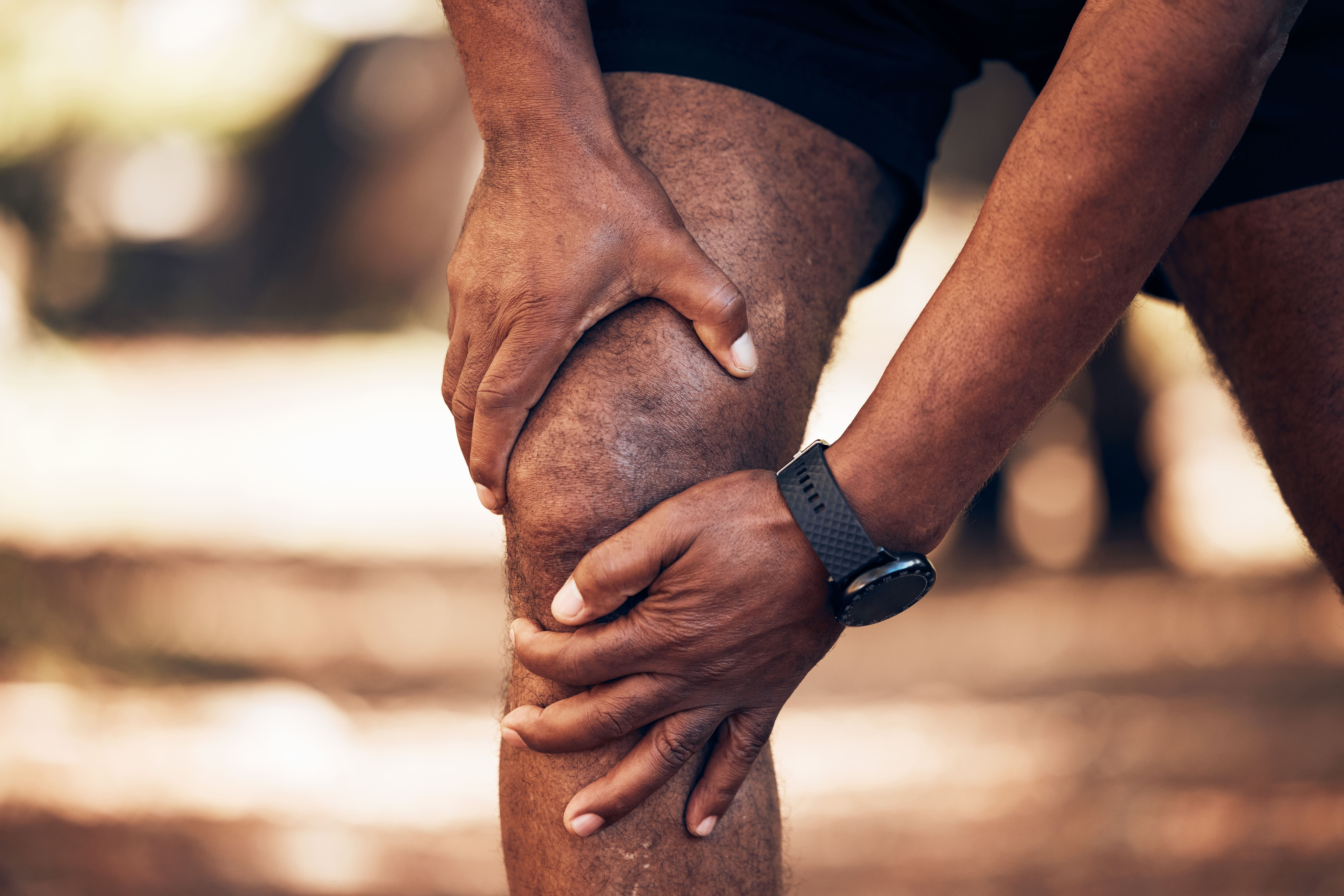
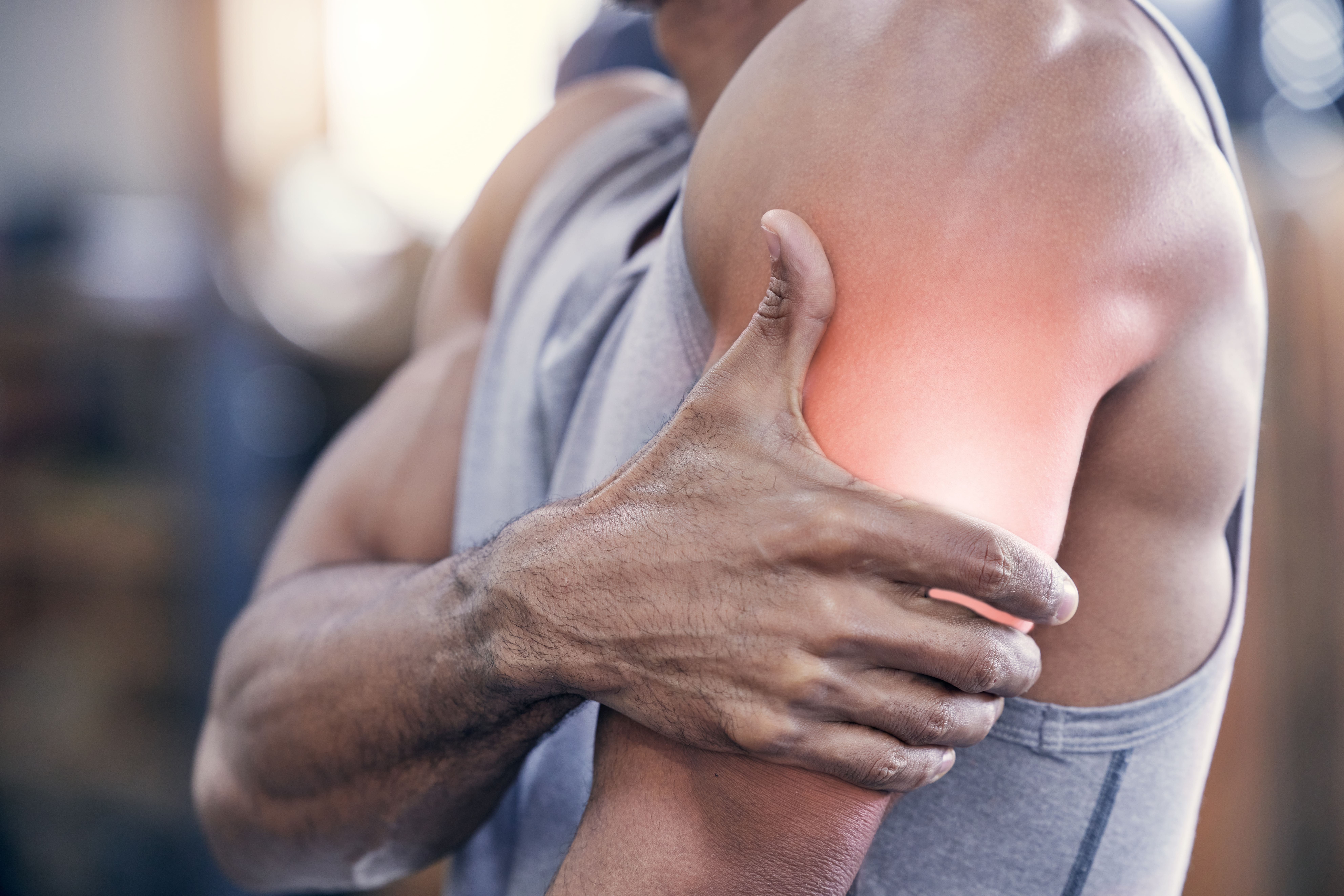
I am Dr. Alexander Jimenez, DC, APRN, FNP-BC, CFMP, IFMCP, ATN, CCST. In musculoskeletal medicine, the burden of disease is massive—over 1.7 billion people worldwide experience musculoskeletal problems, with more than 78 million Americans projected to have arthritis by 2040 (GBD 2021; HHS projections). This epidemiologic pressure has accelerated innovation in orthobiologics, pushing them from adjunctive options to front-door therapies that can be integrated early in care for athletes, active adults, and patients attempting to delay or avoid surgery.
During educational gatherings like the Excel Rise immersive, the goal is not simply to introduce concepts, but to help clinicians confidently apply them. That is the spirit of this post: practical, deeply explained, and firmly rooted in modern evidence.

Five Core Modalities and Two Biological Lenses

We can view the orthobiologic landscape through two lenses:

  • Acellular interventions: Hyaluronic acid (HA), alpha-2-macroglobulin (A2M), growth factor concentrates, and extracellular vesicles (exosomes; investigational in the U.S.).
  • Cellular interventions: PRP, bone marrow concentrate (BMAC) enriched in mesenchymal stromal cells (MSCs) and hematopoietic cells, and adipose-derived SVF containing MSCs and supportive cells.

Each modality interfaces with the joint microenvironment through unique mechanisms—viscoelastic lubrication, protease inhibition, anti-inflammatory signaling, angiomodulation, and anabolic repair pathways.

The Market and Momentum: What Growth Tells Us About Clinical Use

Global market trends mirror clinical adoption. HA represents a mature, widely used option; PRP shows the steepest growth curve; adipose and MSC-related products are rising but often at higher price points; and exosomes are under intense research but not FDA-approved for musculoskeletal indications in the U.S. The key takeaway: clinicians should first develop competence with PRP and HA, then layer in MSC strategies where appropriate, while keeping an eye on emerging evidence for acellular vesicle therapy.

  • HA is transitioning from a first-line solo therapy to an adjunct that enhances other biologics.
  • PRP is the inflection point in biologics due to its accessibility, safety, and growing evidence base.
  • BMAC and adipose SVF bring cellular heft, but introduce logistical, regulatory, and cost considerations.
  • Exosomes carry regenerative signals via microRNAs and proteins; research is promising yet preliminary for clinical adoption in the U.S.

Evidence Landscape: What the Literature Actually Shows

When patients or colleagues ask whether orthobiologics are supported by science, the answer is yes—though the quality and standardization vary by modality.

  • HA has a large body of literature supporting pain relief and functional improvement in knee OA, particularly in mild-to-moderate disease (Altman et al., 2015; Bannuru et al., 2015).
  • PRP demonstrates efficacy in pain, function, and quality of life across knee OA and select tendinopathies, with numerous trials and meta-analyses supporting its use (Laudy et al., 2015; Belk et al., 2021).
  • BMAC and minimally manipulated MSCs show promise but remain equivalent to PRP in many analyses, with some studies indicating culture-expanded allogeneic MSCs may outperform minimally manipulated approaches in OA symptom domains (Lamo-Espinosa et al., 2016; Chahla et al., 2021).
  • Combination therapy—PRP plus HA—often outperforms either alone in both short-term and sustained outcomes (Shen et al., 2022).
  • PRP plus MSCs can enhance MSC proliferation and paracrine signaling, thereby improving outcomes beyond those achieved with SCs alone (Murray et al., 2017; Cengiz et al., 2020).

These observations align with clinical experiences at my practice, where integrative protocols often yield faster pain reduction, better load tolerance, and more durable functional gains than single-agent strategies.

Physiologic Underpinnings: Why These Therapies Work

Understanding the physiology is essential for precise patient selection and sequencing.

Hyaluronic Acid: Lubrication and Mechanotransduction

  • Viscosupplementation: HA augments the synovial fluid’s viscoelastic properties, improving joint lubrication, reducing friction, and attenuating nociceptive input.
  • Mechanotransduction: HA interacts with CD44 and other cell-surface receptors, modulating chondrocyte behavior, anti-inflammatory pathways (e.g., NF-κB), and extracellular matrix synthesis (E.g., Aggrecan, Type II collagen).
  • Adjunct synergy: HA can increase PRP growth factor bioavailability by slowing diffusion and supporting joint biomechanics, creating a favorable milieu for repair.

Platelet-Rich Plasma: Growth Factors and Immunomodulation

  • Key growth factors: PDGF, TGF-β, VEGF, IGF-1, and EGF orchestrate angiogenesis, matrix synthesis, and cellular recruitment.
  • Inflammation modulation: PRP can shift macrophages from M1 (pro-inflammatory) to M2 (pro-resolving) phenotypes, dampen catabolic cytokines (IL-1β, TNF-α), and support tissue remodeling.
  • Leukocyte content: High- vs. low-leukocyte PRP shows equipoise in many OA outcomes. Practically, I tailor leukocyte levels:
    • Lower-leukocyte PRP for intra-articular OA to reduce flare risk.
    • Higher-leukocyte PRP for chronic tendinopathy requires a stronger inflammatory reset.

Bone Marrow Concentrate (BMAC): MSCs, HSCs, and Trophic Support

  • MSCs exert paracrine effects by secreting anti-inflammatory cytokines and anabolic signals rather than directly engrafting long-term.
  • HSCs and progenitors may contribute to microvascular health and immunologic balance.
  • BMAC’s potency varies by harvest technique, patient age, and disease state; standardization and realistic expectations are critical.

Adipose-Derived SVF: Cell Diversity and Immunologic Balance

  • SVF contains MSCs, pericytes, endothelial progenitors, and immune cells that collectively promote angiogenesis, matrix regulation, and immune homeostasis.
  • Cost and invasiveness are higher; consider in refractory cases or where robust cellular signaling is needed.

Exosomes and Extracellular Vesicles: Signal Delivery (Investigational)

  • Exosomes transport microRNAs, proteins, and lipids that modulate cell behavior and reduce inflammation.
  • Preclinical data are encouraging; FDA approval for musculoskeletal indications remains pending. Clinicians should follow the developing guidance closely.

The Multimodal Rationale: Orchestration and Synergy

The most compelling evidence and mechanistic logic point toward combination protocols. Think of biologics as instruments in an orchestra:

  • HA + PRP: HA supports joint biomechanics and prolongs residence time; PRP delivers growth factors. Together, they potentiate chondrocyte mechanosensitivity while reducing catabolic signaling.
  • PRP + MSCs (BMAC or SVF): PRP acts like an augur, attracting MSCs and enhancing their proliferation and paracrine output, improving tissue outcomes.
  • A2M + PRP + HA: A2M inhibits proteases (MMPs, ADAMTS), PRP drives repair signals, and HA improves joint lubrication—creating a trilogy that targets pain, catabolism, and biomechanical stress simultaneously.

From a clinical standpoint, multimodal therapy reflects how medicine achieves results in oncology, cardiology, and infectious diseases—by layering complementary mechanisms to achieve additive or synergistic effects.

Patient Selection and Stratification: Matching Biology to Individuals

A central pillar of modern orthobiologics is patient stratification. Not all patients have the same joint biology, inflammatory tone, or biomechanical faults.

  • Disease stage:
    • Early-to-mid OA responds best to PRP, HA, or PRP + HA.
    • Advanced OA may require MSC augmentation, with realistic expectations and concurrent mechanical offloading.
  • Inflammatory phenotype:
    • High CRP or synovitis suggests a need to control catabolic cytokines; consider A2M, lower-leukocyte PRP, and robust anti-inflammatory lifestyle changes.
  • Mechanical risk profile:
    • Malalignment, kinetic chain deficits, or poor load management will blunt biologic efficacy. This is where integrative chiropractic care becomes central.
  • Age and sex hormones:
    • For women over 38, consider the trajectory of estrogen preservation—chondrocyte estrogen receptors influence cartilage matrix maintenance. Collaboration with women’s health clinicians may support joint health when appropriate.

Integrative Chiropractic Care: The Biomechanical Foundation

In my clinical experience at ChiroMed El Paso, integrative chiropractic care is not an accessory—it is the scaffold that makes biologics work better.

  • Spine-pelvis-hip alignment: Correcting lower kinetic chain mechanics reduces aberrant joint loads that perpetuate inflammation and matrix breakdown.
  • Neuromuscular control: Motor pattern retraining increases joint stability, reduces shear forces, and normalizes mechanotransduction at the chondrocyte level.
  • Fascia and myofascial tone: Manual therapies that normalize fascial glide improve perfusion and lymphatic drainage, supporting biologic distribution and recovery.
  • Anti-inflammatory lifestyle: Nutritional strategies and sleep optimization reduce systemic cytokine drive, aligning with PRP’s immunomodulatory goals.

Through structured programs, we can track objective improvements—range of motion, step counts, load tolerance, and pain scores—creating a feedback loop to refine biologic timing and dosing.

Structured Reports: Turning Data Into Decisions

Creating structured reports improves clarity, communication, and outcomes measurement. Here’s a practical approach:

  • Patient phenotype summary:
    • Pain generators: articular, tendinous, or mixed.
    • Inflammatory markers: CRP, ESR, and synovitis on ultrasound.
    • Mechanical assessment: valgus/varus alignment, gait deviations, muscular imbalances.
  • Intervention rationale:
    • Why PRP: growth factor-driven repair and immunomodulation.
    • Why HA: lubrication, mechanosensitive chondrocyte support.
    • Why MSC adjunct: paracrine potency in advanced cases.
    • Why A2M: protease inhibition to protect cartilage matrix.
  • Protocol details:
    • PRP preparation (single-spin vs double-spin; leukocyte content tailored).
    • HA formulation (molecular weight; crosslinked vs non-crosslinked).
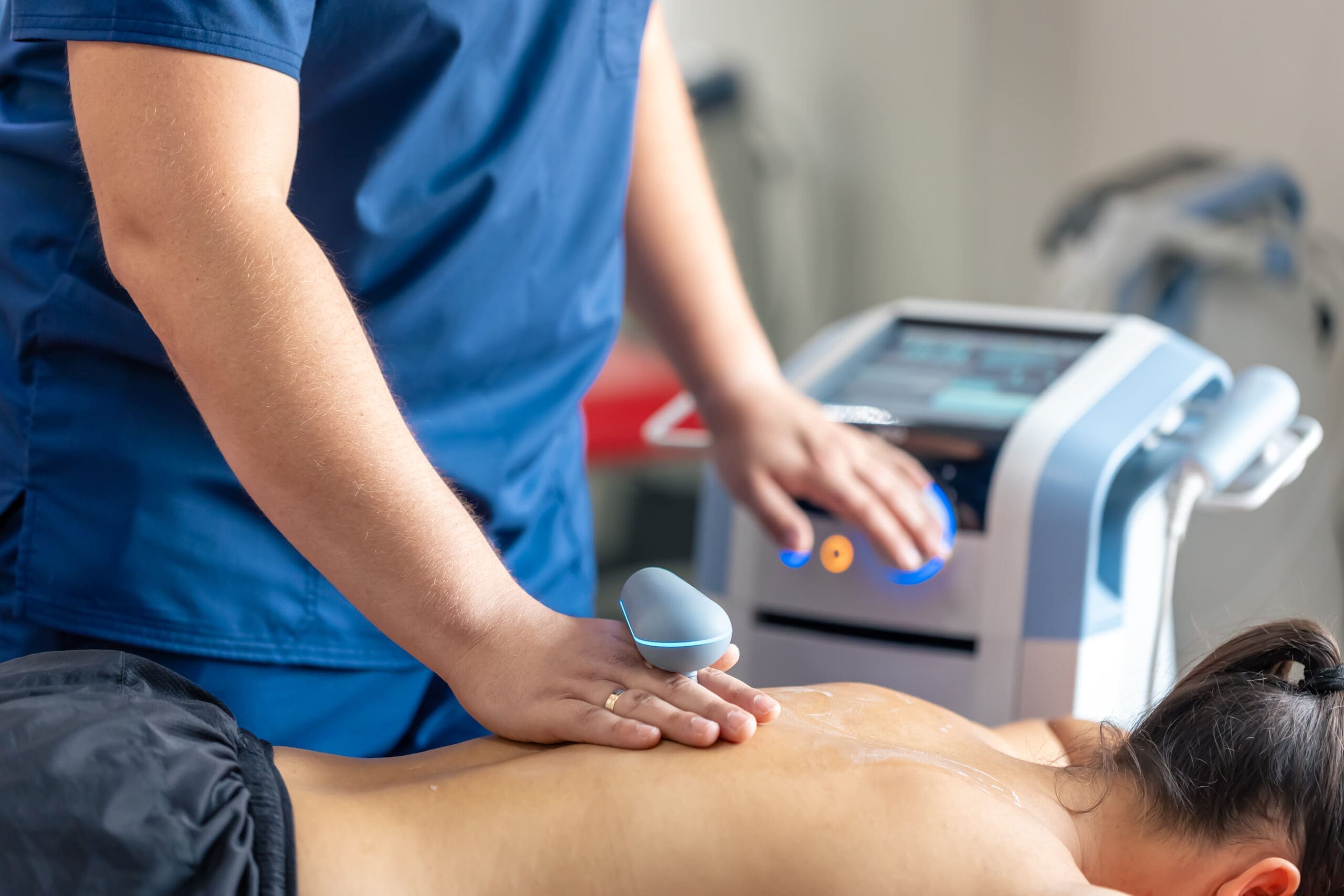
    • Injection strategy (intra-articular vs peri-tendinous; ultrasound-guided precision).
  • Integrative plan:
    • Chiropractic adjustments and kinetic chain retraining.
    • Targeted strengthening and flexibility work.
    • Nutrition and sleep prescriptions to lower inflammatory load.
  • Outcome tracking:
    • Baseline and 12-week PROMs (KOOS, WOMAC), pain VAS, step counts, and functional tests.
    • Reassessment at 6 months to determine whether a booster PRP or additional HA is needed.

Practical Protocols: Stepwise Implementation

Here is how I typically structure care for knee OA patients:

  • Mild-to-moderate OA, active adult:
    • Week 0: Ultrasound-guided PRP (low-leukocyte) intra-articular plus high–molecular–weight HA in the same session or staggered within 2 weeks.
    • Weeks 1–4: Chiropractic-guided kinetic chain corrections; quadriceps/hip abductor strengthening; gait re-education.
    • Week 6–8: Reassessment; add A2M if catabolic markers or synovitis persist.
  • Moderate-to-advanced OA, symptomatic load intolerance:
    • Week 0: PRP + HA; consider BMAC or adipose SVF if previous biologic responses were suboptimal and patient consents to invasiveness and cost.
    • Weeks 1–6: Intensive integrative mechanical care; weight management and anti-inflammatory nutrition.
    • Week 12: Outcomes review; booster PRP if functional gains plateau.

For tendinopathy (patellar, Achilles):

  • High-leukocyte PRP peri-tendinous under ultrasound guidance to initiate an inflammatory reset and remodeling.
  • Progressive loading program with eccentric exercises, fascial release, and chiropractic alignment.

Special Considerations: Hormones, Senescence, and Emerging Agents

  • Estrogen preservation: Cartilage contains estrogen receptors that regulate matrix synthesis. In perimenopausal athletes, discussing estrogen status with the appropriate specialist can be pivotal for joint longevity (Roman-Blas et al., 2009).
  • Senolytics: Cellular senescence contributes to OA progression. Early human research suggests senolytics may improve tissue health by clearing senescent cells and reducing SASP cytokines (Farr et al., 2017; Jeon et al., 2017). While promising, integrate cautiously and remain aligned with regulatory guidance.
  • Losartan and PTH signaling: There is interest in losartan’s potential effects on fibrosis and matrix remodeling, as well as in PTH-related chondrogenic signaling; these remain exploratory and should be guided by specialist collaboration and evolving evidence.

Clinical Observations from My Practice

From my day-to-day work, several patterns consistently emerge:

  • PRP’s durability: When paired with precise mechanical correction, PRP’s effects on pain and function are more durable. Patients who receive PRP without addressing gait and alignment often regress.
  • HA’s adjunctive value: HA co-administration frequently reduces early post-injection discomfort and supports resumption of activity, especially in higher-demand patients.
  • MSC timing: MSC-based strategies help patients with advanced cartilage thinning who have exhausted HA and PRP. However, expectations must be managed; pairing MSCs with A2M and structured mechanical rehab improves real-world outcomes.
  • Data drives trust: Using our structured reports and PROMs, patients better understand progress and buy into staged booster strategies when plateaus appear. This transparency reduces overuse and aligns care with goals.

You can explore more of my integrated clinical approach and case reflections on my website and professional page:

Safety, Regulation, and Ethics

  • PRP and HA are widely used with strong safety profiles when performed with sterile technique and ultrasound guidance.
  • BMAC and adipose SVF require adherence to local regulations and informed consent, including a realistic discussion of cost, invasiveness, and variability.
  • Exosomes remain investigational for musculoskeletal care in the U.S.; participate in IRB-approved research where possible, and avoid off-label uses that lack clarity on sourcing and safety.
  • Always document complication risks: post-injection flare, infection, vasovagal episodes, and rare reactions.

Putting It All Together: A Clinician’s Roadmap

Here is a simple roadmap you can adapt:

  • Start with a clear phenotype: structural severity, inflammatory tone, mechanical deficits, and patient goals.
  • Use PRP as a core for OA and tendinopathy; tailor leukocyte content.
  • Layer HA to enhance lubrication and mechano-biologic signaling.
  • Add A2M when catabolic protease activity seems pronounced.
  • Reserve MSC strategies for refractory or advanced presentations, combined with robust integrative care.
  • Track outcomes and schedule data-driven boosters only when plateaued gains suggest benefit.
  • Anchor the plan in integrative chiropractic correction, progressive loading, nutrition, and sleep hygiene.

Conclusion: From Foundation to Mastery

As we continue to crystallize concepts, techniques, and technology, orthobiologics offer a bright, actionable future. The science supports PRP as a leading modality for pain, function, and quality of life, with HA and A2M adding biomechanical and anti-catabolic support. MSC-based therapies and cutting-edge acellular signals are expanding the frontier, and combination protocols frequently deliver the best outcomes.
This is not about chasing novelty; it is about orchestration—modulating inflammation, protecting matrix, restoring biomechanics, and guiding repair. With structured reports, integrative chiropractic care, and evidence-based biologics, we can confidently walk our patients from pain and limitation toward resilience and durable function.

Key Takeaways

  • Combine PRP + HA for enhanced joint lubrication and repair signaling.
  • Consider A2M when protease-driven matrix loss is suspected.
  • Use PRP + MSCs in advanced cases for synergistic paracrine effects.
  • Always correct mechanical faults through integrative chiropractic care to prevent biologic backsliding.
  • Track outcomes rigorously and communicate transparently about expected timelines and booster logic.

References

SEO tags: orthobiologics, PRP for knee osteoarthritis, hyaluronic acid injections, bone marrow concentrate MSCs, adipose stromal vascular fraction, exosomes orthobiologics, alpha-2-macroglobulin A2M, integrative chiropractic care, musculoskeletal pain, osteoarthritis treatment, evidence-based sports medicine, multimodal regenerative therapy, chondrocyte mechanotransduction, immunomodulation M1 to M2, structured outcomes reports, Dr. Alexander Jimenez

Regenerative Therapy for Auto Accident Injury Recovery

Regenerative Therapy for Auto Accident Injury Recovery

Regenerative Therapy for Auto Accident Injury Recovery

Abstract

Motor vehicle accidents can cause more than quick pain. A crash can injure muscles, ligaments, tendons, joints, nerves, and spinal tissues. Some injuries are felt right away, while others show up days later. For many people, early care can make a major difference in recovery. At a ChiroMed-style integrative clinic, the goal is to look at the whole injury pattern, not just the painful area. Care may include chiropractic treatment, rehabilitation, shockwave therapy, and regenerative options such as platelet-rich plasma (PRP), platelet-poor plasma, plasma-based therapies, and microfragmented adipose tissue (MFAT). These therapies may help support tissue healing, improve movement, reduce pain, and help some patients avoid surgery when appropriate.

Why Motor Vehicle Accidents Can Lead to Chronic Pain

A motor vehicle accident can place sudden force on the body. The neck may snap forward and backward. The lower back may twist. The shoulder, hip, knee, or ankle may absorb impact. Even when there are no broken bones, the soft tissues can still be injured.

Common accident-related injuries may include:

  • Whiplash
  • Neck and back sprains
  • Ligament injuries
  • Tendon injuries
  • Joint pain
  • Muscle strains
  • Disc irritation
  • Nerve irritation
  • Headaches
  • Shoulder, hip, or knee pain
  • Spinal stiffness
  • Reduced range of motion

These injuries can become more difficult to treat when they are ignored. Pain may begin as soreness, then turn into stiffness, weakness, nerve symptoms, or chronic inflammation. This is why early evaluation matters after an accident (Fletcher Family Chiropractic, n.d.; Health Coach Clinic, n.d.).

The ChiroMed Approach to Accident Recovery

ChiroMed is a natural fit for this topic because accident recovery often needs more than one type of care. A patient may need spinal care, soft tissue treatment, rehab, imaging review, and medical coordination. The goal is not only to reduce pain but also to improve how the body moves and heals.

An integrative chiropractic plan may include:

  • A detailed accident history
  • Orthopedic testing
  • Neurological screening
  • Range of motion testing
  • Posture and movement assessment
  • Imaging referrals when needed
  • Chiropractic adjustments
  • Soft tissue therapy
  • Corrective exercises
  • Shockwave therapy
  • Regenerative medicine consultation
  • Ongoing progress exams

This type of care helps connect the injury to the symptoms. It also helps create a clearer recovery plan, especially when the patient has soft-tissue injuries that may not be clearly visible on basic X-rays (Health Coach Clinic, n.d.; Pure Wellness, n.d.).

Why Soft Tissue Injuries Need Special Attention

Soft tissue injuries involve muscles, tendons, ligaments, fascia, and joint-supporting tissues. These structures help stabilize the spine and joints. When they are damaged, the body may become unstable, stiff, painful, or weak.

Soft tissue injuries can be difficult because they may heal slowly. Some tissues have limited blood flow, which can make recovery harder. Ligaments and tendons may also stay irritated if the joint continues to move poorly.

For example, after a rear-end crash, a person may develop whiplash. The neck muscles tighten to protect the spine. Ligaments may be stretched. Small joints in the neck may become irritated. If this pattern is not treated, the patient may develop headaches, limited neck motion, shoulder tightness, or nerve-like symptoms in the arm.

This is where combined care may help. Chiropractic care can improve motion. Rehab can rebuild strength. Shockwave therapy may support soft tissue healing. Regenerative options may be considered when injured tissue needs more support.

PRP Therapy After an Auto Accident

Platelet-rich plasma, or PRP, is a regenerative treatment made from the patient’s own blood. A small blood sample is drawn and processed to concentrate platelets. These platelets contain growth factors that help guide the body’s healing response. The PRP is then placed into the injured area when clinically appropriate (Johns Hopkins Medicine, n.d.).

PRP may be considered for:

  • Ligament sprains
  • Tendon injuries
  • Muscle injuries
  • Joint pain
  • Whiplash-related soft tissue injuries
  • Chronic pain after trauma
  • Pain that has not improved with basic care

PRP does not act like a pain-killing shot. Instead, it is used to support the body’s healing process. Patients still need a full recovery plan that may include chiropractic care, rehab, movement correction, and follow-up exams (MVA MVP, n.d.; Integrative Spine & Sports, n.d.).

PFP and Plasma-Based Regenerative Options

Some clinics discuss platelet-poor plasma, platelet fibrin plasma, or other plasma-based products. The terms can vary depending on how the product is prepared and what the provider is trying to treat. Patients should always ask what type of plasma product is used, why it is recommended, and how it fits into the full treatment plan.

In simple terms, plasma-based therapies are designed to support the tissue environment. After a crash, ligaments, tendons, joints, and spinal tissues may need help calming inflammation and rebuilding healthier function. Plasma-based care may be part of that process when medically appropriate.

For a ChiroMed-style plan, plasma-based care should not stand alone. It works best when the patient is also improving movement, strength, posture, stability, and daily activity habits.

MFAT Therapy for More Complex Soft Tissue and Joint Injuries

Micro-fragmented adipose tissue, or MFAT, uses a small amount of the patient’s own fat tissue. The tissue is processed into tiny fragments and then placed into the injured area. MFAT contains a natural tissue matrix and signaling factors that may help support repair in joints, tendons, ligaments, and other soft tissues (Engelen Sports & Orthobiologics, n.d.; Ortho-Regen, n.d.).

MFAT may be discussed for:

  • Chronic joint pain
  • Partial tendon tears
  • Ligament injuries
  • Meniscus-related problems
  • Degenerative joint changes
  • Soft tissue injuries that are slow to heal
  • Injuries that have not improved with standard care

MFAT is not for every patient. It requires proper evaluation, clear diagnosis, and careful clinical judgment. A 2025 review described MFAT as a developing regenerative option with potential for tissue repair, but also highlighted the need for proper patient selection and further clinical research (Fu & Wang, 2025).

Shockwave Therapy for Post-Accident Pain

Shockwave therapy uses acoustic energy to stimulate injured tissues. It is non-surgical and may be used to support circulation, collagen activity, and tissue remodeling, and to reduce pain. In accident care, shockwave therapy may be beneficial for soft tissue pain, scar tissue, tendon irritation, muscle tightness, and chronic inflammation (Advanced Back & Neck Care, n.d.; Mayo Clinic, n.d.).

Shockwave therapy may help with:

  • Neck and back soft tissue pain
  • Tendon pain
  • Muscle trigger points
  • Scar tissue stiffness
  • Chronic inflammation
  • Reduced mobility
  • Pain that has lasted longer than expected

For many patients, shockwave therapy fits into an integrative plan because it can be paired with chiropractic care and rehab. The goal is to help the tissue respond better while also correcting movement problems that keep stressing the injury.

Why Chiropractic Care Is the Foundation

Regenerative therapies may support tissue repair, but the body also needs proper movement. If a joint is not moving well, the injured area may continue to become irritated. If muscles are weak or tight, the spine may remain under stress. If posture is poor after the crash, pain may continue.

Chiropractic care may help by:

  • Improving spinal motion
  • Reducing joint restriction
  • Calming muscle guarding
  • Supporting better nerve function
  • Improving posture
  • Helping the body move with less stress
  • Supporting better rehab progress

After a motor vehicle accident, chiropractic care is often used for neck pain, back pain, whiplash, headaches, shoulder pain, hip pain, and joint stiffness. When combined with rehabilitation, it can help restore strength and stability, not just temporary comfort (Delaware Back Pain & Sports Rehabilitation Centers, n.d.; Pure Wellness, n.d.).

Dr. Alexander Jimenez’s Clinical Perspective

Dr. Alexander Jimenez, DC, APRN, FNP-BC, has long emphasized a dual-scope approach to personal injury care. This means considering both the structural and medical aspects of an injury. In a motor vehicle accident, the case may involve spinal and joint function, nerve symptoms, soft-tissue damage, inflammation, imaging needs, and clear documentation.

This approach is important because accident injuries can overlap. A patient may experience neck pain, headaches, shoulder tightness, lower back pain, and nerve irritation simultaneously. Treating only one symptom may miss the bigger injury pattern.

A ChiroMed-focused care model can reflect this same goal: evaluate the whole person, identify the injured tissues, restore movement, support healing, and help the patient return to daily life with better function.

Why Early Care Matters After a Crash

The best time to treat injuries from accidents is often early. This does not mean every patient needs advanced treatment right away. It means the patient should be examined before the injury becomes chronic.

Early care may help:

  • Reduce inflammation
  • Protect injured tissue
  • Improve range of motion
  • Prevent stiffness
  • Reduce compensation patterns
  • Identify nerve symptoms
  • Support better documentation
  • Lower the risk of long-term pain

When care is delayed, the body may begin moving around the painful area. This can lead to new problems in the spine, hips, shoulders, knees, and muscles. Over time, the patient may develop chronic pain, weakness, poor posture, or limited mobility.

When Regenerative Care May Be Considered

Regenerative therapies may be considered when a patient has tissue damage that is slow to heal or when the goal is to avoid surgery when possible. These treatments may be useful when standard care has helped only partly or when the tissue injury is more complex.

A provider may consider PRP, PFP, MFAT, or shockwave therapy when there is:

  • Ongoing ligament pain
  • Tendon damage
  • Joint injury
  • Soft tissue trauma
  • Chronic inflammation
  • Pain that returns with activity
  • Reduced function despite basic care
  • A desire to explore non-surgical options

However, regenerative care must be based on diagnosis. It should not be used as a guess. Imaging, exams, and medical history help determine whether the patient is a suitable candidate.

Physical Rehabilitation Completes the Recovery Plan

Rehabilitation is important because healing tissue needs strength and control. A patient may feel better after an adjustment, injection, or shockwave session, but long-term recovery also depends on how well the body moves during daily life.

Rehab may include:

  • Gentle mobility work
  • Stretching
  • Core strengthening
  • Balance training
  • Posture correction
  • Neck stabilization
  • Hip and shoulder strengthening
  • Walking programs
  • Return-to-work conditioning

Physical therapy and rehab can help patients regain confidence after an accident. They also help reduce fear of movement, which is common after painful trauma (RES Physical Medicine & Rehab, n.d.; Fairview Rehab, n.d.).

When Emergency Care Is Needed

Not every accident injury belongs in a chiropractic or regenerative medicine setting first. Some symptoms require emergency medical care.

A person should seek urgent care right away for:

  • Loss of consciousness
  • Severe headache
  • Chest pain
  • Trouble breathing
  • New weakness
  • Loss of bladder or bowel control
  • Severe abdominal pain
  • Numbness that is getting worse
  • Trouble walking
  • Severe neck or back pain after trauma
  • Confusion or memory problems

Safety comes first. Once serious conditions are ruled out, an integrative recovery plan may begin.

A Clear Path Forward With ChiroMed

Motor vehicle accident recovery works best when the care plan is clear, complete, and personalized. Regenerative therapies such as PRP, PFP, and related plasma-based options, as well as MFAT, may help support tissue healing. Shockwave therapy may help improve soft tissue response, circulation, and pain control. Chiropractic care may improve spinal motion, reduce joint stress, and support the nervous system. Rehabilitation helps rebuild strength and long-term stability.

For ChiroMed, the message is simple: accident injuries should not be ignored, and pain should not be treated as a one-size-fits-all problem. A thoughtful integrative plan can help patients understand their injuries, support healing, improve function, and reduce the risk of chronic pain after a motor vehicle accident.


References

Advanced Back & Neck Care. (n.d.). Shockwave therapy for motor vehicle accidents in Lumberton

BenGlassLaw. (n.d.). What is the value of my PRP therapy claim?

Delaware Back Pain & Sports Rehabilitation Centers. (n.d.). Best car accident pain solutions that work

Engelen Sports & Orthobiologics. (n.d.). Microfragmented adipose tissue (MFAT) therapy

Fairview Rehab. (n.d.). What type of post-accident therapy you may need

Fletcher Family Chiropractic. (n.d.). Why seeing a chiropractor after a car accident matters

FoRM Health. (n.d.). MFAT injections

Fu, H., & Wang, C. (2025). Micro-fragmented adipose tissue-An innovative therapeutic approach: A narrative review

Health Coach Clinic. (n.d.). Chiropractic integrative care for motor vehicle accidents

Health Coach Clinic. (n.d.). Regenerative medicine and integrative chiropractic approaches

Imperium Health Center. (n.d.). Holistic chiropractic treatments for injuries

Integrative Spine & Sports. (n.d.). PRP for whiplash: Accelerating recovery and restoring mobility

Jimenez, A. (n.d.). Dr. Alexander Jimenez, DC, APRN, FNP-BC

Jimenez, A. (n.d.). Dr. Alexander Jimenez LinkedIn profile

Johns Hopkins Medicine. (n.d.). Platelet-rich plasma injections

Mayo Clinic. (n.d.). Shockwave treatment: A new wave for musculoskeletal care

MVA MVP. (n.d.). Platelet-rich plasma therapy for vehicle accidents

Ortho-Regen. (n.d.). Microfragmented adipose tissue (MFAT)

Pure Wellness. (n.d.). Treating auto injuries with chiropractic care and regenerative medicine

RES Physical Medicine & Rehab. (n.d.). Road to recovery: The role of physical therapy after a car accident

Whalen Injury Lawyers. (n.d.). What is regenerative care in my motor vehicle accident case?

Ultrasound Therapy Benefits and Uses For The Musculoskeletal System

Find out how ultrasound therapy provides effective solutions for chronic musculoskeletal pain and joint issues.

Abstract

As a clinician with a diverse background in chiropractic, nursing, and functional medicine, I have dedicated my career to integrating the most advanced, evidence-based tools into patient care. This post explores the transformative role of musculoskeletal ultrasound (MSKUS), a powerful, real-time imaging modality that has revolutionized the way we diagnose and treat soft-tissue injuries. We will embark on a journey through the sonographic appearance of various tissues—tendons, muscles, cartilage, ligaments, and nerves—understanding their unique visual signatures. I will share insights from leading researchers and practical clinical pearls from my own practice on interpreting these images, including the critical concept of anisotropy. Furthermore, we will delve into proper probe handling techniques for both diagnostic and procedural applications, emphasizing methods that set clinicians up for success. Finally, I will explain how these advanced diagnostic capabilities integrate with a holistic, integrative chiropractic approach, enabling more precise, effective, and patient-centered treatment plans that support true healing.


Understanding the Language of Ultrasound: Echogenicity Explained

In my practice, I often refer to musculoskeletal ultrasound as a “glorified flashlight” that allows us to peer directly into the body’s anatomy in real time. But to understand what we’re seeing, we must first learn its language. The fundamental concept is echogenicity, which describes how tissues reflect ultrasound waves.

  • Hyperechoic: Tissues that appear bright white on the screen. These structures, like bone, are dense and reflect most ultrasound waves to the probe.
  • Hypochoic: Tissues that appear dark gray. These structures, like muscle or fluid, absorb more ultrasound waves and reflect fewer.
  • Anechoic: Tissues that appear completely black. These are typically fluid-filled structures, such as cysts or bursae, that transmit almost all sound waves.
  • Isoechoic: Tissues that have a similar brightness or echotexture to adjacent structures.

Pattern recognition is the cornerstone of interpreting ultrasound images. Each tissue type has an expected appearance, and deviations from this norm can signal pathology.

Sonographic Signatures of Key Musculoskeletal Tissues

Let’s explore what healthy tissues look like under the lens of an ultrasound probe.

Tendons: The Body’s Strong Cords

Tendons are the strong, fibrous cords that connect muscle to bone. On ultrasound, a healthy tendon has a classic appearance: it’s hyperechoic (bright) and displays a distinct fibrillar pattern—think of it as a tightly packed bundle of cables or parallel stripes.

For example, when we look at the patellar tendon in a long-axis view (aligned with the tendon), we expect to see a bright, organized, striped pattern. Beneath it, we can identify other structures, such as the infrapatellar fat pad (which has a more wavy, less organized appearance) and the hyperechoic surfaces of the patella and tibia. Recognizing this norma, fibrillar architecture is crucial because when a tendon is injured (tendinosis or a tear), it loses this organization, thickens, and appears more hypoechoic (darker).

Muscles: The Engines of Movement

Muscle tissue presents a more complex, mixed-echogenicity pattern. It is generally hypoechoic compared to the bright white of bone. However, within the muscle belly, you’ll see hyperechoic strands of connective tissue, known as the perimysium, which encase the muscle fascicles. This gives healthy muscle a “marbled” or “feathery” appearance.

When viewing a muscle like the bicep or deltoid over the humerus, you can see the dark muscle tissue contrasted against the bright cortical line of the bone. You can even appreciate its structure, tapering towards its tendinous insertion. This visual information helps us identify muscle strains, tears, or atrophy.

Cartilage: Smooth Surfaces and Tough Cushions

Cartilage is a critical tissue, and ultrasound helps us differentiate between its two main types:

  • Hyaline Cartilage: This is the smooth, glassy cartilage that covers the ends of bones within a joint, allowing for low-friction movement. On ultrasound, it appears as a distinct, thin, hypoechoic (dark) line sitting directly on the bright, hyperechoic bone surface. A great example is viewing the posterior aspect of the humeral head in the shoulder joint.
  • Fibrocartilage: This is a tougher, more fibrous type of cartilage found in structures like the meniscus of the knee or the labrum of the shoulder and hip. Unlike hyaline cartilage, fibrocartilage is hyperechoic (brighter) and has a more triangular or wedge-shaped appearance. On the shoulder, you can clearly distinguish the bright, triangular labrum from the dark, linear hyaline cartilage on the humeral head.

Ligaments: The Stabilizers

Ligaments, which connect bone to bone, look very similar to tendons on ultrasound. They are also hyperechoic and have a fibrillar, striated pattern. The key difference is that ligaments are typically more compact and densely packed than tendons.

The true power of ultrasound in evaluating ligaments comes from its real-time, dynamic capabilities. The best way to confirm you are looking at a ligament is to trace it from one bony attachment to another. If it originates from or inserts into a muscle, it’s a tendon. With ligaments such as the Medial Collateral Ligament (MCL) of the knee, we can perform a stress test under direct visualization. By applying a valgus force to the knee, we can watch the ligament in real time to see if there is any “gapping” or separation of its fibers.

A report might read: “The linear probe was placed over the medial aspect of the knee, and the MCL was visualized in a long-axis view. Upon real-time valgus stress, there was observable gapping of the mid-substance fibers with surrounding hypoechoic fluid, consistent with a grade 2 sprain.” This level of detail is impossible with a static MRI.

Nerves: The Body’s Electrical Wiring

Nerves have a unique and fascinating appearance on ultrasound, often described as a honeycomb” in short-axis (cross-section) view. This pattern is created by the hypochoic nerve fascicles (the bundles of nerve fibers) surrounded by the hyperechoic epineurium (the connective tissue sheath).

In a long-axis view, the nerve can look like a bundle of parallel “railroad tracks,” though this view is often less distinct than the honeycomb cross-section. A clinical pearl I share with my students is that nerves are often easier to spot when you scan. The distinct honeycomb pattern moves through the surrounding tissue, catching your eye more readily than the linear patterns of tendons or muscles. The carpal tunnel is the classic location to visualize this, as the median nerve’s honeycomb structure stands out clearly against the adjacent flexor tendons in the forearm.


The Challenge of Anisotropy: A Critical Pitfall to Avoid

One of the most important concepts in MSKUS is anisotropy. This phenomenon occurs when the ultrasound beam is not perfectly perpendicular (at a 90-degree angle) to the structure being imaged, particularly tendons and ligaments. When the beam hits the tissue at an angle, the sound waves are reflected away from the probe instead of back to it. This lack of returning signal causes the normally bright, hyperechoic tissue to appear artifactually hypochoic, or dark.

Why is this so critical? Because a tendon tear also appears as a hypoechoic defect. Anisotropy can mimic pathology, leading to a false-positive diagnosis.

Here’s how we differentiate:

  1. Prove the Pathology: If you see a dark spot in a tendon, like the supraspinatus tendon at its insertion on the humerus, you must prove it’s real.
  2. Toggle the Probe: Carefully “heel-toe” or “toggle” the probe to ensure you are perfectly perpendicular to the tendon fibers at that exact spot.
  3. Observe the Change: If the dark spot disappears and brightens when you adjust the probe angle, it indicates anisotropy. If the dark spot remains dark no matter how you angle the probe, it is more likely to be true pathology, such as tendinosis or a tear.

In my practice, I live by the mantra taught in orthopedic surgery: “One view is no view.” I always confirm a suspected finding from multiple angles, in both long and short-axis views, and correlate it with a dynamic assessment and the patient’s physical exam. This meticulous approach is what separates a novice from an expert operator and ensures diagnostic accuracy.

Mastering the Tool: Proper Probe Handling Techniques

Ultrasound is operator-dependent. Your skill in handling the probe directly impacts the quality of your images and the accuracy of your diagnosis.

The Tripod Grip for Diagnostic Scanning

For diagnostic imaging, stability and fine control are paramount. The “death grip,” where you wrap your whole hand around the probe, is unstable and limits fine motor control. Instead, we use the tripod technique.

  • Hold the probe like a pencil, using your thumb and index finger for control.
  • Brace your remaining fingers (pinky, ring, and/or middle finger) on the patient’s skin.
  • This creates a stable base, allowing subtle, precise movements such as sliding, toggling (heel-toe), and rotating to remain perpendicular to curved structures and eliminate anisotropy.

Your hand should be in contact with the patient. This is a more connected, controlled experience that allows you to feel the anatomy as you visualize it.

Modifying the Grip for Procedural Guidance

When performing an ultrasound-guided injection, the grip must change. Holding the probe with your fingers wrapped around it can physically block your needle’s path. For this reason, I advocate for holding the probe by its edges, which keeps your fingers clear of the sterile field and the needle’s intended path.

  • In-Plane Technique: For this approach, in which the needle is inserted parallel to the probe’s long axis and visualized along its entire length, a pencil-like grip is often effective.
  • Out-of-Plane Technique: In this approach, where the needle is inserted perpendicular to the probe and appears as a bright dot in cross-section, holding the probe by its edges provides the necessary space.

The key is to be facile, comfortable moving the probe in different ways for different tasks. Pre-planning your procedure is essential. My protocol is simple:

  1. Find the Target: Use your scanning skills to locate the exact anatomical target.
  2. Stay Perpendicular: Position the probe directly over the target, perpendicular to the skin. This simplifies your needle trajectory.
  3. Bring Tip to Target: Once you have a clear, stable view of your target, you can confidently guide your needle tip precisely where it needs to go.

This methodical approach minimizes “searching” for the needle or the target, making procedures faster, safer, and more successful.

Integrative Chiropractic Care and Ultrasound Synergy

So, how does this high-tech imaging fit into a chiropractic and functional medicine framework? Perfectly.

At our clinic, we don’t just treat symptoms; we seek to understand and correct the underlying biomechanical and physiological dysfunction. MSKUS is an invaluable tool in this process.

  • Precision Diagnosis: Before I perform a chiropractic adjustment or recommend a course of rehabilitative exercise, I want to know exactly what tissue is injured. Is that shoulder pain from a rotator cuff tear, biceps tendinopathy, or bursitis? Ultrasound tells me instantly, allowing me to tailor my treatment. For instance, if I identify a partial tear in the supraspinatus tendon, I can modify my spinal and extremity adjustments to avoid stressing the injured tissue and instead focus on improving scapular mechanics to offload the tendon.
  • Guiding Soft Tissue Therapies: Many of our treatments involve soft-tissue mobilization, such as Active Release Technique (ART) or the Graston Technique. Ultrasound allows me to visualize fibrotic adhesions or scar tissue and specifically target these areas, making the treatment more efficient and effective.
  • Monitoring Healing: Ultrasound provides objective evidence of tissue healing. We can track the reduction of inflammation, the reorganization of collagen fibers in a healing tendon, or the decrease in fluid within a bursa over time. This helps us advance the patient’s rehabilitation protocol based on actual tissue physiology rather than just subjective pain reports.
  • Patient Education: Showing a patient a real-time image of their injury is incredibly powerful. When they can see the inflamed bursa or the tear in their tendon, it enhances their understanding and improves their adherence to the treatment plan. It transforms the abstract concept of their injury into something tangible.

Ultimately, musculoskeletal ultrasound elevates the practice of integrative chiropractic care. It bridges the gap between a physical exam and a definitive diagnosis, allowing a level of precision previously unattainable in clinical settings. It helps us create highly specific, evidence-based treatment plans that address the root cause of a patient’s pain and dysfunction, accelerating their path back to optimal health and function.

As of May 2nd, 2026, the technology continues to evolve, but its core value remains: it is a safe, dynamic, and profoundly insightful tool that, in the hands of a skilled operator, can truly transform patient outcomes.


References

Jacobson, J. A. (2017). Fundamentals of Musculoskeletal Ultrasound (3rd ed.). Elsevier.

McNally, E. G. (2014). Practical Musculoskeletal Ultrasound (2nd ed.). Elsevier.

The Ultrasound Site. (n.d.). Musculoskeletal Ultrasound. Retrieved from https://www.theultrasoundsite.co.uk/

Ultrasound For Movement Disorders. (n.d.). MSK Resources. Retrieved from https://www.ultrasoundformovementdisorders.com/


SEO Tags: Musculoskeletal Ultrasound, MSKUS, Integrative Chiropractic, Dr. Alexander Jimenez, Echogenicity, Anisotropy, Tendinopathy, Ligament Sprain, Nerve Entrapment, Ultrasound-Guided Injections, Functional Medicine, Chiropractic Care, Sports Medicine, Diagnostic Imaging, Soft Tissue Injury, El Paso Chiropractor, Probe Handling

PRP Injections and Their Benefits for Osteoarthritis

Discover the potential of PRP injections for osteoarthritis treatment and its role in promoting healing and reducing inflammation.

Abstract

As a clinician dedicated to integrative and evidence-based care, I frequently encounter patients suffering from knee osteoarthritis (OA) who are seeking relief. The decision on which injectable treatment to use can be complex, involving a delicate balance of providing rapid pain relief, ensuring long-term joint health, and considering the patient’s individual needs and goals. In this educational post, I will guide you through the latest research on common intra-articular injections for knee OA. We will begin by examining the role and significant risks of corticosteroids, exploring why the medical community is moving toward alternatives. I will then introduce ketorolac, an NSAID injection, as a safer, fast-acting alternative for acute flares. We’ll delve into the science of hyaluronic acid (HA), or viscosupplementation, evaluating its potential for longer-term benefits and its current standing in clinical guidelines. Finally, we will explore the exciting and robust evidence supporting Platelet-Rich Plasma (PRP) as a treatment that not only manages pain but also shows promise as a disease-modifying therapy capable of delaying the need for surgery. Throughout this discussion, I will integrate my clinical observations and emphasize how an integrative chiropractic approach, focusing on biomechanics and holistic patient care, complements these advanced treatments to optimize outcomes for our patients.


Hello, I’m Dr. Alexander Jimenez. With my extensive background in chiropractic and functional medicine, holding titles such as DC, APRN, FNP-BC, CFMP, IFMCP, ATN, and CCST, my practice is rooted in a deep commitment to providing integrative care grounded in the latest scientific evidence. Today, I want to take you on a journey through the landscape of injectable treatments for knee osteoarthritis, a condition I see daily in my clinic. We’ll examine what the research tells us and how we can make the best choices for our patients.

Let’s begin with a common clinical scenario to frame our discussion.

A Common Clinical Scenario: The Acute Knee OA Flare

Imagine a 60-year-old woman who comes into my office with an acute flare-up of her right knee pain. She was diagnosed with mild osteoarthritis two years prior and had managed it well with physical therapy and weight loss. She remains quite active, but this current flare started after a bit more walking than usual. There was no specific injury or trauma. She presents with mild swelling, and her pain is most pronounced when using stairs. Her son’s wedding is just a week away, and she is understandably anxious, requesting an injection to improve her pain and mobility for the event.

On examination, she has a mild antalgic gait (walking with a limp to avoid pain), tenderness along the medial joint line, and a small effusion (swelling within the joint). Her X-rays confirm tricompartmental osteoarthritis with a Kellgren-Lawrence (K-L) grade of 2, which is considered mild to moderate.

So, we have a patient with an acute OA flare, no history of prior injections, and a significant life event approaching. What is the best way to help her? The most conventional response in many practices would be an intra-articular corticosteroid injection. But is that the right answer? My goal today is not to give you a single “correct” answer but to arm you with the evidence so you can make an informed decision, one that aligns with the principles of modern, evidence-based care.

The Double-Edged Sword of Corticosteroid Injections

Why Corticosteroids are so Common

There’s a clear reason why corticosteroids have been the go-to for decades. The primary driver of pain and swelling in an OA flare is synovial inflammation. Corticosteroids are potent anti-inflammatory agents. They work by:

  • Suppressing the infiltration of leukocytes (white blood cells) into the joint.
  • Decreasing the activity of local immune cells within the synovium (the soft tissue lining the joint).
  • Downregulating the expression of genes involved in the inflammatory cascade.

This powerful anti-inflammatory effect leads to decreased synovial membrane inflammation and a reduction in the effusion. The result is rapid pain relief, typically occurring within three to seven days. For our patient with the wedding next week, this timeline is very appealing. While oral NSAIDs or steroids could be considered, they carry significant systemic risks that an injection helps to minimize.

The Mounting Evidence Against Corticosteroids

Despite the short-term benefits, a growing body of evidence urges caution. There is significant concern for chondrotoxicity, meaning the substance is toxic to cartilage cells. Preclinical studies have provided robust evidence that steroids exert dose-dependent deleterious effects on cartilage morphology, histology, and viability. Simply put, the higher the dose of the steroid, the more damage it can cause to the cartilage. Among the different types, dexamethasone and triamcinolone appear to be the least toxic, but the risk remains.

This isn’t just a finding in lab studies. High-level clinical trials support these concerns. A pivotal study published in JAMA in 2017 was a two-year, randomized, placebo-controlled trial comparing injections of triamcinolone to saline in patients with knee OA (McAlindon et al., 2017). Patients received an injection every 12 weeks. The findings were startling:

  • There was no significant difference in pain relief between the steroid group and the placebo (saline) group over the two years.
  • The group receiving repeated steroid injections experienced significantly greater cartilage volume loss compared to the placebo group.

Another powerful retrospective review of over 49,000 patients, published in 2019, found that patients who received even one to three steroid injections (for various hip or knee conditions, not just OA) had a twofold greater risk of needing a knee replacement at the five-year mark (Kompel et al., 2019). The study also revealed a dose-dependent relationship: the risk of total knee arthroplasty (TKA) increased with each subsequent injection.

In my clinical practice at ChiroMed, I’ve observed this pattern. Patients who have received multiple steroid injections over the years often present with more advanced degenerative changes on their imaging than their symptom history might suggest. This is why there is a strong call within the medical community to find safer alternatives. While a single, first-time injection for an acute flare might seem reasonable, we must ask ourselves if we are sacrificing long-term joint health for short-term relief.

Ketorolac: A Safer, Fast-Acting Alternative for Knee Flares

If we want to avoid the chondrotoxic effects of steroids but still provide rapid relief for a patient like the one we discussed, where do we turn? I want to present an excellent alternative: ketorolac.

Ketorolac is a non-steroidal anti-inflammatory drug (NSAID) that can be injected directly into the joint. Its mechanism of action is different from and safer than corticosteroids:

  • As a COX-1 and COX-2 inhibitor, it blocks prostaglandin production, which is a key mediator of inflammation and pain sensitization.
  • Delivering it locally via injection achieves a high concentration in the synovial fluid, providing potent anti-inflammatory effects with minimal systemic exposure compared with oral NSAIDs.
  • Crucially, ketorolac provides anti-inflammatory and analgesic effects without the immunosuppressive and gene-expression-altering effects of steroids.

Most importantly, preclinical models have not shown the deleterious structural effects on cartilage that we see with steroids. Its onset of action is similar, within a few days, and its efficacy often lasts for a few months.

A 2021 systematic review and meta-analysis confirmed that, for knee and hip OA, an intra-articular ketorolac injection provides pain and functional improvements similar to those of corticosteroids from one week to three months post-injection, with minimal adverse events (Saltzman et al., 2021). Our own research has echoed these findings. In a study we conducted on hip pathologies, including OA, we found a trend toward greater pain improvement in the ketorolac group, which was statistically significant for the OA patients. The onset of relief was rapid (around 3 days for ketorolac), and the duration was comparable to that of steroids.

Therefore, ketorolac stands out as a fantastic steroid-sparing option. It addresses the same goal—rapid relief from an inflammatory flare—without the known cartilage toxicity. However, we must still be cautious and avoid its use in patients with contraindications to NSAIDs, such as a history of gastrointestinal ulcers, severe cardiac or kidney disease, or those on anticoagulants.

Hyaluronic Acid for Long-Term Joint Health

What if our patient doesn’t have an acute flare? What if they present with more chronic, persistent pain and want to improve the long-term health of their joint? This is where we shift our focus from just putting out the fire of inflammation to improving the joint’s structural environment. A primary candidate for this goal is hyaluronic acid (HA), also known as viscosupplementation.

The core concept behind HA injections is to augment the natural viscoelastic properties of the synovial fluid. In an osteoarthritic joint, the natural endogenous HA is depleted and degraded. This reduces the fluid’s viscosity and lubricating properties. By supplementing it with an injection, we aim to:

  • Improve joint lubrication and shock absorption.
  • Modulate nociception (pain signaling) and inflammation.

HA works through several biological pathways. It binds to a receptor on synovial cells, CD44, which in turn helps modulate inflammation by decreasing inflammatory cytokines such as IL-1β and cartilage-degrading enzymes such as MMPs. It also appears to stimulate the joint’s own cells to produce more of their natural HA and may have direct chondroprotective effects.

Clinical evidence shows that HA leads to a small but statistically significant reduction in knee OA pain compared to placebo. A comprehensive review by Jevsevar et al. (2015) showed that the pain-relieving effects tend to peak around two months and can last for six months or longer. The treatment appears to be most effective in patients under 65, those with a higher BMI, more severe baseline symptoms, and lower radiographic severity.

It’s important to note that not all HA is created equal. Molecular weight matters. Basic science shows that high-molecular-weight HA has a more profound chondroprotective effect, a greater ability to reduce inflammation through the CD44 pathway, and is better at stimulating the body’s own HA production compared to low-molecular-weight formulations. This is a critical factor I consider when selecting a product for my patients.

Despite this evidence, there is no unified stance on HA. While many European societies accept its use, leading U.S. organizations like the American Academy of Orthopedic Surgeons (AAOS) generally discourage it, citing that the clinical benefit is not consistently large enough. As a result, many insurance companies no longer provide coverage, making it an out-of-pocket expense for many patients. While some studies suggest HA can delay the need for knee replacement surgery, it is not considered a true disease-modifying treatment. This leads us to the next logical step in our journey: biologics.

Platelet-Rich Plasma (PRP): A True Disease-Modifying Potential

If we are looking for an injection that can both treat pain effectively and potentially modify the disease process itself, the strongest evidence currently points to Platelet-Rich Plasma (PRP).

PRP is a concentrate of platelets derived from the patient’s own blood. These platelets are a reservoir of powerful growth factors and signaling molecules that orchestrate tissue healing. When injected into an osteoarthritic knee, PRP has multiple effects:

  • Modulates Inflammation: It influences key inflammatory pathways, such as NF-kappa B, shifting the joint environment from a pro-inflammatory (M1 macrophage) state to an anti-inflammatory and regenerative (M2 macrophage) state.
  • Promotes Tissue Repair: It releases growth factors like VEGF, PDGF, and TGF-beta, which promote angiogenesis (new blood vessel formation) and stimulate local cells to repair damaged tissue.
  • Activates Cellular Signaling: It regulates cell migration, proliferation, and survival, and may even help guide local stem cells toward a cartilage repair lineage.

Preclinical studies in animal models have shown that PRP can decrease cartilage degeneration, increase cartilage thickness, reduce osteophyte (bone spur) formation, and improve subchondral bone health.

The clinical evidence for PRP in knee OA is vast and robust. One of the most comprehensive meta-analyses to date, published in late 2024, included 1900 patients across 16 trials (many of them Level 1 evidence) (Belk et al., 2024). The analysis found that PRP demonstrated a significant advantage over hyaluronic acid, leading to greater symptom improvement and a lower rate of reintervention.

What was particularly compelling about this study was its use of the fragility index, a statistical measure of the robustness of a study’s conclusions. The evidence supporting PRP’s superiority over saline and steroid injections was exceptionally strong. In fact, the evidence base for PRP in this context is more robust than that for over 50% of interventions in general medicine and significantly more robust than that for the average intervention in sports medicine.

A crucial question for any OA treatment is whether it can delay surgery. A 2021 retrospective analysis of nearly 700 patients attempted to answer this (Sánchez et al., 2021). The results were profound:

  • 85% of patients did not undergo a total knee replacement during the five-year follow-up period.
  • For those who did eventually need surgery, the median delay was 5.3 years.
  • Remarkably, 15% of patients were able to delay surgery for more than 10 years.

The Critical Role of Dose in PRP for Regenerative Medicine

The journey with PRP has had its ups and downs. For years, the results of PRP studies were highly variable, leading to considerable confusion. Some studies would report remarkable success, while others, such as a notable study in the Journal of the American Medical Association (JAMA), would conclude that PRP was ineffective. This left us with a critical question: why the discrepancy? The answer, we are now learning, lies in a factor that was often overlooked: dose.

The aforementioned JAMA study used a relatively low platelet concentration—about 325,000 platelets per microliter, for a total dose of approximately 1.6 billion platelets. However, cutting-edge research is revealing that for true cartilage protection and the stimulation of healing processes such as angiogenesis (the formation of new blood vessels), a much higher concentration is required. The target we now aim for is 1.5 to 2 million platelets per microliter.

This concept has been validated by rigorous analysis of the existing literature. A systematic review my colleagues and I analyzed found a stark difference in outcomes based on dosage:

  • Studies with Positive Outcomes: Used an average total dose of 5.5 billion platelets.
  • Studies with Negative Outcomes: Used an average total dose of only 2.2 billion platelets.

This finding strongly suggested a dose-response relationship, meaning that the therapeutic effect of PRP is directly related to the number of platelets administered. A comprehensive meta-regression analysis of 42 knee OA studies confirmed this, showing that a high-dose group (greater than 10 billion platelets) significantly outperformed lower-dose groups on standard pain and function scales. The takeaway is clear: not all PRP is created equal. The effectiveness of the treatment is contingent on the use of a system capable of achieving these high-concentration, high-dose formulations.

PRP vs. Hyaluronic Acid (HA) for Osteoarthritis

Let’s return to a common clinical scenario: a patient with knee OA wants a healthier, long-term solution. The two primary options often considered are PRP and Hyaluronic Acid (HA).

The evidence is now overwhelmingly clear: PRP outperforms HA for the treatment of OA. A meta-analysis of randomized controlled trials, published in Arthroscopy in February 2026, demonstrated that PRP was superior to HA in improving both WOMAC and VAS scores, with the improvements meeting the threshold for Minimally Clinically Important Difference (MCID).

But does this mean HA has no role? Not necessarily. An exciting area of research is the combination of PRP and HA. The rationale is that their mechanisms are complementary:

  • PRP: Works on a biologic level, delivering a high concentration of growth factors that reduce inflammation and stimulate matrix synthesis.
  • HA: Works primarily on a mechanical level, restoring the viscoelastic properties of the synovial fluid to improve joint lubrication.

A compelling 2021 study in Arthroscopy found that the combination of PRP and HA resulted in a greater reduction in inflammatory markers than PRP alone. Clinically, while HA alone lost efficacy after six months and PRP began to wane after a year, the combination group showed continued improvement out to two years. This suggests that HA may act as a bioscaffold, prolonging the presence of PRP growth factors in the joint and thereby extending their therapeutic effect. From a practical standpoint in my clinic, I often consider this combination therapy, particularly if a patient’s insurance covers the cost of hyaluronic acid.

Comparative Look at Knee Injectables for OA

To summarize the key differences, let’s break down how each injectable option functions within the joint:

MechanismPlatelet-Rich Plasma (PRP)CorticosteroidsHyaluronic Acid (HA)Ketorolac (Toradol)
ChondroprotectionExcellent: Stimulates chondrocytes and protects cartilage.Catabolic: Harmful to cartilage cells with repeated use.Mild: Some protective effects.None: No direct effect on cartilage.
Anti-InflammatoryPotent & Biologic: Modulates inflammatory pathways for long-term effect.Potent but Short-Lived: Rapidly reduces inflammation, but the effect is temporary.Mild: Modest anti-inflammatory action.Potent: Blocks COX enzymes for rapid pain and inflammation relief.
Matrix SynthesisExcellent: Upregulates genes for collagen and proteoglycan production.Inhibitory: Suppresses the synthesis of new cartilage matrix.Modest: Small increase in matrix synthesis.None: No benefit to matrix synthesis.
Viscoelastic SupportNone: Does not improve joint lubrication.None: No mechanical benefit.Excellent: The primary mechanism is restoring joint fluid viscosity.None: No mechanical benefit.
Longevity of EffectLongest: Months to years.Short: Weeks.Moderate: 4 to 6 months.Short: Weeks to a month.

The Role of Integrative Chiropractic Care

As a practitioner who integrates chiropractic care into my treatment plans, I see these advanced injections as powerful tools that work synergistically with our philosophy. While PRP helps to heal the joint from the inside, integrative chiropractic care addresses the biomechanical factors that contribute to OA in the first place.

A problem in a joint is often related to biomechanical imbalances, postural deficits, or movement dysfunctions elsewhere in the body. Chiropractic care is essential for:

  • Restoring Proper Biomechanics: Through spinal and extremity adjustments, we correct joint misalignments that place abnormal stress on the knee, addressing the root cause of the overload. Injecting PRP into a misaligned knee is like repaving a road with a faulty foundation; the problem will inevitably return.
  • Improving Neuromuscular Function: Adjustments help normalize nerve function, which is critical for muscle activation patterns, coordination, and proprioception—all of which protect joints from injury.
  • Facilitating Rehabilitation: By combining injectables with a tailored physical rehabilitation program, we create an optimal environment for the injected biologics to work.

This integrative model ensures that we are not just treating the site of pain but are restoring function to the entire kinetic chain, which is paramount for achieving lasting results.

Conclusion: A Personalized, Evidence-Based Approach

So, returning to our 60-year-old patient with the wedding next week, what is the best choice?

  • A corticosteroid injection would offer rapid relief but at the cost of potential long-term cartilage damage.
  • A ketorolac injection would provide similarly rapid relief without the chondrotoxicity, making it a much safer choice for an acute inflammatory flare.
  • If her goal were longer-term management rather than immediate relief, hyaluronic acid would be a reasonable consideration, though its benefits are modest and often not covered by insurance.
  • For a patient seeking not only pain relief but also the potential to slow disease progression and delay surgery, high-dose PRP stands as the most evidence-based option, offering superior and more durable outcomes.

In my practice, the journey doesn’t end with an injection. It’s about creating a comprehensive, personalized treatment plan. This includes functional assessments, nutritional counseling to reduce systemic inflammation, and targeted chiropractic adjustments and rehabilitation to optimize joint mechanics. By combining these advanced injectable therapies with a foundational, integrative approach, we empower our patients not just to manage their pain but to truly improve their joint health and quality of life.


Dr. Alexander Jimenez, DC, APRN, FNP-BC, CFMP, IFMCP, ATN, CCST

References


SEO Tags: knee osteoarthritis, knee pain, intra-articular injections, corticosteroid, chondrotoxicity, ketorolac, hyaluronic acid, viscosupplementation, PRP, platelet-rich plasma, regenerative medicine, integrative chiropractic, Dr. Alexander Jimenez, knee OA treatment, PRP dose, high-dose PRP, non-surgical knee treatment, evidence-based medicine, functional medicine, joint health, cartilage protection

Parking Lot Accident Injuries in El Paso, TX

Parking Lot Accident Injuries in El Paso, TX

Parking Lot Accident Injuries in El Paso, TX

How ChiroMed Supports Safer Recovery

Abstract

Parking lot motor vehicle accidents in El Paso, TX, can look minor, but they can still cause painful injuries. Even at low speeds, a sudden hit can strain the neck, back, shoulders, hips, and soft tissues. Parking lots are risky because drivers, pedestrians, shopping carts, tight spaces, blind spots, poor lighting, and distracted driving all come together in one area. The National Safety Council reports that parking lots and garages experience tens of thousands of crashes each year, resulting in many injuries and hundreds of deaths (National Safety Council [NSC], n.d.).

At ChiroMed – Integrated Medicine in El Paso, patients can receive a patient-centered approach that may include chiropractic care, nurse practitioner services, naturopathy, rehabilitation, nutrition counseling, and acupuncture (ChiroMed, 2026). This type of integrated care can help victims of parking lot accidents address hidden injuries, reduce pain, restore mobility, and support long-term recovery.

Why Parking Lot Accidents Are a Real Concern in El Paso

Many people think parking lot crashes are “small accidents.” The cars may not be moving fast, and the damage may look minor. But the human body can still absorb a sudden force. A quick jolt can cause the head, neck, and spine to move in ways they were not prepared for.

Parking lots can be risky because they are full of activity:

  • Drivers backing out of spaces
  • Pedestrians walking between cars
  • Children, strollers, and shopping carts
  • Drivers looking for open parking spaces
  • Large vehicles blocking the view
  • Distracted drivers using phones
  • Poor lighting or faded parking lines
  • Cars cutting across rows instead of staying in lanes

The National Safety Council states that slow speeds do not automatically make parking lots safe. Drivers still need to stay in lanes, drive slowly, use signals, watch for pedestrians, and be careful when backing out (NSC, n.d.).

This matters in El Paso because local traffic risk is already a concern. KFOX14/CBS4 reported that El Paso ranked 20th on a Forbes list of the worst drivers among the 50 most populated U.S. cities. The ranking examined crashes, fatal crashes, distracted driving, drunk driving, and speeding (Pittock, 2024).

How Parking Lot Crashes Commonly Happen

Parking lot accidents can happen in many ways. Some are simple rear-end crashes. Others involve backing, sideswipes, pedestrians, or unclear right-of-way.

Common parking lot crash patterns include:

  • A driver backs out and hits another vehicle.
  • Two drivers back out at the same time.
  • A car hits a pedestrian walking between vehicles.
  • A driver turns too sharply and sideswipes a parked car.
  • A vehicle cuts across parking rows and hits cross traffic.
  • A driver speeds through the lot and cannot stop in time.
  • A large truck, SUV, or van blocks another driver’s view.

Backing accidents are especially dangerous because blind spots can hide people and vehicles. Backup cameras help, but they are not perfect. The National Safety Council recommends that drivers perform a 360-degree walk-around when possible, look over their shoulders, use mirrors, and avoid relying solely on cameras (NSC, n.d.).

Distracted Driving in Parking Lots

Many drivers relax in parking lots and may start using their phones. That can be a serious mistake. The National Safety Council reported that in one poll, many drivers admitted they would use their phones or other devices while driving through parking lots. This included making calls, programming GPS, texting, using social media, sending emails, and taking photos or videos (NSC, n.d.).

Distraction is dangerous because parking lots change quickly. A child can step out from behind a car. A vehicle can reverse. A pedestrian can cross outside a marked walkway. A driver may only look away for a few seconds, but that can be enough time to cause a crash.

Why Low-Speed Parking Lot Crashes Can Still Cause Pain

A parking lot crash may happen at a lower speed than a highway crash, but the body can still be injured. When a vehicle is hit, the body may twist, bend, brace, or snap forward and backward. This can strain muscles, ligaments, joints, discs, and nerves.

Common injuries after parking lot accidents include:

  • Whiplash
  • Neck pain
  • Low back pain
  • Shoulder pain
  • Hip pain
  • Headaches
  • Muscle spasms
  • Stiffness
  • Tingling or numbness
  • Reduced range of motion
  • Soft-tissue sprains and strains
  • Spinal joint irritation

These injuries may not appear right away. Stress hormones can hide pain after a crash. Some people feel “fine” at the scene, then wake up the next day with stiffness, headaches, or back pain.

Delayed Symptoms After a Parking Lot Accident

Delayed pain is common after car accidents. The body may protect itself by tightening muscles. Over time, that tightness can lead to pain, limited movement, poor sleep, and headaches.

Symptoms to watch for include:

  • Neck stiffness
  • Back tightness
  • Headaches
  • Pain between the shoulder blades
  • Dizziness
  • Numbness or tingling
  • Pain that spreads into the arm or leg
  • Difficulty turning the head
  • Hip or pelvic pain
  • Trouble sitting, standing, or walking normally

If symptoms become severe, or if there is chest pain, trouble breathing, confusion, loss of consciousness, severe headache, weakness, or loss of bladder or bowel control, emergency medical care is needed.

Why Parking Lot Accident Claims Can Be Complicated

Parking lot accidents can be confusing because many happen on private property. This can affect police response, insurance claims, and the review of fault. Universal Law Group notes that officers may not always file reports for private-property accidents unless serious injuries are involved (Universal Law Group, 2025).

Insurance companies may also argue that fault is shared. For example, they may claim both drivers were backing out, both failed to yield, or both were not watching carefully. Texas uses proportionate responsibility rules. Under Texas Civil Practice and Remedies Code § 33.001, a claimant may not recover damages if that person’s percentage of responsibility is greater than 50% (Texas Civil Practice and Remedies Code § 33.001, 2025).

Because of this, documentation is important.

After a parking lot crash, it may help to:

  • Take photos of the vehicles
  • Take photos of the parking lot layout
  • Photograph signs, arrows, lighting, and parking lines
  • Get witness names and phone numbers
  • Ask whether security video exists
  • Report the accident to the property manager
  • Exchange insurance information
  • Seek medical evaluation if pain or symptoms appear
  • Keep a daily pain and activity journal

A clear medical record can help connect the crash to the symptoms and care plan.

How ChiroMed’s Integrated Approach Can Help

ChiroMed – Integrated Medicine in El Paso describes its care model as holistic and patient-centered, offering services such as chiropractic care, nurse practitioner services, naturopathy, rehabilitation, nutrition counseling, and acupuncture (ChiroMed, 2026). This approach can be helpful after a parking lot accident because injuries often affect more than one area of the body.

For example, a person with neck pain may also have:

  • Shoulder tension
  • Mid-back stiffness
  • Headaches
  • Jaw tightness
  • Poor posture
  • Trouble sleeping
  • Nerve irritation

An integrated care plan examines how these problems are connected. Instead of only treating pain, the goal is to improve movement, reduce inflammation, support healing, and help the patient return to normal daily activities.

Clinical Observations From Dr. Alexander Jimenez

Dr. Alexander Jimenez, DC, APRN, FNP-BC, has a dual-scope background that blends chiropractic care with nurse practitioner training. ChiroMed describes Dr. Jimenez as a dual-licensed professional who leads a multidisciplinary team focused on holistic, patient-centered care (ChiroMed, 2026).

In parking-lot accident cases, Dr. Jimenez’s clinical approach focuses on identifying hidden injury patterns. A low-speed crash can still create spinal stress, joint restriction, muscle guarding, nerve irritation, and soft-tissue inflammation. These problems may not always show up as major vehicle damage, but they can affect how the patient moves and feels.

A careful evaluation may include:

  • Health history
  • Accident history
  • Pain location
  • Range-of-motion testing
  • Orthopedic and neurological checks when needed
  • Posture and movement assessment
  • Referral for imaging when red flags are present
  • A treatment plan based on the patient’s findings

This type of care can help connect the patient’s symptoms to the mechanics of the crash.

Chiropractic Care for Whiplash and Spinal Misalignment

Whiplash can happen when the neck moves quickly forward and backward or twists during impact. In a parking lot crash, this can occur when the driver is hit while turning, backing, or looking over the shoulder.

Chiropractic care may help by improving joint motion, reducing muscle tension, and supporting better spinal alignment. Depending on the patient’s condition, care may include:

  • Gentle spinal adjustments
  • Soft-tissue therapy
  • Stretching
  • Corrective exercises
  • Posture training
  • Neck and back mobility work
  • Home care instructions

The goal is not only pain relief. The goal is also better function. Patients often want to turn their neck again, walk without stiffness, sleep better, drive more comfortably, and return to work or daily duties.

Rehabilitation After a Parking Lot Accident

Rehabilitation is important because pain can cause the body to move differently. A person may guard one side, avoid turning the neck, walk with a stiff back, or sit in poor posture to avoid discomfort. Over time, these habits may create more pain.

A rehab plan may include:

  • Gentle range-of-motion exercises
  • Core stability training
  • Hip and shoulder mobility
  • Balance and coordination work
  • Strengthening exercises
  • Guided return-to-activity steps

Rehabilitation helps the body relearn healthy movement. This may lower the risk of chronic pain and repeated flare-ups.

Acupuncture, Nutrition, and Whole-Body Support

Because ChiroMed uses an integrated model, care may also include supportive therapies such as acupuncture, nutrition counseling, and wellness guidance when appropriate. These services may help support pain control, inflammation balance, stress recovery, and overall healing.

After an accident, many patients deal with more than pain. They may also feel tense, anxious, tired, or frustrated. A whole-body approach can help patients feel supported while they recover.

Why Early Evaluation Matters

Early care after a parking lot accident can make a major difference. Waiting too long may allow stiffness, inflammation, and movement problems to worsen. Early evaluation also helps create a clear record of symptoms soon after the crash.

Early care may help:

  • Reduce inflammation
  • Improve range of motion
  • Decrease muscle guarding
  • Identify hidden injuries
  • Support better posture
  • Improve daily movement
  • Reduce the risk of chronic pain
  • Document the injury pattern

Even if the crash seemed minor, pain that lasts more than a short time should not be ignored.

Safety Tips for El Paso Parking Lots

Parking lot accidents are not always preventable, but safer habits can reduce risk.

Helpful tips include:

  • Drive slowly.
  • Stay in marked lanes.
  • Avoid cutting across parking rows.
  • Use turn signals.
  • Look for pedestrians before backing.
  • Do not text or scroll while driving.
  • Watch for children and strollers.
  • Park in well-lit areas.
  • Check mirrors and blind spots.
  • Pull through a parking space when safe and allowed.
  • Do not rely only on backup cameras.
  • Watch for potholes, debris, puddles, and faded lines.

The National Safety Council also warns that poor pavement striping, potholes, cracks, a lack of signs, debris, and poor lighting can increase the risk of injuries in parking lots (NSC, n.d.).

Conclusion

Parking lot accidents in El Paso, TX, should be taken seriously. Even though these crashes often happen at lower speeds, they can still cause whiplash, back pain, neck pain, headaches, soft-tissue injuries, joint stiffness, and reduced mobility. They can also become complicated when the crash happens on private property and insurance companies try to assign shared fault.

ChiroMed – Integrated Medicine offers a patient-centered model that brings together chiropractic care, rehabilitation, nurse practitioner services, nutrition, naturopathy, and acupuncture. For victims of parking lot accidents, this integrated approach can help uncover hidden injuries, restore mobility, reduce pain, and support long-term recovery.

The safest step after a parking lot crash is to pay attention to symptoms, document what happened, and seek an early evaluation when pain, stiffness, headaches, numbness, or reduced movement appear.


References

Angel Reyes & Associates. (n.d.). Parking lot accidents in Texas: Rules & rights

Buckingham & Vega Law Firm. (2021). How common are parking lot accidents?

ChiroMed. (2026). ChiroMed – Integrated Medicine Holistic Healthcare in El Paso, TX

El Paso Back Clinic. (n.d.). Chiropractor for auto injuries? El Paso, TX

El Paso Back Clinic. (n.d.). Integrated chiropractic accident treatment for recovery

El Paso Back Clinic. (n.d.). Integrative chiropractic care benefits in El Paso

El Paso Doctors of Chiropractic. (2025). Chiropractic care in El Paso: How it helps after an accident

Health First Chiropractic. (n.d.). Car accident chiropractor

National Safety Council. (n.d.). Parking lots & distracted driving

Orihuela, J. (2023). Parking lot accidents: Who’s at fault?

Pittock, D. (2024). El Paso ranks 20th on Forbes’ list of U.S. cities with worst drivers

Ruhmann Law Firm. (n.d.). Parking lot injury lawyers in El Paso & Las Cruces

Schilling & Esposito PLLC. (2019). The dangers of parking lot and garage accidents

Synergy Chiropractic. (n.d.). Car accident chiropractic care in El Paso

Texas Civil Practice and Remedies Code § 33.001. (2025). Proportionate responsibility

Texas Municipal Police Association. (2018). Parking and backing basics fact sheet

Universal Law Group. (2025). Don’t get parked: Your guide to Texas parking lot accidents

Autologous Platelet Therapy Benefits for Musculoskeletal Care

Learn about the innovative approaches in musculoskeletal care with autologous platelet therapy and how it benefits patient recovery.

Abstract

In this educational post, I, Dr. Alexander Jimenez, DC, APRN, FNP-BC, CFMP, IFMCP, ATN, CCST, share a practical, step-by-step roadmap for preparing and using modern autologous biologics—specifically platelet-rich plasma (PRP) and protein concentrate (PC)—to treat musculoskeletal pain, tendinopathies, and knee osteoarthritis. I explain, in plain language, how anticoagulants like ACD-A, centrifugation parameters (RCF and time), and careful buffy coat handling determine platelet integrity, leukocyte content, and clinical performance. I also show how integrative chiropractic care—spinal and extremity adjustments, myofascial work, shockwave therapy, laser photobiomodulation, and graded loading—creates the mechanical and neurophysiological context that enables PRP and PC to deliver durable results. Drawing on rigorous, peer-reviewed research and my own clinical observations from practice in El Paso and collaborative networks, I present safety protocols, dosing logic, workflow checklists, and return-to-function pathways that patients and clinicians can use immediately.

Why PRP And PC Belong In Modern Musculoskeletal Care

As a clinician at the intersection of chiropractic medicine, advanced practice nursing, and functional medicine, my goal is to accelerate tissue repair while safeguarding joint integrity and long-term function. Over the past decade, PRP has matured from a promising concept to a therapy with growing support for specific indications, notably chronic tendinopathy and early-to-moderate knee osteoarthritis when protocols are standardized and paired with rehabilitation (Fitzpatrick et al., 2017; Murray et al., 2020). In our El Paso clinic, I increasingly integrate PRP with protein concentrate (PC)—a concentrated fraction derived from platelet-poor plasma—to enhance anti-inflammatory protein density and complement PRP’s growth factor payload.
What PRP does: Platelets deliver a coordinated set of bioactive signals—PDGF, TGF-β, VEGF, IGF-1, and others—that recruit reparative cells, modulate inflammation, stimulate angiogenesis, and upregulate extracellular matrix synthesis in tendons, ligaments, and cartilage (Murray et al., 2020).
What PC adds: By removing water from PPP through a controlled filtration step (typically with ~15 kDa cut-off membranes), we enrich proteins like albumin, fibronectin, and alpha-2-macroglobulin (A2M). This can increase injectate viscosity, potentially inhibit catabolic proteases, and support symptom modulation in degenerative joints—complementing PRP’s anabolic signaling.
Most importantly, biologics work best when the body’s mechanics and neuroimmune balance support healing. This is where integrative chiropractic care amplifies outcomes: restoring joint motion, optimizing load distribution, calming nociceptive drive, and guiding collagen remodeling through progressive, tissue-specific loading.

The Physiology Of PRP: Why Anticoagulants And Spin Settings Matter

Platelets are not just clotting cells; they are mobile drug-delivery systems. Their alpha-granules house the growth factors and cytokines that direct early inflammation, angiogenesis, and matrix deposition. To preserve this potential:
We use ACD-A (acid-citrate-dextrose solution A) to chelate calcium and prevent premature clotting. The acidic environment keeps platelets quiescent, preserving membrane integrity and the kinetics of growth factor release upon tissue exposure (Textor & Taber, 2020).
We standardize centrifugation using RCF (g) rather than rpm because rotor radius affects the rpm-to-g translation. Targeting validated RCF bands yields consistent separation of RBCs, the buffy coat, and PPP. It allows us to choose leukocyte-rich (LR-PRP) or leukocyte-poor (LP-PRP) profiles based on the indication (Chahla et al., 2020).
Why precision is non-negotiable:
Too much g-force or time can pre-activate platelets, spilling growth factors in the tube rather than the tissue.
Too little separation results in an under-concentrated product that may not meet dose thresholds associated with better outcomes (Fitzpatrick et al., 2017).
Leukocyte content shapes the inflammatory milieu. LR-PRP can be helpful for chronic tendinopathy; LP-PRP is often favored for intra-articular administration to reduce synovial flares (Mathesul et al., 2022; Chahla et al., 2020).

Step-By-Step PRP Workflow: From Venous Draw To Final Injectate

I design our workflow to be reproducible in a busy clinic and gentle on the biologic product.
Preparation
Confirm kit integrity, lot numbers, and expiration dates.
Preload ACD-A into the collection system (for example, 5–6 cc in a 60-cc draw, when feasible).
Set up a sterile field and label everything before the draw.
Phlebotomy and Handling
Prefer an 18-gauge or appropriately sized needle to ensure a steady flow without excessive shear.
Mix blood with ACD-A via gentle inversion (5–10 times). Avoid shaking or rapid aspiration that can activate platelets or hemolyze cells.
If venous access is challenging, hydrate the patient in advance and consider a butterfly set with ultrasound guidance.
Centrifugation
Balance paired tubes within 1 gram to limit vibration and shear. Imbalance increases oscillatory forces that can damage platelets.
Use a validated protocol. For many musculoskeletal uses, a single spin at a defined RCF for 10 minutes yields a clean separation into RBCs, a visible buffy coat, and PPP. Systems differ; always standardize by RCF.
If the clinical goal requires a higher concentration or specific leukocyte tailoring, a double-spin protocol can be used judiciously, with activation risk in mind.
Harvesting The Buffy Coat
After spin, the buffy coat is the platelet-rich layer at the RBC-plasma interface. Using a 10 cc syringe, harvest with minimal RBC contamination. A faint salmon hue is acceptable; frank red is not.
For LR-PRP (tendons), dip slightly deeper toward the interface; for LP-PRP (joints), harvest more superficially from the plasma side.
Protein Concentrate From PPP
Process PPP through a pre-moistened filter with a ~15-kDa cut-off. Pre-moistening minimizes nonspecific protein adsorption.
Gently remove ~70–75% of the water content using controlled push-pull through the filter, thereby increasing the relative concentration of beneficial proteins, including A2M and fibronectin.
The resulting PC provides additional injectate volume and a protein-rich milieu that can modulate inflammation and lubricate articular surfaces.
Final Assembly
Decide whether to inject PRP and PC separately or gently homogenize measured volumes using a sterile connector. Mix slowly to avoid shear.
Use ultrasound guidance for precise placement—tendon hypoechoic zones, paratenon planes, or intra-articular spaces.
Clinical reasoning behind each step:
Anticoagulation preserves growth factor payload until the tissue triggers platelet activation.
Centrifuge balance and appropriate RCF protect platelet morphology and reduce unwanted leukocyte shifts.
Leukocyte tailoring aligns the biologic with the tissue’s inflammatory tolerance and remodeling needs.
PC complements PRP by inhibiting proteases and improving the rheology of the injectate, which is especially useful in early OA phenotypes.

Matching PRP Formulation To Pathology: LR-PRP Versus LP-PRP

Deciding between leukocyte-rich and leukocyte-poor PRP is a clinical fulcrum point.
LR-PRP for chronic tendinopathy
Rationale: A brief, targeted inflammatory spark can restart stalled healing, recruit macrophages, and stimulate tenocytes to produce new matrix (Fitzpatrick et al., 2017).
Application: Lateral epicondylalgia, patellar tendinopathy, proximal hamstring tendinopathy, and Achilles tendinopathy in non-irritable phases.
Caveats: Expect more post-injection soreness during days 1–3; plan analgesia and loading accordingly.
LP-PRP for intra-articular applications
Rationale: Minimizes synovial irritation while delivering anabolic signals that support chondrocyte activity and symptom relief (Mathesul et al., 2022).
Application: Knee osteoarthritis and other joints prone to inflammatory flares.
Synergy: LP-PRP + PC can offer additional symptom control via protease inhibition and improved viscoelastic properties.

Protein Concentrate: Extending The Therapeutic Window

After PRP separation, we convert PPP into a protein concentrate to increase the per-injection protein density of albumin, fibronectin, and alpha-2-macroglobulin. Why this matters:
Protease modulation: In osteoarthritic joints, catabolic enzymes degrade the matrix. A2M acts as a broad-spectrum protease inhibitor, potentially shifting the balance toward repair.
Matrix support: Fibronectin aids cell adhesion and matrix assembly, supporting collagen alignment under load.
Symptom modulation: Concentrated proteins can improve injectate lubrication and reduce irritative symptoms between PRP rounds.
In my practice, PC serves as a versatile adjunct—expanding injectable volume for multi-site care without diluting the platelet dose and providing a biologically supportive environment for tissue remodeling.

Patient Preparation, Safety, And Vasovagal Readiness


Real clinics treat real people—some are needle-averse, and a subset are prone to vasovagal syncope. We normalize and prepare:
Screening: Ask explicitly about a history of fainting. If a patient says, “I am a fainter,” we believe them and plan.
Positioning: Supine or semi-recumbent for draws and injections when indicated, with leg elevation available.
Hydration and nutrition: Encourage hydration 24 hours before, unless contraindicated. This improves venous access and stabilizes layers during spin.
Medication review: Avoid NSAIDs and aspirin pre- and post-procedure when clinically appropriate to preserve platelet function (Patrono & Rocca, 2008). Acetaminophen and topical measures are preferred for pain in the early window.
Calm environment: Dim lights, paced breathing, and gentle narration reduce sympathetic spikes and post-episode fatigue.
A smooth experience supports adherence throughout the 6–12-week remodeling arc, which is essential for functional gains.

Integrative Chiropractic Care: The Biomechanical Force Multiplier

Biologics supply the biochemical instructions; movement and alignment tell tissues how to read them. I integrate chiropractic care before and after injections to align cellular and mechanical healing.
Spinal and extremity adjustments
Goal: Restore joint play, reduce aberrant loading, and normalize afferent input. Better arthrokinematics reduce paratenon friction and focal tendon stress.
Physiology: Optimized mechanics improve mechanotransduction, allowing tenocytes and chondrocytes to interpret growth factor signals under appropriate strain (Khan & Scott, 2009).
Myofascial and neurodynamic work
Goal: Restore fascial glide and reduce neural mechanosensitivity that perpetuates guarding and pain.
Payoff: Reduces nociceptive noise and central sensitization, smoothing the early inflammatory-to-proliferative transition post-PRP.
Shockwave therapy
Evidence: Shockwave improves tenocyte activity, neovascularization, and nociceptive modulation in chronic tendinopathy (Laudy et al., 2015).
Timing: Often introduced after the initial inflammatory window, it can reinforce matrix turnover initiated by PRP.
Photobiomodulation (high-intensity laser)
Mechanism: Red/NIR light can enhance mitochondrial respiration via cytochrome c oxidase, improve perfusion, and modulate oxidative stress.
Role: Supports energy demands and pain control during weeks 1–2 when tissues are transitioning into proliferation.
Graded loading and neuromuscular re-education
Sequence: Isometrics for early analgesia, then eccentrics to organize collagen, followed by concentric and plyometric phases as tolerated (Khan & Scott, 2009).
Principle: Tissues remodel along lines of stress. We give them the right stress at the right time.
From my clinic observations at ChiroMed and collaborations documented on my professional channels, patients who receive PRP plus a coherent chiropractic-guided loading plan routinely achieve faster, more durable gains than those who receive injection alone. See clinical updates and case patterns at my practice website and professional profile (Jimenez, n.d.-a; Jimenez, n.d.-b).

Dosing, Volumes, And Timing: Making Every Platelet Count

Does precision matter more than any single concentration number? From a 60 mL whole blood draw, many systems yield roughly 6–7 mL of PRP and an additional 2–4 mL of PC after filtration—enough for a focused tendon program or a knee joint plus peritendinous adjuncts.
Platelet dose: The literature supports aiming for a platelet count threshold associated with improved outcomes in tendinopathy and knee OA, acknowledging system variability (Fitzpatrick et al., 2017; Rabago & Nourani, 2017).
Intervals: Joints commonly respond to 1–3 PRP sessions spaced 4–8 weeks apart, especially when integrated with mechanical correction and strengthening. Symptom relief in knee OA can extend 6–12 months in responders (Saltzman et al., 2016).
PC timing: PC can help maintain symptom control between PRP rounds, particularly in active patients, targeting return-to-play milestones in 4–6 weeks, when mechanics and loading are well controlled.
When a smaller draw (35–40 mL) is all a patient can tolerate, I prioritize the most symptomatic target, amplify the mechanical program, and carefully allocate injectate volume to where it will be most impactful.

Chiropractic Solutions for Osteoarthritis-Video

Post-Procedure Roadmap: From Inflammation To Remodeling

Healing unfolds in phases that we respect and leverage.
Days 0–3: Inflammatory
Expected: Fullness, ache, mild swelling as platelets degranulate.
Plan: Protect the site; avoid NSAIDs unless medically necessary; consider acetaminophen and topical care. Use light mobility and breathing to downshift sympathetic tone.
Days 3–10: Early proliferative
Introduce: Isometrics for tendon analgesia, gentle joint mobilizations as indicated, and low-load tissue exposure.
Weeks 2–6: Proliferative
Progress: Eccentrics and tempo-controlled loading. Add shockwave and manual therapy to restore fascial gliding and reinforce collagen organization.
Weeks 6–12: Remodeling
Integrate: Concentric and energy-storage drills for tendons. Restore kinetic-chain control—hip abductors for knee, scapular control for shoulder, foot intrinsics for Achilles.
Beyond 12 weeks
Return to sport or high-demand tasks with periodic reassessment. Consider a PC boost to modulate symptoms in select OA phenotypes.
This framework capitalizes on PRP’s early signaling and aligns tissue loading with collagen maturation and alignment.

Safety, Ultrasound Guidance, And Documentation

Safety is the floor, not the ceiling.
Aseptic technique: Single-use kits, sterile fields, and ultrasound-guided placement are standard in our clinic.
Ultrasound guidance: Increases accuracy, reduces off-target irritation, and ensures injectate reaches hypoechoic degenerative zones or joint spaces precisely.
Compliance and traceability: We document consent, kit lots, spin parameters (RCF/time), volumes at each stage, and injection details in the EHR. This supports quality assurance and continuous improvement.

Clinical Observations From My Practice

Across my patient panels and collaborative work:
Knee OA with valgus collapse: LP-PRP plus PC fares far better when we also correct foot-ankle mechanics, mobilize the hip, and retrain frontal-plane control. Durable symptom relief tracks with durable biomechanical correction.
Chronic Achilles tendinopathy: Respect tissue irritability. Avoid aggressive stretching immediately post-PRP. Use isometrics for analgesia, progress to heavy-slow resistance, and add plyometrics after week 6, guided by reactivity. Outcomes improve when we follow this cadence.
Desk-bound lateral epicondylalgia: Ergonomics, cervicothoracic mobility, and local tendon loading paired with LR-PRP produce stronger, more durable pain relief than injection alone.
For ongoing case narratives and data-informed reflections, see my clinic resources and professional updates (Jimenez, n.d.-a; Jimenez, n.d.-b).

Troubleshooting And Practical Pearls

Difficult venipuncture: Warm compress, dependent positioning, and ultrasound assistance. Reschedule rather than force a hemolyzing draw.
Clotting in tube: Ensure prompt, gentle mixing with ACD-A; minimize dwell time before spin.
Low platelet yield: Reassess RCF calibration, balance, and harvest technique. Consider double-spin if indicated by protocol.
Patient anxiety: Supine positioning, vibration distraction near the site, and paced breathing reduce sympathetic surges and improve tolerance.
Each solution maps to a physiologic principle: protecting platelet integrity, maintaining layer purity, and stabilizing autonomic balance.

Evidence Snapshot: What Leading Researchers Show

PRP improves pain and function in chronic tendinopathy and offers advantages over comparators in selected knee OA cohorts when properly formulated and delivered with rehab (Fitzpatrick et al., 2017; Saltzman et al., 2016; Rabago & Nourani, 2017).
Leukocyte tailoring matters: LP-PRP generally outperforms or is better tolerated when administered intra-articularly, whereas LR-PRP can benefit recalcitrant tendon pathology (Chahla et al., 2020; Mathesul et al., 2022; Filardo et al., 2018).
Multimodal care—shockwave, photobiomodulation, and structured loading—enhances outcomes beyond injection alone (Laudy et al., 2015; Tumilty et al., 2010; Khan & Scott, 2009).
Standardization is key. Reporting RCF, leukocyte, and platelet counts, and activation status makes methods reproducible and results translatable from clinic to clinic (Chahla et al., 2019).

Putting It All Together: A Patient-Centered, Systems Approach

Our integrative pathway is simple in concept and precise in execution:
Use PRP to catalyze anabolic signaling.
Add PC to concentrate protective proteins and support joint microenvironments.
Deploy ultrasound-guided injections for precision.
Align biomechanics with spinal and extremity adjustments, fascial work, and neurodynamics.
Guide collagen remodeling with staged isometric-to-eccentric-to-plyometric loading.
Support cellular energy and recovery with photobiomodulation, sleep optimization, hydration, and protein-forward nutrition.
When these elements synchronize, I consistently see faster pain relief, stronger functional gains, and longer-lasting results.

References

SEO tags

platelet-rich plasma, PRP, protein concentrate, PPP, ACD-A, leukocyte-poor PRP, leukocyte-rich PRP, centrifugation RCF, buffy coat, ultrasound-guided injection, chiropractic integration, mechanotransduction, shockwave therapy, photobiomodulation, tendon healing, knee osteoarthritis, regenerative medicine, functional rehabilitation, El Paso chiropractor, Dr. Alexander Jimenez

Hormonal Health: What You Need to Know About Sarcopenia


Explore the connection between sarcopenia and hormonal health for better overall vitality and strength in your daily life.

Abstract

Welcome to this in-depth exploration of hormonal health, cellular aging, and the management of chronic diseases like cancer. As a clinician with a diverse background in chiropractic, nursing, and functional medicine, my goal is to bridge the gap between conventional treatments and integrative therapies. In this educational post, I will guide you through the intricate world of hormone replacement therapy (HRT), discussing its profound impact on the body and brain, particularly in the context of aging and menopause. We will delve into the critical roles of hormones like estrogen and progesterone, examining how their balance affects everything from bone density and cognitive function to cancer risk. I will present the latest findings from leading researchers, highlighting the nuanced differences between synthetic and bioidentical hormones and why this distinction matters for long-term health. Furthermore, we will explore the concept of metabolic flexibility and the physiological underpinnings of conditions like insulin resistance, explaining how diet and lifestyle interventions can powerfully influence cellular health. Finally, I will explain how integrative chiropractic care serves as a foundational element in this holistic model, supporting the nervous system and enhancing the body’s innate ability to heal, thereby creating a comprehensive and personalized path to wellness.


The Hormone Conundrum: Understanding the Brain-Body Connection in Aging

In my years of clinical practice, one of the most common and often misunderstood topics I encounter is hormonal change, especially during menopause. Many patients come to me with a sense of inevitability about the associated symptoms—hot flashes, brain fog, sleep disturbances, and a general decline in vitality. A prevalent belief is that these are simply unavoidable consequences of aging. However, modern, evidence-based research tells us a different story.

When a woman’s ovaries cease producing estrogen during menopause, it’s not just a reproductive event; it’s a systemic one that profoundly affects the entire body, most notably the brain. Think of estrogen as a master regulator for cerebral function. It is crucial for neurotransmitter synthesis, glucose utilization, and neuronal protection.

For example, when estrogen levels plummet, the brain’s ability to use glucose—its primary fuel source—is significantly impaired. This metabolic shift can lead to the classic “brain fog,” memory lapses, and even an increased risk for neurodegenerative diseases later in life. This isn’t a temporary state. As soon as a woman stops producing her own ovarian estrogen or discontinues hormone replacement therapy, these neurological changes can manifest. My clinical observations align with this; I’ve seen patients who stop HRT after years of use and report an almost immediate return of cognitive and vasomotor symptoms (like hot flashes), regardless of how long they were on the therapy. The brain doesn’t just “get used to it” and pick up the slack. The hormonal support is either there or it isn’t.

This brings us to a critical point: the notion of “getting off” hormones as a goal. While this might seem prudent based on older, often misinterpreted studies, the physiological reality is that for many, these hormones are replacing a vital substance the body no longer makes. It’s akin to a person with hypothyroidism taking thyroid medication. We don’t advise them to “get off” their medication after a few years; we understand it is replacing a crucial hormone for life. The same logic should be applied to HRT, with careful consideration.


Re-evaluating Hormone Replacement Therapy (HRT): Synthetic vs. Bioidentical

The conversation around HRT is often clouded by fear, largely stemming from the initial reports of the Women’s Health Initiative (WHI) study. This landmark study raised alarms about increased risks of breast cancer and cardiovascular events. However, a deeper dive into the methodology reveals critical flaws that limit its applicability to many women today.

  • The Problem with Progestins: The WHI primarily used a combination of conjugated equine estrogens (derived from horse urine) and a synthetic progestin called medroxyprogesterone acetate (MPA). Research, including a pivotal study by Formby and Wiley (2012), has since demonstrated that synthetic progestins such as MPA can have a proliferative effect on breast tissue, thereby encouraging cancer cell growth.
  • The Power of Bioidentical Progesterone: In stark contrast, bioidentical progesterone—which is molecularly identical to the progesterone our bodies produce—exhibits a different, protective action. It promotes apoptosis, or programmed cell death, in breast cancer cells. This means it helps the body eliminate abnormal cells rather than allowing them to multiply.
  • The Estrogen-Progesterone Dance: Estrogen, when unopposed, can stimulate cell growth (the mitogenic effect). Progesterone’s role is to balance this by signaling for cell differentiation and controlled cell death. When you use a synthetic progestin that fails to provide this apoptotic signal, you lose the protective balance, creating an environment where estrogen’s proliferative effects can dominate. This is a crucial distinction that is often lost in mainstream discussions.

In my practice, I emphasize the importance of using bioidentical hormones. The goal is to replicate the body’s natural hormonal milieu as closely as possible, providing the benefits of estrogen while ensuring the protective counterbalance of progesterone. We don’t just give hormones; we test, monitor, and tailor the dosage to achieve a physiological balance that supports long-term health, not just symptom relief.


The Oncologist’s Perspective: Bridging the Gap with Evidence

One of the greatest challenges my patients face is navigating conversations about HRT with their oncologists, particularly after a cancer diagnosis like breast cancer. The conventional oncology perspective is often one of extreme caution, recommending the avoidance of all hormones. While this stems from a desire to “do no harm,” it is often based on an outdated and incomplete understanding of hormonal physiology.

My approach is to empower my patients with data. We don’t just talk; we test. We use advanced functional testing, such as the DUTCH (Dried Urine Test for Comprehensive Hormones), to map a patient’s hormone metabolites. This allows us to see not just the level of estrogen but how the body is processing it.

  • Protective vs. Risky Metabolites: Estrogen is broken down into several metabolites. Some, like 2-hydroxyestrone (2-OHE1), are considered protective. Others, like 4-hydroxyestrone (4-OHE1) and 16-alpha-hydroxyestrone (16α-OHE1), can have genotoxic effects, meaning they can damage DNA and increase cancer risk.
  • Empowering the Patient-Doctor Dialogue: By presenting an oncologist with a report indicating that a patient’s metabolic pathways favor the protective 2-OHE1 pathway, we can shift the conversation. We can demonstrate, with objective data, that the hormonal environment does not promote cancer. We can show that targeted nutritional support (such as DIM or I3C from cruciferous vegetables) can further enhance these protective pathways.

This transforms the discussion from one based on fear and generalization to one based on the patient’s unique biochemistry. It allows for a collaborative and informed decision-making process, in which the oncologist can see that we are not being reckless but are instead precise and evidence-based in our approach to improving the patient’s quality of life.


*HORMONAL DYSFUNCTIONS* Assessment and treatments-Video


Metabolic Flexibility: The Foundation of Cellular Health

Beyond hormones, the concept of metabolic flexibility is central to my integrative philosophy. This refers to the body’s ability to efficiently switch between burning carbohydrates (glucose) and fats (ketones) for energy. A loss of this flexibility, a condition known as insulin resistance, is at the root of most chronic diseases we face today, from type 2 diabetes and cardiovascular disease to Alzheimer’s and even cancer.

Insulin resistance occurs when our cells, primarily in the muscle, liver, and fat tissue, become “numb” to the effects of insulin. Here’s a simplified breakdown of this complex process:

  1. The Trigger: A diet high in refined carbohydrates and sugars leads to chronically elevated blood glucose.
  2. The Response: The pancreas works overtime, pumping out more and more insulin to try and force glucose into the resistant cells.
  3. The Consequence: This state of hyperinsulinemia (high insulin) is highly inflammatory and metabolically damaging. It promotes fat storage, increases oxidative stress, and impairs the body’s ability to burn its own fat for fuel.

From a cancer perspective, this is particularly dangerous. Many cancer cells have an abundance of insulin receptors and rely heavily on glucose for their rapid growth and proliferation—a phenomenon known as the Warburg effect. By maintaining a state of high blood sugar and high insulin, we are, in essence, feeding the cancer.

My clinical protocol focuses on restoring metabolic flexibility through targeted dietary interventions, such as a well-formulated ketogenic or low-carbohydrate diet. The goal is to lower insulin levels, reduce inflammation, and encourage the body to become efficient at burning fat. This not only helps with weight management but also starves cancer cells of their preferred fuel and creates a less hospitable environment for their growth. We use continuous glucose monitors (CGMs) and regular blood work to track progress and provide patients with real-time feedback, empowering them to take control of their metabolic health.


The Role of Integrative Chiropractic Care in Systemic Wellness

Now, you may be wondering how chiropractic care fits into this complex picture of hormones and metabolism. The connection is profound and lies in the function of the autonomic nervous system (ANS). The ANS is the master control system for all our unconscious bodily functions—heart rate, digestion, immune response, and, crucially, hormone regulation.

The ANS has two main branches:

  • The sympathetic nervous system (the “fight or flight” response).
  • The parasympathetic nervous system (the “rest and digest” response).

In our modern, high-stress world, most people are stuck in a state of sympathetic dominance. This chronic stress state has devastating effects: it elevates cortisol, disrupts sleep, impairs digestion, and contributes directly to insulin resistance and hormonal imbalance.

Chiropractic adjustments are not just about addressing back pain or neck stiffness. At their core, they are a neurological intervention. By correcting spinal misalignments, known as vertebral subluxations, we reduce physical stress on the nervous system. This helps to down-regulate the sympathetic “fight or flight” response and promote a shift toward the healing “rest and digest” parasympathetic state.

At our clinics, we use specialized techniques to assess and improve ANS function. By improving heart rate variability (HRV)—a key marker of autonomic balance—we can enhance the body’s resilience to stress. This creates a physiological foundation upon which all other therapies—be it hormonal, nutritional, or metabolic—can be more effective. A well-regulated nervous system allows for better hormone signaling, improved insulin sensitivity, and a more robust immune response. It is the soil in which the seeds of health can truly flourish.

In conclusion, true health is not achieved by treating symptoms in isolation. It requires an integrative, whole-body approach that honors the intricate connections among our structure, nervous system, hormones, and metabolism. By combining the latest in evidence-based functional medicine with foundational chiropractic care, we can empower our patients to move beyond mere disease management and embark on a journey toward optimal, vibrant health.


References


SEO Tags: Integrative Chiropractic, Functional Medicine, Hormone Replacement Therapy, Dr. Alexander Jimenez, Bioidentical Hormones, Menopause, Estrogen, Progesterone, Insulin Resistance, Metabolic Flexibility, Cancer Metabolism, Autonomic Nervous System, Vertebral Subluxation, DUTCH Test, Chronic Disease Management, Holistic Health, Evidence-Based Care, El Paso Chiropractor.

Bioidentical Hormone Therapy for Energy and Wellness

Bioidentical Hormone Therapy for Energy and Wellness

Bioidentical Hormone Therapy for Energy and Wellness

Abstract

In this educational post, I will explore the transformative potential of Bioidentical Hormone Replacement Therapy (BHRT), focusing on its role in vitality, longevity, and overall quality of life. As a practitioner with a diverse background in chiropractic (DC), nursing (APRN, FNP-BC), and functional medicine (CFMP, IFMCP), I approach patient care through a holistic and integrative lens. We will journey through the physiological underpinnings of hormonal balance, covering the nuances of therapy for both men and women, including key considerations like family planning, menopause, and polycystic ovary syndrome (PCOS). I will detail the evolution of hormone pellet therapy toward a modern, atraumatic approach, explaining the procedural details that ensure patient safety and comfort. Furthermore, I will explain how integrative chiropractic care plays a crucial role in this holistic model by supporting the body’s musculoskeletal and neurological systems, thereby enhancing the benefits of hormonal optimization. Our goal is to empower you with knowledge, clarifying the science behind hormone replacement and helping you understand the path to renewed health.


Hello, I’m Dr. Alexander Jimenez. My work is rooted in a deep passion for understanding the body as an interconnected system. With credentials spanning from Doctor of Chiropractic (DC) and Advanced Practice Registered Nurse (APRN) to certifications in Functional Medicine (CFMP, IFMCP), I have dedicated my career to integrating various healing modalities to achieve optimal patient wellness.

In my practice, we frequently see individuals whose lives are compromised by hormonal imbalances. They come to us with symptoms ranging from fatigue and brain fog to weight gain and low libido. They are looking for a sustainable path to wellness, not just a temporary fix. This is the revolution I see—a demand for a better quality of life, driven by a desire to feel and function at one’s best. By leveraging the latest evidence-based research from leading experts, we can offer solutions like bioidentical hormone replacement therapy (BHRT), which can be truly transformative when approached correctly.

BHRT and Family Planning: A Critical First Step

One of the first and most critical conversations I have with patients considering BHRT revolves around their family planning goals. This is a non-negotiable starting point because hormone therapy directly influences the reproductive system.

  • For Men: Preserving Fertility: If a man tells me he and his partner are planning to have children in the near future, testosterone therapy is immediately off the table. Introducing external testosterone sends a signal to the brain’s pituitary gland to stop producing two key hormones: Luteinizing Hormone (LH) and Follicle-Stimulating Hormone (FSH). These hormones are essential for testicular function. When their production ceases, the testes stop producing their own testosterone and, crucially, sperm production plummets. This effectively renders the man temporarily infertile. While fertility typically returns after stopping therapy, it’s a risk we cannot take if a couple is actively trying to conceive. The conversation is simple: “When are you planning to have children?” If the answer is within the next few years, we explore other avenues.
  • For Women: Navigating Contraception and Intentions: The conversation with women is equally nuanced. A woman’s use of birth control is a significant factor, as it signals a conscious decision not to conceive. This allows us to work more freely with her hormonal state to alleviate other symptoms. However, we always clarify long-term intentions. The fundamental principle is that we must align our treatment with the patient’s life goals. Fertility is a precious biological function, and we must protect it with informed and responsible care.

Hormone Therapy for Women: Navigating Menopause and Beyond

The vast majority of women I see for BHRT are either perimenopausal (the transition years leading up to menopause) or postmenopausal. These are the stages where hormonal support can offer the most significant benefits.

  • Postmenopausal Women: For women who are postmenopausal—defined as having gone 12 consecutive months without a menstrual period—BHRT is often a game-changer. They are no longer concerned with menstrual cycles, making hormonal management more straightforward. It can feel like the best time of their lives once we restore balance.
  • Hysterectomy Patients: A common question is whether women who have had a hysterectomy can benefit from BHRT. The answer is a resounding yes. Most modern hysterectomies involve the removal of the uterus, but the ovaries—the primary producers of estrogen and progesterone—are often left in place. However, if a woman has had a radical hysterectomy where the ovaries were also removed (an oophorectomy), she is plunged into immediate surgical menopause. In these cases, BHRT is essential, as removing the ovaries without replacing their hormones has been linked to accelerated bone density loss, cognitive decline, and other health risks (Shifren & Gass, 2014).
  • Understanding Progesterone and Spotting: When we reintroduce hormones, particularly in perimenopausal women, spotting can occur. This is almost always related to fluctuations in progesterone, which stabilizes the uterine lining. When progesterone levels drop, the lining breaks down, causing bleeding. In BHRT for a woman with a uterus, we sometimes strategically stop progesterone to trigger a “withdrawal bleed.” This safely sheds the uterine lining, preventing a condition called endometrial hyperplasia, a risk factor for cancer. This is a controlled and necessary part of a safe, long-term BHRT protocol.

A Paradigm Shift in Pellet Insertion Technology

In my years of clinical practice, I have always sought advancements that prioritize patient safety and comfort. One of the most exciting developments is in the field of hormone pellet therapy, specifically in moving from traditional, traumatic methods to a modern, atraumatic approach.

  • Simplified, Superior Design: We are moving away from older, multi-piece trocars that relied on a sharp, cutting tip. Today’s modern trocar has a simplified two-piece design, but the crucial innovation is its blunt, conical tip. This tip is engineered not to cut tissue, but to gently separate and dilate it.
  • The “Laying” vs. “Plunging” Method: This new design facilitates a gentler procedure. Instead of forcefully “plunging” pellets into the tissue, we now gently “lay” them into the subcutaneous tract created by the trocar.

The physiological rationale is profound. By separating tissue fibers instead of severing them, we drastically reduce damage to the surrounding microvasculature. This minimizes the initial injury and the subsequent inflammatory cascade. Less trauma means less release of inflammatory mediators, leading to significantly less post-procedural pain, swelling, and risk of complications like hematomas. This atraumatic approach aligns with the core medical principle of “primum non nocere”—first, do no harm.

Mastering the Female Pellet Insertion: Precision and Patient Comfort

The execution of a female pellet insertion has been refined to a science. The primary location is the upper outer quadrant of the gluteal area, a region rich in fatty tissue ideal for the slow release of hormones.

Anatomical Landmarkings: The “Goldilocks” Principle

Finding the perfect spot for insertion is what I call the “Goldilocks” principle—it must be just right.

  • Avoiding the IT Band: Placing pellets too far laterally, near the iliotibial (IT) band, can cause significant pain. The IT band is a thick, fibrous fascial band, and placing pellets near it can cause friction and inflammation with every movement.
  • Steering Clear of the Coccyx: Placement too low or medial, approaching the gluteal cleft and coccyx (tailbone), is also problematic due to pressure when sitting and an increased risk of infection.
  • Respecting the “Tan Line”: A practical yet important consideration is to keep the incision within the typical bikini line to maintain patient confidence.

To achieve this precision, I use the lidocaine syringe and needle as a measuring tool. The needle length matches the trocar. By placing the needle tip where I want the pellets to rest, I can lay it back to see exactly where the hub lands. This marks the ideal location for the incision, ensuring pellets are deposited in the desired fatty pocket, far from sensitive structures.

The Atraumatic Insertion Procedure: A Step-by-Step Guide

With the patient properly positioned and the area anesthetized, the insertion itself is swift and precise.

  1. Creating the Perfect Lidocaine “Wheal”: Anesthesia is key to patient comfort. The procedure begins by inserting the needle just under the epidermis and injecting a small amount of lidocaine to create a visible, blanched bubble on the skin called a wheal. This is the gateway to a painless procedure.
  2. Anesthetizing the Tract: After the wheal is formed, I advance the needle along the predetermined path at a 45-degree angle, injecting lidocaine continuously as the needle advances and as it is withdrawn. This bathes the entire subcutaneous pathway in anesthetic, ensuring the deeper fatty tissue is numb.
  3. Aseptic Technique: We adhere to aseptic procedures using sterile instruments. We meticulously clean the skin with a chlorhexidine gluconate (ChloraPrep) solution, which provides a more robust and longer-lasting antimicrobial effect than alcohol (Lim & Kam, 2008).
  4. The Incision and Trocar Introduction: Using a #11 scalpel blade, I make a tiny incision. I then “bury” the trocar’s conical tip and, with a gentle, wiggling motion, advance it through the anesthetized tract.
  5. Laying the Pellets: Once the trocar is in place, I remove the inner stylet and carefully place the prescribed pellets into the trocar’s chamber. Then, I re-insert the stylet until it contacts the pellets. At this point, I do not plunge. Instead, I firmly hold the inner stylet in place to anchor the pellets and smoothly retract the outer cannula. This action gently “lays” the pellets in a neat stack within the tissue, eliminating traumatic force.

The difference is immediately visible. With this atraumatic technique, the incision site is remarkably clean, with minimal to no oozing—a clear clinical indicator that we have preserved tissue integrity.

The Lifespan of Pellets: Cardiac Output as a Key Determinant

A common question is, “How long will the pellets last?” The answer is intricately linked to an individual’s physiology, specifically their metabolic rate, which we can assess through cardiac output.

Cardiac output is the total volume of blood your heart pumps per minute, calculated as:

Cardiac Output = Stroke Volume x Heart Rate

A higher cardiac output means blood is circulating more rapidly, and hormones delivered via pellets are metabolized, or “burned through,” at a faster rate.

  • Sedentary Individuals: Someone with a lower cardiac output will metabolize hormones more slowly, so the pellets may last closer to 4 or even 5 months.
  • Athletes and Highly Active Individuals: In contrast, those with a high cardiac output are metabolic powerhouses and may burn through their pellets in as little as two to three months.

This variability is normal. At the Chiropractic & Functional Medicine Clinic, we closely monitor each patient’s symptoms to determine the optimal timing for follow-up treatments, ensuring a steady, optimal hormonal state.

Testosterone Therapy: The Advantage of Pellets Over Injections

When it comes to testosterone replacement, the delivery method matters immensely. I strongly advocate for bioidentical hormone pellets over injections for long-term health.

  • Injections and Testicular Atrophy: Testosterone injections flood the body with a large, supraphysiological dose, creating a “roller coaster” of peaks and troughs. This powerful signal tells the brain to almost completely shut down its own production signals (LH and FSH). Over time, this leads to significant testicular atrophy—the testicles shrink and may permanently lose function (Swerdloff & Wang, 2020).
  • The Pellet Advantage: Hormone pellets release a small, steady, physiologic dose over several months, mimicking the body’s natural output. While there is still some minor suppression of natural production, it is far less dramatic and damaging. Function is preserved to a much greater degree, making pellets a safer and more physiologically sound option for long-term therapy.

Integrative Chiropractic Care: A Holistic Framework for Hormonal Health

As a Doctor of Chiropractic, I view the body as an interconnected system. Hormonal balance is not just a chemical state; it is deeply intertwined with our neurological and musculoskeletal health. This is where integrative chiropractic care becomes an invaluable partner to BHRT.

  • Addressing the Root Cause: Chiropractic adjustments can help restore proper nervous system function by correcting spinal misalignments (subluxations). A well-functioning nervous system is essential for the hypothalamic-pituitary-adrenal (HPA) axis, the master controller of hormone production. By optimizing neurological pathways, we help the body better regulate its own endocrine functions.
  • Supporting Musculoskeletal Recovery: As hormone therapy restores muscle mass and improves tissue quality, chiropractic care ensures the body’s framework can support these changes. Adjustments and soft-tissue work can address compensatory patterns, improve posture, and reduce biomechanical stress.
  • Breaking the Cycle of Pain and Inactivity: A patient with chronic back pain is often trapped in a cycle: pain leads to inactivity, which causes muscle atrophy, which destabilizes the spine and worsens pain. BHRT helps rebuild atrophied muscle, while chiropractic care addresses the structural source of the pain. Together, they break the cycle, allowing a return to an active, pain-free life.
  • Preventing Future Degeneration: Strong muscles are the best defense against joint degeneration and arthritis. Research has consistently shown that strong supporting musculature reduces joint load and can slow the progression of osteoarthritis (Goh et al., 2019). By using BHRT to build that muscle and chiropractic care to ensure proper joint mechanics, we are actively working to prevent the chronic diseases of aging.

My clinical observations consistently show that patients who receive concurrent chiropractic care alongside their hormone therapy report faster symptomatic relief, improved physical function, and a greater overall sense of well-being. This integrated approach ensures we are not just replenishing a hormone but restoring the entire system to optimal function.

Post-Procedure Care and Closure: Ensuring Optimal Healing

Proper closure of the incision and clear patient instructions are the final, critical pieces of the puzzle.

  1. Approximating the Wound: We use a Steri-Strip, but its application is key. I apply one side, gently pinch the skin edges together to approximate them, and then pull the strip taut to hold the wound closed.
  2. The Pressure Bandage: Over the Steri-Strip, a folded gauze pad acts as a pressure bandage when taped down securely. This pressure minimizes the risk of a hematoma.
  3. Patient Instructions: Clear communication is essential.
    • The inner Steri-Strip should remain in place for at least three days, ideally until it falls off naturally.
    • The outer pressure bandage can be removed later the same day or the following morning.
    • For at least three days, patients must avoid soaking the area (no hot tubs or baths) and refrain from excessive glute-flexing exercises to allow the site to heal.

By following this meticulous, evidence-based protocol, we elevate the standard of hormone pellet therapy, transforming it into a refined clinical art that prioritizes patient comfort, safety, and superior long-term outcomes. This is a fundamental shift from a disease-management model to a wellness-and-longevity model, empowering you to live a life free from the limitations of pain and chronic illness.


References

Goh, S. L., Persson, M. S., Stocks, J., Hou, Y., Lin, J., Hall, M. C., Doherty, M., & Zhang, W. (2019). Efficacy and potential determinants of exercise therapy in knee and hip osteoarthritis: A systematic review and meta-analysis. Annals of Physical and Rehabilitation Medicine, 62(5), 356–365.

Lim, K. S., & Kam, P. C. A. (2008). Chlorhexidine—pharmacology and clinical applications. Anaesthesia and Intensive Care, 36(4), 502–512.

Shifren, J. L., & Gass, M. L. S. (2014). The North American Menopause Society statement on management of symptomatic vulvovaginal atrophy. Menopause, 21(11), 1145–1162.

Swerdloff, R. S., & Wang, C. (2020). The testis and male hypogonadism, infertility, and sexual dysfunction. In S. Melmed, R. J. Auchus, A. B. Goldfine, R. J. Koenig, & C. J. Rosen (Eds.), Williams Textbook of Endocrinology (14th ed., pp. 646-724). Elsevier.

Chiropractic Care for Speeding Accidents in El Paso

Chiropractic Care for Speeding Accidents in El Paso

Chiropractic Care for Speeding Accidents in El Paso

Why Excessive Speed Crashes Are So Serious

Excessive speed accidents in El Paso, Texas, are often more severe than lower-speed crashes. When a vehicle is moving too fast, the impact is stronger, the driver has less time to stop, and the body absorbs more force. This can lead to painful injuries, long recovery times, and, in the worst cases, permanent disability or death.

Speeding does not only mean driving far above the posted speed limit. A driver may also be going “too fast for conditions.” This can happen during heavy traffic, rain, poor visibility, construction, or on busy roads like I-10, Montana Avenue, McRae Boulevard, Airway Boulevard, and Loop 375.

The National Highway Traffic Safety Administration explains that speeding increases both the chance of a crash and the severity of injuries when a crash happens (National Highway Traffic Safety Administration [NHTSA], n.d.). In El Paso, where major roads carry local traffic, commuters, commercial vehicles, and border-related travel, excessive speed can turn a normal drive into a life-changing event.

Speeding Accidents in El Paso, Texas

El Paso has many high-traffic roads where speeding can become dangerous. Some local crash summaries and legal reports have identified speed as a leading factor in hundreds of crashes in the city. Some 2025 reports have described nearly 750 crashes where speed was believed to be a contributing factor. Because crash numbers can change as reports are updated, official crash data from TxDOT or local law enforcement should always be reviewed when the information is needed for a legal case.

Still, the pattern is clear: speed remains a major safety concern in El Paso.

High-speed crashes may happen in areas such as:

  • I-10 and nearby ramps
  • Montana Avenue
  • McRae Boulevard
  • Airway Boulevard near the airport
  • Zaragoza Road
  • Mesa Street
  • Dyer Street
  • Loop 375
  • Busy intersections with left-turn traffic

Local reporting has also shown how serious speed-related crashes can be. In one East El Paso motorcycle crash, police identified speed and failure to yield as possible factors. Other local reports have described high-speed single-car crashes, rollovers, and deadly crashes on major El Paso roads (KFOX14/CBS4, 2025, 2026).

Why High-Speed Crashes Cause More Damage

Speed changes everything in a crash. The faster a vehicle moves, the more energy it releases at impact. That energy can travel through the body, damaging the neck, back, joints, muscles, nerves, and internal organs.

High-speed collisions often involve:

  • Rear-end crashes
  • T-bone crashes
  • Side-impact collisions
  • Rollovers
  • Motorcycle crashes
  • Multi-vehicle crashes
  • Truck-related crashes
  • Pedestrian or cyclist injuries

Even if a person is wearing a seat belt, the body can still be forced forward, backward, sideways, or twisted. This can strain the spine, stretch ligaments, irritate nerves, and injure soft tissues.

At ChiroMed, this type of injury pattern is important because crash recovery often requires more than a simple pain complaint. A careful evaluation is needed to understand how the crash affected the whole musculoskeletal system.

El Paso’s Vision Zero Plan

The City of El Paso has taken steps to reduce serious crashes through its Vision Zero Action Plan. Vision Zero focuses on reducing and eventually eliminating traffic deaths and serious injuries. The plan recognizes that roadway safety is a shared responsibility between drivers, city planners, road designers, public safety teams, and the community (City of El Paso, n.d.).

Vision Zero matters because it looks at crashes as preventable. Instead of accepting serious injuries as a normal part of driving, the plan focuses on safer roads, safer speeds, safer behavior, and better post-crash care.

This is especially important in El Paso because the city has a mix of fast-moving roads, busy intersections, pedestrians, cyclists, commercial traffic, and high-volume commuter routes.

Texas Law and Driving Too Fast for Conditions

Texas law requires drivers to travel at a speed that is reasonable and safe for the conditions. This means a driver can be considered unsafe even when driving near the posted speed limit if traffic, weather, lighting, or road design makes that speed dangerous (Texas Transportation Code, n.d.).

For example, a driver may be traveling too fast if they fail to slow down:

  • In heavy traffic
  • Near construction zones
  • During rain or poor visibility
  • Around curves
  • Near intersections
  • Near pedestrians or cyclists
  • When approaching stopped traffic
  • While merging onto I-10 or Loop 375

In crash reports and injury claims, these terms may appear:

  • Unsafe speed
  • Failed to control speed
  • Speeding
  • Reckless driving
  • Aggressive driving
  • Too fast for conditions
  • Failure to yield with speed as a factor

These details can matter when a patient needs medical documentation for an injury claim.

Common Injuries After Excessive Speed Accidents

High-speed crashes can injure the body in many ways. Some injuries are obvious right away, while others may take hours or days to appear.

Common injuries after excessive-speed accidents include:

  • Whiplash
  • Neck pain
  • Back pain
  • Herniated discs
  • Sciatica
  • Nerve irritation
  • Shoulder injuries
  • Knee injuries
  • Hip pain
  • Muscle spasms
  • Ligament sprains
  • Headaches
  • Concussions
  • Traumatic brain injuries
  • Chest wall pain
  • Abdominal pain
  • Anxiety after the crash
  • Sleep problems

Whiplash is one of the most common injuries after rear-end and high-impact crashes. Mayo Clinic explains that whiplash happens when the neck is forced backward and forward quickly, injuring muscles, ligaments, and other soft tissues (Mayo Clinic, 2024a).

Traumatic brain injuries can also happen when the head strikes a part of the vehicle or when the brain moves inside the skull from sudden force. The Centers for Disease Control and Prevention explains that motor vehicle crashes are one cause of traumatic brain injury and can lead to serious short-term and long-term health problems (Centers for Disease Control and Prevention [CDC], 2025).

Why Pain May Not Start Right Away

After a crash, many people feel shocked, nervous, or full of adrenaline. This natural stress response can hide pain for a short time. A person may think they are “fine” at the scene, only to wake up the next day with stiffness, headaches, numbness, or severe pain.

Delayed symptoms may include:

  • Neck stiffness
  • Back pain
  • Headaches
  • Dizziness
  • Numbness or tingling
  • Weakness
  • Trouble concentrating
  • Shoulder pain
  • Jaw pain
  • Hip or knee pain
  • Abdominal discomfort
  • Trouble sleeping
  • Anxiety or irritability

This is why it is important to get checked after a high-speed crash. Mayo Clinic notes that whiplash evaluation may include range-of-motion testing, tenderness checks, reflex testing, strength testing, and imaging when needed (Mayo Clinic, 2024b).

How ChiroMed’s Integrative Approach Supports Recovery

ChiroMed’s care model is built around integrative injury recovery. This means the focus is not only on where the pain is felt, but also on how the crash affected the spine, joints, nerves, muscles, movement patterns, and daily function.

Integrative chiropractic care may include:

  • Chiropractic evaluation
  • Spinal adjustments
  • Soft tissue therapy
  • Range-of-motion testing
  • Postural assessment
  • Corrective exercises
  • Rehabilitation planning
  • Functional movement support
  • Imaging review when appropriate
  • Coordination with medical or legal teams when needed

The goal is to help reduce pain, restore motion, improve function, and support long-term healing.

For many El Paso patients, this approach is beneficial because high-speed crashes often cause multiple injuries. A person may experience neck pain, back pain, headaches, shoulder pain, and nerve symptoms simultaneously. Treating only one symptom may miss the bigger injury pattern.

Dr. Alexander Jimenez’s Clinical Observations

Dr. Alexander Jimenez, DC, APRN, FNP-BC, has described motor vehicle accident injuries as complex because they may affect the spine, muscles, ligaments, joints, discs, and nerves. His clinical approach combines chiropractic care with nurse practitioner-level evaluation, helping patients receive a broader assessment of injuries after a crash (Jimenez, n.d.-a).

In his clinical writings, Dr. Jimenez explains that car accident recovery often requires careful documentation, advanced diagnostics when needed, and a clear connection between the crash, the symptoms, the exam findings, and the treatment plan (Jimenez, n.d.-b).

This is especially important after excessive speed accidents because the force of impact can cause injuries that are not always visible on the outside. A patient may look “okay” but still have spinal pain, soft tissue damage, nerve irritation, or functional loss.

Why Medical Documentation Matters After a Speeding Crash

After a high-speed crash, medical documentation can support both recovery and a personal injury claim. It helps explain what happened to the body and how the crash caused the patient’s symptoms.

Important documentation may include:

  • Initial injury history
  • Description of the crash mechanism
  • Pain location
  • Range-of-motion findings
  • Orthopedic tests
  • Neurological findings
  • Muscle spasm findings
  • Imaging results
  • MRI or X-ray reports
  • Treatment plans
  • Progress notes
  • Work or activity restrictions
  • Functional limitations
  • Referrals when needed

This record can help show how the crash affected the patient’s daily life. It may also help attorneys and insurance companies understand the connection between the collision and the injury.

At ChiroMed, this type of injury documentation is important because personal injury care is not only about treating pain. It is also about creating a clear medical record that supports the patient’s recovery journey.

When to Seek Care After a High-Speed Crash

Anyone involved in a high-speed crash should take symptoms seriously. Emergency care is needed right away if there are signs of a serious injury.

Seek immediate medical help for:

  • Loss of consciousness
  • Severe headache
  • Chest pain
  • Abdominal pain
  • Trouble breathing
  • Weakness
  • Numbness
  • Confusion
  • Vision changes
  • Severe neck or back pain
  • Loss of balance
  • Vomiting after head trauma

A chiropractic and integrative injury evaluation may be helpful when symptoms include:

  • Neck stiffness
  • Back pain
  • Headaches
  • Muscle spasms
  • Shoulder pain
  • Hip pain
  • Sciatica
  • Tingling in the arms or legs
  • Reduced range of motion
  • Pain with sitting, standing, or walking
  • Pain that worsens over several days

A Clear Recovery Path After an El Paso Speeding Accident

Recovery after an excessive speed accident should be organized and consistent. Patients often do better when they follow a clear plan instead of waiting for pain to “go away on its own.”

A helpful recovery path may include:

  • Get checked as soon as possible.
  • Report all symptoms, even if they seem small.
  • Follow the recommended treatment plan.
  • Keep all appointments.
  • Track pain and mobility changes.
  • Avoid heavy lifting until cleared.
  • Ask whether imaging is needed.
  • Follow home exercise instructions.
  • Save medical records and crash documents.
  • Speak with an attorney if a personal injury claim is involved.

Healing takes time. The goal is not only to reduce pain but also to restore function, protect the spine, and prevent long-term problems.

Final Thoughts

Excessive speed accidents in El Paso can cause serious injuries because the force of impact is greater. These crashes often occur on busy roads such as I-10, Montana Avenue, Airway Boulevard, McRae Boulevard, and Loop 375. They may lead to whiplash, back pain, herniated discs, nerve irritation, headaches, traumatic brain injuries, and long-term mobility problems.

El Paso’s Vision Zero Action Plan shows that road safety is a major public concern. But when a crash does happen, injured patients need timely care, proper diagnosis, and strong documentation.

ChiroMed’s integrative chiropractic approach supports recovery by combining spinal care, soft tissue therapy, rehabilitation, functional assessment, and injury documentation. With clinical insight from providers like Dr. Alexander Jimenez, DC, APRN, FNP-BC, patients can receive care that looks at both the injury and the whole person.


References

A2X Law. (n.d.). El Paso car crash statistics

Centers for Disease Control and Prevention. (2025). Facts about TBI

City of El Paso. (n.d.). Vision Zero

City of El Paso. (n.d.). Vision Zero progress and data

Farah Law. (2024). Most dangerous roads for car accidents in El Paso

Jimenez, A. (n.d.-a). El Paso injury chiropractor: Your recovery partner

Jimenez, A. (n.d.-b). Auto accident legal support and chiropractic care

KFOX14/CBS4. (2025). Speed, failure to yield identified as factors in deadly East El Paso motorcycle accident

KFOX14/CBS4. (2026). Teen driver killed, passenger hurt in high-speed single-car crash on Montana in El Paso

Mayo Clinic. (2024a). Whiplash: Symptoms and causes

Mayo Clinic. (2024b). Whiplash: Diagnosis and treatment

National Highway Traffic Safety Administration. (n.d.). Speeding and aggressive driving prevention

Texas Department of Transportation. (n.d.). Basic speed law

Texas Legislature. (n.d.). Texas Transportation Code, Section 545.351

Pellet Therapy: What You Need to Know About Subcutaneous Hormones

Get insights into subcutaneous hormones in pellet therapy and its role in enhancing hormonal balance and well-being.

Abstract

In this educational post, I walk you through a clear, step-by-step approach to modern subcutaneous hormone pellet placement and peri-procedural care, drawing from current evidence, practical demonstration using ballistic gel, and my clinical observations in integrated practice. You will learn:
How to prepare the field and orient anatomy for safe, consistent pellet delivery
Why do bevel orientation, trocar locking, and the two-hand technique prevent tissue trauma and pellet migration
How to use the anesthetic weal and track anesthesia to minimize pain and avoid superficial placement
Precise depth, angle, and spacing strategies that reduce encapsulation and extrusion
Post-procedure closure and dressing that supports optimal healing
How integrative chiropractic care complements pellet therapy by optimizing biomechanics, lymphatic flow, autonomic tone, and recovery
Throughout, I translate the latest findings into practical steps, with physiological explanations, so you can understand not just what to do but why each move matters. I also include real-world tips on supplies, alternatives for shortages, and ergonomic technique refinements that improve outcomes for both male and female patients.

Getting Started: Intentional, Patient-Centered Technique

I’m Dr. Alexander Jimenez, DC, APRN, FNP-BC, CFMP, IFMCP, ATN, CCST. As both a chiropractor and nurse practitioner working in an integrative model, my goal is to deliver precise, low-trauma procedures guided by evidence and informed by years of hands-on care. When training practitioners, I often see how good people can drift into habits that subtly increase tissue trauma or pellet-related complications. Today, I focus on building the right habits with a clear, repeatable method you can bring into practice immediately.
Core Principles:
Respect tissue planes and fascia to reduce nociception, inflammation, and fibrotic response.
Use instruments as depth and orientation landmarks.
Maintain consistent force vectors using a two-hand, elbow-locked technique.
Place pellets in stable, well-vascularized subcutaneous fat, not in superficial dermal/fascial layers.
Keep everything within the field of anesthesia to ensure patient comfort.
Close with tension-minimizing techniques to support re-approximation and reduce extrusion.

Why Ballistic Gel Teaches What the Eye Misses

For teaching, I use clear ballistic gel because it behaves like soft tissue while letting you see your mistakes and successes. In live tissue, you feel resistance changes; in gel, you can also see how the trocar tip, bevel, and obturator shift planes, how pellets line up or scatter, and where tissue displacement occurs.
Physiologic takeaway: Human subcutaneous tissue is viscoelastic. If you push with a single hand and an unlocked elbow, force translates into tip wobble, creating micro-tears and uneven tracks. A steady, locked-elbow two-hand technique preserves a clean corridor, reducing inflammatory signaling and later scar formation.

Key Instrument Concepts: Trocar, Obturator, Bevel, and Lock

The obturator is the inner stylet. We remove it to load pellets, then reinsert the delivery pusher as needed—never removing the entire trocar from the track unless we are finished with that pass.
The bevel should be buried and oriented to glide through subcutaneous fat, not to cut into fascia. Twisting a sharp-beveled trocar while advancing can rupture tissue planes—this is a common cause of post-procedure discomfort and fibrosis.
Always ensure the trocar is in its locked position before advancing. Using an unlocked tip forces the blunt end through tissue, increasing trauma and bleeding.

Anatomy Mapping: Landmarks, Planes, and Patient Comfort

For male gluteal placement:
Identify the iliac crest and the lower “lip” of the crest.
Palpate the erector spinae border laterally to appreciate the transition to gluteal fat.
Aim laterally enough to avoid midline structures, but not so far laterally that you drift toward areas with higher shear forces or where the patient might sit directly on the pellets.
For female gluteal/upper buttock placement:
Choose an upper buttock area inside the “tan line” zone where subcutaneous fat is sufficient, and sitting pressure is minimal.
Avoid overly lateral placement to prevent irritation from tight garments and excessive motion.
Place where the patient will not sit on the pellets—this reduces shear, pain, and the risk of extrusion.

Physiology of Good Placement

The subcutaneous fat layer provides a compliant, perfused environment with lower mechanotransduction stress than the superficial dermis/fascia.
Pellets placed too superficially (in the fascial plane) trigger fibroblast activation, collagen deposition, and encapsulation. Patients may palpate tender nodules; absorption can become inconsistent.
Deep, evenly spaced pellets within subcutaneous fat distribute local pressure, reduce frictional shear, and maintain more predictable pharmacokinetics.

Creating a Pain-Free Track: The Anesthetic Weal


I stress the value of a generous, well-placed anesthetic weal:
Create a visible, raised weal in the skin and superficial subcutis at the entry point.
Advance with a small-gauge long needle, infiltrating along the planned track.
Why it works: Local anesthetic blunts nociceptive input, allowing slower, deliberate advancement. It also creates a hydrodissection effect—gently separating tissue planes and reducing the risk of micro-tears as the trocar follows.

Technique Optimization: Angle, Depth, and the Two-Hand Method


Target angle: Approximately 45 degrees relative to the skin surface, adjusted to maintain subcutaneous trajectory without diving into muscle.
Target depth: About 1.0 to 1.5 inches below the skin surface for most patients, guided by palpation and patient habitus; use the anesthetic needle length as a landmark, as many modern delivery systems match trocar length to needle length.
Two-hand method: Lock your elbow against your torso. The non-dominant hand stabilizes and opposes the tissue; the dominant hand advances the trocar. This minimizes longitudinal wobble and prevents pellets from migrating toward the incision.

Spacing Pellets: Laying Them Down in a Track

Load pellets while the trocar is stabilized—do not “syringe-push” with one hand, which creates pellet stacking, tenting, or backflow toward the incision.
After each pellet, slightly retract to the locked position, reorient the tip laterally within the same subcutaneous plane, then advance gently to lay the next pellet just distal to the last.
Aim for even spacing along the track rather than a cluster. Even spacing reduces localized pressure and fosters consistent absorption.

What Happens If You Go Too Superficial?

Superficial placement within the dermal/fascial layers increases the risk of encapsulation: fibroblasts deposit collagen around the pellet in response to mechanical irritation and cytokine signaling (TGF-β, IL-1β). Patients may feel sharp, mobile nodules; sometimes they are visible.
Clinically, superficial pellets can extrude through the incision, especially if closure tension is poor or the patient loads the area early (e.g., sitting, exercise).
In my practice, when a patient reports palpable, sharp pellets near the incision after a prior procedure, it is almost always a depth and plane issue. This is correctable with better mapping, adequate weal/track anesthesia, and disciplined two-hand delivery.

Male vs Female Considerations

Male patients often have thicker fascia and variable fat distribution. Use a slightly deeper approach, but stay within the subcutaneous fat. Avoid the fascial layer beneath the dermis; that’s where nodularity and pain start.
Female patients often have adequate upper-buttock subcutaneous tissue; place pellets in an area where sitting pressure is minimal. Ensure the angle and depth maintain a subcutaneous trajectory to avoid penetration of the gluteal muscle.

Instrument Handling: The “Lock” and the “Cup”

Always verify the trocar is locked before advancing. If the tip is out of the lock, the blunt end abrades tissue.
Use your non-dominant hand to “cup” under the loading hand during pellet insertion. This stabilizes the device and prevents micro-movements that displace pellets.
Replace the obturator or pusher carefully and avoid withdrawing completely from the track until you are done with that run of pellets.

Closure That Protects Your Work

After pellet delivery, apply gentle pressure to express any excess fluid without milking pellets toward the incision.
Approximate the edges using a skin adhesive with a small, tension-minimizing pattern, then apply a pressure dressing.
I often use a two-stage closure:
An inner approximation with a skin adhesive strip or sterile adhesive mesh that holds the dermal edges together.
A short-term pressure bandage arranged in a “T” configuration over the incision to counter early shear and reduce hematoma.
Post-care instruction matters: Keep the area dry; for 3 days, avoid immersion (e.g., tub baths) and strenuous gluteal loading. Breathable adhesive can be left until it releases naturally.

Why Triamcinolone-Containing Pellets May Reduce Tissue Reaction

Some modern pellets contain a microdose of corticosteroid (e.g., triamcinolone) designed to reduce local inflammation and modulate fibroblast collagen deposition around the implant.
Mechanism: Corticosteroids downregulate pro-fibrotic pathways (e.g., TGF-β/SMAD) and diminish local cytokine-driven edema.
Clinical payoff: Lower rates of palpable fibrosis and encapsulation; smoother recovery.

A Word on Supplies, Shortages, and Safe Substitutes

Occasionally, clinics encounter shortages of chlorhexidine or specific kits. Practical options:
Skin prep: Chlorhexidine-alcohol remains superior for microbial kill rates, but povidone-iodine or 70% isopropyl alcohol are acceptable alternatives when used correctly (allow full contact time).
Needles and kits: Verify sterility and length parity with your trocar system. If ordering from third-party vendors, check lot numbers and packaging integrity.


Avoiding Common Errors

One-handed “syringe” push: Tends to jet pellets forward or backward, creating stacking or back-migration toward the incision.
Over-rotation of the trocar: Can lacerate fascia and create a painful track.
Shallow track creation: Leads to visible pellets, patient tenderness, and an increased risk of extrusion.
Over-advancement beyond an anesthetized field: Increases pain and sudden patient movement.

Troubleshooting In Real Time

If pellets are drifting toward the incision, you are pushing without stabilizing. Re-establish the two-hand lock, retract to the lock position, and re-advance gently.
If tissue tents: You are too superficial or pushing too hard; pause, deepen slightly to subcutaneous fat, and slow the advance.
If the patient reports a sharp “zing,” You may have approached fascia or nerve-rich areas; reorient more laterally within the subcutaneous tissue and proceed after re-anesthetizing the track if needed.

Integrative Chiropractic Care: Enhancing Outcomes Around Pellet Placement

As a chiropractor and functional medicine clinician, I integrate musculoskeletal care before and after pellet placement to improve circulation, lymphatic drainage, and autonomic balance.
Why chiropractic integration helps:
Biomechanics: Balanced pelvic and lumbosacral mechanics reduce shear on the pelvic site and mitigate asymmetric tension on the gluteal fascia.
Lymphatic flow: Gentle soft-tissue and lymphatic techniques promote efficient interstitial fluid movement, reducing edema around the insertion site.
Autonomic tone: Parasympathetic-enhancing strategies (breathing drills, rib/thoracic mobility work) reduce sympathetic drive and pain perception, supporting smoother recovery.
Clinical observations from practice:
Patients receiving targeted lumbopelvic adjustments and myofascial work pre-procedure consistently report lower post-procedure soreness and demonstrate fewer superficial adhesions at follow-up.
Coordinating insertion on the less-loaded side (based on gait analysis) tends to reduce early shear forces.
Light, graded gluteal mobility work begins after the initial 72-hour window to encourage pliability in subcutaneous planes without disrupting the track.
For more on our integrated approach and case-driven outcomes, see my clinical updates and practice insights on my website and professional profile:
ChiroMed: https://chiromed.com/
Professional insights: https://www.linkedin.com/in/dralexjimenez/

Evidence-Based Rationale: Pain, Inflammation, and Tissue Mechanics

Nociception and procedural pain: Minimizing needle/trocar torque and staying within anesthetized fields dampens C-fiber activation. The anesthetic weal and hydrodissection reduce mechanical coupling to nociceptors.
Edema control: Clean tracks and precise closure limit exudate accumulation. Pressure dressings reduce dead space and shear, lowering the risk of seroma and extrusion.
Fibrosis prevention: Avoiding fascial disruption and using microdose steroid pellets (when the product design includes them) reduces fibroblast activation. Even pellet spacing prevents localized pressure necrosis and inflammatory signaling.

Step-by-Step Summary You Can Use Tomorrow

Map landmarks: Iliac crest, erector spinae margin, upper buttock safe zone.
Prep and drape: Use chlorhexidine-alcohol when available; alternatives include povidone-iodine or alcohol.
Create a robust anesthetic weal at the entry; infiltrate along the entire intended subcutaneous track.
Incision: A small, controlled incision aligned with the planned track to reduce shear across the wound.
Trocar entry: Tip locked, bevel buried, 45-degree approach to maintain subcutaneous depth.
Two-hand technique: Elbow locked to torso; non-dominant hand opposes and cups; dominant hand advances.
Pellet loading: Keep within the anesthetized field; retract to lock between pellets; space evenly along the track.
Final check: Ensure no superficial tenting; gently compress to clear fluid without expressing pellets.
Closure: Approximate edges with skin adhesive/strips; apply a pressure “T” bandage; give clear aftercare instructions.
Integrative follow-up: After 72 hours, resume gentle mobility and, if indicated, integrative chiropractic care to optimize biomechanics and lymphatic function.

Why This Method Works

Every element here reduces variables:
Angle and depth keep you in the right tissue plane.
The weal-and-track anesthesia reduces pain and defensive muscle guarding.
Two-hand stability protects the track and pellet spacing.
Proper closure reduces the risk of shear and extrusion.
This is modern, precision-guided tissue management—simple, reproducible, and kind to the body.

Patient Education Talking Points

You should not feel sharp pellets close to the skin. If you do, contact us.
Mild soreness is normal; avoid soaking the site and heavy gluteal loading for three days.
Keep the dressing clean and dry; let adhesive strips fall off naturally.
Report any increasing redness, warmth, or drainage.

From Training Room to Clinic Floor

In training, I often have clinicians work in a cadence: five pellets, then one, practicing the lock-retract-advance rhythm. Starting centrally and spreading laterally within the same plane produces consistent results. When in doubt, slow down, re-check the wheel, confirm the lock, and honor the tissue.

Closing Thoughts

Great outcomes follow great habits. With disciplined instrument handling, a respect for tissue planes, and an integrative approach to recovery, pellet procedures can be consistent, comfortable, and durable. Blend these steps with your clinical judgment, use your tools as landmarks, and keep your patient’s comfort at the center of every move.

References


SEO tags: hormone pellets, subcutaneous pellet placement, trocar technique, obturator, anesthetic weal, encapsulation prevention, pellet extrusion, gluteal anatomy, integrative chiropractic, lymphatic drainage, pressure dressing, chlorhexidine prep, triamcinolone microdose, tissue mechanics, fascia, subcutaneous fat, pain control, procedural ergonomics, Dr. Alexander Jimenez, DC, APRN FNP-BC, CFMP IFMCP ATN CCST