Explore the connection between sarcopenia and hormonal health for better overall vitality and strength in your daily life.
Abstract
Welcome to this in-depth exploration of hormonal health, cellular aging, and the management of chronic diseases like cancer. As a clinician with a diverse background in chiropractic, nursing, and functional medicine, my goal is to bridge the gap between conventional treatments and integrative therapies. In this educational post, I will guide you through the intricate world of hormone replacement therapy (HRT), discussing its profound impact on the body and brain, particularly in the context of aging and menopause. We will delve into the critical roles of hormones like estrogen and progesterone, examining how their balance affects everything from bone density and cognitive function to cancer risk. I will present the latest findings from leading researchers, highlighting the nuanced differences between synthetic and bioidentical hormones and why this distinction matters for long-term health. Furthermore, we will explore the concept of metabolic flexibility and the physiological underpinnings of conditions like insulin resistance, explaining how diet and lifestyle interventions can powerfully influence cellular health. Finally, I will explain how integrative chiropractic care serves as a foundational element in this holistic model, supporting the nervous system and enhancing the body’s innate ability to heal, thereby creating a comprehensive and personalized path to wellness.
The Hormone Conundrum: Understanding the Brain-Body Connection in Aging
In my years of clinical practice, one of the most common and often misunderstood topics I encounter is hormonal change, especially during menopause. Many patients come to me with a sense of inevitability about the associated symptoms—hot flashes, brain fog, sleep disturbances, and a general decline in vitality. A prevalent belief is that these are simply unavoidable consequences of aging. However, modern, evidence-based research tells us a different story.
When a woman’s ovaries cease producing estrogen during menopause, it’s not just a reproductive event; it’s a systemic one that profoundly affects the entire body, most notably the brain. Think of estrogen as a master regulator for cerebral function. It is crucial for neurotransmitter synthesis, glucose utilization, and neuronal protection.
For example, when estrogen levels plummet, the brain’s ability to use glucose—its primary fuel source—is significantly impaired. This metabolic shift can lead to the classic “brain fog,” memory lapses, and even an increased risk for neurodegenerative diseases later in life. This isn’t a temporary state. As soon as a woman stops producing her own ovarian estrogen or discontinues hormone replacement therapy, these neurological changes can manifest. My clinical observations align with this; I’ve seen patients who stop HRT after years of use and report an almost immediate return of cognitive and vasomotor symptoms (like hot flashes), regardless of how long they were on the therapy. The brain doesn’t just “get used to it” and pick up the slack. The hormonal support is either there or it isn’t.
This brings us to a critical point: the notion of “getting off” hormones as a goal. While this might seem prudent based on older, often misinterpreted studies, the physiological reality is that for many, these hormones are replacing a vital substance the body no longer makes. It’s akin to a person with hypothyroidism taking thyroid medication. We don’t advise them to “get off” their medication after a few years; we understand it is replacing a crucial hormone for life. The same logic should be applied to HRT, with careful consideration.
Re-evaluating Hormone Replacement Therapy (HRT): Synthetic vs. Bioidentical
The conversation around HRT is often clouded by fear, largely stemming from the initial reports of the Women’s Health Initiative (WHI) study. This landmark study raised alarms about increased risks of breast cancer and cardiovascular events. However, a deeper dive into the methodology reveals critical flaws that limit its applicability to many women today.
The Problem with Progestins: The WHI primarily used a combination of conjugated equine estrogens (derived from horse urine) and a synthetic progestin called medroxyprogesterone acetate (MPA). Research, including a pivotal study by Formby and Wiley (2012), has since demonstrated that synthetic progestins such as MPA can have a proliferative effect on breast tissue, thereby encouraging cancer cell growth.
The Power of Bioidentical Progesterone: In stark contrast, bioidentical progesterone—which is molecularly identical to the progesterone our bodies produce—exhibits a different, protective action. It promotes apoptosis, or programmed cell death, in breast cancer cells. This means it helps the body eliminate abnormal cells rather than allowing them to multiply.
The Estrogen-Progesterone Dance: Estrogen, when unopposed, can stimulate cell growth (the mitogenic effect). Progesterone’s role is to balance this by signaling for cell differentiation and controlled cell death. When you use a synthetic progestin that fails to provide this apoptotic signal, you lose the protective balance, creating an environment where estrogen’s proliferative effects can dominate. This is a crucial distinction that is often lost in mainstream discussions.
In my practice, I emphasize the importance of using bioidentical hormones. The goal is to replicate the body’s natural hormonal milieu as closely as possible, providing the benefits of estrogen while ensuring the protective counterbalance of progesterone. We don’t just give hormones; we test, monitor, and tailor the dosage to achieve a physiological balance that supports long-term health, not just symptom relief.
The Oncologist’s Perspective: Bridging the Gap with Evidence
One of the greatest challenges my patients face is navigating conversations about HRT with their oncologists, particularly after a cancer diagnosis like breast cancer. The conventional oncology perspective is often one of extreme caution, recommending the avoidance of all hormones. While this stems from a desire to “do no harm,” it is often based on an outdated and incomplete understanding of hormonal physiology.
My approach is to empower my patients with data. We don’t just talk; we test. We use advanced functional testing, such as the DUTCH (Dried Urine Test for Comprehensive Hormones), to map a patient’s hormone metabolites. This allows us to see not just the level of estrogen but how the body is processing it.
Protective vs. Risky Metabolites: Estrogen is broken down into several metabolites. Some, like 2-hydroxyestrone (2-OHE1), are considered protective. Others, like 4-hydroxyestrone (4-OHE1) and 16-alpha-hydroxyestrone (16α-OHE1), can have genotoxic effects, meaning they can damage DNA and increase cancer risk.
Empowering the Patient-Doctor Dialogue: By presenting an oncologist with a report indicating that a patient’s metabolic pathways favor the protective 2-OHE1 pathway, we can shift the conversation. We can demonstrate, with objective data, that the hormonal environment does not promote cancer. We can show that targeted nutritional support (such as DIM or I3C from cruciferous vegetables) can further enhance these protective pathways.
This transforms the discussion from one based on fear and generalization to one based on the patient’s unique biochemistry. It allows for a collaborative and informed decision-making process, in which the oncologist can see that we are not being reckless but are instead precise and evidence-based in our approach to improving the patient’s quality of life.
*HORMONAL DYSFUNCTIONS* Assessment and treatments-Video
Metabolic Flexibility: The Foundation of Cellular Health
Beyond hormones, the concept of metabolic flexibility is central to my integrative philosophy. This refers to the body’s ability to efficiently switch between burning carbohydrates (glucose) and fats (ketones) for energy. A loss of this flexibility, a condition known as insulin resistance, is at the root of most chronic diseases we face today, from type 2 diabetes and cardiovascular disease to Alzheimer’s and even cancer.
Insulin resistance occurs when our cells, primarily in the muscle, liver, and fat tissue, become “numb” to the effects of insulin. Here’s a simplified breakdown of this complex process:
The Trigger: A diet high in refined carbohydrates and sugars leads to chronically elevated blood glucose.
The Response: The pancreas works overtime, pumping out more and more insulin to try and force glucose into the resistant cells.
The Consequence: This state of hyperinsulinemia (high insulin) is highly inflammatory and metabolically damaging. It promotes fat storage, increases oxidative stress, and impairs the body’s ability to burn its own fat for fuel.
From a cancer perspective, this is particularly dangerous. Many cancer cells have an abundance of insulin receptors and rely heavily on glucose for their rapid growth and proliferation—a phenomenon known as the Warburg effect. By maintaining a state of high blood sugar and high insulin, we are, in essence, feeding the cancer.
My clinical protocol focuses on restoring metabolic flexibility through targeted dietary interventions, such as a well-formulated ketogenic or low-carbohydrate diet. The goal is to lower insulin levels, reduce inflammation, and encourage the body to become efficient at burning fat. This not only helps with weight management but also starves cancer cells of their preferred fuel and creates a less hospitable environment for their growth. We use continuous glucose monitors (CGMs) and regular blood work to track progress and provide patients with real-time feedback, empowering them to take control of their metabolic health.
The Role of Integrative Chiropractic Care in Systemic Wellness
Now, you may be wondering how chiropractic care fits into this complex picture of hormones and metabolism. The connection is profound and lies in the function of the autonomic nervous system (ANS). The ANS is the master control system for all our unconscious bodily functions—heart rate, digestion, immune response, and, crucially, hormone regulation.
The ANS has two main branches:
The sympathetic nervous system (the “fight or flight” response).
The parasympathetic nervous system (the “rest and digest” response).
In our modern, high-stress world, most people are stuck in a state of sympathetic dominance. This chronic stress state has devastating effects: it elevates cortisol, disrupts sleep, impairs digestion, and contributes directly to insulin resistance and hormonal imbalance.
Chiropractic adjustments are not just about addressing back pain or neck stiffness. At their core, they are a neurological intervention. By correcting spinal misalignments, known as vertebral subluxations, we reduce physical stress on the nervous system. This helps to down-regulate the sympathetic “fight or flight” response and promote a shift toward the healing “rest and digest” parasympathetic state.
At our clinics, we use specialized techniques to assess and improve ANS function. By improving heart rate variability (HRV)—a key marker of autonomic balance—we can enhance the body’s resilience to stress. This creates a physiological foundation upon which all other therapies—be it hormonal, nutritional, or metabolic—can be more effective. A well-regulated nervous system allows for better hormone signaling, improved insulin sensitivity, and a more robust immune response. It is the soil in which the seeds of health can truly flourish.
In conclusion, true health is not achieved by treating symptoms in isolation. It requires an integrative, whole-body approach that honors the intricate connections among our structure, nervous system, hormones, and metabolism. By combining the latest in evidence-based functional medicine with foundational chiropractic care, we can empower our patients to move beyond mere disease management and embark on a journey toward optimal, vibrant health.
Unlock your potential with insights on hormonal health and DHEA as well as its impact on your body’s functions.
Abstract
As a clinician in integrative musculoskeletal and metabolic health, I have spent decades helping patients navigate hormone optimization, metabolic dysfunction, and chronic symptoms that defy quick fixes. In this educational post, I share an evidence-based, first-person roadmap that blends functional endocrinology, integrative chiropractic care, and primary care protocols. I cover how and why sex hormone binding globulin (SHBG) modifies testosterone bioavailability, why we generally avoid suppressing SHBG, and how to navigate SHBG-driven symptoms clinically. I explain polycystic ovary syndrome (PCOS) through a gut–metabolic–endocrine lens, including practical treatment sequencing with GLP-1s, metformin, spironolactone, thyroid hormone, and progesterone optimization, along with nutrition, probiotics, and careful testosterone dosing where appropriate. For men considering testosterone therapy, I outline modern prostate-specific antigen (PSA) strategies that reduce unnecessary biopsies, emphasizing percent-free PSA, PSA velocity, and prostate MRI. Finally, I detail the central nervous system and immunometabolic roles of DHEA, how to test and dose it, and how to integrate it safely into comprehensive hormone care. Throughout, I share clinical observations from my practice and colleagues, focusing on how integrative chiropractic care supports these protocols through autonomic regulation, movement prescription, and anti-inflammatory strategies.
Introduction: Building A Foundation For Smarter Hormone Care
I learned early in my career that “just dosing the pellet” or “just raising the lab number” isn’t enough. My real training came while managing patients over months and years—especially those with “great labs” but persistent fatigue, brain fog, low libido, acne, hirsutism, or sleep disruption. When a patient’s serum looks ideal, yet they still do not feel well, physiology is telling us to widen the lens. Core lesson from experience: Hormone signaling depends on more than the hormone molecule. It depends on receptor expression and sensitivity, membrane and nuclear co-activators, nutrient status, thyroid conversion, inflammatory tone, insulin, and the microbiome. Patients with optimal total testosterone can feel poorly if free fractions are low, androgen receptors are dysregulated by inflammation, or if thyroid and vitamin D are suboptimal. A vivid case taught me the leverage of micronutrients. Years ago, a long-time patient told me her hormone therapy “just wasn’t working.” Her labs were good; her symptoms were not. We discovered she had stopped taking her vitamin D. I asked her to restart it daily, and if she felt no improvement within three to four months, I promised a refund. She returned about three and a half months later, noticeably improved. “I will never stop vitamin D again.” That experience mirrors the literature showing that vitamin D is a co-regulator of hormone receptor activity and immune tone, impacting how hormones “land” at the tissue level. In this guide, I’ll walk you through the why beneath the what, so each clinical step is anchored to physiology and research. I’ll also show how integrative chiropractic care fits: regulating autonomic balance, improving movement and sleep, reducing nociceptive input, and lowering systemic inflammation—all of which support endocrine therapies.
Understanding Sex Hormone Binding Globulin SHBG) and Testosterone Bioavailability
Why SHBG Matters
SHBG binds circulating androgens and estrogens—particularly testosterone—governing how much hormone is free and bioactive. High SHBG can trap testosterone, lowering free testosterone and causing symptoms despite normal or high total testosterone. Low SHBG often signals metabolic dysfunction. It correlates with insulin resistance, risk of fatty liver, and cardiometabolic disease.
Key Physiology
SHBG is produced in the liver. It is upregulated by estrogens, hyperthyroidism, low insulin, alcohol intake, and lower body mass; downregulated by androgens, insulin, obesity, and hepatic steatosis. SHBG acts as more than a passive binding protein. Several studies have associated low SHBG with increased risk of type 2 diabetes and all-cause mortality, suggesting it serves as a biomarker of metabolic risk and possibly as a modulator of steroid signaling in hepatocytes and peripheral tissues (Ding et al., 2009; Laaksonen et al., 2004).
Clinical Reasoning: Do Not Reflexively Lower SHBG
Because low SHBG is linked to metabolic syndrome and increased cardiometabolic risk, attempting to suppress SHBG to “raise free T” can be counterproductive. Instead, we: Optimize total testosterone within evidence-based ranges to “outcompete” high SHBG. Address contributors to high SHBG (excess estradiol, alcohol, low protein intake, hyperthyroid states, certain medications) when appropriate. Improve receptor sensitivity and steroid signaling (thyroid, vitamin D, inflammation, insulin sensitivity). In selected cases, use targeted nutraceuticals that support androgen economy and estrogen metabolism.
Practical Strategies to Overcome High SHBG
Raise testosterone dose carefully and symptom-guided while monitoring free T and estradiol. Support hepatic estrogen metabolism and androgen bioavailability: Nutrients such as diindolylmethane DIM and shilajit may assist estrogen metabolism and mitochondrial function. In my own n-of-1 testing with a compound containing shilajit and DIM, I observed improved free testosterone near the trough period. While anecdotal, this aligns with data indicating that DIM supports phase I estrogen metabolism and that shilajit may influence mitochondrial dynamics and steroidogenesis (Zhu et al., 2020; Pacchetti et al., 2021). Address lifestyle levers: Moderate alcohol, ensure adequate dietary protein, optimize thyroid status, and maintain resistance training to enhance androgen receptor density and insulin sensitivity.
Why Integrative Chiropractic Care Helps Here
By reducing musculoskeletal pain and improving movement patterns, we lower sympathetic overdrive. Chronic sympathetic dominance elevates cortisol levels and impairs signaling along the gonadal axis. Manual therapies, nerve glides, and graded exercise can improve sleep quality and inflammatory tone, enhancing hormone receptor sensitivity over time. In practice, we see better outcomes when patients combine hormonal optimization with structured movement, fascial care, and recovery protocols.
SHBG As A Metabolic Biomarker
Low SHBG often precedes elevations in A1c and fasting glucose, flagging early insulin resistance (Perry et al., 2010). In women, higher SHBG is associated with lower insulin resistance risk; the opposite trend is observed with low SHBG and high BMI (Ding et al., 2009).
Takeaway
Use SHBG diagnostically, not just therapeutically. Let it inform your metabolic plan. Avoid “chasing free T” by artificially suppressing SHBG; treat the person, not just the lab.
PCOS Root-Cause Thinking: Gut Dysbiosis, Insulin Resistance, Androgen Excess
The Modern PCOS Lens
PCOS is the most common endocrine disorder in women and is frequently misdiagnosed. Not all patients present with the classic triad of obesity, hirsutism, and oligomenorrhea. About half are not overweight. Many women display a PCOS-like phenotype without ovarian cysts: hyperandrogenic symptoms, acne, irregular cycles, infertility, and insulin resistance. The Rotterdam criteria: diagnosis requires two of three: Oligo/anovulation Clinical or biochemical hyperandrogenism Polycystic ovarian morphology
Physiology: Gut–Immune–Endocrine Crosstalk
Emerging evidence implicates gut dysbiosis, increased intestinal permeability, and metabolic inflammation as upstream drivers that worsen insulin resistance, elevate LH relative to FSH, and promote ovarian androgen excess (Qi et al., 2019; Lindheim et al., 2017). Hyperinsulinemia lowers SHBG and directly stimulates ovarian theca cells to produce androgens, increasing free testosterone despite “normal” total testosterone. Vitamin D, thyroid function, and micronutrients influence androgen receptor function and ovarian steroidogenesis.
Clinical Picture I See Often
Baseline total testosterone is low-to-normal, but free testosterone is disproportionately high because SHBG is suppressed by insulin. LH: FSH ratio may be >2:1 in some patients. Although the literature debates its reliability, it can be supportive when considered alongside other features. Symptoms: acne, hirsutism, hair shedding, irregular cycles, subfertility, mood changes, and abdominal weight gain.
An Integrative Treatment Plan That Works
Fix the gut basics first. Ensure regular bowel movements, basic elimination diet counseling, and introduce a quality probiotic. While patients vary in readiness for diet change, I begin with a high-quality, multi-strain probiotic and foundational nutrition coaching. Our team has observed favorable outcomes with formulas enriched for Lactobacillus and Bifidobacterium species that support barrier integrity and short-chain fatty acid production. As noted in our nutrition education resources, formulations designed to support the GI barrier and immune crosstalk can accelerate symptom relief. Why this works Reducing dysbiosis and LPS translocation lowers systemic inflammation and insulin resistance, thereby reducing ovarian androgen output and raising SHBG, which decreases free androgen excess. Improved gut function enhances the absorption of micronutrients (iodine, selenium, zinc, magnesium) necessary for thyroid hormone conversion and steroidogenesis. Target insulin resistance Metformin: titrate slowly to 2,000 mg/day as tolerated. Start at 500 mg with the evening meal, then stepwise add 500 mg every 1–2 weeks to minimize GI upset. The goal is 1,000 mg twice daily, extended-release when possible. GLP-1/GIP receptor agonists: semaglutide, tirzepatide, or class peers, if accessible and clinically appropriate. These agents reduce appetite, weight, and inflammation, and improve insulin sensitivity, thereby raising SHBG and lowering free testosterone. Why this works Lower insulin levels reduce theca cell androgen production, increase SHBG synthesis in the liver, and restore ovulatory signaling. Over time, menses regularity and ovulatory function return. In my practice, I have seen cycle normalization and improved fertility after 12–36 months of diligent metabolic and hormonal care. Manage androgenic symptoms while root causes are addressed Spironolactone for hirsutism and acne in PCOS: Typical PCOS dose: 100 mg/day. This is one of the few contexts where I use 100 mg in women because androgen excess is both a symptom generator and a psychosocial burden. For non-PCOS androgenic symptoms, I generally avoid >50 mg/day to prevent excessive androgen blockade and sexual side effects. Topical options can support acne management. Expect 6–12 months before a significant improvement in hirsutism due to hair cycle biology. Protect pregnancy and fertility. Progesterone support is critical. PCOS patients are frequently progesterone-deficient during early gestation. I often target at least 200 mg nightly micronized progesterone; in some cases, an additional 100 mg during the day is required. I aim for luteal progesterone levels above 20 ng/mL, with 24 ng/mL often providing greater clinical reassurance when measured appropriately during the cycle. Thyroid optimization matters. Subclinical hypothyroidism can disrupt ovulation and increase miscarriage risk. Target symptom-guided euthyroidism with appropriate T4/T3 conversion support, ferritin >50–70 ng/mL, selenium 100–200 mcg/day, and vitamin D optimization. Testosterone therapy in women with possible PCOS phenotype If testosterone is indicated for symptomatic women who “look like PCOS” or have insulin resistance, start low and go slow. In my practice, I avoid starting doses above approximately 75–87.5 mg when using implants in such patients and titrate carefully. These women are more sensitive to free T spikes due to low SHBG and hair follicle sensitivity. Overshooting increases acne and hirsutism. Lifestyle and integrative chiropractic care Sleep: normalize circadian rhythm to lower cortisol and improve insulin sensitivity. Movement: emphasize resistance training and low-impact aerobic conditioning to increase GLUT4 signaling and androgen receptor density in skeletal muscle. Chiropractic integration: manual therapy and corrective exercise downregulate pain signaling and sympathetic tone, improving adherence to activity and nutrition. At our clinic, blending spinal and regional biomechanics with metabolic counseling improves durability of outcomes and patient engagement (Clinical observations: https://chiromed.com/; https://www.linkedin.com/in/dralexjimenez/).
PCOS Outcomes
With sustained care for the gut, metabolism, and hormones, many women regain regular cycles and ovulation over 12–36 months. I have followed patients who conceived naturally after years of infertility once insulin and inflammation were reduced, thyroid and progesterone were optimized, and lifestyle became sustainable.
PSA, Percent-Free PSA, PSA Velocity, And Prostate MRI In Men On Or Considering Testosterone
What Changed in the Last Decade
PSA alone is an imperfect cancer biomarker: specific but not sensitive. Many nonmalignant factors raise PSA: prostate massage, ejaculation, cycling, prostatitis, and benign prostatic hyperplasia BPH. Percent-free PSA improves sensitivity. A lower percent-free PSA indicates a higher likelihood of prostate cancer. PSA velocity matters. A rapid rise from baseline is more concerning than an isolated value.
How I Screen and Refer
Baseline PSA before initiating testosterone therapy in men, with shared decision-making consistent with American Urological Association guidance (AUA, 2023). If PSA is elevated or rises rapidly, automatically reflex to percent-free PSA when the lab allows. Many laboratories can set an auto-reflex rule when PSA exceeds 4.0 ng/mL; you can request this configuration.
Interpreting Percent-Free PSA
Percent-free PSA <10%: higher likelihood of malignancy; urology referral and/or prostate MRI is strongly considered. Percent-free PSA 10–25%: intermediate zone; evaluate for prostatitis symptoms, consider empiric management and repeat testing, and consider MRI based on shared decision-making. Percent-free PSA >25%: lower likelihood; monitor and reassess.
Remember Finasteride
5-alpha-reductase inhibitors (finasteride/dutasteride) reduce PSA by ~50%. Double the measured PSA to estimate the “true” value for risk assessment.
PSA Velocity Example
A jump from 0.9 to 2.9 ng/mL over a year represents a significant increase associated with a higher risk. Some urology practices may not act on a “low” absolute PSA, but the velocity and low percent-free PSA can justify expedited evaluation.
Multi-parametric has become the preferred next step
Multi-parametric prostate MRI is now a gold-standard triage tool. It detects clinically significant lesions, grades risk with PI-RADS, and can identify prostatitis or prominent BPH. MRI can reduce unnecessary biopsies and better target biopsies when indicated (Ahmed et al., 2017; Kasivisvanathan et al., 2018). MRI is not confounded by recent ejaculation or prostate manipulation in the way total PSA can be. Percent-free PSA also remains stable relative to such perturbations.
Clinical Pathway I Use
Baseline PSA and DRE as indicated. If PSA is above the threshold or velocity is high: Order percent-free PSA. If percent-free PSA <10% or MRI PI-RADS suggests a clinically significant lesion: refer to urology for targeted biopsy. If MRI shows prostatitis/BPH without suspicious lesions, treat and monitor; repeat PSA/percent-free PSA after an appropriate interval. Testosterone therapy after prostate cancer workup Current guidance allows resumption or initiation of testosterone therapy in select men with a normalizing PSA and no active disease, via shared decision-making with urology (AUA, 2018 update; Pastuszak & Khera, 2015). The dogma of indefinite deferral has softened with better risk stratification.
DHEA: Beyond A Precursor—Neurosteroid, Immunomodulator, And Metabolic Ally
What We Now Know
Dehydroepiandrosterone DHEA and its sulfated form DHEA-S are not merely precursors. DHEA acts as a neurosteroid with receptors and modulatory effects in the central nervous system and immune system (Maninger et al., 2009; Labrie et al., 2005). DHEA declines steeply with age—more sharply than testosterone—and this decline correlates with changes in mood, immune robustness, bone turnover, and cardiometabolic health.
Physiology Highlights
Source: adrenal zona reticularis and, to a lesser degree, CNS synthesis. Conversion: DHEA interconverts with androstenedione and downstream sex steroids; however, DHEA exerts independent effects on GABAergic, glutamatergic, and sigma-1 receptors, and modulates neuroinflammation. Immune: DHEA enhances natural killer cell activity and can counter-regulate cortisol’s catabolic and immunosuppressive effects (Kharigaokar et al., 2022). Vascular: associations with endothelial function and modulation of atherosclerosis risk have been reported, especially in women (Shufelt et al., 2010).
Clinical Uses I Have Found Most Impactful
Residual low energy, blunted libido, and low resilience despite optimized thyroid and sex steroids—especially in women—often reflect low DHEA-S. Chronic stress phenotype with central adiposity, sleep disruption, and anxiety may show high cortisol/low DHEA-S. Repleting DHEA-S can rebalance the cortisol–DHEA axis and improve stress tolerance.
Testing and Target Ranges
Test DHEA-S, not just DHEA. DHEA-S is more stable and better reflects adrenal throughput. Laboratory “normal” ranges are wide and population-based. I individualize within the upper-normal tertile for symptom relief while monitoring for androgenic side effects. Women: I often aim for mid-to-upper range appropriate for age, not exceeding the lab’s upper limit without a clear rationale. Men: similar philosophy—optimize within age-adjusted upper-normal if symptomatic and low at baseline.
Dosing Strategy
Start low, reassess, titrate slowly. For compounded prescription-grade DHEA, I prefer quality-controlled products to ensure accurate dosing. Women: 5–25 mg/day, commonly 10–20 mg/day. Start at the lower end in younger women or those prone to acne/hair shedding. Men: 25–50 mg/day, commonly 25–40 mg/day. Recheck DHEA-S in 6–8 weeks and monitor lipids, liver enzymes, and androgenic symptoms. Limitations: In PCOS, DHEA-S may already be elevated; avoid adding DHEA without a documented deficiency. Watch for acne, oily skin, or hair changes; these suggest excess conversion to DHT.
Why It Works
DHEA’s neurosteroid effects can improve motivation and sexuality beyond what testosterone alone provides. DHEA also contributes to local intracrine androgen/estrogen balance in tissues, including the brain, bone, and vaginal mucosa (Labrie et al., 2017). In my practice, layering DHEA into a well-structured program has repeatedly improved libido and mood in patients (especially women) who were otherwise optimized on thyroid and sex steroids.
Integrative Chiropractic Care: The Missing Link In Hormone Outcomes
The Autonomic–Endocrine Connection
Pain, poor sleep, and immobility drive sympathetic dominance and HPA axis activation. Elevated cortisol impairs gonadal function, thyroid conversion, and insulin sensitivity. By restoring joint mechanics, reducing nociceptive signaling, and promoting diaphragmatic breathing and parasympathetic tone, integrative chiropractic care improves the neuroendocrine environment in which hormone therapies can work.
How We Implement It
Manual therapy to reduce segmental dysfunction and myofascial tension. Individualized corrective exercise to build strength and insulin sensitivity, particularly gluteal and posterior-chain dominance for metabolic health. Recovery protocols: sleep hygiene, vagal stimulation through paced breathing, and light exposure strategies. Nutrition and supplementation guidance: vitamin D sufficiency, omega-3 intake, magnesium repletion, and protein adequacy—all essential for hormone receptor function and musculoskeletal repair. Observed benefits in the clinic Patients marrying hormone therapy with structured musculoskeletal care report more stable energy, better sleep, superior adherence to resistance training, and more durable symptom control. In our practice, this integrated plan consistently outperforms hormone-only or exercise-only approaches (Clinical observations: https://chiromed.com/; https://www.linkedin.com/in/dralexjimenez/).
Putting It All Together: A Stepwise Protocol
Assessment History and goals; menstrual and fertility history; sexual function; sleep, pain, stress. Labs: CBC, CMP, fasting insulin, fasting glucose, A1c, lipid panel, and hs-CRP. Thyroid panel with TSH, free T4, free T3, thyroid antibodies as indicated. 25-hydroxyvitamin D. Total testosterone, free testosterone, estradiol, SHBG. DHEA-S. In men: PSA with reflex percent-free PSA if available; note finasteride. Body composition and blood pressure; consider continuous glucose monitoring for insulin resistance phenotypes. Interventions Gut and lifestyle: Regular bowel movements, probiotic initiation, fiber 25–35 g/day, protein 1.2–1.6 g/kg/day, omega-3 repletion, and vitamin D to 40–60 ng/mL. Resistance training 2–4x/week; low-impact cardio; sleep 7.5–8.5 hours; alcohol moderation. Integrative chiropractic care to decrease pain, normalize movement, and support autonomic balance. Insulin resistance: Metformin was titrated to 2,000 mg/day as tolerated. GLP-1 or GLP-1/GIP agonists where appropriate and accessible. Androgen management: For PCOS: spironolactone 100 mg/day for hirsutism/acne; expect 6–12 months for maximal hair effects. Testosterone in women with PCOS phenotype: start low-dose and titrate cautiously; monitor free T and symptoms. Thyroid and progesterone: Optimize thyroid status; address ferritin, selenium, and zinc. Progesterone support in PCOS, especially if pregnancy is a goal; aim for luteal adequacy. DHEA: Add if DHEA-S is low and symptoms persist; start low and titrate based on lab and symptom feedback. Monitoring Reassess labs at 8–12 weeks for medication changes; 3–6 months for broader interventions. In men on testosterone: PSA and percent-free PSA per guideline intervals; consider MRI if risk signals appear. Track patient-reported outcomes: energy, libido, sleep, menses regularity, skin/hair changes, and training capacity. Why This Works: The Physiology In One View Lower insulin raises SHBG and dampens ovarian and adrenal androgen excess. Vitamin D and thyroid hormones optimize receptor transcription and mitochondrial function, amplifying the hormonal signal. DHEA restores neurosteroid tone and immune balance, reducing the “stress drag” on the HPG axis. Movement and manual care improve insulin sensitivity and vagal tone, lowering cortisol and improving receptor responsiveness. PSA strategies that include percent-free PSA and MRI provide safer testosterone care for men by reducing false positives and unnecessary biopsies.
Closing Thoughts
I began this work focused on “getting the number right.” Over the years, I learned that the patient gets better when we get the physiology right. That means connecting the gut and liver to hormones, sleep to insulin, vitamin D to receptors, pain to cortisol, and movement to mitochondrial health. When you put these pieces together—root-cause metabolic care, precise hormone management, DHEA where it belongs, modern PSA strategy, and integrative chiropractic support—the results compound.
Citations
Ahmed, H. U., El-Shater Bosaily, A., Brown, L. C., Gabe, R., Kaplan, R., Parmar, M.K., multi-parametric M. (2017). Diagnostic accuracy of multi-parametric MRI and TRUS biopsy in prostate cancer PROMIS: a paired validating confirmatory study. The Lancet. https://doi.org/10.1016/S0140-6736(16)32401-1
Ding, E. L., Song, Y., Malik, V. S., & Liu, S. (2009). Sex differences of endogenous sex hormones and risk of type 2 diabetes: a systematic review and meta-analysis. JAMA. https://doi.org/10.1001/jama.2009.130
Kasivisvanathan, V., Rannikko, A. S., Borghi, M., Panebianco, V., Mynderse, L. A., Vaarala, M. H., … & PRECISION Study Group. (2018). MRI-targeted or standard biopsy for prostate cancer diagnosis. The New England Journal of Medicine. https://doi.org/10.1056/NEJMoa1801993
Labrie, F., Luu-The, V., Labrie, C., & Simard, J. (2005). DHEA and intracrinology. The Journal of Steroid Biochemistry and Molecular Biology. https://doi.org/10.1016/j.jsbmb.2005.08.002
Labrie, F., Archer, D. F., Koltun, W., Vachon, A., Young, D., Frenette, L., … & Plante, M. (2017). Efficacy of intravaginal DHEA on moderate to severe dyspareunia. Menopause. https://doi.org/10.1097/GME.0000000000000801
Laaksonen, D. E., Niskanen, L., Punnonen, K., Nyyssönen, K., Tuomainen, T. P., Valkonen, V. P., … & Salonen, J. T. (2004). Sex hormones, SHBG, and metabolic syndrome in middle-aged men. Diabetes Care. https://doi.org/10.2337/diacare.27.5.1036
Maninger, N., Wolkowitz, O. M., Reus, V. I., Epel, E. S., & Mellon, S. H. (2009). Neurobiological and neuropsychiatric effects of dehydroepiandrosterone DHEA and DHEA-sulfate DHEAS. CNS Drugs. https://doi.org/10.2165/00023210-200923070-00004
Perry, J. R., Weedon, M. N., Langenberg, C., Jackson, A. U., Lyssenko, V., Sparsø, T., … & Frayling, T. M. (2010). Genetic evidence that raised sex hormone binding globulin SHBG) Levels reduce the risk of type 2 diabetes. Human Molecular Genetics. https://doi.org/10.1093/hmg/ddq316
Qi, X., Yun, C., Pang, Y., & Qiao, J. (2019). The impact of the gut microbiota on the reproductive system. Molecular Human Reproduction. https://doi.org/10.1093/molehr/gaz013
Shufelt, C., Bretsky, P., Almeida, C. M., Johnson, B. D., Shaw, L. J., Azziz, R., & Bairey Merz, C. N. (2010). DHEA-S levels and cardiovascular disease mortality in postmenopausal women. The Journal of Clinical Endocrinology & Metabolism. https://doi.org/10.1210/jc.2010-0302
Zhu, B. T., Lee, A. J., & Conney, A. H. (2020). Effects of indole-3-carbinol and its dimer diindolylmethane on estrogen metabolism. Journal of Cellular Biochemistry. https://doi.org/10.1002/jcb.29488
Pacchetti, B., Ghezzi, L., & Galimberti, D. (2021). Shilajit: a herbo-mineral exudate for mitochondrial health. Frontiers in Pharmacology. https://doi.org/10.3389/fphar.2021.656924
Refermulti-parametric
Ahmed, H. U., et al. Diagnostic accuracy of multi-parametric MRI and TRUS biopsy in prostate cancer PROMIS: a paired validating confirmatory study. The Lancet. https://doi.org/10.1016/S0140-6736(16)32401-1
Ding, E. L., et al. Sex differences of endogenous sex hormones and risk of type 2 diabetes: a systematic review and meta-analysis. JAMA. https://doi.org/10.1001/jama.2009.130
Kasivisvanathan, V., et al. MRI-targeted or standard biopsy for prostate cancer diagnosis. The New England Journal of Medicine. https://doi.org/10.1056/NEJMoa1801993
Perry, J. R., et al. Genetic evidence that raised sex hormone binding globulin SHBG) Levels reduce the risk of type 2 diabetes. Human Molecular Genetics. https://doi.org/10.1093/hmg/ddq316
Shufelt, C., et al. DHEA-S levels and cardiovascular disease mortality in postmenopausal women. The Journal of Clinical Endocrinology & Metabolism. https://doi.org/10.1210/jc.2010-0302
Zhu, B. T., et al. Effects of indole-3-carbinol and its dimer diindolylmethane on estrogen metabolism. Journal of Cellular Biochemistry. https://doi.org/10.1002/jcb.29488
In this educational post, I take you through a practical, clinician-tested roadmap to understanding and treating hormone-related metabolic dysfunctions across the lifespan—particularly the interplay among sex hormone–binding globulin (SHBG), insulin resistance, polycystic ovary syndrome (PCOS), DHEA dynamics, and prostate-specific antigen (PSA) decision-making for men’s health. Drawing on current research and my clinical observations at Chiromed and in integrative practice, I explain why SHBG is not your enemy, how gut-driven insulin resistance amplifies androgen effects, how to identify PCOS phenotypes that do not look “typical,” and how to merge modern therapeutics (GLP-1s, metformin, spironolactone) with lifestyle, nutrition, and integrative chiropractic care to restore function. I also walk through PSA interpretation using percent free PSA and velocity, and when to order a 3T multiparametric prostate MRI. You will find physiologic context, step-by-step reasoning, and practical protocols you can apply immediately.
Key topics that follow
SHBG physiology, clinical meaning, and why chasing a lower SHBG is usually counterproductive
Insulin resistance, the gut–ovary axis, and PCOS phenotypes and treatment logic
Practical dosing pearls for metformin, GLP-1 receptor agonists, and spironolactone
DHEA physiology, neurological roles, and targeted use in men and women
PSA, percent free PSA, velocity, and the role of 3T multiparametric MRI
Where integrative chiropractic, movement therapy, and neuromusculoskeletal care fit into endocrine-metabolic care plans
Understanding SHBG, Free Testosterone, and Metabolic Health
I often meet patients who are symptomatic for low testosterone despite “normal” total testosterone. The missing piece is frequently sex hormone–binding globulin (SHBG)—a carrier protein synthesized in the liver that binds androgens (with a higher affinity for testosterone than for estradiol) and regulates the amount of hormone that is free and bioavailable to occupy intracellular receptors.
Core physiology, clearly explained
SHBG binds circulating androgens. Bound hormone is transport-ready but not freely available to cross the cell membrane and activate intracellular androgen receptors.
The fraction that remains free (or loosely albumin-bound) is bioavailable and exerts physiologic effects in target tissues (muscle, brain, bone, skin, reproductive organs).
Hepatic SHBG synthesis is modulated by insulin, estrogen, and thyroid status. Hyperinsulinemia suppresses SHBG; estrogen and thyroid hormone tend to raise it.
Clinically, a low SHBG often signals insulin resistance, while a higher SHBG is frequently associated with favorable metabolic profiles.
Why this matters clinically
Patients with low SHBG often present with features of metabolic syndrome—even when A1c still looks “fine.” Multiple cohorts show that low SHBG is a predictive marker for insulin resistance, dysglycemia, and cardiometabolic risk in both women and men (Ding et al., 2009; Selva et al., 2007).
Chasing a lower SHBG to “free up” testosterone usually misses the root cause and may worsen risk. Raising insulin (e.g., by overeating refined carbohydrates) can drop SHBG, but at a clear metabolic cost.
Evidence snapshot
Prospective data indicate that low SHBG predicts incident type 2 diabetes in women and men independent of BMI and baseline glucose (Ding et al., 2009).
Mechanistically, hepatic insulin signaling downregulates SHBG gene expression (Selva et al., 2007), providing a direct pathway from insulin resistance to low SHBG.
Treatment logic you can trust
Goal: Improve insulin sensitivity and the liver’s metabolic set point rather than artificially forcing SHBG down.
When symptomatic hypogonadism coexists with low SHBG, you may need to “saturate” androgen receptors by optimizing total testosterone so that the available free fraction reaches clinical effectiveness. The parallel, long-term fix is to address metabolic drivers that normalize SHBG.
Integrative chiropractic fit
In our practice, optimized movement patterns, resistance training, and autonomic balance through chiropractic care and neuromusculoskeletal rehabilitation improve insulin sensitivity, lower systemic inflammation, and support hepatic health—mechanisms that indirectly help normalize SHBG. I find that restoring spinal mechanics and reducing pain enables patients to engage in consistent physical activity, a cornerstone for improving insulin signaling (see my practice observations at Chiromed).
PCOS, Insulin Resistance, and the Gut–Ovary Axis
PCOS is one of the most common endocrine disorders in women of reproductive age. Yet, it is easy to miss because many patients lack the classic triad of obesity, acne, and hirsutism. I routinely see athletic women with irregular cycles, dysmenorrhea, or infertility—sometimes the only obvious clue—who nonetheless have the hormonal signature of PCOS.
Current diagnostic framework
Rotterdam criteria: Diagnose PCOS when at least 2 of 3 are present:
Oligo- or anovulation (e.g., irregular or skipped cycles)
Polycystic ovarian morphology (PCOM) on ultrasound
Note: Not all patients have ovarian cysts, and total testosterone may be normal while free testosterone is elevated due to low SHBG.
Useful lab patterns
Elevated LH: FSH ratio (often >2:1) in some premenopausal patients.
Low or low-normal SHBG, elevated free testosterone; often high DHEA-S in adrenal-dominant phenotypes.
Early insulin abnormalities and low SHBG can precede changes in A1c.
Why insulin resistance drives PCOS
Hyperinsulinemia stimulates theca cells in the ovary to increase androgen production while simultaneously suppressing hepatic SHBG synthesis, thereby increasing free androgens (Escobar-Morreale, 2018).
Gut dysbiosis and endotoxemia (LPS exposure) promote low-grade inflammation and worsen insulin signaling, propagating ovarian dysfunction (Zhang et al., 2019).
Atypical PCOS phenotypes I see
Lean, athletic women with:
Severe dysmenorrhea or irregular cycles
Elevated LH: FSH
High free T with normal total T
High DHEA-S
Minimal or no hirsutism/acne
This pattern demands a gut–metabolic workup even when body composition appears healthy. I frequently include stool microbiome testing when symptoms suggest dysbiosis.
Evidence-Based Treatment Algorithms for PCOS
My approach integrates metabolic therapy, targeted pharmacology, nutrition, and neuromusculoskeletal care.
Normalize insulin signaling
Metformin: Start low (e.g., 500 mg nightly) and titrate slowly to 1,500–2,000+ mg/day as tolerated to reduce hepatic gluconeogenesis and improve insulin sensitivity. GI side effects often attenuate with gradual titration and extended-release forms (Rena et al., 2017).
GLP-1 receptor agonists (e.g., semaglutide, exenatide): Improve glucose-dependent insulin secretion, delay gastric emptying, reduce appetite, and facilitate weight loss; randomized trials show improved metabolic and reproductive outcomes in PCOS (Kahal et al., 2021; Elkind-Hirsch et al., 2008).
Manage androgenic symptoms while root-cause care takes hold
Spironolactone: An aldosterone antagonist with androgen receptor–blocking activity; effective for hirsutism, acne. Typical doses 50–100 mg/day; allow 6–12 months for maximal effect (Brown et al., 2009).
Combined oral contraceptives (COCs) with antiandrogenic progestins (e.g., drospirenone-containing formulations) can raise SHBG and reduce free T; useful for cycle control and symptom relief when pregnancy is not desired (Teede et al., 2018).
Caution: Symptom control does not correct the insulin–ovary axis; keep metabolic therapy central.
Nutrition, gut health, and inflammation
Anti-inflammatory, Mediterranean-style diet with adequate protein, fiber, and omega-3 fatty acids improves insulin sensitivity and reduces ovarian androgen production (Barrea et al., 2019).
Intermittent fasting (time-restricted eating) may improve insulin sensitivity and weight in appropriately selected patients; ensure adequate caloric intake and avoid in those with disordered eating tendencies (Patterson & Sears, 2017).
Microbiome support: Address dysbiosis, SIBO, and intestinal permeability where indicated; diet, prebiotic fiber, and evidence-based probiotics can improve metabolic parameters.
Movement and integrative chiropractic
Consistent resistance training and aerobic exercise improve GLUT4 translocation, mitochondrial function, and insulin sensitivity. In my clinic, we pair individualized spinal and joint care with corrective exercise to reduce pain-related movement avoidance and enhance adherence.
Autonomic balance matters: Many PCOS patients show sympathetic dominance; hands-on care and breathing-based neuromuscular retraining can reduce allostatic load and support ovulatory recovery.
Fertility trajectory
Expect cycles and ovulation to normalize over months to years as insulin sensitivity improves. I have seen patients regain regular ovulation and conceive after systematic, sustained metabolic and gut care—even in those previously considered “lean and healthy.”
Clinical pearls and cautions
Start androgen therapy cautiously in PCOS or insulin-resistant women with low SHBG. Given the higher free fraction, standard doses can overshoot, increasing the risk of side effects. Start low and titrate slowly if testosterone therapy is clinically indicated for other reasons.
Obtain LH and androgen panels in premenopausal patients with menstrual complaints or infertility—even if phenotype is nonclassic.
Consider GI testing (e.g., stool analysis) when symptoms or history suggest dysbiosis, IBS, or food-triggered inflammation.
SHBG: What to Avoid and What to Embrace
Common misconception
“Lower SHBG to increase free T.” This treats the lab number, not the disease process.
What to avoid
Strategies that raise insulin (e.g., high refined carbohydrate load) just to lower SHBG.
Unnecessary suppression of SHBG may worsen cardiometabolic risk.
What to embrace
Improve insulin sensitivity through nutrition, exercise, sleep optimization, stress modulation, and gut care.
Use medications like metformin and GLP-1 receptor agonists to shift the metabolic field when lifestyle alone is insufficient.
In my practice, when we prioritize insulin sensitivity and inflammation control, SHBG trends upward into healthier ranges, free testosterone normalizes relative to total testosterone, and symptoms improve without chasing lab artifacts.
PSA, Percent Free PSA, and Prostate MRI: Smarter Men’s Health
PSA screening has evolved. A single total PSA value is an imperfect signal. Two tools improve decision-making:
Percent free PSA (%fPSA): The fraction of PSA not bound to serum proteins. Lower %fPSA indicates a higher likelihood of malignancy at a given total PSA.
PSA velocity: The year-over-year change in PSA. Faster rises suggest higher risk.
How I interpret PSA in practice
If total PSA is elevated (e.g., >4.0 ng/mL), I obtain percent free PSA. General rules supported by meta-analyses:
%fPSA <10% = higher probability of prostate cancer
%fPSA 10–20% = intermediate zone; consider prostatitis treatment if symptomatic and retest in ~3 months
Consider PSA velocity: An increase >0.35–2.0 ng/mL/year—context-dependent—merits further evaluation even if the absolute PSA is “within range” (Vickers et al., 2011).
Many benign factors elevate total PSA—intercourse, cycling, digital stimulation, BPH, prostatitis—but they do not significantly affect %fPSA, which is why I lean on percent free PSA for triage.
Imaging that changes outcomes
If risk remains concerning (low %fPSA, rapid velocity, suspicious DRE, or persistent PSA elevation), I order a 3 Tesla multiparametric prostate MRI (mpMRI). This modality improves lesion detection and helps target biopsies, reducing unnecessary procedures (Ahmed et al., 2017).
Most patients prefer an MRI over immediate biopsy, and mpMRI adds diagnostic clarity, including detection of chronic or acute prostatitis—a common cause of PSA bumps that I diagnose frequently.
Practical pearls
Finasteride lowers total PSA by roughly ~50% but does not meaningfully change %fPSA—interpretation should be adjusted accordingly.
Counsel patients to avoid prostate stimulation (e.g., ejaculation, vigorous cycling) for 48–72 hours before PSA sampling to reduce noise in total PSA.
If PSA and %fPSA suggest low risk, recheck in 3 months rather than rushing to biopsy.
Testosterone therapy timing
When PSA and urologic evaluation are reassuring, testosterone therapy can proceed with routine monitoring. I coordinate closely with urology, recognizing that practice styles vary.
DHEA Physiology, Brain Receptors, and When to Treat
Dehydroepiandrosterone (DHEA) and its sulfated form, DHEA-S, are produced primarily by the adrenal cortex and function as both endocrine prohormones and neurosteroids, with receptors and actions in the brain. Levels peak in the 20s and decline steadily with age. In both sexes, suboptimal DHEA can present as low vitality, depressed mood, impaired stress tolerance, and reduced sexual function—even when testosterone looks “good.”
Why DHEA matters
Neurosteroid action: DHEA modulates GABAergic and glutamatergic tone, supporting mood, cognition, and arousal (Maninger et al., 2009).
Peripheral conversion: DHEA can be converted to androgens and estrogens via tissue-specific enzymes; in women, a portion is converted to DHT in peripheral tissues, contributing to libido and sexual response.
Immunometabolic effects: DHEA has anti-inflammatory properties and may influence endothelial function and bone metabolism.
Clinical patterns I see
Women with adequate total and free testosterone who remain symptomatic for low libido or anorgasmia sometimes have low DHEA-S in the double digits. Carefully titrated DHEA supplementation often improves sexual function and overall well-being.
In men and women with persistent fatigue and low mood despite thyroid/hormone optimization, DHEA can be the missing link.
Dosing logic
I typically optimize thyroid and sex hormones first; DHEA often rises when metabolic stress decreases.
If DHEA-S remains suboptimal:
Women: 5–10 mg/day compounded DHEA; reassess at ~6 weeks
Men: 20 mg/day compounded DHEA; reassess at ~6 weeks
Over-the-counter options vary in potency; when used, I start around 25 mg/day with close follow-up.
Monitor for androgenic side effects, especially in PCOS (who often already have high DHEA-S); avoid in hyperandrogenic phenotypes.
Evidence notes
Studies link low DHEA-S to reduced well-being, depression, and sexual dysfunction, with improvements seen in targeted supplementation cohorts (Arlt et al., 1999; Wierman et al., 2014). Age-associated decline is robust and correlates with multiple health outcomes.
Why Integrative Chiropractic Care Belongs in Endocrine-Metabolic Programs
The neuromusculoskeletal system interfaces with the endocrine and immune systems through shared inflammatory and autonomic pathways. Here is how integrative chiropractic care fits, based on observations from my clinic and the scientific literature:
Mechanistic bridges
Inflammation: Chronic pain amplifies IL-6 and TNF-α signaling, worsening insulin resistance. By reducing nociceptive drive and improving joint mechanics, manual therapies can lower inflammatory load and facilitate activity.
Autonomic balance: Spinal and rib mechanics influence sympathetic/parasympathetic tone. Improved thoracic mobility and diaphragmatic function promote vagal activity, which supports glycemic control and gut motility—both key to the gut–ovary axis.
Movement competency: Targeted strength and mobility programs enhance GLUT4 activity in skeletal muscle, thereby improving insulin sensitivity and supporting healthy SHBG levels.
In practice at Chiromed
We build individualized plans that synchronize:
Spinal and extremity joint care to enable pain-free training
Progressive resistance training emphasizing posterior chain and hip mechanics
Aerobic conditioning at sustainable intensities
Breathing retraining and sleep hygiene to normalize cortisol rhythms
This approach improves adherence to metabolic prescriptions, enabling the nutrition and pharmacology to “land” in real life.
Search-optimized section title Practical Protocols and Case-Style Reasoning
Putting it all together, here is how I apply the logic in daily care.
When SHBG is low, and symptoms suggest androgen deficiency
Address insulin resistance first-line with nutrition, exercise, sleep, and stress management; consider metformin and/or GLP-1 RAs.
If symptoms persist, carefully optimize testosterone with awareness that low SHBG increases free fraction—start low, titrate to symptom relief and physiologic targets.
When PCOS is likely, but the phenotype is atypical
Order LH, FSH, total and free T, SHBG, DHEA-S, fasting insulin/glucose, and consider stool testing.
Begin metabolic therapy plus symptom-directed therapy (spironolactone or COCs if appropriate and pregnancy not desired).
Integrate resistance training and chiropractic-guided movement plans to accelerate insulin sensitivity and ovulatory recovery.
When initiating or adjusting DHEA
Confirm suboptimal DHEA-S and symptom alignment (low mood, libido, vitality).
Start low, reassess in 6–8 weeks, and monitor for androgenic side effects.
Avoid in hyperandrogenic PCOS unless clearly indicated and monitored.
When PSA is elevated or changing fast
Obtain percent free PSA and calculate velocity.
If %fPSA <10% or velocity is concerning, proceed to 3T mpMRI; if prostatitis is suspected, treat and retest.
Collaborate with urology based on mpMRI and clinical findings; delay testosterone changes until evaluation clarifies risk.
Why We Use Each Technique: The Physiology Behind the Protocols
Metformin: Reduces hepatic gluconeogenesis and improves peripheral insulin sensitivity via AMPK activation; lowers insulin, allowing SHBG to normalize and free T to calm down.
GLP-1 receptor agonists: Enhance glucose-dependent insulin secretion, reduce appetite, and reduce systemic inflammation; improved ovulatory function reported in PCOS.
Spironolactone: Direct androgen receptor blockade plus inhibition of 5α-reductase at higher doses; symptom relief while metabolic causes are corrected.
DHEA: Restores neurosteroid tone and supports sexual function with selective peripheral conversion; used when clinically and biochemically indicated.
Integrative chiropractic and movement: Improves neuromechanics and reduces pain, enabling training volume and intensity that improve insulin sensitivity; enhances autonomic balance affecting gut and endocrine axes.
Final Takeaways for Patients and Providers
Think metabolically first: Low SHBG is often a metabolic distress signal, not a target to suppress.
PCOS can be lean and subtle: Free T, LH: FSH, and DHEA-S mapping, plus gut assessment, can catch atypical cases.
Combine symptom control and root-cause therapy: Use spironolactone or COCs for hirsutism/acne while you restore insulin sensitivity and gut health.
Use smarter PSA strategies: Percent free PSA and PSA velocity reduce unnecessary biopsies and guide timely imaging with 3T mpMRI.
Integrate care: When manual therapy, structured exercise, and metabolic medicine are aligned, recovery timelines shorten and outcomes improve.
Barrea, L., Marzullo, P., Muscogiuri, G., Di Somma, C., De Alteriis, G., Colao, A., & Savastano, S. (2019). Nutritional aspects of PCOS: an update. Advances in Nutrition, 10(2), 270–292.
I am Dr. Alexander Jimenez, DC, APRN, FNP-BC, CFMP, IFMCP, ATN, CCST. In this educational post, I guide you through the science and practice of optimizing hormones by treating the gut–liver–hormone axis and reinforcing micronutrient and mitochondrial foundations. I explain how dysbiosis, intestinal permeability, and microbial enzymes like beta-glucuronidase reshape estrogen metabolism and influence conditions such as PCOS, endometriosis, and autoimmunity, and how lipopolysaccharide (LPS) and short-chain fatty acids (SCFAs) affect insulin sensitivity, mood, and inflammation. I translate current research on vitamin D, K2, iodine, selenium, methylated B vitamins, DIM, and shilajit into clinic-ready protocols, and I show where integrative chiropractic care fits by supporting vagal tone, motility, neuromusculoskeletal dynamics, and autonomic balance. You will find practical frameworks, dosing concepts, lab-monitoring advice, and rationale for each intervention, with citations to leading researchers.
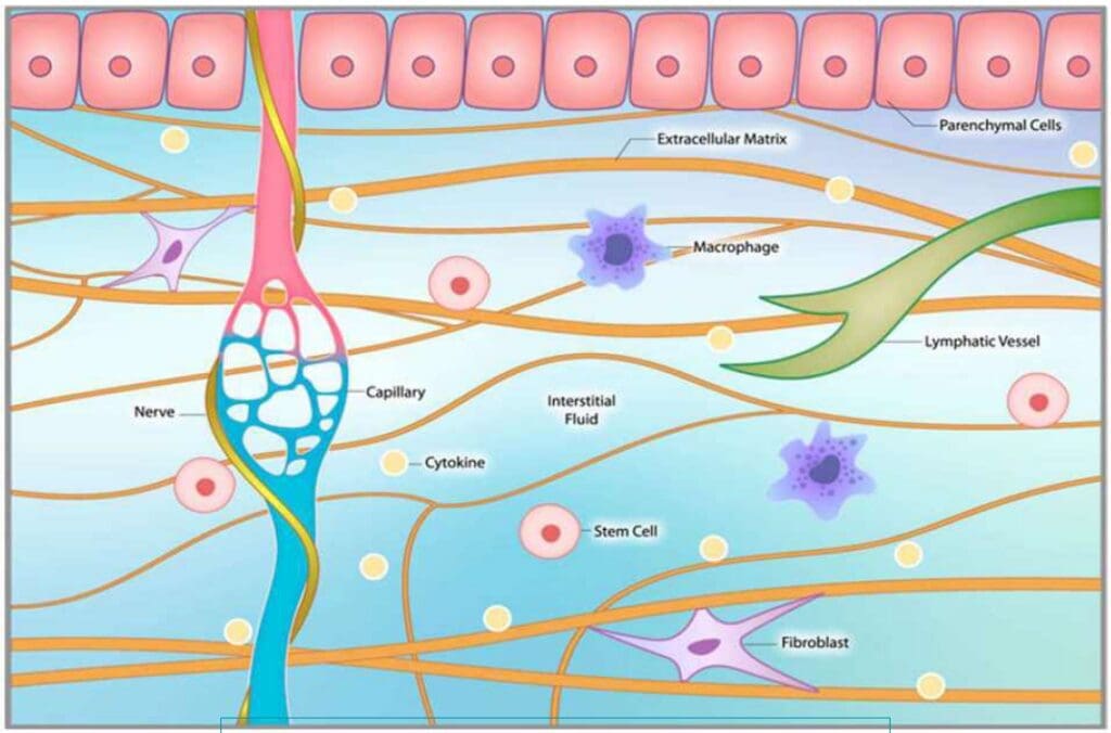
Why Hormones Are Microbiome-Dependent: The Gut–Liver–Hormone Axis
When I first connected hormone symptoms to gut physiology, I saw a pattern: many “hormone” problems began as microbiome and barrier problems. The gut microbiome—a complex community of bacteria, viruses, fungi, and archaea—regulates digestion, immune tolerance, barrier integrity, and the enterohepatic circulation that clears estrogens. From the earliest studies linking metabolic endotoxemia to insulin resistance, it has become clear that LPS-driven inflammation can disrupt cardiometabolic and reproductive health (Cani et al., 2007).
When the microbiome is balanced, commensals generate SCFAs (notably butyrate) that nourish colonocytes, tighten junctions, and reduce inflammatory signaling.
When dysbiosis develops, beta-glucuronidase-producing taxa expand, and LPS permeates, amplifying NF-κB cytokine cascades that alter hormone receptors, hepatic detoxification, and insulin signaling (Fasano, 2012; Slyepchenko et al., 2017).
Clinically, if you manage estrogen symptoms, insulin resistance, or autoimmune patterns, you are managing the microbiome—whether you realize it or not.
Dysbiosis and Leaky Gut Explained: Distinct Problems that Reinforce Each Other
Two related but distinct issues commonly coexist:
Dysbiosis: A shift away from beneficial microbes, with loss of diversity and expansion of pathobionts. Consequences include increased LPS, altered bile acid signaling, and elevated beta-glucuronidase.
Leaky gut (increased intestinal permeability): Disruption of tight junction proteins (occludin, claudins, ZO-1) allows antigens and endotoxins to enter circulation, thereby increasing systemic inflammation and immune activation (Fasano, 2012).
Why that matters for hormones:
LPS activates TLR4–NF-κB, increasing TNF-α, IL-1β, and IL-6—cytokines that reduce insulin signaling and alter steroid hormone receptor function (Cani et al., 2007).
Permeability increases immune load and oxidative stress, thereby consuming methyl donors and glutathione needed for safe phase II detox (methylation, glucuronidation, sulfation) of estrogens.
I screen for these drivers whenever patients report PMS, heavy cycles, PCOS features, endometriosis pain, acne or hair loss, mood changes, fatigue, or autoimmune flares. Correcting the gut often increases the safety and efficacy of hormone therapy.
Estrogen Metabolism 101: Enterohepatic Circulation and the Estrobolome
The liver metabolizes estrogens via phase I hydroxylation (CYP1A1, CYP1B1) and phase II conjugation (COMT methylation, glucuronidation, sulfation). Conjugated metabolites pass into bile and should be excreted. In dysbiosis, microbial beta-glucuronidase deconjugates these estrogens, promoting reabsorption and recirculation—the biochemical basis of “estrogen dominance,” even with careful dosing (Plottel & Blaser, 2011).
2-hydroxylation generally produces less proliferative metabolites.
4- and 16α-hydroxylation yield more proliferative or potentially genotoxic metabolites if methylation and conjugation are suboptimal.
In complex cases or when there is a family history of estrogen-dependent cancers, I consider urinary metabolite testing to map pathways and guide targeted support.
PCOS, Endometriosis, and Autoimmunity: What the Microbiome Adds
Recent studies sharpen the microbiome’s role:
PCOS: Dysbiosis with fewer SCFA producers and higher LPS correlates with insulin resistance, hyperandrogenism, and impaired GLP-1 signaling (Lindheim et al., 2017; Qi et al., 2019). Restoring butyrate producers improves metabolic tone.
Endometriosis: Altered microbiota, increased permeability, and immune activation correlate with symptom severity. Increased beta-glucuronidase raises estrogen recirculation that can exacerbate lesions and pain (Chen et al., 2017; Jiang et al., 2017).
Autoimmunity: Barrier dysfunction and loss of tolerogenic species permit pathobiont translocation and molecular mimicry, priming autoimmune activity (Manfredo Vieira et al., 2018).
Clinical translation: Addressing the gut can reduce hormone dosing requirements, expand the therapeutic window, and stabilize mood, sleep, and metabolism.
The Simple Question with Big Impact: Are You Pooping Daily?
I ask every patient: “Do you have a daily bowel movement?”
Estrogen metabolites exit via bile and stool. Constipation increases residence time, giving beta-glucuronidase more opportunity to deconjugate and recirculate estrogens.
Correcting bowel habits is a core risk-reduction strategy for estrogen-driven conditions.
Practical steps I use:
Increase hydration and electrolytes.
Ramp fiber to 25–35 g/day; add PHGG (partially hydrolyzed guar gum) 4–6 g/day for low-bloat prebiotic support.
Add magnesium glycinate or citrate at night for stool regularity and sleep.
Encourage postprandial walks and vagal toning (slow exhale breathing, humming).
A 3-by-3 Framework for Gut Repair: Remove, Replace, Repair
To keep things doable, I use a 3-by-3 approach:
Remove/Reduce Irritants
Clean up the diet: favor whole foods; limit alcohol, ultra-processed items, added sugars; consider a gluten-light or gluten-free trial for sensitive individuals.
Medication review: minimize NSAIDs and PPI overuse when clinically safe.
Stress load: hard-wire breath work, walks, and sleep hygiene.
Replace and Restore
Fiber and prebiotics: 25–35 g/day total fiber; add PHGG for gentle SCFA support.
Probiotics: multi-strain Lactobacillus and Bifidobacterium blends (e.g., L. rhamnosus GG, B. lactis) for barrier and immune balance.
Digestive support: bitters and meal hygiene for hypochlorhydria/slow motility; phosphatidylcholine and balanced fats for bile flow.
L-glutamine fuels enterocytes and accelerates barrier recovery.
Berberine improves the microbial balance and activates AMPK to improve insulin sensitivity.
Nutrient Foundations for Receptor-Level Hormone Action: D, K2, A, Magnesium, Iodine, Selenium, and Methylation
I frequently see patients with robust serum hormones but poor tissue effects. The missing link is often receptor signaling, cofactors, and membranes.
Vitamin D3 behaves like a secosteroid hormone that modulates transcription through the VDR. Low vitamin D is associated with all-cause and cardiovascular mortality and can blunt androgen signaling even when total testosterone appears normal (Pilz et al., 2011; Holick, 2017).
Magnesium is a cofactor for D activation (25- and 1α-hydroxylases); deficiency dampens VDR signaling (Rosanoff et al., 2016).
Vitamin K2 directs calcium into bone and away from soft tissues by activating matrix Gla protein and osteocalcin; it complements D to protect vessels and build bone (Schurgers & Vermeer, 2000; Beulens et al., 2013).
Vitamin A supports epithelial integrity, immune balance, and nuclear receptor synergy with vitamin D.
I often use an ADK formula (D3 with K2 and A) alongside magnesium to safely improve receptor-mediated effects, while monitoring 25(OH)D, calcium, and PTH (Rosen et al., 2012).
Thyroid resilience: iodine and selenium synergy
Iodine is essential for T4/T3 synthesis, but safe utilization depends on selenium-dependent enzymes (glutathione peroxidases, thioredoxin reductases) to quench the H2O2 generated during iodide organification (Ventura et al., 2017).
Inadequate selenium increases oxidative stress at the thyroid, raising the risk of autoimmunity when iodine intake rises (Gartner & Gasnier, 2003).
I pair iodine (200–400 mcg) with selenium (100–200 mcg selenomethionine) and often zinc (10–30 mg), titrated to labs and symptoms (Zimmermann & Boelaert, 2015).
Methylation for estrogen safety
Methylated B vitamins—methylfolate and methylcobalamin—support COMT-mediated methylation of catechol estrogens, reducing genotoxic stress and stabilizing phase II clearance.
These micronutrients are the bedrock that allows hormones to “dock” and trigger healthy cellular responses.
DIM and Estrogen Metabolites: Steering Toward Safer Pathways
Diindolylmethane (DIM) shifts estrogen metabolism toward 2-hydroxylation and away from 16α- and 4-hydroxylation pathways associated with proliferative and genotoxic risk (Zeligs et al., 2006; Reed et al., 2006). Preclinical studies suggest that DIM may also upregulate BRCA1 signaling and promote apoptosis in cancer cell lines (Fan et al., 2009; Li et al., 2010).
How I apply it:
Women with estrogen-dominant symptoms or unfavorable metabolite profiles: 150–300 mg/day, adjusted to labs and tolerance.
Men with prostate risk or aromatization-driven symptoms: 300–600 mg/day, personalized.
I pair DIM with omega-3s, iodine/selenium, and fiber/probiotics to support the entire estrobolome–liver–stool axis.
Rationale: By changing metabolite balance and supporting conjugation, DIM decreases receptor overstimulation and DNA-adduct risk while improving symptom stability.
Shilajit for Free Testosterone and Mitochondrial Support
Some patients—particularly young males—present with high total testosterone but low free testosterone and low vitality. Shilajit, a purified, fulvic-acid–rich resin, has randomized data showing increases in total (~31%), free (~51%), and DHT (~37%) over ~90 days at 250 mg twice daily (Pandit et al., 2016). Mechanisms likely include improved mitochondrial function, nutrient transport, and hypothalamic–pituitary–gonadal signaling.
How I use it:
In those seeking endogenous support without exogenous hormones, I combine shilajit with vitamin D, magnesium, zinc, B12, and iodine/selenium when indicated, then track changes in free T, SHBG, energy, and body composition.
Why this works: Enhancing mitochondrial ATP and cofactor availability raises tissue responsiveness; changes in binding dynamics can increase the bioactive fraction without pushing total testosterone to excessive levels.
Vitamin D as a Systemic Modulator: Barrier, Immunity, and Receptors
I routinely optimize vitamin D because it acts at the intersection of immunity, barrier integrity, and endocrine signaling. Observational data tie suboptimal 25(OH)D to higher risks across diseases (Bouillon et al., 2019). Mechanistically, D supports tight junction proteins, cathelicidin, and endocrine receptor sensitivity. Clinically, many patients feel “stuck” until D is restored to an optimal range; I often target 60–80 ng/mL with appropriate monitoring to avoid hypercalcemia (Holick, 2017; Rosen et al., 2012).
Integrative Chiropractic Care: The Neuroimmune–Endocrine Interface
As a chiropractor and nurse practitioner, I see daily how autonomic balance, fascial mobility, and pain modulation determine whether patients can absorb nutrients, move consistently, and sleep well—foundations for endocrine success.
Vagal tone and motility: Gentle spinal and cervical adjustments can influence autonomic balance, improving gut motility, secretory IgA, and anti-inflammatory vagal pathways. Patients with low vagal tone present with constipation, bloating, and poor stress resilience.
Fascia and diaphragm: Thoracolumbar fascial restrictions and diaphragmatic stiffness impair breathing mechanics and lymphatic flow, promoting sympathetic overdrive. Mobility restores circulation and reduces pain.
Pain reduction without NSAIDs: Lowering nociception decreases cortisol and protects the mucosa from NSAID-induced permeability.
Behavioral activation: When pain decreases, patients walk, train, and sleep—activities that increase SCFAs, improve insulin sensitivity, and stabilize mood.
These neurophysiologic effects align with published observations on autonomic modulation and musculoskeletal care (Pickar, 2002; Lehman et al., 2012) and help nutrition and endocrine strategies “stick” in daily life.
For examples of how we operationalize this, see my resources at Chiromed and my professional updates on LinkedIn.
A Phased, Clinic-Ready Protocol for Gut and Hormone Optimization
I layer care to build momentum and safety.
Phase 1: Stabilize and Build Trust (Weeks 0–4)
Ensure daily bowel movements; add PHGG, hydration, and magnesium as needed.
Start a multi-strain probiotic (Lactobacillus + Bifidobacterium).
Begin vitamin D3 with K2 and magnesium; consider ADK formulations.
Introduce walks after meals and fixed sleep schedules.
Provide chiropractic adjustments and diaphragmatic work to normalize autonomics and reduce pain.
Phase 2: Targeted Gut Repair and Hormone Pathways (Weeks 4–12)
Add L-glutamine 5 g/day for barrier support when indicated.
Short berberine course for endotoxemia/dysbiosis; replete with probiotics.
Add DIM if clinical or metabolite data show proliferative pathways.
Start a methylated B complex to support COMT and phase II detox.
Maintain chiropractic care cadence for autonomic and biomechanical resilience.
Phase 3: Personalize, Monitor, and Maintain (Months 3+)
Reassess symptoms, bowel habits, and targeted labs; titrate to the lowest effective doses.
Reinforce lifestyle anchors: fiber intake, movement, sleep, and stress practices.
Schedule periodic tune-ups for the spine, fascia, and breath mechanics to sustain vagal tone and support recovery.
This sequencing respects physiology and behavior: patients feel better first, then commit to more significant changes—resulting in better adherence and durable outcomes.
Special Focus: PCOS and Endometriosis
PCOS
Emphasize insulin sensitization through fiber, postprandial walks, resistance training, and, where appropriate, berberine.
Reduce LPS: probiotics, polyphenols, and barrier repair to lower endotoxemia.
Consider inositols for ovulatory support alongside gut therapy.
Monitor androgenic symptoms as gut protocols progress; improvements often track with better bile acid and SCFA signaling.
Endometriosis
Reduce beta-glucuronidase pressure via probiotics and fiber to limit estrogen recirculation.
Calm neuroimmune inflammation with omega-3s, curcumin, and sleep optimization.
Use gentle movement and manual therapy to address pelvic floor tension and diaphragm mobility; sympathetic downshift reduces pain tone.
Coordinate with gynecology; gut protocols augment, not replace, indicated care.
Case Reflection: High Total Testosterone, Low Vitality
I saw an 18–19-year-old male with low mood, low energy, weight gain, and “low-T” symptoms. His total testosterone was ~900 ng/dL—clearly not low. What we found: very low vitamin D, low B12, and signs of micronutrient insufficiency. I started a robust B-complex, ADK (D3 + K2 + A), iodine paired with selenium, and magnesium. At follow-up, his mother said, “He’s a totally different person.” Energy, mood, and cognition improved, and multiple medications were discontinued. The physiology: hormones were present, but receptor signaling and cellular machinery were underpowered. Restoring micronutrients enabled the hormones to “work.”
In other young men with high total but low free testosterone, I have added shilajit and structured resistance training. Free fractions improved, and vitality followed—without pushing total testosterone into excess.
Safety, Lab Monitoring, and Personalization
Monitor: 25(OH)D, calcium, PTH for vitamin D repletion; thyroid panel and antibodies for iodine–selenium strategies; ferritin, B12, folate, magnesium, zinc, selenium, CRP for micronutrient and inflammatory status; sex hormones including free testosterone and SHBG.
Adjust doses to labs and symptoms. If vitamin D stays low despite oral dosing, assess bile flow, fat absorption, and adherence; consider supervised loading.
Cautions:
Vitamin A: avoid hypervitaminosis; use caution in pregnancy.
Iodine: go slowly with autonomous nodules or hyperthyroidism; collaborate with endocrinology.
Zinc: long-term high dosing can lower copper; keep the balance.
DIM and shilajit: use third-party-tested products; personalize the dose.
Berberine: short targeted courses; watch for GI sensitivity and drug interactions.
How Integrative Chiropractic Care Complements Endocrine and Gut Strategies
Mechanistically, chiropractic-informed care bridges biochemistry and behavior:
Reduces nociception and sympathetic overdrive, lowering cortisol drag on thyroid conversion and gonadal axes (Lehman et al., 2012).
Improves respiratory mechanics and fascial glide, supporting lymphatic flow, nutrient delivery, and waste clearance.
Enhances vagal tone, supporting motility, secretory IgA, and peristalsis—foundations for microbiome stability.
Facilitates movement prescriptions (resistance training, mobility, aerobic intervals) that reduce aromatase activity, improve insulin sensitivity, and raise androgen receptor density.
In my practice, patients combining endocrine protocols with spinal–fascial optimization report better sleep, steadier energy, more predictable lab trajectories, and lower required doses—an elegant synergy of systems biology and hands-on care. Explore our integrative approach at Chiromed and my professional notes on LinkedIn.
Why Each Technique Matters: Systems Biology Rationale
Fiber/PHGG: Feeds SCFA producers, tightens junctions, and supports GLP-1 signaling.
Probiotics: Reduce beta-glucuronidase, improve barrier integrity, and temper endotoxemia.
L-glutamine: Primary fuel for enterocytes; accelerates epithelial repair.
Berberine: Reshapes the gut microbiota, lowers LPS levels, and activates AMPK to improve insulin sensitivity.
Methylated B vitamins: Enable COMT activity and conjugation; reduce genotoxicity of catechol estrogens.
Vitamin D + K2 + A + Mg: Align receptor signaling and calcium kinetics; protect vessels and bone.
Iodine + selenium: Support thyroid synthesis while detoxifying H2O2 to prevent autoimmune escalation.
Shilajit: Enhances endogenous androgens via mitochondrial and HPG-axis support.
Chiropractic care: Normalizes autonomic function, reduces pain, and supports movement habits that sustain microbiome and endocrine gains.
Each intervention nudges a different lever; together, they realign the system.
Clinical Observations from Practice
Across patient cohorts at my clinic, we see reproducible patterns:
Resolving constipation reduces PMS and “estrogen rollercoaster” symptoms within weeks.
Regular adjustments correlate with improved sleep and stress tolerance, enabling consistent training and meal timing that benefit the microbiome.
Vitamin D optimization often coincides with improved mood, less joint pain, and better responses to both gut and hormone protocols.
These observations are consistent with the mechanistic and clinical literature, reinforcing the rationale for why foundational steps deliver outsized results. For more, visit Chiromed and my LinkedIn updates.
Learn how bio-regulatory medicine for cellular health can enhance your health and support your body’s natural functions.
Abstract: An Introduction to Advanced Cellular Restoration and Regenerative Medicine
Hello, I’m Dr. Alexander Jimenez, and I am honored to share my clinical experience and insights with you today. As a practitioner holding dual qualifications as a Doctor of Chiropractic (DC) and a Family Nurse Practitioner (FNP-APRN), my approach is rooted in a comprehensive, systems-based understanding of human physiology. My practice is dedicated to integrating the latest findings from leading researchers in the field, employing modern, evidence-based research methods to navigate the complex landscape of chronic illness, aging, and performance optimization. This educational post is designed to move beyond surface-level discussions and delve into the intricate biochemical and physiological mechanisms that govern our health. My goal is to present this information not as a rigid lecture but as an educational dialogue, sharing insights from modern, evidence-based research in a narrative format that is both comprehensive and relatable. In the sections that follow, we will examine several key conceptual areas in detail. We will begin by exploring the pleiotropic nature of natural molecules and therapies, likening them to “Swiss Army knives” for their multifaceted ability to modulate inflammation, reduce fibrosis, protect cells, and orchestrate healing. We’ll explore why a single approach is often insufficient and how combining therapies such as peptides, light therapy, and advanced oxygen therapies can create a powerful synergistic effect. Central to this is the extracellular matrix (ECM) and the role of myofibroblasts in healing versus fibrosis. Next, we will navigate the practical yet challenging aspects of clinical practice. I’ll share my personal methodology for ensuring patients can access necessary and affordable lab testing without jeopardizing their future insurability, emphasizing patient education and advocacy in today’s medical economy. We will also address the operational hurdles of integrating advanced therapies, including the prohibitive cost of certain equipment and the critical importance of proper technique and patient safety. This includes a deep dive into Extracorporeal Shockwave Therapy (ESWT), differentiating it from ultrasound and explaining how its mechanotransduction properties reboot stalled healing processes.
A significant portion of our discussion will be dedicated to the cutting edge of regenerative medicine. We will explore novel treatments, such as Dezawa MUSE cells, and clarify what makes these stress-enduring stem cells unique. This leads to an exploration of cellular memory and energetic transfer, examining how information can be stored in tissues. We will then apply these principles to clinical scenarios, including a detailed case study on managing post-traumatic osteoarthritis using a hierarchical approach that prioritizes foundational support before escalating to agents such as Thymosin Beta-4 (TB-500) and Body Protection Compound 157 (BPC-157). This underscores the principle of treating the individual rather than the diagnosis. Finally, we will synthesize these concepts in a comprehensive, interactive case study analysis of a 45-year-old woman with a complex constellation of symptoms. We will deconstruct her case, analyze her lab work, and construct a logical, evidence-based treatment plan from the ground up, demonstrating how to identify root causes and apply targeted interventions such as Selank, Tesamorelin, and GLP-1 agonists. We will conclude by reviewing the remarkable clinical outcomes achievable with an integrated, patient-centered approach, demonstrating the profound impact of moving beyond symptom management to restore foundational health. This post is a comprehensive resource that offers a deeper appreciation of the intricate, interconnected nature of the human body and the powerful tools we have to foster true healing and vitality.
The Critical Importance of Sourcing and Clinical Integrity in Regenerative Medicine
Before we delve into the core scientific concepts, I feel it’s imperative to address a fundamental aspect of my practice and a cornerstone of responsible medicine: the integrity of our tools and the nature of the practitioner-patient relationship. In the world of regenerative therapies, especially with substances like peptides, the source is everything. My journey into this field wasn’t just academic; it was a boots-on-the-ground investigation. It began over a decade ago when a representative from an online peptide company unexpectedly appeared in my office. He was selling powders, and this was long before peptides were a common topic of discussion in clinical circles. My curiosity was piqued not only by the products but also by the person and his story. This encounter set me on a path. I felt a profound responsibility to understand exactly what I might one day offer my patients. This wasn’t a responsibility I could delegate. I couldn’t just trust a website or a salesperson’s word. So, I traveled. I accompanied this individual to MIT, where I connected with distinguished scientists at the Broad Institute in Cambridge. They were deeply immersed in translational medicine, working on bringing laboratory discoveries to clinical practice. They recognized my potential as a collaborator, a clinician willing to bridge that gap. But I knew I had to proceed with the utmost caution. I couldn’t afford to put my patients or my license at risk. This led me down a literal and figurative path of exploration—visiting facilities, sometimes in back alleys, to see the steel tables and the synthesis processes firsthand. I needed to know the conditions under which these compounds were produced to assess the quality control, or lack thereof. My search for knowledge and quality extended to Europe, where I began meeting peptide science founders and researchers in 2016 and 2017. These were the pioneers, the people who had been studying these molecules for decades. I wasn’t just an attendee at a conference; I immersed myself in their world, becoming a friend and a trusted clinical colleague. They were researchers, and I was often the only clinician in the room, bringing real-world patient challenges to the table. For instance, I was working with a small, desperate group of patients with massive brain injuries who had exhausted all other options. I began using peptides such as BPC-157, then attended meetings with leaders in the field and learned that they had been using Thymosin peptides in humans since the 1960s. They had governmental permission in other countries to use these to keep children with DiGeorge’s Syndrome, a severe immunodeficiency, alive. This entire experience solidified a core principle: I must know my source. I need to work with pharmacists and manufacturers who can guarantee purity, explain peptide isomers, and don’t change their formulations arbitrarily. This is why I build personal relationships with my suppliers. I visit them. It’s the same level of personal commitment I bring to my patients. If I am going to offer a treatment, I must be confident that I am providing the highest-quality, safest option available—the same Tender Loving Care (TLC) I would want for myself or my family.
Empowering Patients Through Education and Shared Decision-Making
This brings me to the second cornerstone: the patient’s role. I do not make decisions for my patients. My role is to educate, lay out the options, risks, benefits, and the current state of scientific understanding. The final decision always rests with the patient. This is the essence of informed consent. I work with the medical board and am consistently impressed by their diligence, but I’m also aware of the risks practitioners take, sometimes without a full appreciation of the dangers. I once had a conversation with a colleague, a clinician and co-owner of a practice, who casually mentioned taking a powerful medication without a second thought. To me, this demonstrated a lack of appreciation for the profound biological impact of these substances. It’s far more dangerous than a simple cortisol shot, a procedure that itself is fraught with peril. I’ve witnessed the devastating consequences of seemingly routine procedures. I think of a patient, a vibrant man of Italian heritage, a passionate tennis player, who developed a knee problem. His doctors, intending to help, administered a steroid injection. And then another. Within two months, the tissue had degraded so severely that he required open-knee surgery. This surgery, performed on a body now in a state of malnourishment from the catabolic effects of the steroids, initiated a downward spiral. He developed a bleeding disorder, became emaciated, and ultimately, he passed away. This tragic story is a stark reminder that even well-intentioned interventions can have catastrophic, unforeseen consequences. Therefore, my approach is one of partnership. I never convince anyone to do anything. When a patient is hesitant or upset, I don’t push. I calmly state, “I understand that there are things you don’t yet understand, and that is okay. You are on your own journey. We need to determine where you want to be on that path. Right now, it seems we are not aligned. And if we’re not aligned, it’s not the right time for me to treat you.” I would love to help, but the foundation must be a participatory dialogue. When the patient is ready to have that conversation and engage as an equal partner, we can move forward. In medicine, especially when exploring the frontiers of regenerative therapies, we are always managing risk. The best we can do is to ensure that every decision is fully informed, deeply considered, and truly shared.
Fascia: The Unsung Hero of Healing and Communication
For decades, medical textbooks depicted fascia as little more than biological shrink-wrap—a passive, inert barrier that held our muscles and organs in place. We now know this view is profoundly outdated. Modern research, through advanced imaging and biomechanical studies, has revealed fascia as a dynamic, intelligent, and communicative system. It is a tensegrity structure, a complex, body-wide web of connective tissue that is intricately involved in every aspect of our physiology. Fascia is not a passive barrier. It is the fundamental fabric of our extracellular matrix (ECM), the environment in which all of our cells live. This matrix is a bustling neighborhood composed of cells, signaling molecules, and structural proteins. The cells within the fascia, what they secrete, and the structural components they produce—such as collagen and elastin—collectively organize how our bodies heal. This process determines whether we successfully restore tissue integrity or are left with a dysfunctional scar. Understanding this is absolutely critical for any effective therapeutic intervention.
Myofibroblasts: The Architects and Potential Saboteurs of Healing
Within this fascial universe, a specific cell type plays the leading role in wound repair: the myofibroblast. When you sustain an injury—a cut, a tear, a strain—your body initiates a beautifully orchestrated inflammatory cascade. Local cells release signals that call fibroblasts to the site of injury. These fibroblasts are the workhorses of tissue repair. In response to specific mechanical and chemical cues, they differentiate into myofibroblasts. These specialized cells are remarkable. They contain contractile filaments, much like those in smooth muscle cells, which allow them to physically pull the edges of a wound together. They are the architects of healing, diligently spinning a new scaffold of collagen to form a fibrin clot and bridge the damaged tissue. A little bit of this organized scarring is essential for survival. It provides a quick, strong patch to maintain structural integrity. The problem arises when this process doesn’t turn off. If the inflammatory signals persist—due to chronic injury, systemic inflammation, or metabolic dysfunction—the myofibroblasts remain activated. They continue to churn out collagen relentlessly. This is when healing goes awry. The organized, functional scaffold of a healthy scar devolves into a dense, disorganized, and restrictive mass of fibrotic tissue. This is the cytokine storm that creates a vicious cycle. The thick tissue restricts blood flow, trapping waste products and preventing nutrients from reaching the cells. This hypoxic and toxic environment triggers more inflammation, which in turn activates more myofibroblasts, leading to more fibrosis. The tissue loses its elasticity and glide, becoming stiff and painful. This process isn’t limited to musculoskeletal injuries. It happens in our organs as well. Pulmonary fibrosis, liver cirrhosis, and the stiffening of cardiac tissue in heart failure are all manifestations of this same underlying pathology: chronic myofibroblast activation and runaway fibrosis.
A Clinical Case Study: The Systemic Impact of Localized Fibrosis
To illustrate how devastating this fibrotic state can be, let me share the story of a dear friend and patient. He is a brilliant man who, for various reasons, has been managing his health in a fragmented way. I had seen his labs years ago and noted some concerning markers—signs of anemia and kidney stress. But he didn’t follow up consistently. He called me recently, five times in one week, his voice filled with despair. He had just seen his cardiologist, who informed him that his heart failure had significantly worsened. He was heartbroken and terrified. Listening to him, I immediately recognized the pattern. His body was in a state of hypercoagulation, a fibrogenic state strikingly similar to a systemic condition called Disseminated Intravascular Coagulation (DIC). In DIC, the body’s clotting mechanisms go into overdrive, creating micro-clots throughout the circulatory system, which can lead to organ failure. In my friend’s case, the primary organ was the heart, but the underlying principle is the same. The fibrinogen levels in his blood were likely elevated—a marker I often see alongside high ferritin in inflammatory states. This sticky, clot-promoting environment is a direct consequence of the systemic inflammation that drives fibrosis. Our circulatory system is not separate from our tissues; it’s an information highway. Inflammatory cytokines and fibrotic signaling molecules produced in one area circulate throughout the body, triggering dysfunction elsewhere. His heart wasn’t failing in a vacuum; it was failing within a systemic environment that was primed for fibrosis and clotting. I asked him a simple question: “What was your NT-proBNP level?” This is a crucial blood marker used to track the severity of heart failure. A healthy level is very low. In heart failure, it rises dramatically. With effective treatment, you can see this number drop from, say, 1,500 pg/mL down to 300 pg/mL, indicating that the strain on the heart is decreasing. He didn’t know his number. He hadn’t been tracking it, despite my recommendation a year prior. He was looking for a cure, a magic bullet. But medicine rarely offers cures for chronic conditions like this. What we can do is manage the underlying physiology. We can work to turn off the inflammatory signals, break the fibrotic cycle, and support the body’s innate capacity for repair. His situation is a powerful, albeit tragic, example of how a localized problem—in this case, the heart—is deeply enmeshed in a systemic web of inflammation and fibrosis, demonstrating the critical need for a holistic, systems-based approach to healing.
Harnessing Sound for Healing: An In-Depth Look at Shockwave Therapy
Given this understanding of fascia, fibrosis, and the devastating cycle of chronic inflammation, the logical question is: how can we intervene? How can we communicate with the tissue to break this cycle and promote true regeneration? This is where modalities that act at the biophysical level become incredibly powerful. One of the most effective and well-researched of these is Extracorporeal Shockwave Therapy (ESWT). My friend and colleague, Dr. P, does fantastic work with frequency-specific microcurrent and other vibrational therapies. Our conversations always circle back to the same conclusion: it’s not an “either/or” situation; it’s a “this and” approach. We are, at our core, electrical beings. Our tissues exhibit piezoelectricity, meaning they generate an electrical charge in response to mechanical stress. Our bodies are constantly interpreting and responding to physical forces. ESWT leverages this principle to restart a stalled healing process.
The Science of Shockwave: Beyond Ultrasound
First, let’s be clear about what a shockwave is and isn’t. People often confuse it with therapeutic ultrasound, but they are fundamentally different.
Ultrasound waves are biphasic and continuous. As you can see in the upper diagram on the screen, they have a symmetrical sine-wave pattern with both positive (compressive) and negative (tensile) phases. These continuous oscillations primarily generate a thermal (heating) effect in the tissue.
Shockwaves, in contrast, are monophasic. They are single-pulse acoustic waves characterized by a very rapid, high-pressure positive phase followed by a much smaller, longer negative phase. As the lower diagram illustrates, there is a dramatic, near-instantaneous rise to peak pressure. This is not a continuous oscillation. This unique physical property is what creates a strong mechanotransduction effect—the process by which cells convert a mechanical stimulus into a biochemical response.
ESWT is a non-invasive technology that uses a device to generate these high-intensity sound waves and deliver them to a specific area of the body. You might see it called Acoustic Wave Therapy or a similar name, but the principle is the same. It’s about delivering a precise mechanical force to stimulate a biological cascade. The technology has its roots in urology, where it has been used for decades as lithotripsy to break up kidney stones. It later found application in orthopedics for treating non-union fractures, in which bones have failed to heal on their own. Now, its application in musculoskeletal and regenerative medicine is exploding.
Radial vs. Focused Shockwave: Choosing the Right Tool for the Job
There are two main types of shockwave therapy, and they are not interchangeable. They have different methods of generation, physical characteristics, and clinical applications.
Radial Shockwave Therapy (rESWT): This is the more common type you’ll see. The device has a handpiece that looks a bit like a pistol. Inside, a projectile is accelerated by compressed air and strikes a metal applicator. The energy created at the applicator head then radiates outwards into the tissue in a divergent, or radial, pattern.
Characteristics: Energy is highest at the surface and dissipates as it moves deeper. It’s less precise and covers a broader area. The sensation is often described as a strong, percussive tapping.
Best Use: Radial shockwave is excellent for treating larger, more superficial areas. I often use it as a preparatory treatment. It’s gentler and more diffuse, making it perfect for “loosening up” the surrounding fascial tissue before targeting a specific lesion. It helps improve circulation and downregulate hypersensitive nerve endings across a wide area.
Focused Shockwave Therapy (fESWT): This technology is more advanced and was available in Europe long before it was widely adopted in the U.S. It uses an electrohydraulic, piezoelectric, or electromagnetic source to generate the wave, which is then focused by a lens or reflector to a specific point deep within the tissue, much like a magnifying glass focusing sunlight.
Characteristics: The energy converges at a precisely adjustable focal point, enabling treatment of deep structures without significantly affecting the overlying tissue. It can penetrate much deeper than a radial shockwave.
Best Use: Focused shockwave is the tool of choice for targeting specific deep pathologies, such as tendinopathies, non-union fractures, or trigger points within a muscle belly.
In my practice, I find the most effective approach is to use both together. I might start a treatment plan with radial shockwave to prepare the entire kinetic chain—calming the nervous system and improving tissue compliance. Then, in subsequent sessions, I can introduce focused shockwave therapy to target the primary lesion with greater precision and energy.
The Physiological Cascade: How Shockwave Reboots Healing
When a shockwave enters the tissue, it creates a cascade of biological effects that effectively reboot the healing process, shifting the tissue from a chronic, degenerative state to an acute, regenerative one.
Releases Substance P and Induces Analgesia:Substance P is a neuropeptide that acts as a primary neurotransmitter for pain. Chronic pain conditions are often associated with high concentrations of Substance P. The intense pulses of shockwave therapy cause a depletion of Substance P in the local nerve endings and inhibit its resynthesis. This leads to a significant and often immediate reduction in pain—an analgesic effect.
Enhances Circulation (Angiogenesis): Mechanical stress triggers the release of key angiogenic growth factors, such as Vascular Endothelial Growth Factor (VEGF). This stimulates angiogenesis and neovascularization—the formation of new blood vessels. You can see this in the diagrams showing tissue before and after treatment. The “before” tissue has sparse blood flow, while the “after” tissue shows a rich new capillary network. This is crucial. Improved blood flow brings oxygen and nutrients essential for healing and, just as importantly, flushes out metabolic waste products and inflammatory mediators that perpetuate the chronic state.
Stimulates Collagen Production and Breaks Down Calcification: The therapy initiates a controlled, pro-healing inflammatory response. This functionally stimulates fibroblast proliferation. It encourages them to produce fresh, healthy Type I and Type III collagen, the building blocks of healthy tendons and ligaments. For conditions like “calcific tendinitis,” the mechanical force of shockwaves can physically break up calcium deposits, which the body can then clear through the lymphatic system and improved circulation. I’ve seen remarkable reductions in calcified scar tissue.
Increases Cell Membrane Permeability and Restores Balance: At the cellular level, the shockwave’s shear force temporarily increases cell membrane permeability. This helps re-equilibrate the flow of ions such as sodium, potassium, and calcium, which is fundamental to restoring normal cell function, nerve signaling (action potentials), and mitochondrial energy production. You are essentially restoring the balance between tension and compression at the micro level, allowing the cells to breathe and function properly again.
Mobilizes Stem Cells: Research has shown that shockwave therapy can stimulate the mobilization and migration of the body’s own mesenchymal stem cells to the site of injury. These are the master repair cells, capable of differentiating into various tissue types to facilitate regeneration. By using shockwave, we are essentially ringing the dinner bell and calling the body’s intrinsic repair crew to the job site.
The Principle of Hormesis: Therapeutic Stress for Adaptation
It’s vital to understand that the magic of shockwave—and many other regenerative therapies like Hyperbaric Oxygen Therapy (HBOT) or intermittent fasting—lies in the principle of hormesis. Hormesis is a biological phenomenon in which a beneficial effect results from exposure to a low dose of an agent that is otherwise toxic or lethal at higher doses. When you apply shockwave to tissue, you create a controlled microtrauma. The therapy itself is the stressor. But the real healing happens after the treatment is over. The body perceives this controlled stress and mounts a powerful adaptive response. It up-regulates its own anti-inflammatory and regenerative systems. This response doesn’t just last an hour; it can last days or weeks. It’s the same principle as HBOT. When you are in the chamber at high pressure (e.g., 2.0 ATA), your plasma becomes supersaturated with oxygen (hyperoxia). But when you come out, your body experiences a state of relative hypoxia. This swing is the hormetic trigger that stimulates the production of stem cells and other healing factors. Even low-pressure HBOT (e.g., 1.3 ATA) is now showing profound effects, especially in neurological healing, because it’s the change in oxygen tension that drives adaptation. The body loves adaptation. Shockwave therapy is a perfect example of using a targeted physical stressor to provoke a robust, positive biological adaptation.
Practical Application and Clinical Evidence: From Head to Toe
The applications for shockwave therapy are vast. Hundreds of peer-reviewed scientific studies have validated its efficacy for a wide range of musculoskeletal conditions, making it a highly cost-effective and evidence-based treatment.
Plantar Fasciitis: This is one of the most well-studied indications, with success rates often exceeding 80-90%, even in chronic cases that have failed all other conservative treatments.
Tendinopathies: Achilles tendinopathy, patellar tendinopathy (“jumper’s knee”), rotator cuff tendinopathy, and lateral epicondylitis (“tennis elbow”) all respond exceptionally well. ESWT is effective at breaking down the disorganized, degenerative tissue and stimulating the production of healthy new tendon fibers.
Post-Stroke Spasticity: Shockwave therapy can be used to treat muscle spasticity and contractures after a stroke, improving range of motion and function.
Bone Healing: As mentioned, it is highly effective for delayed union or nonunion fractures, stimulating bone-forming cells (osteoblasts) to resume activity. A case study on the screen shows the healing of a horse’s leg fracture, comparing a screw-only repair to a screw-plus-shockwave repair. The shockwave group shows significantly faster and more robust healing, getting the athlete back to play sooner.
Myofascial Pain and Scar Tissue: It is excellent for breaking up dense scar tissue, whether from surgery or chronic injury, and for deactivating stubborn trigger points. I have even used it to break up cosmetic cellulite, which is fundamentally a problem of fibrotic bands pulling down on the skin.
I once treated my own hand after a particularly nasty injury. I made some Platelet-Rich Plasma (PRP), performed shockwave therapy on the area, and then injected the PRP. The initial response was intense—my hand was incredibly red and painful. I was worried I had overdone it. But I trusted the process and continued to work with it gently. The result was a complete recovery, with no residual scar tissue or dysfunction. I was shocked by how well it worked, especially in areas prone to adhesions, such as dentists’ hands or manual therapists’.
Important Contraindications and Precautions
While incredibly safe, shockwave therapy is not for everyone. There are some key contraindications:
Active Malignancy: You never want to apply shockwave over a known tumor, as it could potentially encourage metastasis by increasing circulation.
Pregnancy: It should not be used over the abdomen or the lower back of a pregnant woman.
Coagulation Disorders or Anticoagulant Use: If a patient has a known clotting disorder or is taking blood thinners such as Warfarin, caution is essential. If my heart failure patient had a known clot in his heart (an atrial thrombus), I would absolutely not perform shockwave anywhere on his body.
Over Open Growth Plates: It should not be used over the epiphyseal plates of growing children.
Recent Corticosteroid Injection: Be cautious. Steroids weaken tissue. I recommend waiting at least six weeks after a steroid injection before applying shockwave to that area to avoid the risk of tissue rupture.
In conclusion, shockwave therapy represents a paradigm shift in how we treat chronic musculoskeletal and fibrotic conditions. By moving away from purely biochemical or suppressive interventions (like NSAIDs or steroids) and embracing a biophysical approach, we can communicate directly with the body’s tissues in a language they understand—the language of force and adaptation. We can break the cycle of chronic pain and degeneration and empower the body to do what it does best: heal itself.
A2M (Alpha-2-Macroglobulin): The Molecular “Mop” for Joint and Tissue Inflammation
In my practice, I am constantly seeking therapies that are not only effective but also work in harmony with the body’s natural physiology. One of the most remarkable tools in our regenerative arsenal is Alpha-2-Macroglobulin(A2M). This isn’t a synthetic drug or an external compound; it’s a large plasma glycoprotein that your own body produces, primarily in the liver. Think of it as the body’s innate cleanup crew, a molecular “mop” specifically designed to seek out and neutralize the agents of tissue destruction. To truly appreciate the power of A2M, we must first understand the battlefield of a chronically inflamed joint, like an osteoarthritic knee. Within that joint space, a vicious cycle is at play. The initial injury or age-related wear and tear triggers an inflammatory response. This response, if not properly resolved, leads to the overproduction of a class of enzymes known as proteases. These include catabolic proteases like matrix metalloproteinases (MMPs) and ADAMTSs (A Disintegrin and Metalloproteinase with Thrombospondin Motifs).
In a healthy state, these enzymes are crucial for normal tissue remodeling—breaking down old, damaged cartilage so it can be replaced. However, in a state of chronic inflammation, they become hyperactive and unregulated. They begin to relentlessly chew away at the healthy cartilage, degrading the very collagen and proteoglycan structures that give the joint its cushion and smooth-gliding surface. This enzymatic destruction is a primary driver of pain, stiffness, and the progressive loss of function we see in osteoarthritis and other degenerative conditions. This is where A2M makes its dramatic entrance. A2M is a massive protein with a unique structure, often described as a “venus flytrap.” Its primary function is to act as a pan-protease inhibitor, meaning it can trap and inactivate a broad spectrum of these destructive enzymes. Here’s a step-by-step breakdown of its sophisticated mechanism:
Detection and Baiting: The A2M molecule contains a “bait region.” When a destructive protease, such as an MMP, approaches, it is attracted to the bait region and cleaves it, thinking it’s just another protein to degrade.
The “Trap” Mechanism: This cleavage triggers a massive conformational change in the A2M molecule. It essentially collapses around the protease, physically trapping it within a molecular cage. This is a covalent bond, meaning it’s incredibly strong and irreversible.
Neutralization and Clearance: Once trapped, the protease is completely neutralized. It can no longer access and degrade cartilage or other tissue components. Scavenger receptors then recognize the entire A2M-protease complex on cells like macrophages, which engulf and clear it from the joint space, effectively removing the destructive agent from the environment.
By concentrating A2M directly in an inflamed joint or soft-tissue area, we are essentially giving the body a super-dose of its own natural defense mechanism. We are stopping the chemical source of the breakdown. This is why I find A2M so valuable, particularly as a preparatory step before other regenerative procedures. Imagine you are trying to plant a new garden. If the soil is full of weeds and toxins (the proteases), your new seeds (stem cells, for example) will struggle to grow. A2M therapy clears the soil. It creates a healthier, less inflammatory, and more pro-regenerative microenvironment. If I am considering a stem cell or PRP procedure for a patient with significant joint degeneration, I will often recommend a preliminary A2M injection. By first reducing the levels of these hyperactive proteases, we ensure that the valuable growth factors and signaling molecules introduced by the subsequent procedure aren’t immediately degraded. It gives the regenerative cells a much better chance to survive, differentiate, and orchestrate the repair process. The procedure itself involves a significant blood draw from the patient, as A2M needs to be concentrated from a large volume of plasma. My practice is equipped with the specialized centrifuge and filtration system required for this. While the kits and the process can be costly, the investment is often justified by the profound and long-lasting results. We are not just masking pain; we are intervening in the core biochemical pathway of tissue destruction. For patients suffering from neuropathic pain, joint degeneration, or chronic soft tissue inflammation, A2M offers a powerful, evidence-based strategy to halt damage and pave the way for true healing.
Photobiomodulation (PBM): Harnessing Light to Energize Cellular Healing
I firmly believe that we are, at our core, electrical and energetic beings. Our bodies are constantly humming with biochemical and bioelectrical activity. It stands to reason, then, that we can use external energy sources to influence and optimize these internal processes. This is the fundamental principle behind Photobiomodulation (PBM). This therapy uses non-ionizing light sources, including lasers, LEDs, and broadband light, in the visible and near-infrared spectrum to stimulate healing, relieve pain, and reduce inflammation. For too long, medicine has been dominated by a purely chemical paradigm. PBM reintroduces the physics—specifically, the photoelectric effect—into our understanding of healing. This concept, for which Einstein won a Nobel Prize, describes how light, acting as a particle (a photon), can transfer its energy to an electron when it strikes a material. In the context of our bodies, this “material” is a specific molecule within our cells called a chromophore. The primary chromophore targeted by PBM is Cytochrome C Oxidase, a crucial component of the mitochondrial electron transport chain. Think of the mitochondria as the power plants of our cells, responsible for producing Adenosine Triphosphate (ATP), the universal energy currency of the body. Here’s how PBM sparks this cellular revitalization:
Light Absorption: When photons of a specific wavelength (typically in the red and near-infrared range, roughly 600-1000nm) penetrate the tissue, they are absorbed by Cytochrome C Oxidase within the mitochondria.
Nitric Oxide Dissociation: In stressed or inflamed cells, a molecule called Nitric Oxide (NO) can bind to Cytochrome C Oxidase, competitively inhibiting oxygen and effectively “clogging up” the ATP production line. The energy from the absorbed photon is just enough to break this bond, causing the NO to dissociate and release.
Restoration of Oxygen Flow & ATP Production: With the NO “blockage” removed, oxygen can once again bind freely, restoring the electron transport chain to its full efficiency. This results in a significant increase in ATP production. More ATP means more energy for the cell to perform its essential functions, including repair and regeneration.
Signaling Cascades: The release of NO isn’t just a side effect; it’s a therapeutic event in itself. The transient burst of NO acts as a powerful signaling molecule, leading to vasodilation (widening of blood vessels). This improves local circulation, bringing more oxygen and nutrients to the injured area and facilitating the removal of waste products. Furthermore, this process initiates downstream signaling cascades that activate transcription factors such as NF-κB (Nuclear Factor kappa B), leading to the production of a range of proteins involved in cell proliferation, migration, and tissue repair.
Reduction of Oxidative Stress: PBM has also been shown to modulate Reactive Oxygen Species (ROS). While high levels of ROS cause oxidative stress and damage, the modest, transient increase induced by PBM appears to act as a beneficial signaling mechanism, upregulating the body’s own endogenous antioxidant defenses.
The clinical applications of these mechanisms are vast. In my practice, I utilize a variety of light therapy devices, from targeted wands to whole-body systems like a BioCharger, which combines multiple forms of energy. For a patient with peripheral neuropathy, for instance, applying PBM to the affected limbs can help reduce pain and inflammation by increasing circulation and providing the damaged nerves with the ATP they desperately need to repair their myelin sheaths. For a joint injury, it reduces swelling and pain. In the context of brain health, transcranial PBM is being explored to improve cognitive function by enhancing mitochondrial activity in neurons. It’s crucial to understand that we are constantly bombarded by various energies in our modern environment, many of which are detrimental. Using targeted, therapeutic energies like PBM helps counteract this and actively support our body’s electrical nature. It is a modern, elegant, and deeply physiological approach that provides pain relief, reduces inflammation, improves circulation, and supports healing in a wide variety of neurological and musculoskeletal conditions. It’s a foundational therapy that helps power the very engine of cellular life.
The Foundational Trinity: Nourish, Measure, and Personalize
While advanced technologies like A2M and Photobiomodulation are powerful tools, they are most effective when applied to a body that has the fundamental building blocks it needs for repair. You cannot build a sturdy house with rotten wood and missing bricks. Similarly, you cannot expect the body to regenerate without the proper nutritional foundation. My approach to patient care is always anchored in this principle: nourish, nourish, nourish. The ultimate goal is to nurture the extracellular matrix (ECM). The ECM is the intricate, non-cellular three-dimensional network that surrounds all our cells. I often describe it to patients as the “soil” in which our cellular “seeds” (including stem cells) are planted. This soil is composed of a complex mesh of proteins like collagen and elastin, as well as glycoproteins and proteoglycans. It provides structural support, but far more importantly, it’s the medium through which cells communicate, receive nutrients, and get signals to grow, migrate, or differentiate. If this matrix is inflamed, dehydrated, or deficient in key nutrients, cellular function will be compromised. Stem cells will not receive the proper signals, waste products will accumulate, and the entire regenerative process will stall. Therefore, my priority is to ensure this “soil” is rich and fertile.
Measure, Don’t Guess: The Power of Advanced Diagnostics
This is where my philosophy diverges sharply from a one-size-fits-all approach. I never make things up. I need objective data to guide my clinical decisions because the risk of being wrong is too high when you are simply guessing. I rely on advanced functional testing to create a precise, personalized roadmap for each patient. A cornerstone of this is the Organic Acid Test (OAT). This comprehensive urine test provides a snapshot of the body’s metabolic processes. It can reveal incredible detail about:
Mitochondrial Function: Are you efficiently converting food into energy?
Neurotransmitter Levels: Are there imbalances in dopamine or serotonin metabolites that could be contributing to mood or pain perception?
Detoxification Pathways: Is your body effectively clearing toxins? The OAT measures markers for glutathione production, a master antioxidant crucial for detoxification.
Nutrient Deficiencies: This is a critical one. The OAT can show functional deficiencies in key vitamins and minerals. It tells me whether the body has enough zinc, selenium, B vitamins, and amino acids, such as glycine, to perform its necessary enzymatic reactions.
When I see a low glutathione level, I know the body’s ability to handle oxidative stress is compromised. This isn’t just an abstract finding; it has direct clinical implications. It means the patient is more susceptible to inflammation and cellular damage. My intervention is then highly targeted: I will support glutathione production with its precursors, such as N-acetylcysteine (NAC), glycine, and selenium.
Personalized Supplementation: The Right Tools for the Right Job
Based on this detailed testing, I can recommend a highly specific nutrient protocol. This isn’t about throwing a generic multivitamin at the problem. It’s about surgical precision.
Essential Fatty Acids: Everybody needs some form of omega fatty acids, but the type and ratio matter immensely. Based on their lab work and clinical presentation, a patient might need more EPA/DHA from fish oil for its potent anti-inflammatory effects. Another patient, perhaps with skin issues or hormonal imbalances, might benefit more from GLA (Gamma-Linolenic Acid), an omega-6 fatty acid. I measure, so I know.
Phospholipids: Phosphatidylcholine is a vital component of every cell membrane in your body. It ensures membrane fluidity, which is essential for proper cell signaling and nutrient transport. For patients with neurological issues or liver detoxification challenges, supporting cell membrane health with phosphatidylcholine can be a game-changer.
Minerals: Zinc is a cofactor in over 300 enzymatic reactions in the body. As we will see in the case study, a deficiency can have widespread effects, from impaired immune function to hair loss. Selenium is a critical cofactor for the enzyme glutathione peroxidase. Without it, your primary antioxidant system cannot function properly.
My approach is flexible and patient-centered. If a patient is overwhelmed, we start with just one thing. Let’s fix the most critical deficiency first. My team and I provide extensive follow-up, acting as partners and guides. We check in, monitor progress, and once the first change has been integrated, introduce the next. It’s a step-by-step process of rebuilding the body from the ground up. Sometimes, we start with hormones if that is the patient’s most pressing concern. Other times, we start with the gut. The entry point can be anywhere, just like reading a book—you don’t always have to start on page one. The key is to be confident in your understanding of the underlying physiology and to use objective data as your compass. The ultimate goal remains the same: to create a nutrient-rich, well-supported internal environment where the body’s own regenerative potential can be fully unleashed.
The Pleiotropic Power of Nature’s Molecules: The Swiss Army Knife Approach to Healing
In the world of functional and regenerative medicine, we often look to nature for inspiration. We find that the most effective molecules and therapeutic interventions rarely have just one job. Instead, they exhibit pleiotropic effects, meaning that a single agent can produce multiple, often seemingly unrelated, biological responses. Think of these compounds not as a simple key fitting a single lock, but as a sophisticated Swiss Army knife, equipped with a variety of tools to address a complex problem from multiple angles. Let’s consider a common flavonoid found in many plants. This single small molecule might simultaneously act as a powerful antioxidant, neutralizing damaging free radicals. At the same time, it could modulate inflammatory pathways such as NF-κB, effectively turning down the “volume” of systemic inflammation. It might also inhibit enzymes that contribute to scar tissue formation (fibrosis), promote the detoxification of harmful compounds, and subtly influence neurotransmitter balance in the brain. This multi-pronged action is not an accident; it’s a hallmark of biological efficiency. The body’s systems are deeply interconnected, and a problem in one area—say, chronic inflammation—inevitably spills over to affect others, like metabolic health, immune function, and neurological integrity. A therapy that only targets one specific point in this complex web is often fighting an uphill battle. This is where the “Swiss Army knife” approach becomes so valuable. When a patient presents with a constellation of issues, I ask myself a series of questions:
What is the primary driver of their dysfunction? Is it unchecked inflammation?
Am I trying to modulate an overactive immune response or elevate a suppressed one?
Is there a need to reduce fibrosis and break down restrictive scar tissue, perhaps in a joint or an organ?
Do I need to protect the cells from ongoing oxidative stress, a process known as cytoprotection?
Is the goal to modulate the intricate signaling of the gut-brain axis?
The beauty of pleiotropic therapies is that we can often address several of these needs simultaneously. This principle extends beyond plant molecules. Consider some of the advanced tools we use in our practice:
Peptide Therapy: Peptides are short chains of amino acids that act as signaling molecules. A peptide like BPC-157 is a prime example of a pleiotropic agent. It is renowned for its ability to heal the gut lining. Still, it also promotes angiogenesis (the formation of new blood vessels), reduces inflammation, protects organs, and accelerates the healing of tendons and ligaments. It doesn’t just “fix” one thing; it orchestrates a broad, systemic healing response. It is the quintessential Swiss Army knife in our therapeutic toolkit.
Light Therapy (Photobiomodulation): Exposing the body to specific wavelengths of red and near-infrared light can trigger a cascade of beneficial effects. It enhances mitochondrial function to produce more ATP (cellular energy), reduces inflammation, improves circulation, and stimulates collagen production. You might use it for skin rejuvenation, but it also helps reduce joint pain and improve muscle recovery.
Hyperbaric and Intermittent Hypoxic-Hyperoxic Therapy (IHHT): The “hypoxic-hyperoxic paradox” involves strategically alternating between low-oxygen and high-oxygen environments. This process stimulates the production of Hypoxia-Inducible Factor 1-alpha (HIF-1α), a master regulator of cellular adaptation. The result? Growth of new blood vessels, mobilization of stem cells, enhanced mitochondrial efficiency, and a powerful anti-inflammatory effect. It’s a systemic “upgrade” driven by a simple, powerful stimulus.
The key takeaway is that these therapies are fundamentally modulatory. They don’t typically force a pathway into overdrive or shut it down completely, as many pharmaceuticals do. Instead, they work with the body’s innate intelligence to restore balance, or homeostasis. They provide the signals and resources the body needs to heal itself. This is why a peptide like BPC-157, or a therapy like IHHT, can be beneficial for such a wide range of conditions. They are not treating the disease; they are restoring the function of the underlying systems that have gone awry. By understanding and leveraging these pleiotropic, modulatory actions, we can create more elegant, effective, and holistic treatment plans that honor the complexity of the human body.
Navigating the System: Strategic Lab Testing and Patient Advocacy
One of the most significant challenges in modern medicine is not a lack of knowledge or technology, but the bureaucratic and economic maze of the healthcare system. As a clinician committed to evidence-based practice, I rely heavily on comprehensive laboratory testing to get an accurate picture of a patient’s physiology. However, obtaining these tests can be a minefield of insurance denials, exorbitant “patient responsibility” bills, and the looming threat of future insurance discrimination. A crucial part of my role has become that of a strategist and advocate, helping my patients navigate this system to get the care they need without facing financial ruin or future penalties. The problem often begins with diagnostic codes. Every lab test ordered must be justified by an ICD-10 code, which represents a specific diagnosis or symptom. If an insurance company deems the test “not medically necessary” for the given code, they will deny the claim, leaving the patient with a potentially massive bill. Furthermore, certain diagnoses—even if used to justify a test—can be used by life or disability insurance companies to deny coverage or charge prohibitive premiums. So, what is the solution? I’ve developed a meticulous strategy. When I determine a patient needs a panel of tests—perhaps to assess nutrient levels, hormonal balance, inflammation, and metabolic markers—I don’t use a single, highly specific (and potentially problematic) code. Instead, I carefully review the patient’s entire symptom profile and medical history to identify multiple, less alarming, and fully justifiable codes. For example, I may use codes for:
Myositis (muscle inflammation) to justify testing for inflammatory markers like C-Reactive Protein (CRP) or creatine kinase.
Anemia (e.g., B12 deficiency anemia or iron-deficiency anemia) to justify tests for a complete blood count (CBC), ferritin, vitamin B12, and folate. This is a common, low-risk diagnosis.
Vitamin D Deficiency is another common and non-threatening code.
General symptoms like fatigue, malaise, or abnormal weight loss/gain.
I have compiled a master list of these “safe” and versatile codes, which my team uses to process lab requisitions. The goal is to find the most general yet accurate reason for the test that will not be held against the patient later. The primary reason I’m testing ferritin is that I suspect iron-deficiency anemia. Perhaps not. The primary reason might be to assess it as a critical inflammatory marker in the context of a complex chronic illness. But if the patient also has symptoms consistent with anemia, using that code is both ethically sound and strategically wise. I also believe in radical transparency with my patients. I educate them about the “game” of health insurance. I explain that if we bill their insurance and the claim is denied, the lab will often charge them a retail price that is three or four times higher than the direct cash price. The lab cannot legally go back and offer the cash price after the insurance has been billed. Therefore, for some tests, it’s far cheaper to bypass insurance altogether. A Vitamin B12 test might cost $150 through insurance but only $12 if paid directly. Why a Vitamin D test can cost $50 is another mystery of healthcare pricing, but knowing the cost upfront empowers the patient. I tell my patients, “I am going to recommend the tests I believe are clinically necessary for us to create an effective plan. I will also help you find the most affordable way to get them, whether it’s through a specific lab that offers better cash prices or by using your Health Savings Account (HSA). The choice is always yours. We can proceed with a plan based on your symptoms and clinical presentation—an educated guess—or we can test and know for sure. Here is the cost associated with each path.” This approach was born from hard-learned lessons. Early in my practice, I worked with a lab company under the assumption that they would handle the billing responsibly. I sent the patient’s insurance information and the appropriate codes. Later, I received a massive bill for thousands of dollars in “outstanding” lab fees. The company claimed that because the patients did not respond to their calls or emails to settle the denied claims, I, as the ordering physician, was responsible. This experience solidified my resolve to protect both my patients and my practice from the predatory aspects of the medical-industrial complex. My advice to patients today is often counterintuitive: consider getting a lower-premium insurance plan and saving the difference to pay for diagnostics and treatments directly. Use your HSA wisely. Be a savvy consumer of healthcare. My job is not just to diagnose and treat, but also to be your guide and advocate in this challenging environment, helping you make wise choices about your health and resources.
The Realities of Advanced Therapies: Cost, Safety, and Clinical Application
Integrating cutting-edge regenerative therapies into a clinical practice is an exciting prospect, but it comes with a host of practical challenges that are rarely discussed in academic papers. From the prohibitive cost of equipment to the steep learning curve and critical importance of patient safety, these are the on-the-ground realities that clinicians must master. A perfect example is the financial barrier to entry for many advanced modalities. I’ve often looked at a new piece of equipment—say, a machine for a specific type of extracorporeal blood treatment—and been astounded by the price tag. The manufacturer wants me to buy their machine, but then they make the disposable, single-use kits required for each treatment absurdly expensive. I once had a machine that sat in my office for six years, a constant reminder of this frustrating dynamic. I even challenged my engineering student interns, “Go figure out how to make these disposables cheaper. It’s absurd that a single treatment kit costs $500. It’s simply not sustainable for the patient.” This financial burden creates a significant barrier to access, limiting these powerful therapies to only the very wealthy. Beyond cost, there is the critical issue of technique and patient safety. A therapy is only as good as the person administering it. I’ve seen firsthand how a powerful treatment can go wrong in inexperienced hands. Let’s talk about Extracorporeal Blood Oxygenation and Ozonation (EBOO), a procedure where blood is drawn from one arm, passed through a dialysis filter where it is oxygenated and ozonated, and then returned to the other arm. It’s a powerful systemic detoxification and anti-inflammatory treatment. However, it requires significant skill to manage the patient, the equipment, and the IV access. I’ve had patients tell me they would rather have a knee injection any day of the week than go through another EBOO treatment. Why? Because a poorly executed procedure is uncomfortable and stressful. You need to cannulate two veins, the patient is tethered to a machine, and the blood flow must be carefully managed. I even witnessed a cardiothoracic surgeon, a brilliant man in his own field, attempt to perform this procedure on a patient in my office. He hooked the patient up, and then we all left the room. A few moments later, I heard the patient coughing and gasping for air. I rushed in to find him in distress. The surgeon had inadvertently turned off the machine. I had to intervene immediately. It was a stark reminder that even the most accomplished medical professionals need specific training and hands-on experience for these specialized procedures. This is not a “set it and forget it” therapy. This is why I am so meticulous about vetting both the technology and the practitioners I collaborate with. I have sought out and trained with clinicians who are true masters of their craft. For example, I spent days training with a remarkable physician who has developed his own refined techniques for blood filtration and has healed countless patients. I’ve had these procedures performed on myself. I’ve seen the difference that expertise makes. The bottom line is this: there is no single silver bullet in medicine. If there were, none of us would need to be here, constantly learning and refining our approach. The effectiveness of any advanced therapy—whether it’s EBOO, MUSE cell administration, or peptide injections—depends on a combination of factors:
The right patient: Is this therapy appropriate for this individual’s specific condition and physiology?
The right protocol: What is the correct dosage, frequency, and duration of treatment?
The right technique: Is the practitioner highly skilled and experienced in administering the therapy safely and effectively?
The right combination: Is the therapy being used in synergy with other supportive treatments to address the root cause of the problem?
For example, some of the most advanced protocols I’ve seen involve a sequence of therapies. A clinician might perform an EBOO treatment to “clean the canvas”—to reduce the patient’s inflammatory load and improve the microcirculatory environment. Then, immediately afterward, they might administer a high dose of exosomes or Dezawa MUSE cells, knowing that these regenerative agents will now enter a much more receptive and less hostile biological environment. This thoughtful, synergistic sequencing is where the real art and science of regenerative medicine lie. It’s about more than just owning the latest machine; it’s about mastering the “how,” the “when,” and the “why” for each unique patient.
Body Signals Decoded- Video
The Vanguard of Regenerative Medicine: Understanding Dezawa MUSE Cells
In the rapidly evolving field of regenerative medicine, the term “stem cells” is often used as a broad, catch-all phrase. However, this oversimplification does a great disservice to the nuanced and highly specific science that underpins this discipline. Not all stem cells are created equal, and one of the most exciting and rigorously studied types is the MUSE cell, which stands for Multilineage-differentiating Stress Enduring cell. To truly appreciate their therapeutic potential, it’s essential to understand what they are and why the source and processing method are critically important. MUSE cells were discovered and characterized by a team of researchers led by Dr. Mari Dezawa in Japan. Her extensive work, documented in over 200 published scientific papers, has established these cells as a unique and powerful tool for regeneration. What makes them so special?
Stress Enduring: As their name implies, MUSE cells have a remarkable ability to survive severe cellular stress. When tissue is damaged—whether by trauma, lack of oxygen (ischemia), or inflammation—most cells die. MUSE cells, however, can endure this hostile environment. They are naturally present in our bodies in small numbers, acting as a “first responder” repair crew that activates in response to injury.
Homing Capability: When administered intravenously, MUSE cells have an innate ability to “home” to sites of injury and inflammation. They are guided by the chemical distress signals (chemokines) released by damaged tissues. This means they travel through the bloodstream and accumulate precisely where they are needed most, rather than distributing randomly throughout the body.
Multilineage Differentiation: Once they arrive at the site of damage, MUSE cells can differentiate into a wide variety of cell types to replace those lost. They are pluripotent, meaning they can become cells of all three primary germ layers: endoderm (e.g., liver, pancreas), mesoderm (e.g., bone, muscle, cartilage), and ectoderm (e.g., neurons, skin). This versatility makes them applicable to a vast range of degenerative conditions.
Non-Tumorigenic: Unlike embryonic stem cells, MUSE cells are non-tumorigenic. They integrate into damaged tissue and stop dividing once the repair is complete, posing a very low risk of forming tumors.
Immune-Privileged: MUSE cells have low immunogenicity, meaning they are less likely to be rejected by the recipient’s immune system. This makes allogeneic (donor-sourced) MUSE cell therapy a viable option without the need for harsh immunosuppressive drugs.
Given these remarkable properties, it’s clear why the name “Dezawa” is so important. When a clinician refers to “Dezawa MUSE cells,” they mean the cells have been isolated, cultured, and processed according to the specific, patented protocols developed by Dr. Dezawa and her team. This is a critical distinction in quality control. Many labs may claim to offer “stem cells.” Still, if they are not specifically Dezawa MUSE cells, they lack the same body of scientific evidence supporting their identity, safety, and efficacy. So, when I consider using a cellular therapy product, my first question is always: “Are these authentic Dezawa MUSE cells?” The applications are profound. In my practice and in the broader research community, we’re seeing their use in complex cases ranging from neurodegenerative diseases to autoimmune conditions and post-COVID recovery. When you’re dealing with a patient who has a high inflammatory load and significant tissue damage, administering a high dose of these targeted, stress-enduring cells can provide the raw materials for a level of repair that the body can no longer achieve on its own. Furthermore, we are seeing these cells being used in combination with other powerful therapies. As mentioned earlier, a protocol might first “clear the ground” with a treatment such as EBOO to reduce systemic inflammation, and then introduce the MUSE cells into a more favorable environment. Some protocols also incorporate other types of regenerative cells, such as trophoblastic stem cells, which are sourced from the placenta and are also known for their powerful regenerative and immunomodulatory properties. The key is to understand that we are moving into an era of highly specific, targeted regenerative medicine. It’s not just about injecting “stem cells”; it’s about choosing the right type of cell, from the right source, processed in the right way, and administered in the right context to address the patient’s specific pathology. The work of pioneers like Dr. Dezawa has given us a powerful, evidence-based tool, and it is our responsibility as clinicians to use it with the precision and respect it deserves.
The Energetic Blueprint: Cellular Memory, Tissue Intelligence, and Intergenerational Transfer
One of the most profound and mind-bending concepts emerging from the frontiers of biology is the idea that our tissues possess a form of memory. This is not memory in the cognitive sense, such as recalling a childhood event, but a deeper, energetic, and informational imprint stored within the very fabric of our cells and the extracellular matrix. This concept challenges the purely mechanistic view of the body and opens the door to understanding how experiences, exposures, and even ancestral information can be physically encoded and transmitted. We often talk about the body in electrical terms. Our nervous system runs on electrical impulses, our heart has an electrical conduction system, and every cell maintains an electrical potential across its membrane. But the concept of tissue memory suggests a more subtle form of energy and information storage. How else can we explain phenomena that defy simple biochemical explanations? Consider the field of psychoneuroimmunology, which studies the interactions among our psychological processes, nervous system, and immune system. We know that chronic stress and trauma can lead to tangible, long-term changes in immune function and inflammation. The “memory” of that trauma isn’t just in the brain; it appears to be held in the body’s tissues, creating a state of chronic hypervigilance and inflammation. This is why body-based therapies like Somatic Experiencing or certain types of bodywork can be so effective in releasing trauma—they are working directly with the information stored in the tissues.
A more recent and controversial example comes from research surrounding the COVID-19 vaccines. Studies have emerged that found evidence of vaccine-induced spike protein in the sperm of vaccinated fathers. This information was then subsequently detected in their newborns. How is this possible? The conventional view struggles to explain how a protein fragment could be so durably stored and then transferred via germ cells. However, if we adopt an energetic and informative framework, it makes more sense. The body is not just a collection of chemical reactions; it is a dynamic, resonant field of information. Tissues, and the water-rich extracellular matrix that surrounds them, may function like a liquid crystal, capable of storing and transmitting information via vibrational patterns and electromagnetic fields. The spike protein, or more accurately, the data of the spike protein, could be encoded into this biological matrix. This energetic signature, this “memory,” can then be passed on through the energetic continuum of the germ line. This is a paradigm-shifting idea. It suggests that our cells and tissues are “listening” to our experiences and environment on a level we are only just beginning to comprehend. It gives a plausible mechanism for phenomena that have long been observed but dismissed as anecdotal, such as:
The “memory” of an injury that aches when the weather changes.
The transfer of tastes or preferences in organ transplant recipients.
The persistence of “phantom limb” pain, where the energetic blueprint of the limb remains even after the physical structure is gone.
This concept has profound implications for how we approach healing. It means that true, deep healing may require more than just correcting biochemical imbalances. It may require us to address the informational and energetic imprints held within the body. Therapies that work on this level include:
Peptide Therapy: Peptides are signaling molecules; they are pure information. They can introduce new, corrective messages into the system to override faulty or “stuck” informational loops.
Frequency and Light Therapies: These modalities use specific frequencies of sound, light, or electromagnetic fields to interact with and restore coherence to the body’s own energetic field.
Detoxification Protocols: By clearing heavy metals, toxins, and other disruptive elements from the extracellular matrix, we can improve the clarity and fidelity of the body’s internal communication system.
The idea that information and memory can be stored in our very tissues and passed down through generations is not science fiction. It is the leading edge of a new biology, one that recognizes the body as an intelligent, interconnected system of energy and information. As we continue to explore this “bio-energetic” framework, we will unlock even more powerful ways to understand and heal the human body, moving beyond the physical to address the energetic blueprint that underlies our health.
A Hierarchical Approach to Post-Traumatic Osteoarthritis in the Young
When a young person presents with osteoarthritis (OA) that is significantly advanced for their age, it’s almost always a case of post-traumatic OA. This can result from a single major injury, such as a ligament tear or fracture, or from the cumulative effects of repetitive microtrauma and surgeries. The conventional approach often jumps directly to managing symptoms with anti-inflammatory drugs and, eventually, joint replacement. My approach, however, is hierarchical and foundational. I believe we must first address the underlying systemic issues preventing the joint from healing before we bring in high-tech regenerative tools. There is a definite role for peptides like Thymosin Beta-4 (TB-500) and BPC-157, but they are not the first step. Here is how I would structure the treatment plan for a young individual with post-traumatic OA, building from the ground up:
Step 1: Re-mineralize and Balance the Terrain
The first and most overlooked aspect of joint health is the body’s mineral composition. We are fundamentally mineral-based beings. The cartilage, bone, and synovial fluid that make up a joint require a rich and diverse array of minerals to maintain their structure and function. We have become hyper-focused on a few key players, like magnesium and calcium. Still, we often forget the vast spectrum of trace minerals that are essential for enzymatic processes, collagen synthesis, and the control of inflammation. Our modern food supply is notoriously depleted of these vital minerals due to soil degradation. Therefore, my first intervention is to ensure the patient has an abundant supply of a full spectrum of minerals. This means looking beyond standard supplements. We must consider the periodic table of elements. Yes, some aspects of that table are toxic in large amounts, but many are required in minute, trace amounts for optimal physiological function. We often find that patients with degenerative conditions are deficient in elements such as silica, boron, manganese, copper, and many others that are no longer present in their diet. Alongside re-mineralization, I assess and address the body’s pH balance. A state of chronic, low-grade metabolic acidosis creates an internal environment that is hostile to healing. Acidic stress contributes to the breakdown of cartilage and bone, as the body leaches alkaline minerals from these tissues to buffer the acid load. Correcting this with dietary changes, proper hydration, and targeted alkaline-forming supplements is a non-negotiable first step.
Step 2: Protect the Core and Provide the Building Blocks
The next step is to protect the body’s primary regenerative engine: the bone marrow. The bone marrow is the source of our hematopoietic and mesenchymal stem cells, the very cells that are responsible for tissue repair. If the bone marrow is suppressed by chronic inflammation, toxicity, or nutrient deficiencies, the body’s ability to heal is profoundly compromised. Therapies that support bone marrow health, such as certain peptides and nutrient protocols, are crucial. Simultaneously, we must provide the raw materials for repair. A joint is primarily made of collagen, which is made of protein. I will ensure the patient’s diet is rich in high-quality protein and supplemented with specific amino acids that are the precursors to collagen, such as glycine, proline, and lysine. Using a targeted secretagogue, which is a substance that stimulates the pituitary gland to release its own growth hormone, can also be beneficial here. A gentle, pulsatile release of endogenous growth hormone is a powerful signal for tissue repair without the risks of using exogenous hormones. A peptide like CJC-1295/Ipamorelin could be considered at this stage.
Step 3: Introduce Targeted Regenerative Peptides
Once the foundation is laid—the mineral stores are replenished, the pH is balanced, and the basic building blocks are available—we can introduce the more specialized tools. This is where peptides like BPC-157 and Thymosin Beta-4 (TB-500) shine.
BPC-157: As our “Swiss Army knife,” BPC-157 will work systemically and locally to reduce inflammation in the joint, promote the formation of new blood vessels (angiogenesis) to bring nutrients to the damaged cartilage, and accelerate the healing of ligaments and tendons that provide stability to the joint. It is a master orchestrator of the healing cascade.
Thymosin Beta-4 (TB-500): TB-500 primarily promotes cell migration, differentiation, and tissue regeneration. It is particularly effective at stimulating the progenitor cells that give rise to cartilage (chondrocytes) and at reducing inflammation within the joint capsule. It acts as the “foreman” on the construction site, directing the raw materials and cellular workers to the right place to rebuild the damaged structure.
In some cases, I might also consider a more advanced injectable therapy, such as Extracellular Matrix (ECM) products, which provide a biological scaffold for new tissue to grow on, or a bioregulatory peptide designed to support cartilage health. The key to this hierarchical approach is patience and diligence. We are not looking for a quick fix. We are systematically rebuilding the body’s internal environment and its innate capacity for healing. By addressing foundational issues first, we ensure that when we introduce powerful regenerative agents like BPC-157 and TB-500, they can work their magic in a receptive, supportive biological environment, leading to more profound, lasting results. This is the essence of true regenerative medicine.
The Uniqueness of the Individual: A Tale of Two Twins
One of the most profound lessons in my clinical practice—and indeed, in my personal life—is the undeniable uniqueness of each individual. The principle of biochemical individuality is not just a theoretical concept; it is a fundamental truth that I see play out every single day. Even identical twins, who share 100% of their DNA, can have dramatically different health journeys and responses to life. I have a unique perspective as the mother of identical twin daughters. For the first two decades of their lives, their synchronicity was astonishing. They looked the same, of course, but it went far deeper than that. They broke the same fingers on the same day. They lost their baby teeth within hours of each other. Their growth charts were so perfectly aligned that they would both grow half an inch in the same week. They slept in the same bed, shared everything, and even, as toddlers, would suck on each other’s thumbs. They were, for all intents and purposes, a single entity in two bodies. All the “experts” who studied them were fascinated by their similarities. They had the same diet, the same environment, the same upbringing. And then, life happened. They went off to college and, for the first time, had truly different experiences. One of my daughters entered into a situation that was incredibly damaging—emotionally, psychologically, and even physically. She experienced a level of trauma that her sister did not. And in their mid-20s, everything changed. The perfect synchronicity was shattered. The invisible trauma had imprinted itself on her biology. Their health trajectories, their emotional responses, and their physiological needs diverged completely. They were no longer the same. This experience with my own daughters, who are now 28, informs my approach to every patient who walks into my office. If identical twins, with similar genes and nearly identical upbringings, can be so profoundly different due to their unique experiences, how can we possibly treat any two unrelated patients with the same protocol? When a patient comes to me, I see more than a diagnosis. I see a unique biography written into their physiology. The fraternal twins I treat are unique individuals from birth. The siblings I treat require different approaches. My son, who is just 15 months older than my twins, has distinct needs. This principle becomes particularly poignant when a family is in crisis. Recently, the daughter who endured the trauma required an intensive intervention. It was a serious event that demanded a massive mobilization of resources—emotional, financial, and temporal. I had to sit down with my other children and have a frank conversation. “Right now,” I told them, “all of my resources have to go to your sister. We are in survival mode. I am here for you, I love you, but there is only so much of me to go around. I need you to pull your own stuff together and let me know if you are in a true crisis. The squeaky wheel is getting the oil right now.” And I reminded them, “If it were you, and when it has been you in the past, you received the same focused attention.” This is a microcosm of clinical practice. We must triage and prioritize. When a patient presents with a complex case, we must identify the “squeaky wheel”—the system in the most acute state of dysfunction—and direct our initial efforts there. Is it the gut? The adrenal glands? The immune system? We must address the most pressing fire first, all while keeping the whole person in view. This is why I reject one-size-fits-all protocols. BPC-157 might be a go-to peptide for gut healing, but it’s not the right tool for everyone in every situation. A ketogenic diet might be miraculous for one person’s brain health and disastrous for another’s hormonal balance. The art of medicine lies in the ability to listen to the patient’s story, their symptoms, and the subtle signals of their body—and to tailor a truly individualized plan that honors their unique journey, biochemistry, and needs at that specific moment in time. My daughters taught me that in a way no textbook ever could.
Case Study Series: Applying Integrative Principles in Complex Conditions
To truly illustrate how these principles converge in a clinical setting, I want to share a series of cases that are powerful testaments to the body’s interconnectedness and its profound capacity for healing when the right underlying issues are addressed.
Case Study 1: Healing Alopecia, Inflammation, and Distress in a Young Boy
This case is one of my earliest and most memorable patients, and it solidifies my belief in an integrative, systems-based approach. The patient was an 11-year-old boy. His parents brought him to me in a state of deep distress. The presenting issues were alarming: alopecia areata (patchy, autoimmune hair loss) that was progressing rapidly, persistent nocturnal enuresis (bedwetting), and significant emotional upset. For a boy on the cusp of adolescence, losing his hair and wetting the bed was socially and emotionally devastating.
His parents explained that they had been on a frustrating journey, seeking answers from conventional medicine. They had been to a major children’s hospital where he was given a diagnosis of Lyme disease and treated with a course of minocycline. However, his condition did not improve; in fact, it worsened. The family came to me in January, feeling lost and desperate.
The Diagnostic Deep Dive: Uncovering the Roots
My first step, as always, was to look deeper. We ran a comprehensive panel of labs to investigate the underlying functional imbalances. The conventional tests for celiac disease, CRP (C-Reactive Protein, a general marker of inflammation), CBC (Complete Blood Count), and CMP (Comprehensive Metabolic Panel) were all largely unremarkable. This is a common scenario; standard labs often miss the functional disturbances that drive chronic illness. However, two critical findings stood out from our more specialized testing:
Low Zinc: His zinc level was significantly low. As I mentioned earlier, zinc is a vital mineral cofactor for hundreds of biological processes. It is essential for a healthy immune system, proper growth and development, and the integrity of skin and hair. A deficiency in a growing boy is a major red flag.
Low BPC-157: We measured his low endogenous BPC-157 levels. BPC-157 (Body Protective Compound-157) is a peptide—a short chain of amino acids—naturally found in human gastric juice. It is a master repair signaling molecule, known for its potent systemic healing effects. It plays a crucial role in gut health, wound healing, angiogenesis (the formation of new blood vessels), and modulating inflammation. Knowing his genetic history and his parents’, I suspected a predisposition that might affect his ability to produce or utilize this critical peptide. Low levels would certainly contribute to systemic inflammation and poor tissue repair.
Here we had two concrete, actionable pieces of data. His body was lacking a fundamental building block (zinc) and a key repair signal (BPC-157). The alopecia and bedwetting were not two separate problems; they were likely downstream manifestations of a core systemic inflammatory process and a compromised ability to heal.
A Multi-Pronged Treatment Protocol
Armed with this information, we designed a multi-faceted treatment plan. This was not about treating the alopecia or the bedwetting in isolation; it was about treating the boy and restoring his body’s systemic balance.
Nutritional Support: The first and simplest step was to correct the zinc deficiency. We significantly increased his zinc supplementation, from the low dose he was already taking to a more therapeutic level.
Peptide Therapy – BPC-157: We began subcutaneous (subQ) injections of BPC-157. The goal was to replenish his low levels of this master repair peptide. By providing an external source, we aimed to quell the systemic inflammation, promote healing in his gut (which is often the epicenter of autoimmunity), and send a powerful “repair” signal throughout his entire system.
Local Regenerative Therapy – PRP: To directly target the areas of hair loss, we performed Platelet-Rich Plasma (PRP) injections into the dermis of his scalp. This procedure involves drawing the patient’s own blood, concentrating the platelets, and injecting this platelet-rich solution into the target tissue. Platelets are a reservoir of powerful growth factors that stimulate tissue regeneration and activate local stem cells. In this case, the goal was to awaken the dormant hair follicles.
The results were astonishing. Five weeks after his first treatment, he returned to the office, and the change was dramatic. New hair was visibly starting to grow in the bald patches. His parents were overjoyed. More importantly, the bedwetting had stopped completely. This confirmed that we were on the right track—we were treating a systemic issue, and the improvements were body-wide.
Phase Two: Enhancing the Healing Cascade with TB4
While the initial progress was remarkable, there were still areas of alopecia. To build on our success, we initiated a second phase of treatment. We continued PRP on the scalp to further stimulate follicles and added methionine, an essential amino acid crucial for hair health. Crucially, we added another therapeutic peptide: Thymosin Beta-4 (TB4). TB4 is another systemically active repair peptide known for promoting cell migration, stimulating the formation of new blood vessels, and strongly downregulating inflammation. It works in beautiful synergy with BPC-157. While BPC-157 is a potent stabilizer and protector, TB4 is a primary promoter of actin mobilization—the very process by which cells move to sites of injury to begin repair. I often think of TB4 as a key signal for “recruiting the troops” to the battlefield. He started taking TB4 subcutaneously every day. Over ten years ago, when I treated him, our understanding of peptide dosing was still evolving. But I knew the physiology. I knew that by providing both BPC-157 and TB4, we were giving his body an unparalleled combination of signals to protect, repair, and regenerate. The final results were a complete success. His mom sent me pictures showing a full, healthy head of hair. This case was a profound learning experience that solidified my belief in an integrative, systems-based approach.
Case Study 2: The Mouth-Body Connection and Oral Regeneration
The human body is not a collection of disconnected parts; it is a deeply integrated system. Nowhere is this more evident than in the relationship between oral health and systemic health. One of the most critical and underappreciated issues in dentistry is the formation of cavitations. A cavitation is an area of dead or decaying bone, often occurring in the jaw at the site of a previous tooth extraction. These areas become chronic, low-grade septic pockets that continuously leak inflammatory cytokines and bacterial toxins into the bloodstream. I will state this unequivocally: if we could effectively identify and fix the cavitations in people’s mouths, we could heal a significant portion of heart disease. The mouth is a direct source of organisms and inflammation that seeds the rest of the body. Let me share a case that highlights a regenerative approach to oral health. This patient first came to see me around 2019, dealing with a failing dental implant due to fragile bone. Her bone density was insufficient to support the implant. I advised her to consult her local oral surgeon, and in 2020, we began a collaborative effort. Her surgeon was Dr. Arun Garg, a talented practitioner I’ve had the pleasure of working with. Our treatment plan was based on stimulating her body’s own healing processes within the jaw.
Platelet-Rich Plasma (PRP): We used her own blood to prepare a platelet-rich plasma concentrate containing growth factors. Platelets release a host of growth factors that orchestrate the healing cascade: they attract stem cells, stimulate cell proliferation, and promote the formation of new blood vessels. We injected PRP throughout the soft tissue and into the bone surrounding the failing implant.
Phosphatidylcholine (PPC): This is another key element of her protocol. PPC is a fundamental component of all our cell membranes. When administered, often intravenously, it helps to repair damaged cell membranes throughout the body, supporting cellular health and robust tissue regeneration. She takes it orally or via another route based on her preference and schedule.
This tailored treatment protocol empowers her to take control of her health. Today, her oral health is the best it has ever been. The bone has regenerated, the implant is stable, and she is thriving. This is a perfect example of what can be achieved when we combine skilled surgical intervention with biological therapies that support the body’s innate wisdom.
Case Study 3: Accelerating Musculoskeletal Recovery in Athletes
The principles of regenerative medicine have transformative applications in sports medicine. Let’s explore a few cases. The Young Athlete with a Hamstring Tear: This case involves a 26-year-old male I’ll call George. He had just recovered from a significant health crisis when he sustained an acute hamstring tear while sprinting for a soccer ball. He came into the office right away. Our approach was twofold:
Shockwave Therapy: We administered ESWT twice a week. The shockwaves create micro-trauma, jumpstarting the body’s healing response by increasing blood flow, stimulating growth factor release, and reducing pain.
Peptide Therapy: To complement the physical modality, we used BPC-157 and TB4. BPC-157 is renowned for its healing effects on muscle and tendon, while TB4 is crucial for cell migration and stem cell activation. Together, they create a powerful synergistic effect.
The results were swift. He healed completely, getting back on his feet far quicker than with conventional rest and ice.
The Student with Chronic Joint Injuries: This 26-year-old student came to me in 2021 from California with chronic injuries in his left shoulder and right knee that had persisted for two years despite physical therapy. This is a classic picture of a chronic, non-healing state with fibrosis and low-grade inflammation. Our treatment plan included:
Peptide Injections: We injected a combination of BPC-157 and TB4 directly into both the shoulder and knee joints to reduce inflammation, break down fibrosis, and stimulate regeneration.
Shockwave Therapy: We used shockwave on both joints to stimulate blood flow and trigger the healing cascade.
Altitude Therapy: Intermittent Hypoxic-Hyperoxic Training (IHHT), in which a patient breathes air with alternating low and high oxygen concentrations. This process creates a mild stress that forces mitochondria to become more efficient, enhancing systemic healing capacity.
The outcome was a complete success. He regained full strength and, since 2021, has been attending Harvard Business School, even running the Boston Marathon. These cases underscore a key principle: tissue wants to heal; you have to give it what it needs.
Decoding Long COVID: A Deep Dive into Systemic Inflammation and Autoimmunity
We now face a global health challenge of unprecedented scale: Post-Acute Sequelae of SARS-CoV-2 infection (PASC), more commonly known as Long COVID. This condition presents with a bewildering array of over 200 possible symptoms, affecting nearly every organ system. I want to walk you through a comprehensive case of a young woman with Long COVID to illustrate the profound systemic dysregulation that can occur and how we can begin to address it.
The Patient Presentation
This 22-year-old patient came to see me from Wisconsin. She is from a family of physicians; her mother is a pediatrician. For a year and a half, she had been incredibly sick. Her primary issue was profound exertional fatigue. This wasn’t just feeling tired; this was a complete crash after even minimal physical or mental effort, a hallmark symptom known as post-exertional malaise (PEM). Her case was a classic Long Hauler’s story. Her medical history was complex:
Initial Infection: March 2020, early in the pandemic.
Reinfection: Two years later.
Co-morbidities: A history of Irritable Bowel Syndrome (IBS), chronic elbow tendonitis, and recurrent skin issues.
Systemic Symptoms: She was heavier than her normal weight, had developed disordered eating patterns, and was struggling with significant anxiety and depression.
My task was to get her out of the “tunnel” of chronic illness.
Lab Analysis: Uncovering the Roots of Dysfunction
To understand what we were dealing with, we needed a deep dive into her biochemistry. Her labs told a story of profound immune dysregulation and metabolic chaos.
Complete Blood Count (CBC): A Window into the Immune System
The first and most glaring finding was on her basic CBC with differential.
White Blood Cell (WBC) Count:1.7 K/uL (Normal range is typically 4.0-11.0). This is a state of severe leukopenia (low white blood cells).
Neutrophils: Her neutrophil count was dangerously low (neutropenia). Neutrophils are our primary defense against bacterial infections.
Platelets: Her platelet count was low at 175 K/uL (normal range is roughly 150-450). This is a concern in Long COVID and is often related to endothelial damage.
Clinical Insight: Forget the subtle details. The bottom line was leukopenia. Her immune system was suppressed and exhausted. This is the central problem that needs to be resolved.
Inflammatory and Metabolic Markers: The Fire Within
C-Reactive Protein (CRP): 3.7 mg/L. Elevated, indicating a persistent inflammatory state.
Fibrinogen: Slightly elevated. A sign of inflammation and potential micro-clotting.
Insulin and HOMA-IR: Elevated, indicating metabolic dysfunction and insulin resistance.
Coenzyme Q10 (CoQ10): A significant deficiency. CoQ10 is critical to the electron transport chain in our mitochondria, which generates ATP. A deficiency means her cellular energy production was severely impaired—a direct biochemical explanation for her profound fatigue.
Cortisol: Her morning cortisol level was 29 ug/dL. This is extremely high, reflecting severe, chronic stress.
Genetics and Immunology: The Attack on Self
Genetics: She had genetic markers indicating a predisposition to high oxidative stress and issues with her glutathione pathway, the body’s master antioxidant.
Cytomegalovirus (CMV): Her IgG was positive, indicating a past infection. Reactivation of latent viruses like CMV and Epstein-Barr Virus (EBV) is a common feature of Long COVID.
Brain Autoimmunity: The most concerning finding was evidence of brain autoimmunity and brain inflammation. Her symptoms were not just “in her head”; they were the result of an inflammatory process affecting her central nervous system.
The Physiology of Long COVID: A Cascade of Dysfunction
Let’s synthesize this information. The SARS-CoV-2 spike protein binds to the ACE2 receptor, triggering a cascade of events:
Endothelial Damage: The virus attacks the endothelium, the inner lining of blood vessels, leading to endotheliitis, inflammation, and a pro-clotting state.
Mitochondrial Hijacking: The virus hijacks the mitochondria to replicate, draining the cell of energy and creating massive oxidative stress. This is the root of the profound fatigue.
Immune Dysregulation: The immune response becomes chaotic, leading to exhaustion (leukopenia) and the production of autoantibodies that attack the body’s own tissues, including the brain.
Neuroinflammation: The spike protein can cross the blood-brain barrier, triggering inflammation that injures regions such as the area postrema, a brainstem chemosensor. This leads to brain fog, headaches, dizziness, and dysautonomia.
This is a complex, vicious cycle. Inflammation drives mitochondrial dysfunction, which creates more inflammation.
A Phased Treatment Protocol: Rebuilding from the Ground Up
Given this complexity, the treatment must be comprehensive, personalized, and phased.
Phase 1: Foundational Support and Calming the System
Oral Supplementation:
Nutritional Support: A comprehensive medical food shake and a multivitamin.
Mitochondrial Cocktail: High-dose CoQ10, L-carnitine, D-ribose, and magnesium.
Adrenal Support: An adrenal adaptogen formula and potassium.
Peptide Therapy: We started with gentle peptides.
KPV: A small peptide fragment that is powerfully anti-inflammatory, especially in the gut.
Thymosin Alpha-1 & Beta-4: To begin immune modulation and tissue repair.
Photobiomodulation (Light Therapy): To support mitochondrial function systemically.
Phase 2: Deeper Healing and Cellular Regeneration
Phosphatidylcholine (PPC) + Glutathione (GSH): A cornerstone IV therapy to repair damaged cell membranes and combat oxidative stress.
Hyperbaric Oxygen Therapy (HBOT): She did 40 sessions. In an HBOT chamber, you breathe 100% oxygen under pressure. This floods tissues with oxygen, reducing inflammation, promoting new blood vessel growth (angiogenesis), mobilizing stem cells, and healing the brain.
BioCharger: Before each HBOT session, she used the BioCharger, which uses light, frequencies, and PEMF to charge the body’s cells.
This multi-modal approach is not a quick fix. It is a systematic process of removing dysfunction while providing the raw materials and energetic support the body needs to heal itself, from the mitochondria up.
Case Study Workshop: Deconstructing Complexity
Now, let’s roll up our sleeves and apply these principles to a real-world scenario. This is the kind of complex case that walks into my office every day.
The Patient Profile
Our patient is a 45-year-old woman in a strained marriage, a significant source of chronic stress. She presents with a long list of symptoms and an even longer list of supplements.
Chief Complaints & History:
Weight Gain: “Fluffy” weight gain around the middle.
Stress & Eating Habits: She is a self-professed stress eater who craves bowls of cereal or nachos.
Fatigue & Energy Dysregulation: Low energy in the middle of the day.
Sleep Disruption: Wakes frequently at night.
Gastrointestinal Issues: “Always” constipated, significant bloating, and floating stools.
Sinus & Allergy Symptoms: Chronic sinus problems, itching, and watery eyes—a major red flag for gut issues.
Cognitive & Mood Issues: Anxiety and “perseverates” on thoughts.
Current Regimen (The “Throw Everything at It” Approach):
Phentermine: An amphetamine-like appetite suppressant.
Caffeine: Consumed throughout the day.
BPC-157, CJC-1295 / Ipamorelin, a “Vegan Cleanser,” Melatonin, and a laundry list of other supplements.
This is a classic case of a highly motivated patient who is completely lost. She is throwing dozens of interventions at the problem without a coherent strategy, and nothing is really working.
Initial Laboratory Findings & Analysis
We ran comprehensive labs, including a DUTCH test and a blood panel.
DUTCH Test (Hormones): Her cortisol pattern is dysregulated—likely high at night (poor sleep) and blunted during the day (midday fatigue). She’s in a state of HPA axis dysfunction, or “adrenal fatigue.”
Blood Panel:
Thyroid Peroxidase (TPO) Antibodies: Elevated at 8.34 (top of range is 9.0), indicating she is on the cusp of Hashimoto’s thyroiditis.
Analysis: She is stuck in a classic “wired and tired” cycle driven by chronic stress. She is using stimulants (Phentermine, caffeine) to function and a sedative (Melatonin) to sleep. Her gut is a mess, driving systemic inflammation that is now manifesting as autoimmunity against her thyroid.
Clinical Approach
Let me walk you through the protocol we actually implemented. Phase 1: Stop the Noise and Calm the System
Dampen the Stress Response: First, we got her off stimulants and managed her cortisol levels. We started her on Selank, a neuropeptide known for its potent anti-anxiety effects. Why Selank? Her stress eating is a classic cortisol-induced carbohydrate craving. By calming the stress response with Selank, we reduce cravings at their source.
Address the Gut Dysbiosis: Her gut is a disaster. We initiated a “kill” phase using antimicrobial agents like berberine and grapefruit seed extract. This was followed by a “repair” phase with L-glutamine, zinc carnosine, and BPC-157 (used at the right time!).
Improve Insulin Sensitivity: Cortisol dysregulation has made her insulin-resistant. We used a combination of Alpha-Lipoic Acid (ALA), Chromium, and Berberine to help her body handle carbohydrates more effectively.
Identify Food Triggers: Given her symptoms, food sensitivities are a certainty. We recommended a comprehensive food allergy/sensitivity test to identify and remove inflammatory triggers.
Phase 2: Rebuilding and Optimizing
Once the inflammation was down, we moved on to rebuilding, which included:
Hormone Modulation: Carefully balancing her estrogen, progesterone, and testosterone.
Thyroid Support: Providing nutrients like iodine and selenium.
Targeted Peptide Therapy: Strategically using peptides like Tesamorelin, a GHRH analog that is particularly effective at reducing visceral adipose tissue—the dangerous fat around the organs driven by cortisol.
This systematic, hierarchical approach is key. We didn’t just add more supplements. We removed triggers, calmed the system, addressed root causes in order of priority (Stress/HPA Axis -> Gut -> Insulin Resistance), and rebuilt on a solid foundation.
The Power of GLP-1 Agonists: A Modern Tool for Metabolic Reset
In our discussion of metabolic health, it’s impossible to ignore one of the most significant breakthroughs in recent years: the class of medications known as GLP-1 (Glucagon-Like Peptide-1) receptor agonists. These include drugs like Semaglutide (Ozempic/Wegovy) and Tirzepatide (Mounjaro). While often sensationalized as simple “weight loss drugs,” their physiological effects are far more profound. Let’s look at a case that illustrates their impact. This is a male patient with classic signs of metabolic syndrome. Baseline Labs (Before GLP-1 Therapy):
A1c: 6.1% (pre-diabetic)
Fasting Glucose: 105 mg/dL (impaired)
Triglycerides: 201 mg/dL (high)
Total Testosterone: 296 ng/dL (low)
SHBG (Sex Hormone-Binding Globulin): 38 nmol/L (high, binding up his testosterone)
This is a picture of severe insulin resistance. The high insulin is driving down his testosterone and driving up his SHBG, creating a vicious cycle: low testosterone leads to more fat and less muscle, which worsens insulin resistance, which further suppresses testosterone. Now, let’s look at his labs after treatment with a GLP-1 agonist, combined with lifestyle changes. Follow-Up Labs (After GLP-1 Therapy):
A1c: 5.2% (optimal)
Fasting Glucose: 85 mg/dL (optimal)
Triglycerides: 75 mg/dL (excellent)
Total Testosterone: 550 ng/dL (significant improvement)
SHBG: 25 nmol/L (lowered, freeing up more testosterone)
The transformation is remarkable. GLP-1 agonists work through several key mechanisms:
They Mimic a Natural Gut Hormone: GLP-1 signals the pancreas to release insulin, suppresses glucagon, and slows gastric emptying, making you feel full longer.
They Work on the Brain: They activate receptors in the hypothalamus, the brain’s appetite-control center, thereby directly reducing hunger signals and cravings.
They Improve Insulin Sensitivity: By reducing the glucose load and promoting weight loss, they dramatically increase the body’s sensitivity to insulin.
In this patient, breaking the cycle of insulin resistance restored his body’s natural hormonal cascade. As his insulin came down, his SHBG dropped, his pituitary sent a stronger signal to his testes, and his testosterone production came back online. The weight loss was a sign of a fundamental metabolic reset. It’s crucial to understand that these are powerful tools that must be used as part of a comprehensive program. But for the right patient, they can provide the leverage needed to break free and regain control of their health.
Summary, Conclusion, and Key Insights
Summary
This educational post has navigated a broad and complex landscape of modern regenerative and functional medicine from my perspective as Dr. Jimenez (DC, FNP-APRN). We began by establishing the foundational concept of pleiotropism, illustrating how natural molecules and advanced therapies act as “Swiss Army knives” to produce multifaceted healing responses. We transitioned to the pragmatic realities of clinical practice, detailing the strategic necessity of navigating diagnostic codes and the operational challenges of implementing advanced therapies like Extracorporeal Shockwave Therapy (ESWT) and A2M injections. The core of the post focused on applying these principles through a series of detailed case studies. We explored the successful treatment of alopecia areatawith peptides such as Thymosin Beta-4 (TB4) and BPC-157, the regeneration of bone around a dental implant withPRP, and the rapid recovery of musculoskeletal injuries in athletes. We undertook an extensive analysis of a complex Long COVID case, uncovering severe leukopenia, mitochondrial dysfunction, and neuroinflammation, and outlined a multi-phased treatment protocol incorporating peptides, nutritional support, and Hyperbaric Oxygen Therapy (HBOT). Finally, we deconstructed a multi-symptom case in a 45-year-old woman, demonstrating a systematic approach to identify root causes such as HPA axis dysregulation and gut dysbiosis, and to build a logical treatment plan incorporating Selank and GLP-1 agonists.
Conclusion
The landscape of medicine is shifting from a model of disease suppression to one of physiological restoration. The future of medicine lies not in finding a single magic bullet, but in a deeper understanding of the body as an interconnected, intelligent system. True healing comes from a holistic and hierarchical approach that honors this complexity. It begins with re-establishing the foundations of health: providing essential nutritional building blocks, balancing the body’s internal terrain, and calming the chronic stress responses that drive modern disease. Only then can we effectively leverage the power of advanced regenerative tools—whether they are peptides, specialized stem cells, or novel metabolic drugs—to guide the body back to a state of balance and vitality. Pathological fibrosis and chronic inflammation are not irreversible endpoints but dynamic processes that can be influenced. The role of the clinician is evolving into that of a master integrator, a strategist, and a patient advocate, skillfully combining evidence-based science with the art of individualized care to unlock the profound healing potential that resides within each person.
Key Insights
Fascia as a Communicative Organ: The most critical shift in understanding is to view fascia not as inert tissue, but as a body-wide sensory and communication system that actively directs healing and biomechanical function.
Myofibroblasts are a Double-Edged Sword: While essential for acute healing, the chronic activation of myofibroblasts is the central villain in the story of fibrosis and many chronic pain syndromes. The primary therapeutic goal should be to turn off these “on” signals.
Hormesis is the Master Principle of Healing: The body adapts and grows stronger in response to controlled stress. Regenerative therapies like ESWT and HBOT work by applying a targeted stressor that provokes a powerful, positive adaptive response. The healing is in the reaction, not the stimulus itself.
Peptides as Master Regulators: Peptides like Thymosin Alpha-1, Thymosin Beta-4, and BPC-157 are sophisticated signaling molecules that can precisely modulate the immune system, orchestrate complex tissue repair, and reduce inflammation.
Mitochondrial Health is Paramount: Profound fatigue and systemic dysfunction, especially in chronic conditions like Long COVID, are often rooted in mitochondrial damage. Therapies that support mitochondrial function are critical for restoring vitality.
Long COVID is a Multi-System Disease: a complex syndrome driven by a vicious cycle of endothelial damage, mitochondrial dysfunction, immune dysregulation, and neuroinflammation that requires a comprehensive, multimodal approach.
Biochemical Individuality is Paramount: Even genetically identical twins diverge based on life experiences, mandating that all treatment plans must be uniquely tailored to the individual’s biography and physiology.
Systemic Problems Require Systemic Solutions: Seemingly unrelated symptoms often stem from a common root of systemic inflammation. Effective treatment requires looking beyond the site of pain to address the entire physiological environment.
References (Illustrative Examples of Supporting Literature):
d’Agostino, M. C., Craig, K., Tibalt, E., & Respizzi, S. (2015). Shock wave as biological therapeutic tool: From mechanical stimulation to recovery and healing, through mechanotransduction. International Journal of Surgery, 24, 147-153.
Gabbiani, G. (2003). The myofibroblast in wound healing and fibrocontractive diseases. Journal of Pathology, 200(4), 500-503.
Goldstein, A. L., Hannappel, E., & Kleinman, H. K. (2007). Thymosin β4: a multi-functional regenerative peptide. Expert Opinion on Biological Therapy, 7(5), 671-678.
Lechner, J., & von Baehr, V. (2014). RANTES and fibroblast growth factor 2 in jawbone cavitations: triggers for systemic disease? International Journal of General Medicine, 7, 277–290.
Mattson, M. P. (2008). Hormesis defined. Ageing Research Reviews, 7(1), 1-7.
Pretorius, E., Venter, C., & Laubscher, G. J. (2021). Prevalence of symptoms and comorbidities, and their association with T-cell activation and platelet-leukocyte aggregates, in a cohort of convalescent COVID-19 patients. Cardiovascular Diabetology, 20(1), 1-15.
Robbins, T., Glyn, M., Bouteleux, C., & et al. (2022). Hyperbaric oxygen therapy for the treatment of long COVID: a randomised controlled trial. The Lancet Regional Health – Europe, 21, 100462.
Schleip, R., Jäger, H., & Klingler, W. (2012). What is ‘fascia’? A review of different nomenclatures. Journal of Bodywork and Movement Therapies, 16(4), 496-502.
Seiwerth, S., Birač, K., Vukojević, J., Kos, K., & Sikirić, P. (2021). Brain-gut axis and pentadecapeptide BPC 157: Theoretical and practical implications. CNS & Neurological Disorders-Drug Targets, 20(4), 304-314.
Wang, S., Wei, X., Zhou, J., Zhang, J., Li, K., & He, C. (2014). Identification of α2-macroglobulin as a master inhibitor of cartilage-degrading enzymes in obscene, and its synergistic anti-inflammatory effects with platelet-rich plasma. Arthritis & Rheumatology, 66(7), 1843-1853.
Wirth, K., & Scheibenbogen, C. (2021). A Unifying Hypothesis of the Pathophysiology of Myalgic Encephalomyelitis/Chronic Fatigue Syndrome (ME/CFS): A Vicious Circle of Low-Grade Neuroinflammation and Dysfunctional Autoimmunity. Journal of Clinical Medicine, 10(15), 3418.
Dezawa, M., et al. (2004). Specific Isolation of Multilineage-differentiating Stress Enduring (Muse) Cells from Human Bone Marrow. Journal of Stem Cells & Regenerative Medicine.
Disclaimer
The information provided in this post is for educational and informational purposes only and is not intended as medical advice. The content is not a substitute for professional medical advice, diagnosis, or treatment. The content reflects the clinical opinions and perspectives of Dr. Jimenez, based on her education, training, and experience. It should not be interpreted as a recommendation for a specific treatment plan, product, or course of action. All individuals must obtain recommendations for their personal health situations from their own licensed medical providers. Do not disregard professional medical advice or delay in seeking it because of something you have read in this post. Reliance on any information provided here is solely at your own risk. The case studies presented are individual experiences and do not guarantee similar outcomes for others. Always consult your physician or another qualified health care provider with any questions you may have about a medical condition.
Understand the importance of cellular health and fascia in managing systemic inflammation. Enhance your wellness journey today.
By Dr. Alex Jimenez, DC, FNP-APRN
Introduction: Charting the Course for a Longer, Healthier Life
Welcome to this in-depth exploration of the new frontier in longevity and cellular health. As a practitioner dedicated to both the structural and systemic aspects of human wellness, holding credentials as a Doctor of Chiropractic (DC) and a Family Nurse Practitioner (FNP-APRN), I have always been driven by a singular mission: to integrate the most advanced, evidence-based research into practical, effective clinical strategies. This post is the culmination of that mission, designed to distill complex, cutting-edge science into a clear, actionable framework for health. We are moving beyond the traditional, fragmented view of the body and embracing a holistic, interconnected systems-based approach. We will not be talking in hypotheticals; instead, we will present the latest findings from leading researchers in the field, showcasing their work through modern, evidence-based research methods to illuminate the path toward a longer, more vibrant life.
In the sections that follow, we will embark on a comprehensive journey into the very fabric of our being. We will begin by demystifying the concept of aging itself, reframing it not as an inevitable decline but as a modifiable process rooted in what modern science calls the “Hallmarks of Aging.” A central theme will be the concept of “inflammaging,” the chronic, low-grade inflammation that silently accelerates the aging process and underlies nearly every chronic disease. We will discuss why establishing a baseline of your unique biological data from a young age is no longer a luxury but a necessity for predictive and personalized medicine.
From there, our exploration will dive deep into the fascinating world of the fascial system—the body’s ubiquitous connective tissue network. Once dismissed as mere “packing material,” fascia is now recognized as a primary sensory and communication organ, a “body-wide web” that influences everything from our posture and movement to our immune function and cellular health. We will unpack the intricate physiology of fascia, its relationship with the extracellular matrix (ECM), and the pivotal role of its health in optimal cellular signaling, nutrient exchange, and waste removal. You will learn how the fascial system can become dense and fibrotic due to injury, stress, and inflammation, creating a “cellular prison” that perpetuates dysfunction and pain.
A significant portion of our discussion will focus on the immune system’s intimate relationship with the fascial network and the ECM. We will examine how immune cells, such as macrophages, lymphocytes, and mast cells, reside within and are influenced by this matrix. We will explore the Cell Danger Response (CDR), a universal metabolic response to threat, and how a persistent CDR can lead to chronic inflammation and a breakdown in immune tolerance, setting the stage for autoimmune conditions and accelerated aging. This will lead us to a critical analysis of modern metabolic health, particularly the hidden dangers of hyperinsulinemia and its devastating impact on cellular function, even in individuals with “normal” blood sugar. We’ll present a compelling case for why measuring fasting insulin alongside glucose is a non-negotiable aspect of any true health assessment.
Finally, we will translate this deep physiological understanding into practical, evidence-based interventions. We will critically evaluate therapies such as cryotherapy (cold treatment) and thermotherapy (heat therapy), moving beyond simplistic advice to provide nuanced guidelines for their proper application in acute injury versus chronic remodeling. We will also touch on the powerful potential of targeted interventions such as peptides (e.g., BPC-157) and photobiomodulation (red light therapy) to support tissue repair, modulate the immune system, and restore cellular homeostasis. Throughout this post, my goal is to empower you with knowledge—to help you understand the why behind the what, so you can become a more informed and active participant in your own health journey. This is not about chasing fads; it is about building a foundation of resilient health based on the profound and elegant principles of human physiology.
Redefining Aging: From Inevitability to a Modifiable Process
For centuries, we’ve viewed aging as a one-way street of inevitable decline. However, a seismic shift is occurring in medical science. We are beginning to understand aging not merely as the passage of time, but as a specific, definable biological process characterized by a collection of interconnected dysfunctions known as the “Hallmarks of Aging.” This perspective is revolutionary because it reframes aging as a condition that can be studied, understood, and, most importantly, modified.
The conversation has moved from “how long we live” to “how well we live” for the duration of our lives—our healthspan. The goal is no longer to add years to life, but to add life to our years. This involves actively working to re-function, regenerate, and create resilience within our own biology.
The Critical Importance of a Biological Baseline
One of the most foundational principles of this new paradigm is establishing a biological baseline. Imagine trying to navigate a complex journey without a map or a starting point. That’s precisely what we do when we wait for a disease to manifest before taking a deep look at our health.
When I work with patients, whether they are young children or their aging parents, the first and most crucial step is to capture a comprehensive snapshot of their current physiological state. This isn’t just a standard physical; it’s a deep dive into their molecular and cellular world. This baseline becomes our immutable point of reference.
For example, if a patient comes to me today, in 2024, and we run a comprehensive panel of biomarkers, that data is locked in. If they return in 2026 after experiencing a health challenge or simply as part of a proactive monitoring plan, we don’t have to guess what “normal” looks like for them. We can compare their new results directly to their unique baseline. This allows us to detect subtle shifts and deviations long before they snowball into a full-blown clinical diagnosis. It’s the essence of predictive and preventative medicine.
Ideally, this process should begin in youth. By understanding an individual’s genetic predispositions and establishing their unique physiological “signature” early on, we create a roadmap for a lifetime of personalized health optimization. This is about being in a constant state of readiness. Like a well-prepared military, we aren’t waiting for a crisis to happen; we are building the resources and intelligence to anticipate and mitigate threats before they escalate.
Aging as an Informational Problem: Senescence and Geriatric Genes
At its core, aging can be viewed as an informational problem. Our DNA is the blueprint, but it’s the epigenome—the layer of chemical tags that tells our genes when to turn on and off—that acts as the software. Over time, due to environmental insults, lifestyle factors, and metabolic dysfunction, this “software” can become corrupted.
This leads to a phenomenon called cellular senescence, where cells lose their ability to divide and function properly. These “zombie cells” don’t just sit there quietly; they secrete a cocktail of inflammatory signals known as the Senescence-Associated Secretory Phenotype (SASP), which poisons the surrounding tissue environment and accelerates the aging of neighboring cells.
This is an informational breakdown. The cell’s internal programming has been disrupted. For example, in an aging liver or ovary, the epigenetic signals can begin to turn on what we might call “geriatric genes”—genes that promote fibrosis, inflammation, and a loss of functional capacity. Our goal is to maintain the integrity of that original, youthful genetic “software” for as long as possible. Understanding a person’s baseline is the first step in monitoring and protecting that precious informational code.
The Fascial System: Your Body’s Intelligent, Interconnected Web
If we are to understand health and longevity truly, we must look beyond individual organs and systems and appreciate the tissue that connects them all: fascia. For too long, fascia was dismissed in anatomy labs as the white, fibrous “stuff” that you had to cut through to get to the “important” structures like muscles and organs. Modern research, however, has unveiled fascia as one of the most vital and intelligent systems in the body.
Think of it as a continuous, body-wide tensional network that exists from head to toe, from skin to bone. It’s not just a series of disconnected sheets; it’s a single, uninterrupted web. This web has three main layers:
Superficial Fascia: Located just beneath the skin, rich in fat, nerves, and blood vessels.
Deep Fascia: The dense, fibrous layer that envelops and separates muscles, bones, and organs, forming compartments and transmitting mechanical force.
Visceral Fascia: The layer that surrounds and suspends our organs within their cavities.
But it goes even deeper. Fascia continues into the structures it surrounds. The connective tissue wrapping a muscle (epimysium), a muscle bundle (perimysium), and even a single muscle fiber (endomysium) are all part of this continuous fascial matrix. It even extends to the covering of our nerves (epineurium, perineurium, endoneurium). It is, in every sense, the fabric that holds us together.
Fascia as a Primary Sensory and Communication Organ
Perhaps the most groundbreaking discovery is that fascia are among our richest sensory organs. It is densely populated with mechanoreceptors—nerve endings that sense pressure, tension, and movement. In fact, it’s estimated that the fascial network contains a staggering number of sensory nerve endings, potentially rivaling or even exceeding that of the retina. Some leading researchers, like Dr. Robert Schleip, posit that up to 80% of our interoceptive information—the sense of our body’s internal state—originates from the sensory nerves embedded in our fascia, not just from our muscles.
This has profound implications. When you feel “stiff,” “tight,” or have a poor sense of your body’s position in space, you are receiving signals from your fascial network. This system is constantly communicating with the central nervous system, providing a real-time feedback loop on our mechanical status, hydration levels, and overall physiological well-being. It is the physical substrate of our mind-body connection. Energy and information travel through this network at incredible speed. While we are roughly 70% water by weight, the molecules that make up our bodies are 99% water molecules. This aqueous, crystalline matrix of the fascia is the perfect medium for conducting bioelectric and mechanical signals.
Fascial Lines and the Transmission of Force
Fascia is not arranged randomly. It organizes itself along lines of tension, creating what pioneers like Tom Myers have termed “Anatomy Trains” or myofascial meridians. These are continuous lines of fascial connection that link different parts of thebody.
A classic example is the Superficial Back Line, which runs from the bottom of your feet, up the calves and hamstrings, over the sacrum, up the erector spinae muscles, and over the top of the skull to your eyebrows. This explains why tension in your feet can contribute to headaches, or why tightness in your hamstrings can cause low back pain.
Another crucial line is the Spiral Line, which loops around the body like a double helix, connecting, for example, the left shoulder to the right hip. This is the line that governs rotational movements, like throwing a ball or walking. An imbalance or restriction anywhere along this line will compromise the efficiency and fluidity of the entire chain. This is why a therapist might work on your hip to resolve a shoulder problem—they are not treating the site of pain, but the source of the dysfunction within the interconnected fascial web. This interconnectedness is the very reason why a holistic approach to the body is not just a philosophy, but a physiological necessity.
The Extracellular Matrix (ECM): The Cellular Neighborhood
To understand how fascia influences health at the most fundamental level, we must zoom in from the macroscopic fascial planes to the microscopic environment surrounding every cell in our body: the Extracellular Matrix (ECM). The ECM is the non-cellular component of all tissues. If the cells are the residents of a neighborhood, the ECM is the entire infrastructure—the roads, communication lines, waste-disposal systems, and public parks.
The ECM is a complex, gel-like substance primarily produced and maintained by cells called fibroblasts. It is composed of a rich “cocktail” of molecules, including:
Collagen: The primary structural protein, providing tensile strength and stability. There are many types, with Type I being the most abundant and providing rigidity. At the same time, Type III (reticular collagen) is finer and more flexible, often found in new tissue and during the early stages of wound healing.
Elastin: A protein that allows tissues to stretch and recoil, providing elasticity.
Proteoglycans and Glycosaminoglycans (GAGs): These are large molecules (like hyaluronic acid) that attract and hold vast amounts of water, creating the hydrated, gel-like consistency of the ECM. This hydration is critical for shock absorption and for facilitating the diffusion of nutrients and signaling molecules.
The ECM is not passive scaffolding. It is a dynamic, biologically active environment in constant, bidirectional communication with the cells living within it. Cells use the ECM to impart strength and shape to tissues, but the ECM, in turn, dictates cell behavior. It provides physical and biochemical cues that influence cell differentiation, migration, proliferation, and survival. Hormones, growth factors, and cytokines all travel through and are modulated by the ECM to reach their target cells.
When the Neighborhood Goes Bad: Fibrosis and the Cell Danger Response
In a healthy state, the ECM is a fluid, adaptable, and resilient environment. However, following injury, chronic inflammation, or metabolic stress, this neighborhood can become a very hostile place.
This is where the Cell Danger Response (CDR) comes into play. The CDR, a concept brilliantly articulated by Dr. Robert Naviaux, is a universal, evolutionarily conserved metabolic response that a cell initiates when it perceives a threat—be it a virus, a toxin, or a physical injury. The cell essentially shifts its priorities from “peacetime” functions (growth, repair, social interaction with other cells) to “wartime” functions (defense). It hunkers down, hardens its membrane, and changes its metabolism.
If this threat is acute and resolved quickly, the cell returns to its normal state. But if the danger is chronic—persistent inflammation, ongoing metabolic stress, unresolved emotional trauma—the CDR can get “stuck” in the “on” position. This has devastating consequences for the ECM.
In a state of chronic CDR, cells like fibroblasts are signaled to go into overdrive. They begin to churn out excessive amounts of collagen, particularly the thick, rigid Type I collagen. They also produce enzymes that cross-link these fibers, making the ECM dense, stiff, and fibrotic. The once-fluid, gel-like matrix becomes more like hardened cement.
This densification and fibrosis of the fascia and ECM create a “cellular prison.”
Impaired Communication: The stiff matrix physically blocks the flow of signaling molecules, nutrients, and oxygen to the cells.
Waste Accumulation: Metabolic waste products cannot be efficiently cleared, creating a toxic local environment. This further lowers the local pH, making the tissue more acidic, which in itself is a powerful inflammatory signal.
Mechanical Entrapment: Nerves and blood vessels become compressed and entrapped within this fibrotic tissue, leading to pain, numbness, and ischemia (lack of blood flow).
Perpetuating Inflammation: The stiff ECM itself sends pro-inflammatory signals back to the cells, creating a vicious, self-perpetuating cycle of inflammation and fibrosis. This is a key driver of conditions like hyperalgesia (an amplified pain response) and sustained inflammation seen in chronic pain syndromes.
This process is not limited to musculoskeletal injuries. It is the same fundamental pathology we see in a fibrotic liver (cirrhosis), hardened arteries (atherosclerosis), and the tissue damage following chemotherapy or radiation. Understanding how to address and remodel this dysfunctional ECM is a cornerstone of true healing and longevity.
The Immune System’s Role in Fascia and the ECM
The immune system and the fascial/ECM network are inextricably linked. The ECM is not just a passive scaffold; it is a primary residence and playground for a vast array of immune cells. This is where the body’s surveillance and defense operations are headquartered.
Key immune cells that reside within the fascial matrix include:
Macrophages: the “clean-up crew” of the immune system. They patrol the ECM, engulfing cellular debris, pathogens, and senescent cells. They are also master regulators, capable of shifting their phenotype (behavior) from a pro-inflammatory (M1) state to an anti-inflammatory and pro-repair (M2) state. The state of the ECM heavily influences this shift.
Mast Cells: These cells are packed with granules containing potent signaling molecules like histamine and cytokines. When they degranulate in response to an injury or allergen, they initiate the inflammatory cascade, increasing blood vessel permeability and recruiting other immune cells to the site.
Lymphocytes (T cells and B cells): These are the cells of the adaptive immune system. They infiltrate tissues from the bloodstream in response to specific threats, orchestrating targeted attacks and creating immunological memory.
Dendritic Cells: These are the “scouts” that sample the environment for foreign invaders. They capture antigens and present them to T cells in lymph nodes, thereby initiating a specific immune response.
In a healthy state, these cells work in a beautifully orchestrated symphony. Following an acute injury, they mount a controlled inflammatory response to clear the damage and then transition to a pro-resolving phase to facilitate healing and remodeling of the ECM. This process is called immune tolerance and resolution.
Breaking the Tolerance: From Acute Inflammation to Chronic Disease
The problem arises when this process becomes dysregulated. In the context of a chronically stuck Cell Danger Response and a fibrotic ECM, the immune system’s behavior changes dramatically.
Vicious Cycle: The stiff, acidic, and hypoxic (low oxygen) ECM sends danger signals that keep macrophages in a pro-inflammatory M1 state. These M1 macrophages, in turn, release cytokines that stimulate fibroblasts to produce even more fibrotic tissue, perpetuating the cycle.
Loss of Tolerance: The normal resolution process fails. The immune system remains on high alert. T regulatory cells, which are supposed to pump the brakes on the immune response by releasing anti-inflammatory signals such as IL-10, become suppressed or ineffective.
Auto-reactivity: Chronic inflammation and tissue damage can expose “self-antigens”—proteins normally hidden from the immune system. This can trigger a case of mistaken identity, where the immune system begins to attack the body’s own tissues, leading to autoimmune diseases.
This breakdown of immune tolerance is a central driver of aging and chronic disease. It is the link between a local injury and systemic dysfunction. For example, a “leaky gut” (intestinal barrier hyperpermeability) allows bacterial components, such as lipopolysaccharide (LPS), to enter the bloodstream. This systemic inflammatory trigger can then break down the blood-brain barrier, allowing inflammation to spill into the central nervous system and activate microglia (the brain’s resident immune cells), contributing to neuroinflammation, brain fog, and chronic fatigue. The principles are universal: a breakdown in a barrier, a loss of immune tolerance, and a vicious cycle of inflammation.
The Case of the 19-Year-Old Woman: A Lesson in Immune Dysregulation
To make this tangible, let me share a clinical example. I recently worked with a 19-year-old young woman whose parents were concerned about her neurodivergent tendencies and a general lack of vitality. Her standard blood work was largely unremarkable, but a deeper dive revealed a story of profound immune dysregulation.
Her neutrophil count was persistently elevated. While neutrophils are our first responders to acute infection, chronically high levels suggest a state of sterile, low-grade inflammation. A closer look at her lymphocyte subsets and viral antibody panels told the real story. She had sky-high IgG antibodies to Epstein-Barr Virus (EBV).
Now, many people have been exposed to EBV. But her pattern was different. She had elevated antibodies to multiple viral components (VCA, EA, and EBNA), indicating a chronic, poorly controlled viral reactivation. Her immune system was “stuck” fighting a ghost. This constant battle was consuming vast amounts of energy and resources, contributing to her fatigue and neurological symptoms. Her immune system was unable to achieve resolution. It was locked in a state of perpetual, ineffective warfare, and her fascial and extracellular matrix environment was undoubtedly paying the price, becoming progressively more inflamed and dysregulated. This case highlights why we cannot look at any one system in isolation. Her neurological symptoms were a direct reflection of her immune dysregulation.
Metabolic Health: The Unseen Driver of Cellular Dysfunction
No discussion of cellular health and longevity is complete without a deep dive into metabolism, and specifically, the role of insulin. We live in an epidemic of metabolic dysfunction, and much of it is hidden, lurking beneath the surface of “normal” blood sugar readings.
The standard American diet, laden with processed carbohydrates and sugars, forces the pancreas to pump out large amounts of insulin to shuttle glucose out of the bloodstream and into cells. Over time, cells become resistant to this constant hormonal shouting. They “turn down the volume” on their insulin receptors. This is insulin resistance.
In response, the pancreas has to shout even louder, producing even more insulin to get the job done. This condition is called hyperinsulinemia (high insulin levels). For years, even decades, this compensatory mechanism can keep blood glucose levels in the “normal” range. The person’s A1C might be 5.5, and their fasting glucose might be 95 mg/dL. Their doctor tells them everything is fine.
But everything is not fine.
The Hidden Opportunity for Intervention
This period of “normoglycemic hyperinsulinemia” is a massive, missed opportunity for intervention. Insulin is a potent pro-growth and pro-inflammatory hormone. Chronically high levels of insulin are a powerful driver of the negative changes we’ve been discussing:
It promotes fat storage, particularly in the form of inflammatory white adipose tissue (WAT).
It directly stimulates inflammatory pathways.
It contributes to cell proliferation and can accelerate cancer growth.
It damages the endothelium (the lining of blood vessels), driving atherosclerosis.
It fuels the inflammatory processes within the ECM.
This is why it is absolutely critical to measure fasting insulin alongside fasting glucose and HbA1c. A fasting insulin level above 8 μIU/mL, and certainly above 10, is a major red flag, even if glucose is normal. I once had a physician patient whose fasting glucose was only 100 mg/dL, but his fasting insulin was over 30. He was on the brink of a metabolic catastrophe and didn’t even know it. This wasn’t a knowledge gap; it was a measurement gap. By addressing his profound hyperinsulinemia with targeted dietary changes, we averted a crisis.
The popular GLP-1 agonists work by improving insulin sensitivity and promoting satiety. While they can be powerful tools, they should never be used in a vacuum. If the underlying lifestyle and dietary habits that drive insulin resistance are not addressed, these drugs become a mere crutch. The foundation must be restoring the body’s natural insulin sensitivity through whole foods, proper nutrient timing, and movement. For example, simply adding glycine, an amino acid that can improve insulin signaling, can be a supportive measure alongside these broader strategies.
The Non-Surgical Approach to Wellness with Chiropractic Care- Video
Practical Interventions: Modulating the Fascial-Immune-Metabolic Axis
Understanding this complex interplay among the fascial system, the immune system, and our metabolism enables us to be much more strategic and precise in our interventions. The goal is to break the vicious cycles of inflammation and fibrosis and restore the body’s innate capacity for healing and resolution.
Heat and Cold: A Nuanced Approach
Thermotherapy (heat) and cryotherapy (cold) are ancient and powerful tools, but they are often used indiscriminately. Their effects are profoundly different, and their application must be timed correctly.
Cold Therapy (Cryotherapy): Cold causes vasoconstriction (narrowing of blood vessels) and has a potent acute anti-inflammatory effect. It slows down metabolic processes and reduces the initial swelling and pain signals immediately following an injury. Therefore, cold is best used for acute problems. Think of an athlete who just sprained their ankle. A short, targeted cold application can be very beneficial in the first 24-48 hours.
However, chronic, long-term use of cold can be counterproductive to healing. By persistently suppressing inflammation, you also suppress the signals necessary for repair and remodeling. You are essentially hitting the “pause” button on the healing process. Short-term application is key.
Heat Therapy (Thermotherapy): Heat causes vasodilation (widening of blood vessels), increasing blood flow to an area. This is crucial for the remodeling phase of healing. Increased blood flow brings in the oxygen and nutrients needed for fibroblasts to lay down new, healthy ECM. It also helps to flush out metabolic waste products that have accumulated in the area.
Therefore, heat is best used for chronic conditions, stiffness, and to promote the later stages of tissue repair after the acute inflammatory phase has subsided. It helps to make the fascial matrix more pliable and supports the long-term process of restoring tissue quality.
Clinical Guideline:
Acute Phase (0-72 hours post-injury): Use short-term, intermittent cold therapy to manage pain and swelling.
Subacute/Chronic Phase (After 72 hours): Transition to heat therapy to promote blood flow, tissue relaxation, and remodeling. Avoid daily, habitual cold plunging if your goal is tissue repair and adaptation.
Beyond Temperature: Photobiomodulation and Peptides
Modern science offers even more targeted ways to influence this system.
Photobiomodulation (PBM) / Red Light Therapy: This involves exposing the body to specific wavelengths of red and near-infrared light. The mitochondria, the powerhouses of our cells, absorb this light energy. The primary effect is to stimulate ATP production (cellular energy) and, in a controlled manner, transiently increase reactive oxygen species (ROS), triggering the body’s antioxidant and repair mechanisms. PBM can reduce inflammation, stimulate fibroblast activity for healthy collagen production, improve circulation, and modulate the immune response. It is a powerful tool for changing the phenotypic expression of cells—shifting them from a “danger” state to a “healing” state.
Peptides: Peptides are short chains of amino acids that act as precise signaling molecules. They offer a way to give the body specific instructions. For example, BPC-157 (Body Protection Compound-157) is a peptide that has been shown in extensive research to accelerate the healing of a wide variety of tissues—muscle, tendon, ligaments, and even the gut lining. It appears to work by promoting angiogenesis (the formation of new blood vessels), modulating growth factor signaling, and protecting the endothelial barrier. When dealing with a “leaky gut” or a chronic tendon injury, BPC-157 can be a remarkable tool for restoring barrier integrity and facilitating repair.
The ultimate strategy involves a multi-pronged approach. We must change the terrain. This means cleaning up the diet to reverse hyperinsulinemia, using strategic movement to hydrate and mobilize the fascia, managing stress to calm the nervous system, and then layering in targeted therapies like PBM or peptides to provide the specific signals the body needs to break out of chronic dysfunction and re-engage its powerful, innate healing programs.
Disclaimer
The information contained in this educational post is for informational purposes only and is not intended to be a substitute for professional medical advice, diagnosis, or treatment. It is not intended for self-diagnosis or to replace a qualified healthcare professional’s consultation. Dr. Alex Jimenez, DC, FNP-APRN, is not your medical provider. Always seek the advice of your physician or other qualified health provider with any questions you may have regarding a medical condition. Never disregard professional medical advice or delay in seeking it because of something you have read on this web page. All individuals must obtain recommendations for their personal situations from their own medical providers. Reliance on any information provided in this post is solely at your own risk.
Summary, Conclusion, and Key Insights
Summary
This educational post has provided a comprehensive overview of a modern, systems-based approach to health, longevity, and the treatment of chronic conditions. We began by redefining aging as a modifiable biological process, emphasizing the critical need for establishing a physiological baseline early in life for predictive and personalized medicine. We then delved into the profound importance of the fascial system and the Extracellular Matrix (ECM), recasting them as a dynamic, body-wide sensory and communication network rather than passive structural elements. We explored how this network’s health is determined by the behavior of cells such as fibroblasts, and how chronic inflammation and the Cell Danger Response (CDR) can lead to fibrosis, creating a “cellular prison” that impairs function and perpetuates disease. The intimate relationship between the fascial/ECM environment and the immune system was highlighted, explaining how immune dysregulation and a loss of tolerance can drive chronic inflammatory and autoimmune conditions. Furthermore, we identified hyperinsulinemia as a key, often hidden, metabolic driver of this systemic inflammation. Finally, we translated this complex physiology into a practical intervention framework, discussing the nuanced application of heat and cold therapies and introducing advanced modalities such as photobiomodulation and peptides as targeted tools to restore cellular function, modulate the immune response, and promote tissue remodeling.
Conclusion
The future of medicine lies in moving beyond siloed symptom-based treatment and embracing a holistic understanding of the body’s interconnected systems. The fascial network, the immune system, and our metabolic state are not separate entities; they are in constant, dynamic conversation. Health and vitality are emergent properties of a well-functioning, resilient biological system, while chronic disease results from a system stuck in a vicious cycle of dysfunction. By understanding the underlying physiology—from the densification of the ECM to the subtleties of immune cell signaling and the devastating impact of metabolic dysregulation—we can shift our focus from merely managing disease to actively cultivating health. The ultimate therapeutic strategy addresses the root causes: it cleans up the cellular environment, restores proper signaling, provides the necessary resources for repair, and empowers the body’s innate intelligence to heal itself. This evidence-based, integrative approach is the most powerful and promising path toward extending not just our lifespan, but our healthspan.
Key Insights
Aging is Modifiable: View aging not as a fixed timeline but as a collection of biological processes (the “Hallmarks of Aging”) that can be influenced by lifestyle, environment, and targeted interventions.
The Baseline is Everything: Proactively establishing a comprehensive biological baseline is the cornerstone of preventive and personalized medicine, enabling early detection of physiological deviations.
Fascia is an Intelligent System: Your fascial network is a primary sensory and communication organ, not just structural “stuff.” Its health is paramount for overall well-being, movement, and cellular communication.
The ECM Dictates Cellular Health: The state of the Extracellular Matrix—whether it is hydrated and fluid or dense and fibrotic—directly controls the function, survival, and behavior of every cell in your body.
The Cell Danger Response (CDR) is a Vicious Cycle: A chronically activated CDR leads to a self-perpetuating cycle of inflammation and fibrosis, which is a root cause of many chronic pain and disease states.
Measure Fasting Insulin: “Normal” blood sugar can mask dangerous hyperinsulinemia, a potent driver of chronic inflammation and metabolic disease. Measuring fasting insulin is a critical, non-negotiable health metric.
Interventions Must Be Strategic: Therapies like heat, cold, PBM, and peptides are powerful but must be applied with a nuanced understanding of their physiological effects and the specific stage of tissue healing to be effective. The goal is to break the cycle of dysfunction and restore the body’s innate healing capacity.
López-Otín, C., Blasco, M. A., Partridge, L., Serrano, M., & Kroemer, G. (2013). The hallmarks of aging. Cell, 153(6), 1194-1217.
Schleip, R., Jäger, H., & Klingler, W. (2012). What is fascia? A review of different nomenclatures. Journal of Bodywork and Movement Therapies, 16(4), 496-502.
Naviaux, R. K. (2014). Metabolic features of the cell danger response. Mitochondrion, 16, 7-17.
Myers, T. W. (2014). Anatomy Trains: Myofascial Meridians for Manual and Movement Therapists. 3rd ed. Churchill Livingstone.
Franceschi, C., & Campisi, J. (2014). Chronic inflammation (inflammaging) and its potential contribution to age-associated diseases. The Journal of Gerontology: Series A, 69(Suppl_1), S4-S9.
Crofts, C. A., Zinn, C., & Wheldon, M. (2015). The case for a low-carbohydrate diet in the management of type 2 diabetes. Expert Review of Clinical Pharmacology, 8(3), 263-265. [Note: Thematic reference for hyperinsulinemia concept].
Hamblin, M. R. (2017). Mechanisms and applications of the anti-inflammatory effects of photobiomodulation. AIMS Biophysics, 4(3), 337-361.
Seiwerth, S., Sikiric, P., et al. (2018). BPC 157 and standard angiogenic growth factors. Synergistic effects of BPC 157 and VEGF. Current Pharmaceutical Design, 24(18), 1972-1989.
Bordoni, B., & Mahabadi, N. (2021). Fascia, Function, and Medical Applications. In StatPearls. StatPearls Publishing.
Langevin, H. M., & Sherman, K. J. (2007). Pathophysiological model for chronic low back pain integrating connective tissue and nervous system mechanisms. Medical Hypotheses, 68(1), 74-80.
Explore metabolic health with effective strategies to manage insulin resistance. Learn about the biology and solutions now.
Abstract
As a clinician bridging chiropractic functional medicine and advanced nursing practice, I have spent decades guiding patients through the complex terrain of metabolic health—where excess adiposity, insulin resistance, chronic stress, mitochondrial inefficiency, and circadian misalignment converge to drive weight gain, cardiometabolic disease, fatigue, and impaired cognitive sharpness. This educational post synthesizes contemporary evidence from leading research teams, including randomized controlled trials, prospective cohorts, mechanistic physiology, multi-omics (genomics, proteomics, metabolomics), and translational studies, to build an actionable, systems biology approach to metabolic resilience. I write in the first person to share how I assess, plan, and implement care, explaining the physiology underlying each recommendation and why specific tactics work. We begin by clarifying the interconnected axes of metabolism: the stress-cortisol rhythm that shapes insulin signaling and thyroid conversion; the glucose-insulin axis that governs energy storage and endothelial function; the mitochondrial axis that determines whether fuel is burned cleanly or leaks into oxidative byproducts; the immune-inflammatory axis where cytokines (IL-6, TNF-α, NF-κB) impair receptor signaling; the circadian-sleep axis that coordinates hormonal timing and appetite; and the nutrient status axis, where deficits in magnesium, chromium, zinc, B vitamins, protein, vitamin D, and omega-3s hinder energetic throughput and repair. I also unpack adipose biology—white, beige, and brown fat phenotypes—and explain how thermogenic capacity affects metabolic flexibility and basal energy expenditure. A focus of this post is practical, evidence-based guidance for individuals using and transitioning off GLP-1 receptor agonists. I describe the mechanisms behind appetite suppression, glycemic improvement, and gastric emptying, as well as the risks—especially lean mass loss when protein intake and resistance training are inadequate. I outline a GLP-1 exit strategy that I employ clinically: protein lock-in, strength training, structured meals, micronutrient sufficiency, sleep and stress stabilization, and hunger protocols that maintain satiety while minimizing reward-driven eating. I provide a detailed clinical decision-tree rubric to evaluate metabolic health holistically: anthropometrics and body composition, glucose and insulin dynamics (fasting glucose, fasting insulin, HOMA-IR, fructosamine, postprandial checks), inflammatory markers (hs-CRP, ferritin), kidney and liver function, thyroid and sex hormones, micronutrients, gut and microbiome assessment, mitochondrial patterning, environmental exposures (arsenic and metals), medications (SSRIs, antipsychotics, steroids, beta-blockers), and behavioral skills. Throughout, I explain why “eat less, move more” is insufficient for many adults over 30–40 due to sarcopenia, hormonal shifts, sleep debt, stress load, and hidden deficiencies. We explore healthy aging by addressing sarcopenia and bone loss in both men and women, nighttime circadian disruption, COVID-related cytokine and microbiome shifts, and oxidative stress markers (oxLDL, MPO, LDH) that reflect redox imbalance. I discuss clinical tactics to improve mitochondrial biogenesis (SIRT1/3, AMPK, PGC-1α), repair membranes before pushing electron transport, enhance adiponectin while reducing leptin resistance, and personalize protocols by HRV-guided training and recovery. Finally, I translate complex mechanisms into relatable plans anchored in daily life—protein-forward meals, post-meal walks, structured training, environment control, stress rituals, and accountability—so that patients can sustain weight loss, stabilize glucose, and regain cognitive clarity.
This is not medical advice; it is an educational resource grounded in modern evidence, intended to help you collaborate with your medical providers and co-create personalized plans that respect your biology, context, and goals.
Foundations of Systems Biology in Metabolic Health — Understanding the Interconnected Axes
In my clinical approach, I start with the premise that metabolic health behaves as a multi-node network rather than a single switch. The physiology that drives weight change, energy level, mood, and long-term disease risk emerges from the interplay of distinct yet synchronized axes. When a patient asks, “Why am I gaining weight despite dieting and exercising?” I look across the network to identify mismatches between biological and behavioral processes. The traditional “eat less, move more” mantra often falls short because it addresses energy intake and expenditure without calibrating the underlying system.
The systems model uses the concept of physiological axes to guide assessment:
The Stress–Cortisol Axis: Chronic stress elevates cortisol and can flatten the diurnal rhythm. This dysregulation reduces insulin sensitivity, suppresses T4→T3 conversion, increases visceral adiposity, and heightens food salience under reward-seeking states.
The Glucose–Insulin Axis: Frequent hyperglycemia/hyperinsulinemia impairs receptor sensitivity; hyperinsulinemia becomes a driver of fat storage, endothelial strain, and neurocognitive changes.
The Thyroid Axis: Inflammation and nutrient deficits (selenium, zinc, iron) reduce deiodinase function and T3 activity at the tissue level, lowering mitochondrial throughput and energy.
The Sex Hormone Axis: Post-menopausal declines in estradiol and altered testosterone availability change adiposity distribution, muscle protein synthesis, and mitochondrial density.
The Circadian–Sleep Axis: Misalignment and sleep debt elevate appetite (ghrelin), dampen satiety (leptin), lower insulin sensitivity, and alter gut microbiome composition.
The Immune–Inflammatory Axis: Cytokines (IL-6, TNF-α) and NF-κB activation blunt insulin receptor signaling (IRS-1/2), reduce GLUT4 translocation, and increase barrier permeability and systemic inflammation.
The Mitochondrial Axis: Membrane integrity, electron transport chain efficiency, and mitochondrial biogenesis (regulated by SIRT1/3, AMPK, and PGC-1α) determine the balance between clean fuel utilization and ROS generation.
The Nutrient Status Axis: Deficits in magnesium, chromium, zinc, B vitamins (especially B1), protein, vitamin D, and omega-3s impair enzymatic activity and signaling fidelity.
The Microbiome–Gut Axis: Dysbiosis alters short-chain fatty acid production, incretin signaling, immune tone, and appetitive drive.
When someone transitions off GLP-1 receptor agonists, these axes must be protected proactively. Appetite signals rebound, stress rises, and if lean mass was lost during pharmacologic therapy, resting metabolic rate (RMR) drops—creating a physiologic pull toward rapid regain. The solution is multisystem: preserve lean mass, design meal structure, stabilize sleep and stress, and correct micronutrient deficits. Why this works: tuning all axes simultaneously restores metabolic flexibility, enabling the body to use glucose and fat efficiently, maintain satiety signaling, and reduce inflammatory brake patterns on insulin receptors. This is the essence of systems biology care—interweaving physiology and life context to create durable outcomes.
Why “Eat Less, Move More” Fails After 30–40 — Physiological Shifts That Demand Precision
In the first decades of life, caloric restriction paired with activity improvements often yields noticeable results. But beyond age 30–40, physiology moves. Even without sharp changes in lifestyle, many adults notice weight creeping upward, energy thinning, and training that “doesn’t work as it used to.” Here’s why:
Sarcopenia begins subtly: Without consistent resistance training and adequate protein, lean mass declines. Muscle is the largest glucose sink and a critical determinant of RMR. Lose muscle, and the caloric burn drops—making maintenance tougher even with similar intake.
Hormonal transitions change the map: Declines in estradiol and shifts in testosterone affect adipose distribution, lipolysis, and muscle protein synthesis. These changes favor visceral fat, which is metabolically active and inflammatory.
Sleep debt and circadian drift impair insulin sensitivity, elevate ghrelin levels to increase appetite, reduce leptin levels to reduce satiety, and destabilize energy rhythms. Night shift work or frequent late nights compounds these effects.
Chronic stress flattens the cortisol curve: A high sympathetic tone raises food salience, increases cravings, lowers thyroid conversion, and distorts recovery. Many patients run high-intensity workouts while under-sleeping—fueling an overtrained, under-recovered physiology.
Micronutrient deficits accumulate: Gradual shortfalls in magnesium, B1, zinc, chromium, vitamin D, and omega-3s impair receptor signaling and mitochondrial enzymes, diminishing response to diet and training.
Thus, a simple caloric deficit without systems support can produce paradoxical outcomes: weight plateaus, regain, fatigue, hair shedding, and mood volatility. The answer is not moral effort but precise physiology—protect muscle, align sleep, reduce stress, correct deficits, and modulate insulin dynamics while tailoring activity to recovery.
GLP-1 Physiology, Lean Mass Risk, and Exit Strategy — Designing Durable Outcomes
GLP-1 receptor agonists (e.g., semaglutide) reduce appetite, delay gastric emptying, and improve glycemic control—excellent tools within a comprehensive plan. Yet, risks arise when therapy occurs in isolation:
Lean mass loss: Appetite suppression often reduces total intake and protein intake specifically. Without deliberate protein dosing (1.2–1.6 g/kg/day) and resistance training (2–4 sessions/week), patients lose lean mass, lowering RMR and increasing the likelihood of rebound.
Stress and hunger rebound: Discontinuation can reactivate “food noise” and amplify cravings. Cortisol rises, insulin sensitivity dips, and satiety cues weaken—especially if sleep debt and high-intensity training persist.
Nutrient gaps: Reduced intake can produce deficits (protein and micronutrients), leading to fatigue, hair thinning, poor recovery, and reduced detoxification capacity.
GI adaptation: Changes in gastric emptying alter meal timing and tolerance. Reintroducing normal structure post-therapy requires gradual transitions, fiber, and gut support.
My GLP-1 exit strategy starts before therapy: protect lean mass, calibrate protein intake, build a stress-regulation plan, optimize sleep, and establish structured meals with fiber-rich foods. Post-therapy, we maintain protein targets, prioritize full-body strength (legs/posterior chain), fix meal timing, and use volumetric satiety foods (soups, salads, broths) to reduce hedonic overdrive. Monitoring lipase/amylase helps catch pancreatic stress early. Why this works: lean mass preservation stabilizes RMR and glucose disposal; structured meals and micronutrient sufficiency restore satiety and energy; stress and sleep harmonization rebuild autonomic balance; and post-therapy hunger protocols prevent reward-driven relapse.
Clinical Decision-Tree Rubric for Comprehensive Weight Management — Precision Assessment
To aim interventions precisely, I use an integrated decision-tree. This rubric identifies dominant drivers and ensures coherence rather than scattershot fixes.
History and Context
Personal timeline: pregnancy, menopause/andropause, concussion or head trauma, sleep changes, night-shift work.
Social determinants: family food culture (pizza nights, celebrations), childcare stressors, work demands, commute time, screen exposure.
Coping patterns: smoking, alcohol, binge tendencies, reward-seeking behaviors, prior disordered eating. Not a moral judgment—physiology under stress seeks accessible dopamine.
Traumatic stress: hypervigilance and emotional eating link; we consider counseling.
Anthropometrics and Body Composition
DEXA or bioimpedance for body fat percentage and lean mass; trends matter more than snapshots.
Fasting glucose: incremental increases (e.g., 95→99 mg/dL) matter clinically; cohort data show that steps upward correlate with long-term diabetes risk.
Cooking routines, meal planning, shopping, and food environment.
Stress management, literacy, and social support.
Exercise preferences, barriers, opportunities.
Why this works: the rubric illuminates root causes—insulin dynamics, inflammation, endocrine shifts, nutrient deficits, sleep/stress patterns, gut integrity, environmental exposures—so interventions become targeted, layered, and sustainable.
Stress, Cortisol, and Appetite (“Food Noise”) — How Autonomic Patterns Drive Eating Behavior
Patients pushing intense workouts on short sleep and high stress often report ravenous evening hunger and frustration. The physiology is straightforward:
Cortisol elevation and curve flattening: Early high stress followed by persistent evening activation dampens diurnal oscillation. Over time, HPA axis resilience declines and the body maintains a “wired and tired” state—high sympathetic drive, low parasympathetic tone.
T4→T3 conversion drops: Stress reduces deiodinase activity, lowering tissue T3 levels; energy throughput declines, fat loss stalls.
Enteric inflammation and permeability: Stress elevates gut cytokines and loosens tight junctions, increasing translocation and food sensitivity patterns; cravings intensify as the brain seeks quick dopamine relief.
Post-GLP-1, these effects can magnify: appetite returns, stress rises, and cravings escalate. My strategy depowers physiology triggers first—normalize sleep, enforce structured meals, prioritize protein and fiber, replete magnesium and other cofactors—and only then escalate exercise intensity with periodization. Why this works: restoring autonomic balance reestablishes hormonal timing and appetite regulation; micronutrient sufficiency improves receptor fidelity; structured meals stabilize glycemia, reducing reward-driven seeking.
Glucose and Insulin Regulation — Central Levers for Weight, Longevity, and Vascular Health
Glucose and insulin dynamics sit at the heart of metabolic health. Cohort data show stepwise increases in fasting glucose predict long-term diabetes risk. Layering fasting insulin, HOMA-IR, and fructosamine sharpens risk estimation. Beyond numbers, mechanisms matter:
Postprandial spikes increase endothelial stress and cognitive fluctuations, reflecting microvascular strain and oxidative stress.
Chronic exposure suppresses lipolysis, increases visceral fat, and distorts energy flux.
Clinical tactics:
Protein-forward meals: Protein attenuates glycemic response and promotes muscle protein synthesis, supporting lean mass preservation.
Carbohydrate quality: Choose fiber-rich, minimally processed carbs; pair with protein and healthy fats to slow absorption and reduce spikes.
Meal timing: Consistent windows aligned to circadian cues reduce variability; avoid late-night eating to protect insulin sensitivity.
Movement micro-bursts: 10–15 minutes of light walking after meals lowers postprandial glucose excursions.
Why this works: blunting spikes reduces oxidative stress and endothelial activation; protein preserves GLUT4 capacity in muscle; regular movement improves insulin signaling and glucose disposal.
Lean Mass Preservation — The Anchor of Long-Term Weight Maintenance and Metabolic Flexibility
I tell patients: you cannot see lean mass on a bathroom scale, but it is your metabolic bank account. Lose it, and the body wastes energy. GLP-1 therapy accelerates lean mass loss when protein is inadequate and strength training is absent.
Protein targets:
Aim for 1.2–1.6 g/kg/day for adults seeking fat loss while maintaining lean mass, or for those gaining lean mass. Higher ranges can be considered for older adults or those in aggressive training, tailored to kidney health.
Distribute evenly across meals (roughly 25–40 g per meal, with leucine-rich sources) to maximize muscle protein synthesis.
Resistance training:
2–4 weekly sessions focusing on compound lifts or bodyweight progressions (squats, deadlifts, presses, rows).
Progressive overload and periodization tailored to recovery; track strength scores and energy to avoid overreaching.
Mitochondrial support:
Build aerobic base and strength to enhance mitochondrial biogenesis and substrate use.
Avoid “biohack-only” approaches that focus solely on NAD+ without addressing membrane repair; combine nutrition, sleep, and progressive exercise for durable mitochondrial restoration.
Why this works: muscle increases basal energy consumption, stabilizes glucose, and raises RMR; training signals drive GLUT4 translocation and mTOR activation; adequate protein supports repair and enzymatic function.
Magnesium and Micronutrients — The Hidden Cofactors of Insulin Signaling and Energy Metabolism
In patients with metabolic disease, magnesium deficiency is common and consequential. It is essential for ATP-dependent enzymes, insulin receptor phosphorylation, and sleep quality. The literature consistently links magnesium insufficiency to impaired glucose regulation, hypertension, and adiposity.
Magnesium supports enzymatic fidelity and reduces inflammatory tone; it often corrects subtle sleep fragmentation that undermines recovery and appetite regulation.
Chromium enhances insulin receptor complex function and glucose handling.
Zinc supports insulin storage and receptor function and is integral to thyroid conversion and immune balance.
B1 (thiamine) is critical for carbohydrate metabolism; deficiency impairs pyruvate dehydrogenase, leading to increased lactate and fatigue.
Vitamin D and omega-3 fatty acids modulate immune tone and insulin sensitivity.
Protein—while a macronutrient—is functionally essential for lean mass, enzymes, transport proteins, and hormones.
Why this works: correcting micronutrient deficits restores intracellular signaling fidelity, improves mitochondrial enzymes, and stabilizes hormonal rhythms—enabling dietary and training strategies to produce their intended results.
Environmental Toxicants and Metabolic Burden — Metals, Endocrine Disruptors, and Hidden Roadblocks
Environmental exposures can derail metabolic regulation. In stubborn cases where behavior is strong but results lag, I screen for burden:
Arsenic exposure is associated with insulin dysregulation and increased diabetes risk in some populations; water sources and occupational factors matter.
Other metals can impair thyroid enzymes and mitochondrial function.
Air and water quality elevate oxidative burden; filtration and remediation may be necessary.
Why this works: uncovering and addressing environmental load reduces inflammatory tone, protects endocrine axes, and restores mitochondrial throughput—unlocking progress when standard strategies stall.
Circadian Biology, Night Shift, and Meal Timing — Aligning Daily Rhythms to Metabolic Needs
We are circadian organisms. Night shift work disrupts hormonal timing, increases appetite, reduces insulin sensitivity, and alters microbiome composition. Perfect alignment may be impossible, but optimization within constraints matters:
Anchored meals: Fix meal timing relative to sleep windows even on night shift; consistency reduces circadian mismatch.
Light management: Bright light during the active phase; dim light before sleep; minimize blue light exposure in the pre-sleep window.
Sleep hygiene: Dark, cool environments, pre-sleep routines, and noise reduction.
Post-shift nutrition: Avoid large, high-carb meals immediately before sleep; favor protein and fiber earlier in the active period.
Why this works: stable timing helps synchronize peripheral clocks (pancreas, liver, adipose), improving insulin secretion rhythms, appetite cues, and energy regulation.
COVID-19, Cytokines, and Metabolic Shifts — Immune Perturbations and Recovery Strategies
Since COVID emerged, I have seen clinically significant shifts in metabolic tone among patients with previously stable health. Mechanisms likely include cytokine dysregulation, microbiome perturbations, and immune recalibration:
Cytokine elevation increases insulin resistance and appetite dysregulation; hyperinflammatory states distort autonomic balance.
Microbiome changes disrupt incretin signaling and short-chain fatty acid production, increasing gut permeability.
Post-viral fatigue reduces exercise capacity; graded activity with careful recovery is required.
Clinical strategy: stabilize with sleep normalization, micronutrient sufficiency, low-inflammatory diets, gentle movement, and gut support. Build intensity gradually, guided by HRV, to avoid relapse. Why this works: restoring immune balance reduces NF-κB activity, improves insulin signaling, and rebuilds training tolerance.
Medication-Induced Weight Gain — Understanding Drug Metabolic Signatures and Mitigation
Medications can influence weight and metabolic dynamics:
SSRIs (e.g., sertraline): Some patients gain weight despite reduced intake; consider alternatives or mitigation strategies when appropriate.
Antipsychotics, steroids, beta-blockers: Known metabolic impacts; evaluate necessity and dosing.
Antihistamines: Sedation and appetite changes can drive intake.
Contraceptives and hormone therapies: Affect fluid, fat distribution, and mood.
Why this works: collaborating with prescribers to choose metabolically friendlier options and implementing compensatory lifestyle tactics (protein-first meals, resistance training, sleep optimization) reduces downstream weight gain.
From Biohacking to Coherent Strategy — Building a Plan That Outlasts Trends
Patients arrive confused by disparate tactics: cold plunges, sauna, red light, NAD, fasting—stacked without sequence or rationale. While these tools have merit, the lack of a coherent plan leads to burnout. My framework anchors fundamentals first:
Sleep and circadian alignment.
Protein and micronutrient sufficiency.
Progressive resistance and aerobic conditioning.
Structured meal timing and glycemic management.
Environmental hygiene.
Why this works: fundamentals build resilience. Once sleep, protein, and training consistency are established, add targeted supports (green tea extract, resveratrol, alpha-lipoic acid) based on labs and recovery metrics. Without foundations, advanced tactics yield inconsistent or transient results.
Building a Lifestyle That Keeps Weight Off — Habit Architecture and Environment Control
Nobody regrets maintaining results; frustration arises when weight rebounds. Maintenance requires embedding behaviors into daily routines:
Social support: family agreement on food culture, peer accountability, and community.
Skill-building: Quick protein options, fiber-rich sides, batch cooking, travel strategies.
Relatable example: A parent wakes at 6 a.m., does 15 minutes of resistance band work, eats a 30 g protein breakfast, takes a 10–15 minute walk after lunch, and keeps dinner early with vegetables and lean protein. Over months, this steady structure beats sporadic boot camps. Why this works: consistency beats intensity. Behavioral scaffolding reduces friction, prevents drift, and sustains physiologic alignment.
Post-Menopause and Andropause — The Inflammatory Shift and Metabolic Implications
After estradiol declines, cellular tone moves from anti-inflammatory to pro-inflammatory. Men may experience declining testosterone and changes in body composition. Both contexts elevate visceral adiposity risk and complicate weight loss. Clinical adjustments:
Higher protein intake to preserve lean mass.
Resistance training emphasis to counter sarcopenia; full-body compound lifting with progressive overload.
Omega-3 and polyphenol-rich diets reduce inflammatory tone and support endothelial function.
Sleep support and stress regulation protect the cortisol rhythm.
Careful evaluation of thyroid conversion and micronutrient status (selenium, zinc, iron).
Why this works: restoring anti-inflammatory balance and anabolic signaling rebuilds metabolic flexibility; muscle becomes a reliable glucose sink and supports bone via mechanical loading.
Reading the Data — Clinically Relevant Metrics and Thresholds for Decision-Making
Numbers guide interventions:
Fasting glucose: incrementals (e.g., 95–99 mg/dL) are not benign when paired with elevated fasting insulin or fructosamine.
Insulin: fasting and postprandial values contextualize glucose; high fasting insulin with normal glucose suggests early resistance.
Triglycerides and HDL: high TG/low HDL patterns point to insulin resistance and poor lipid handling.
eGFR: early declines signal metabolic stress; protect kidney microvasculature with glycemic stability and improved endothelial function.
hs-CRP: persistent elevation reflects inflammatory burden and associates with vascular risk.
Why this works: Integrating metabolic, inflammatory, and functional markers yields a more accurate picture of disease trajectory—informing a more precise strategy and monitoring plan.
Lean Mass, Strength Scores, and Everyday Load — Integrating Movement into Daily Life
Strength scores quantify functional capacity—grip strength, squat depth, push capacity, and carry distance. I show patients how everyday tasks (pushing a lawn mower, carrying groceries, climbing stairs) can match or exceed gym exertion when leveraged intentionally.
Increase step counts and embed micro-movements (stairs, walking errands).
Use walking meetings and family walks to bond and move at the same time.
Track simple performance markers to reinforce progress: more push-ups, longer carries, and a steadier heart rate during submaximal effort.
Why this works: integrating movement reduces the psychological barrier to exercise and smooths energy use across the day—shifting energy balance sustainably.
Functional Medicine’s Influence Beyond The Joints- Video
Transitioning Off GLP-1s — A Stepwise, Protective Plan
The GLP-1 exit period is vulnerable. My plan includes:
Protein lock-in: Anchored at 1.2–1.6 g/kg/day before titrating down.
Resistance training: 2–4 sessions/week, with leg and posterior chain emphasis to activate large muscle groups.
Hunger protocols: Volumetric foods; protein-first strategy; minimize ultra-processed reward foods; use soups and salads as satiety bridges.
Support and tracking: Weekly check-ins, appetite journal, strength and energy metrics, lipase monitoring if indicated.
Why this works: lean mass protection stabilizes energy use; structured meals reduce variability and cravings; micronutrient sufficiency ensures enzymatic integrity; stress and sleep optimization restore autonomic balance; hunger protocols prevent reward-driven overeating.
Protein in Practice — Flexible, Real-Life Strategies for Satiety and Muscle
Not everyone wants meat thrice daily. I built flexible options:
Greek yogurt bowls with seeds and berries (high-protein, probiotic support).
Cottage cheese with tomatoes, olive oil, and herbs (protein and healthy fats).
Plant protein blends (pea/rice) calibrated to leucine thresholds to trigger mTOR.
Egg-based meals; tofu stir-fries; rotating fish/seafood for omega-3s.
Legume soups and stews with lean proteins for satiety and fiber synergy.
Why this works: varied textures and cultural preferences improve adherence; protein-first structures glycemic response and support muscle protein synthesis while fitting diverse tastes.
Metaflammation, Adipose Biology, and Insulin Receptor Dynamics — Breaking the Feedback Loop
Adipose tissue is not inert; it is hormonally active:
Inflammatory adipokines (TNF-α, IL-6) increase with visceral fat, impair insulin receptor signaling, promote serine phosphorylation of IRS-1/2, and reduceGLUT4 translocation.
Leptin resistance blunts satiety and increases inflammatory tone.
Adiponectin declines, reducing insulin sensitivity and endothelial protection.
Chronic hyperinsulinemia downregulates receptor density and function. Fat oxidation declines, glycolytic bias increases, and lactate rises—amplifying fatigue and limiting training tolerance. We reverse this by:
Reducing inflammatory load via anti-inflammatory nutrition (omega-3s, polyphenols).
Improving mitochondrial function (aerobic base, resistance training, sleep).
Aligning meal timing to reduce hyperinsulinemia and postprandial spikes.
Why this works: detoxification capacity lowers oxidative stress and inflammatory signaling, restoring receptor sensitivity and improving energy metabolism.
Metabolic Coaching — Translating Physiology into Daily Rituals
Willpower alone fails against physiology in a mismatch. We structure behaviors:
Fixed breakfast: 30–40 g protein.
Planned resistance training on set days; aerobic based on alternate days.
10–15 minute post-meal walks.
Pre-commitments: grocery list defaults, meal prep routines.
If–then plans: “If late meeting → protein shake and nuts; if craving → volumetric soup first.”
Why this works: rituals create predictability; reducing friction increases adherence; physiology receives consistent energy and recovery cues.
Dopamine, Reward, and Non-Stigmatizing Strategies — Rewiring for Resilience
Some patients have lower basal dopamine tone or histories of compulsive behaviors. I approach this compassionately:
Provide alternate dopamine sources: movement, sunlight, social connection, creative pursuits.
Reduce exposure to ultra-processed foods that hijack reward pathways.
Use consistent meal timing and protein-first strategies to blunt reward-driven hunger.
Refer to counseling when trauma or compulsive patterns are present.
Why this works: reestablishing healthy reward circuits reduces reliance on food for dopamine; structured meals prevent crashes that trigger hedonic seeking.
Preventing Relapse After Goal Weight — Anchoring Maintenance to Physiology
Relapse is predictable if the plan ends at the goal. We pre-empt by:
Scheduling maintenance training.
Maintaining protein targets.
Keeping meal timing constant.
Monitoring stress and sleep.
Refreshing micronutrients regularly.
Sustaining community and accountability.
Why this works: the maintenance phase is a programmed state that protects lean mass, stabilizes hormones, and preserves glycemic control—preventing the slide that leads to regain.
Case-Based Scenarios — Translating Science into Real Lives
Composite examples illustrate the approach:
Early-morning boot camper: Wakes at 4 a.m., trains hard, sleeps 6 hours, craves chips at night. We reduce intensity, move workouts later, increase protein, add magnesium, and anchor sleep. Food noise decreases; weight loss resumes.
Post-GLP-1 transitioner: Stops medication; appetite surges; hair thinning from low protein. We lock protein at 1.4 g/kg/day, emphasize resistance training, structure meals, and supplement zinc and B vitamins. Lean mass stabilizes; maintenance holds.
Night-shift nurse: Eats during circadian “night,” struggles with weight. We anchor meals to sleep, fix protein at the start of the active period, use light management, and post-meal walks. Insulin sensitivity improves; weight trends downward.
Post-menopause professional: Belly fat and fatigue. We stabilize sleep, build resistance training, elevate omega-3s and magnesium, and monitor thyroid conversion and insulin. Visceral fat decreases; energy increases.
Why these work: personalized sequencing respects life context, physiological readiness, and recovery capacity—turning complex science into practical routines.
Practical Food and Movement Tactics — High-Impact, Low-Friction Strategies
Pair carbohydrates with protein and fiber to blunt glycemic spikes.
Build lunches around lean proteins plus large salads or vegetable soups.
Add 10–15 minute walks after meals to lower postprandial glucose.
Keep protein-forward snacks available (eggs, yogurt, shakes).
Stack habits: combine family time with evening walks, use walking phone calls at work.
Default dinners: fish or chicken, vegetables, and healthy fats.
Why this works: low-friction habits implemented daily outperform intermittent intensity; small steps compound into meaningful physiologic change.
Metaflammation and Integrated Clinical Strategies — Linking Inflammation to Metabolic Rigidity
I use the term metaflammation to describe persistent, low-grade inflammation driven by excess adiposity, stress, poor sleep, and environmental exposures. In adipose tissue, macrophage infiltration and cytokine signaling (IL-6, TNF-α) blunt IRS-1/2 and GLUT4 function—creating insulin resistance and metabolic rigidity. Endothelial dysfunction increases vascular risk; mitochondrial overload raises ROS, peroxidizes lipids (oxLDL), and fuels NF-κB. Clinical markers:
LDL particle size and number, oxLDL, MPO, and hs-CRP.
Resistance training increases GLUT4 and improves insulin sensitivity; aerobic base enhances endothelial function.
Sleep and stress regulation normalize cortisol curves, reducing inflammatory signaling.
Why this works: reducing inflammatory signaling restores receptor fidelity and mitochondrial efficiency—recovering metabolic flexibility and lowering disease risk.
Brown and Beige Fat Thermogenesis — Unlocking UCP1 to Raise Basal Expenditure
Adipose types differ:
White adipose tissue (WAT) stores energy.
Brown adipose tissue (BAT) contains abundant mitochondria and UCP1, enabling thermogenesis.
Beige adipocytes (within WAT) can be induced to express UCP1 and become thermogenic in response to specific cues.
Many with obesity fail to recruit beige-to-brown transformation due to chronic inflammation, sympathetic dysregulation, low thyroid tissue activity, and inactivity. Enhancing thermogenesis increases basal energy expenditure, improves metabolic flexibility, and supports fat loss. Strategies:
Resistance training and interval exercise to upregulate myokines.
Optimizing thyroid status to improve mitochondrial biogenesis.
Nutritional support for mitochondrial cofactors (iron, copper, coenzyme Q10, carnitine as indicated).
Why this works: thermogenesis increases energy expenditure independent of conscious effort, complements dietary changes, and improves glucose handling through enhanced mitochondrial oxidation.
Oxidative Stress and Redox Balance — Simple Assessments, Precision Interventions
Oxidative stress reflects an imbalance between ROS generation and antioxidant defenses. In insulin resistance, mitochondrial overload increases ROS, which damages lipids (oxLDL), proteins, and DNA, and fuels NF-κB. Markers:
Oxidized LDL, MPO, and hs-CRP.
Elevated LDH suggesting glycolytic bias and lactate overflow.
Interventions should restore redox balance rather than over-supplement blindly:
Dietary polyphenols (berries, olives, green tea).
Adequate magnesium and B vitamins to support mitochondrial enzymes.
Sequenced mitochondrial support (repair membranes first, then consider NAD+ strategies).
Why this works: targeted interventions lower the oxidative burden without prematurely pushing electron transport, reducing inflammatory signaling and restoring efficient energy production.
Thyroid and Testosterone Axes — Tissue-Level Metabolism and Mitochondrial Function
Normal TSH/T4 does not guarantee adequate tissue T3. Inflammatory cytokines and nutrient deficiencies reduce deiodinase activity, lowering intracellular T3 levels in muscle, adipose tissue, liver, and kidney. Clinically, this presents as fatigue, cold intolerance, poor recovery, and weight gain. Similarly, low or suboptimal testosterone impairs lean mass accretion, reduces basal metabolic rate, decreases mitochondrial density, and limits lipolysis. Both axes influence IRS-1/2 signaling and GLUT4 trafficking. Rationale for correction:
Restoring physiologic T3 and testosterone levels improves mitochondrial biogenesis, oxidative capacity, and glucose disposal.
Use evidence-based guidelines, monitor hematocrit and lipids, and integrate lifestyle supports (sleep, stress, resistance training).
Why this works: correcting endocrine insufficiencies removes systemic brakes on metabolic signaling, enabling the body to respond to nutrition and training.
Systems Biology Protocols — Sequencing Interventions for Maximum Impact
Sequencing matters. I structure protocols to prevent overload and maximize adaptation:
Stabilize circadian rhythms: a consistent sleep-wake schedule and morning sunlight.
Improve diet quality: fiber-rich, minimally processed foods; adequate protein and polyphenols.
Initiate movement: resistance training first, then build aerobic base; add intervals only when readiness metrics (sleep, HRV) support.
Repair membranes: targeted phospholipids for mitochondrial integrity.
Support redox: magnesium, green vegetables, hydration; monitor morning urine pH trends with clinical oversight.
Activate sirtuin/AMPK pathways: green tea extract, resveratrol, alpha-lipoic acid under clinician guidance.
Personalize via labs: adjust thyroid, iron, vitamin D, and insulin markers.
Evaluate medications: minimize metabolic side effects; mitigate with lifestyle and monitoring when pharmacologic changes are not possible.
Why this works: orderly sequencing respects cellular priorities—repair first, then upgrade signaling—producing stable, sustainable improvements.
Sleep, Stress, HRV, and Cortisol — Restoring Autonomic Balance for Metabolic Recovery
Stress physiology shapes insulin sensitivity and thyroid conversion. Autonomic balance and HPA axis function are foundational:
Evaluate resting heart rate and HRV to gauge stress and recovery.
Aim for 7–9 hours of restorative sleep; fragmented sleep is associated with metabolic dysregulation.
Align meal and alcohol timing with circadian rhythms; avoid late, heavy meals.
Why this works: parasympathetic dominance improves glucose control and recovery; sleep normalizes hormonal rhythms and reduces nocturnal cytokine surges.
Sarcopenia and Bone Loss — Muscle and Skeletal Health Across the Lifespan
Sarcopenia diminishes glucose disposal capacity and functional independence. Bone loss is increasingly observed in men and women, exacerbated by medications (PPIs, statins), low testosterone, stress, undernutrition, and low mechanical loading. Interventions include:
DEXA scans when indicated.
Protein sufficiency, vitamin D, calcium, and magnesium attention.
Resistance and impact training to stimulate bone remodeling.
Address malabsorption and endocrine issues.
Why this works: muscle and bone are linked through mechanical signals; building muscle preserves function, reduces fracture risk, and improves insulin sensitivity.
Iron–Ferritin Disparities and Hepcidin — Interpreting Inflammation’s Signature
Chronic inflammation modulates iron trafficking via hepcidin. Disparities (high iron/low ferritin or vice versa) reflect altered storage and mobilization. Iron is essential for deiodinase function and mitochondrial respiration; dysregulation of iron metabolism drives fatigue and cold intolerance. Interventions:
Treat root inflammatory drivers first.
Assess for occult blood loss, malabsorption, or excessive supplementation.
Replete iron only when indicated and safe; monitor to avoid oxidative stress.
Why this works: correcting iron handling improves thyroid conversion and oxygen transport, restoring energy and thermogenesis.
LDH, Glycolytic Bias, and the Warburg Lens — Metabolic Shifts in Health and Disease
Elevated LDH indicates glycolytic bias, which in oncology correlates with the Warburg effect. In non-cancer metabolic dysregulation, elevated LDH levels can reflect reduced mitochondrial oxidative capacity, overtraining, tissue injury, or inflammation. Interventions:
Aerobic base building and resistance training.
Nutrient sufficiency (B vitamins, magnesium).
Reducing inflammatory stress; improving sleep.
Why this works: restoring oxidative capacity reduces lactate overflow, improving endurance and recovery.
Urine pH, Renal Redox, and Practical Alkalinization — Tracking Trends with Clinical Oversight
Morning urine pH provides a low-cost window into systemic acid load and potential trends in oxidative stress. Persistently low values below 6.5 in diabetics can correlate with oxidative burden and microvascular stress. Practical steps:
Track morning pH with reliable strips; review patterns with your clinician.
Increase dietary alkalinity via greens and mineral-rich foods.
Focus on magnesium sufficiency and hydration.
Medical strategies (e.g., sodium bicarbonate) are reserved for advanced disease under nephrology oversight.
Why this works: improved redox poise protects microvasculature and reduces renal stress; dietary shifts are foundational and safe when supervised.
Perfusion, Oxygen Delivery, and Microvascular Integrity — Lessons from Diabetes
Microvascular complications in diabetes (retinopathy, nephropathy, neuropathy) reveal the importance of oxygen delivery and endothelial health. Improving endothelial function via aerobic exercise, omega-3s, nitric oxide pathways, and glycemic variability reduction preserves capillary networks. Why this works: mitochondrial integrity and thyroid sufficiency (tissue T3) enhance oxygen utilization; carnitine may assist fatty acid transport when indicated, but priority remains hormonal and mitochondrial repair.
Lifestyle Extremes — Overnutrition, Starvation, Alcohol, and Overtraining Risks
Extremes aggravate metaflammation:
Severe caloric restriction leads to bone loss, menstrual disruption, thyroid suppression, and lean mass loss; it is counterproductive long-term.
Overnutrition and ultra-processed foods elevate insulin and cytokines, overwhelm mitochondria, and promote fat storage.
Alcohol patterns disrupt sleep and cortisol rhythms; dose matters even with “clean” spirits.
Overtraining without adequate recovery increases cortisol, injury risk, and paradoxically worsens metabolic markers. HRV-guided training helps titrate load safely.
Why this works: avoiding extremes preserves hormonal balance, mitochondrial function, and reduces inflammatory burden—creating a hospitable environment for sustainable change.
Whole-food patterns with high fiber and polyphenols.
Protein balance is typically 1.2–1.6 g/kg/day in active individuals; adjust based on kidney function and goals.
Distribute carbohydrates around training when insulin sensitivity allows; otherwise, lower glycemic load and adapt gradually.
Movement:
Resistance training 2–3 days/week minimum, full-body compound lifts adjusted for joint health.
Aerobic base 150–300 minutes/week, moderate intensity, progressing carefully.
Interval work 1–2 days/week, only when sleep and HRV support readiness.
Recovery:
Sleep 7–9 hours, consistent timing.
Stress regulation daily (10–20 minutes).
HRV monitoring to titrate training load and detect overreaching.
Lab-Guided Adjustments:
If oxLDL and MPO are high, intensify anti-inflammatory diet, consider omega-3s, increase aerobic base, assess for sleep apnea when appropriate.
For thyroid, address selenium, iron, iodine; adjust medications per guidelines.
For testosterone, evaluate causes (sleep apnea, obesity, medications); prioritize lifestyle, then pharmacology when indicated and monitored.
Environmental:
Screen for toxic metals when history suggests exposure; consider chelation/binding only under medical supervision.
Improve indoor air quality and address occupation-specific risks.
Why this works: personalized dosing matches physiology and life context; stepwise escalation maintains safety and coherence.
Clinician’s Perspective — Iterative Care and Patient Empowerment
Care is a living process. Biomarkers guide us, but the lived experience—energy, mood, pain, sleep quality, performance, recovery—matters equally. Education empowers patients to understand the rationale behind interventions and carry them out consistently. We iterate based on feedback and labs, building momentum and resilience over time. Why this works: optimally dosing interventions depends on real-time data from the body and life; patient understanding drives adherence and shared decision-making.
Summary
Metabolic health is a networked physiology that requires synchronized tuning across stress-cortisol rhythms, glucose-insulin dynamics, thyroid conversion, sex hormone transitions, immune-inflammatory signaling, mitochondrial capacity, circadian alignment, micronutrient sufficiency, gut integrity, and environmental hygiene. Metaflammation—low-grade, persistent inflammation—impairs IRS-1/2 and GLUT4 signaling, decreasing insulin sensitivity and pushing cells toward glycolysis and lactate overload. Adipose biology matters: inflamed, hypertrophic adipocytes produce IL-6 and TNF-α, reduce adiponectin, and fuel visceral fat accumulation, elevating cardiometabolic risk. I use a systems biology decision-tree to personalize care: anthropometrics and body composition, fasting and postprandial glucose-insulin measures (HOMA-IR, fructosamine), lipid particle analysis (LDL-P, oxLDL, ApoB), inflammatory markers (hs-CRP, ferritin patterns), kidney and liver trends, thyroid and sex hormones, micronutrients (magnesium, B1, zinc, chromium, vitamin D, omega-3s), and gut health. Sleep architecture, HRV, and cortisol curves guide recovery and training dose; environmental exposures (arsenic, metals) and medications are explored for metabolic signatures. For GLP-1 use and transitions, we mitigate risks by locking protein at 1.2–1.6 g/kg/day, emphasizing resistance training, structuring meals and fiber intake, stabilizing sleep and stress, and using hunger protocols that reduce ultra-processed reward-seeking. We monitor lipase/amylase for pancreatic stress and replete micronutrients to restore enzymatic fidelity. Thermogenic strategies (inducing beige/brown fat via UCP1) increase basal expenditure; mitochondrial improvement (repair membranes, then activate SIRT1/3–AMPK–PGC-1α) restores oxidative capacity. We reduce NF-κB activity and improve endothelial function through anti-inflammatory nutrition, sleep, and exercise. Practical tactics include protein-first meals, post-meal walks, pantry control, travel strategies, and habit stacking. Maintenance plans prevent relapse by embedding routines—such as scheduled training, consistent meal timing, stress rituals, and community accountability. This approach is not a single “diet” but an iterative, personalized framework grounded in modern, evidence-based methods. By harmonizing physiology and life context, patients regain energy, cognitive sharpness, weight stability, and long-term cardiovascular and metabolic resilience.
Conclusion
Lasting metabolic health cannot be reduced to calorie arithmetic or isolated hacks. It is the outcome of coherent, systems biology care that restores signaling fidelity, mitochondrial function, hormonal orchestration, and circadian timing, while protecting muscle and microvasculature. By correcting micronutrient deficits, aligning sleep and stress, structuring meals to minimize glycemic volatility, and periodizing resistance and aerobic training, we re-enable insulin receptors and optimize fuel use. Thoughtful protocols for GLP-1 transitions—anchored in lean mass preservation and appetite regulation—ensure durable results. A compassionate, iterative partnership with patients, guided by data and practicality, transforms frustration into metabolic resilience and healthy aging.
Key Insights
Lean mass preservation is the anchor of maintenance; protect it with adequate protein and progressive resistance training.
Cortisol, insulin, thyroid conversion, and inflammatory signaling form an interconnected web; treat them together rather than piecemeal.
Magnesium and key micronutrients are frequently deficient; repletion restores enzyme function, insulin signaling, sleep, and energy.
Circadian alignment and quality sleep are essential; night shift requires tailored timing of meals, light exposure, and training.
GLP-1 therapies must be embedded in whole-person plans; design the exit before the start to prevent rebound.
Environmental exposures, medications, and microbiome shifts can stall progress; screen and address methodically.
Consistency beats intensity; habit architecture and environment control sustain outcomes and prevent relapse.
References
Hotamisligil GS. Inflammation, metaflammation, and immunometabolic disorders. Nature Reviews Immunology.
Shoelson SE, Herrero L, Naaz A. Obesity, inflammation, and insulin resistance. J Clin Invest.
Cannon B, Nedergaard J. Brown adipose tissue: function and physiological significance. Physiol Rev.
Petersen KF, Shulman GI. Mechanisms of insulin action and insulin resistance. Physiol Rev.
Ridker PM. hs-CRP in cardiovascular risk assessment. N Engl J Med.
Ross R. Atherosclerosis—an inflammatory disease. N Engl J Med.
Pedersen BK. Muscles and myokines: endocrine functions of skeletal muscle. Nat Rev Endocrinol.
Warburg O. On the origin of cancer cells. Science.
Arnlov J et al. Low testosterone and cardiovascular risk. Circulation.
Van Cauter E, Spiegel K. Sleep and metabolic regulation. Lancet.
Chouchani ET, et al. Mitochondrial dysfunction in metabolic disease: mechanisms and therapeutics.
Cantó C, Auwerx J. Targeting sirtuin-AMPK-PGC-1α axis to improve metabolism.
Rosen ED, Spiegelman BM. Adipocyte biology and energy balance.
Bass J, Lazar MA. Circadian rhythms and metabolic regulation.
Turnbaugh PJ, et al. Microbiome in obesity and metabolic syndrome.
Lean MEJ, et al. GLP-1 therapy and clinical monitoring.
Esposito K, et al. Mediterranean diet and inflammation.
Vlasova AN, et al. Maternal microbiome and infant immunity.
This educational content is for informational purposes only and should not be used as medical advice. All individuals must obtain recommendations for their personal situations from their own medical providers.