Give us a Call
+1 (915) 412-6680
Send us a Message
[email protected]
Opening Hours
Mon-Thu: 7 AM - 7 PM
Fri - Sun: Closed

Integrative Treatment and Management for Hyperparathyroidism

Learn about holistic approaches to hyperparathyroidism integrative treatment that promote overall well-being and balance

Educational Abstract: Integrative Insights on Primary Hyperparathyroidism, Quality of Life, and Multidisciplinary Care

Welcome to this comprehensive educational guide on hyperparathyroidism, a condition often characterized by the overproduction of parathyroid hormone (PTH), which plays a crucial role in regulating calcium levels in our blood and bones. In this post, we will embark on a detailed, evidence-based journey to understand the intricacies of hyperparathyroidism, starting with its primary symptom: hypercalcemia, or elevated calcium levels. I will explain how calcium, vitamin D, and PTH regulate human physiology; how laboratory and imaging modalities clarify diagnosis; and how surgical and medical strategies are selected, sequenced, and personalized. We will delve into the different types of hyperparathyroidism—primary, secondary, and tertiary—and discuss the clinical criteria that guide treatment decisions, particularly the indications for surgery, which remains the only definitive cure.
This post will also cover medical management options, including medications like Cinacalcet (Sensipar) and bisphosphonates, and highlight the importance of an integrated, multidisciplinary approach. I will explain how our unique practice model at the Injury Medical Clinic PA in El Paso, Texas, brings together the expertise of Dr. Maria Guadalupe Cardenas, MD, a highly experienced board-certified internist serving as our Medical Director, with my own background in chiropractic, functional medicine, and as a Family Nurse Practitioner. This collaborative framework allows us to provide holistic, evidence-based care that addresses the musculoskeletal, metabolic, and systemic aspects of hyperparathyroidism. Join me as we explore the latest findings from leading researchers, translate them into actionable clinical protocols, and share real-world observations from my practice to turn “calcium chaos” into clarity.

Calcium Chaos Made Understandable: A First-Person Educational Journey

I have spent decades observing how subtle shifts in calcium physiology reverberate through the nervous system, musculoskeletal integrity, kidney function, and cognitive well-being. What may appear as a simple “high calcium” on a routine lab can conceal years of bone remodeling imbalance, silent nephrolithiasis, accelerating vascular calcification, and progressive neurocognitive changes. When I first encountered patients with unexplained fatigue, recurrent musculoskeletal pain, and vague gastrointestinal symptoms, I noticed a pattern—”normal” ranges did not explain their day-to-day experience. That realization led me to dig into the physiology behind calcium chaos and build a multidisciplinary pathway with medical, functional, and chiropractic expertise, guided by rigorous research.
In this educational post, I will:

  • Define primary hyperparathyroidism and differentiate it from secondary and tertiary forms.
  • Explain the physiological roles of calcium, vitamin D (D3 and D2), PTH, and phosphorus.
  • Discuss laboratory tests including serum calcium, albumin-adjusted calcium, ionized calcium, PTH, 25-hydroxyvitamin D, 1,25-dihydroxyvitamin D, urinary calcium excretion, and phosphorus.
  • Review imaging modalities: neck ultrasound, sestamibi scintigraphy, 4D-CT, and MRI.
  • Outline medical management strategies, including hydration, antiresorptives, and cinacalcet.
  • Detail indications and protocols for parathyroidectomy, with modern minimally invasive methods.
  • Illustrate how integrative chiropractic care fits into comprehensive management—addressing posture, pain, fracture prevention, and rehabilitation.
  • Integrate functional medicine frameworks for root-cause investigation, including nutrition, microbiome, and stress physiology.
  • Present personal injury considerations and case-style narratives connecting physiological insights to clinical decisions.
  • Cite leading researchers, synthesize current literature, and provide APA-7 in-text citations with linked references.

My goal is to make this journey readable and relatable: to take you step-by-step across the terrain of hyperparathyroidism and its practical, integrative care.

Meet Our Collaborative Team: Internal Medicine and Chiropractic in an Integrative Clinic

At the Injury Medical Clinic PA, also known as Mission Plaza Injury Medical Clinic, in El Paso, Texas, we have cultivated a unique, multidisciplinary environment dedicated to providing comprehensive and patient-centered care. A cornerstone of our practice is the collaborative relationship between me, Dr. Alex Jimenez, and our esteemed Medical Director and Collaborative Physician, Dr. Maria Guadalupe Cardenas, MD. Dr. Cardenas is a board-certified internist with an impressive career spanning over 40 years (NPI #1164426749, Texas MD License #J2933). Her vast experience and deep understanding of internal medicine provide the essential medical oversight that underpins our integrative model.
This partnership allows us to blend the best of multiple disciplines. My expertise as a Doctor of Chiropractic (DC) and an Advanced Practice Registered Nurse (APRN) specializing as a Family Nurse Practitioner (FNP-BC), combined with advanced certifications in functional and integrative medicine, focuses on the body’s structure, function, and underlying metabolic pathways. Dr. Cardenas’s role as the supervising physician is crucial. She provides the medical direction necessary to diagnose and manage complex internal conditions like PHPT, prescribe medications, order labs and imaging, and ensure that all aspects of our patients’ care meet the highest medical standards. This setup is common in leading-edge integrative and injury care clinics, where the synergistic expertise of a Medical Doctor (MD) and a Doctor of Chiropractic (DC) creates a powerful therapeutic alliance.
Together, our team integrates:

  • Medical Diagnosis and Management (Dr. Cardenas): Providing primary medical assessment, oversight of complex endocrine conditions, and prescription management.
  • Chiropractic Care (Dr. Jimenez): Focusing on spinal and joint alignment, nervous system function, and biomechanical health to reduce pain and improve mobility.
  • Functional Medicine (Dr. Jimenez): Investigating the root causes of chronic illness by analyzing genetics, lifestyle, and environmental factors to create personalized health strategies.
  • Rehabilitation and Personal Injury Care: Developing customized programs to help patients recover from injuries, restore function, and prevent future issues.
  • Nutritional and Lifestyle Counseling: Guiding patients toward optimal health through diet, exercise, and stress management techniques.

This integrated model ensures that when a patient presents with a condition like hyperparathyroidism, we can address it from multiple angles. While Dr. Cardenas oversees the medical diagnosis and management of the endocrine disorder, I can implement supportive chiropractic and functional medicine protocols to manage associated symptoms like bone pain, muscle weakness, and fatigue, and to optimize overall metabolic health. This holistic approach empowers our patients on their journey to wellness.

The Historical Perspective: Early Recognition and Surgical Milestones

The first diagnosis of hyperparathyroidism was documented in 1926 by Dr. Eugene Dubois, whose patient, Captain Charles Martel, suffered progressive skeletal disease and repeated surgeries until a parathyroid adenoma was discovered. His case, dramatic and tragic, foreshadowed the intimate link between parathyroid pathology and bone. Historically, before the widespread use of routine biochemical screening that began around 1970, this condition was known by the memorable triad of “bones, stones, and abdominal groans.” Patients were often symptomatic, presenting with bone pain, kidney stones, and gastrointestinal issues. Today, surgical finesse and biochemical understanding enable earlier identification and curative procedures, reducing the severe bone and renal complications that tormented early patients.

Identifying High Calcium: The First Step in the Diagnostic Journey

When we begin to investigate conditions like hyperparathyroidism, our journey often starts with a simple, yet profoundly important, lab finding: hypercalcemia, or an elevated level of calcium in the blood. While hyperparathyroidism is a primary suspect, it is by no means the only cause. As a clinician, my first responsibility is to consider a broad differential diagnosis to ensure we are not overlooking other serious conditions. High calcium levels, particularly those exceeding 12 mg/dL, demand a thorough and systematic evaluation.
Here are some of the critical conditions we must consider when a patient presents with hypercalcemia:

  • Hypercalcemia of Malignancy: This is one of the most serious considerations. Certain cancers can produce a substance called parathyroid hormone-related peptide (PTHrP), which mimics the action of PTH and causes calcium to be released from the bones. Other malignancies, such as multiple myeloma or cancers that have metastasized to the bone, can directly destroy bone tissue, releasing calcium into the bloodstream.
  • Vitamin D or Vitamin A Toxicity: In our modern world of supplements, it’s not uncommon for individuals to overdo it. Excessive intake of vitamin D can increase calcium absorption from the gut, resulting in hypercalcemia. Similarly, though less common, vitamin A toxicity can also affect bone metabolism and elevate calcium levels.
  • Milk-Alkali Syndrome: This condition arises from the ingestion of large amounts of calcium and absorbable alkali, typically from over-the-counter antacids. I recall a recent case in our clinic that perfectly illustrates this point. A patient presented with significantly high calcium levels. During our detailed consultation, a crucial part of the functional medicine approach, we explore every aspect of a patient’s lifestyle, including supplements. When I asked her specifically about any over-the-counter products she was taking, she mentioned using Tums for indigestion. The follow-up question is always about quantity. When I asked how many she was taking, she revealed she was consuming about thirty Tums per day. This crucial piece of information immediately pointed us toward the cause of her hypercalcemia. It underscores the absolute necessity of asking detailed, quantified questions during the patient history.
  • Other Potential Causes: The list of possibilities extends further. We also consider:
    • Prolonged Immobilization: Patients who are bedridden for long periods can experience increased bone resorption, leading to a release of calcium.
    • Hyperthyroidism: An overactive thyroid gland can accelerate bone turnover and cause mild hypercalcemia.
    • Granulomatous Diseases: Conditions like sarcoidosis or tuberculosis (TB) can lead to the formation of granulomas, which can activate vitamin D and subsequently raise calcium levels.
    • Medications: Certain drugs like thiazide diuretics and lithium can disrupt calcium homeostasis.
    • Adrenal Insufficiency: A deficiency in adrenal hormones can sometimes be associated with hypercalcemia.
    • Total Parenteral Nutrition (TPN): Patients receiving all their nutrition intravenously can sometimes develop imbalances in their calcium levels.

Understanding this broad landscape is the first step in a precise and effective diagnostic process.

Clinical Pearls for Navigating an Elevated Calcium Result

When routine lab work reveals an elevated serum calcium level, it’s essential not to jump to conclusions. A calm, methodical approach is key. Here are some “clinical pearls”—practical steps and considerations—that I follow in my practice to navigate this finding effectively.

The First Step: Recheck and Confirm

The very first action I take when I see an isolated elevated serum calcium result is simple: recheck it. Lab errors can happen, and transient fluctuations can occur. I will have the patient come back for a repeat blood draw, ideally under controlled conditions (e.g., fasting). If the repeat test comes back normal and there are no other red flags in the patient’s history or exam, we can often monitor the situation. I might say, “Let’s move on for now, but we’ll keep an eye on this in future lab panels.” If the elevation reappears on a subsequent test down the line, then a more thorough investigation is warranted. However, if a single repeat test normalizes, we can often avoid an unnecessary and potentially costly workup.

The Second Step: Deepen the Investigation

If the repeat calcium level is also elevated, our investigation deepens. This is the point where we need more data to understand the underlying physiology. The next logical and crucial test to add is a parathyroid hormone (PTH) level. This single test is the most important differentiator. The relationship between calcium and PTH tells us a story:

  • High Calcium + High PTH: This combination strongly suggests primary hyperparathyroidism, where the parathyroid glands are autonomously overproducing the hormone.
  • High Calcium + Low PTH: This pattern indicates that the parathyroid glands are functioning correctly—they are trying to shut down production in response to the high calcium. This points us toward other causes, such as malignancy or vitamin D toxicity.

Considering Ionized Calcium

You might also consider ordering an ionized calcium level. Total serum calcium, which is what is measured in a standard metabolic panel, is about 50% bound to proteins, primarily albumin. The other 50% is the “free” or “ionized” calcium, which is the biologically active form. Ionized calcium directly interacts with the calcium-sensing receptor (CaSR) on parathyroid chief cells, guiding PTH secretion. Changes in albumin levels can affect the total calcium measurement without changing the active ionized calcium. For instance, in a dehydrated patient with high albumin, the total calcium might appear falsely elevated.
We often use a formula to calculate a “corrected calcium” based on the albumin level. However, ordering a direct ionized calcium test can sometimes provide a more accurate picture, especially when albumin is abnormal, acid-base status is unstable, or when clinical symptoms diverge from total calcium readings. It’s important to remember, though, that in a relatively healthy individual with a normal acid-base status, the corrected calcium is often sufficient.

The Physiology of Calcium: Roles, Regulation, and Resonance

Calcium is more than a bone mineral; it is a signaling ion orchestrating countless processes.

  • Cardiac conduction: Calcium influx drives the plateau phase of action potentials; abnormalities can precipitate arrhythmias.
  • Neuromuscular function: Calcium binds troponin C, enabling muscle contraction. Hypercalcemia can alter excitability, causing weakness or fatigue.
  • Neurotransmission: Presynaptic calcium influx triggers synaptic vesicle release; dysregulation affects cognition and mood.
  • Coagulation: Calcium (Factor IV) is essential for activating various clotting cascades.
  • Bone remodeling: Calcium combines with phosphate in hydroxyapatite; osteoclast and osteoblast balance hinges on PTH and vitamin D.

Serum calcium exists in three fractions:

  • Protein-bound (mostly to albumin): Not bioactive; shifts with albumin changes.
  • Complexed (bound to anions such as citrate, phosphate).
  • Ionized (free): The bioactive fraction; tightly regulated, it reflects the true physiologic impact.

Symptoms of Hypercalcemia: Thresholds and Clinical Nuance

  • Mild elevations (around 10–11 mg/dL): Often asymptomatic or subtle—fatigue, constipation, mild mood changes.
  • Moderate elevations (>11 mg/dL): Nausea, abdominal pain, anorexia, cognitive changes, dehydration, decreased exercise tolerance.
  • Severe elevations (≥12–14 mg/dL): Neurocognitive impairment, acute kidney injury, arrhythmias, altered mental status; can become life-threatening.

In emergency settings, patients may be obtunded with acute renal compromise. Rapid stabilization—IV hydration, loop diuretics (after volume repletion), calcitonin, and consideration of bisphosphonates—is essential. Our integrated team prioritizes assessment of the underlying cause: PHPT, malignancy, granulomatous disease, thyrotoxicosis, drug-induced hypercalcemia, immobilization, vitamin A toxicity, and milk-alkali syndrome.

Understanding the Types of Hyperparathyroidism: Primary, Secondary, and Tertiary

Hyperparathyroidism is not a single entity. It is categorized into three distinct types—primary, secondary, and tertiary—each with a different underlying cause and physiological mechanism. Understanding these distinctions is fundamental to accurate diagnosis and treatment.

Primary Hyperparathyroidism

Primary hyperparathyroidism (PHPT) is the focus of our discussion. In this condition, the problem originates within the parathyroid glands themselves. One or more of the four parathyroid glands develops a benign tumor, called an adenoma, or, less commonly, becomes enlarged, a condition known as hyperplasia. This abnormal gland tissue becomes autonomous and produces an excessive amount of parathyroid hormone (PTH), irrespective of the body’s actual calcium levels.
The normal feedback loop is broken. In a healthy individual, high blood calcium would signal the parathyroid glands to stop producing PTH. In PHPT, the adenoma continues to churn out PTH, leading to a classic biochemical signature:

  • High Serum Calcium
  • High or Inappropriately Normal PTH

Secondary Hyperparathyroidism

In secondary hyperparathyroidism, the parathyroid glands are not the source of the problem. They are actually functioning correctly, but they are responding to another underlying disease state that is causing low calcium levels (hypocalcemia). The glands become enlarged and overproduce PTH as a compensatory mechanism to try and raise the blood calcium back to normal.
The most common causes of secondary hyperparathyroidism are:

  • Chronic Kidney Disease (CKD): This is the most prevalent cause. As kidney function declines, the kidneys are unable to excrete phosphate effectively and lose the ability to activate vitamin D. The combination of low calcium and high phosphate creates a powerful stimulus for the parathyroid glands to produce massive amounts of PTH.
  • Vitamin D Deficiency: A severe and prolonged deficiency of vitamin D leads to poor calcium absorption from the diet, triggering the parathyroid glands to ramp up PTH production.

The biochemical profile of secondary hyperparathyroidism is typically: Low or Normal Serum Calcium and High PTH.

Tertiary Hyperparathyroidism

Tertiary hyperparathyroidism is a complication that arises from long-standing, severe secondary hyperparathyroidism, most often in patients with end-stage renal disease. After years of constant stimulation, the parathyroid glands become so enlarged and hyperplastic that they begin to function autonomously, similar to a primary adenoma. They continue to secrete massive amounts of PTH even if the original problem is corrected (e.g., after a kidney transplant), leading to significant hypercalcemia.
The biochemical profile of tertiary hyperparathyroidism is: High Serum Calcium and Very High PTH.

Differentiating the Causes of Elevated Calcium and PTH

When we have confirmed that a patient has both elevated calcium and elevated parathyroid hormone (PTH), we have significantly narrowed our differential diagnosis. The primary culprit we focus on is primary hyperparathyroidism. However, before finalizing this diagnosis, it’s crucial to rule out other factors that can create a similar lab profile.

The Role of Medications

  • Thiazide Diuretics: Medications like hydrochlorothiazide (HCTZ) increase the reabsorption of calcium from the urine back into the blood. This can lead to elevated serum calcium levels and a slightly elevated PTH. In such cases, if clinically appropriate, I work with the patient’s primary care provider to temporarily hold the diuretic and recheck labs.
  • Lithium: This medication, used for bipolar disorder, increases the “set point” for calcium, desensitizing the parathyroid glands to calcium. The result is both hypercalcemia and elevated PTH. Managing this requires close collaboration with the patient’s mental health provider.

Rare Genetic Conditions

  • Familial Hypocalciuric Hypercalcemia (FHH): This is a benign autosomal dominant genetic disorder that affects the calcium-sensing receptor (CaSR). This leads to lifelong mild hypercalcemia, inappropriately normal or mildly elevated PTH, and low urinary calcium excretion (hypocalciuria). The key diagnostic test is a 24-hour urine calcium collection. A very low result (calcium-to-creatinine clearance ratio < 0.01) points to FHH, a condition that does not benefit from parathyroid surgery.
  • Familial Hyperparathyroidism and Multiple Endocrine Neoplasia (MEN) Syndromes: If I encounter a patient with primary hyperparathyroidism who is under the age of 30, a red flag is immediately raised. This early onset suggests a possible genetic predisposition, such as Multiple Endocrine Neoplasia (MEN) type 1 (Parathyroid, Pituitary, Pancreatic tumors) or type 2A (Medullary thyroid cancer, Pheochromocytoma, Parathyroid hyperplasia). These patients often have multi-gland involvement and require genetic testing and lifelong screening.

A Visual Guide to Interpreting Lab Results

To simplify the process of differentiating these conditions, I find visual aids to be incredibly helpful. The following breakdown provides a clear framework for interpreting the key lab values: calcium and PTH.

Serum CalciumParathyroid Hormone (PTH)Probable Diagnosis
HighHigh or NormalPrimary Hyperparathyroidism or Tertiary Hyperparathyroidism
NormalHighNormocalcemic Primary Hyperparathyroidism or Secondary Hyperparathyroidism
LowHighSecondary Hyperparathyroidism
HighLowHypercalcemia of Malignancy, Vitamin D Toxicity, etc.
LowLowHypoparathyroidism

A Closer Look at Normocalcemic Primary Hyperparathyroidism

One of the more challenging diagnostic puzzles is Normocalcemic Primary Hyperparathyroidism (NPHPT). This is a distinct subgroup of patients who have persistently elevated PTH levels but consistently normal total and ionized calcium levels. Diagnosing NPHPT requires a careful and systematic process of exclusion. We must be certain that we are not dealing with a case of secondary hyperparathyroidism.
The key criteria for diagnosing NPHPT are:

  1. Normal Calcium Levels: Confirmed over several tests.
  2. Elevated Parathyroid Hormone (PTH): Confirmed on at least two separate occasions.
  3. Exclusion of All Secondary Causes: This is the most critical step and involves a comprehensive workup to rule out Vitamin D Deficiency (level must be >30 ng/mL), Renal Insufficiency (GFR must be >60 mL/min), and the influence of medications or malabsorption issues.

Only after this exhaustive process can we confidently diagnose NPHPT. It is considered an early form of primary hyperparathyroidism, and studies have shown that a significant percentage of these patients will eventually progress to develop hypercalcemia over time.

The Essential Diagnostic Workup for Primary Hyperparathyroidism

Once we suspect primary hyperparathyroidism, a structured and comprehensive workup is necessary to confirm the diagnosis, assess its severity, and determine the best course of treatment.

Crucial Laboratory Tests

  1. Comprehensive Metabolic Panel (CMP): Provides Serum Calcium, Albumin (to calculate corrected calcium), and Renal Function (Creatinine/eGFR).
  2. Intact Parathyroid Hormone (PTH): The cornerstone test. An elevated or “inappropriately normal” PTH level in the presence of high calcium confirms the diagnosis.
  3. 25-Hydroxy Vitamin D: Essential to rule out vitamin D deficiency as a cause of secondary hyperparathyroidism.
  4. Serum Phosphorus: Often low or low-normal in PHPT because PTH causes the kidneys to waste phosphate.
  5. 24-Hour Urine Collection for Calcium and Creatinine: Vital to assess kidney stone risk and to differentiate PHPT from Familial Hypocalciuric Hypercalcemia (FHH).

Essential Imaging Tests

These imaging studies are not used to diagnose the condition, but to stage its severity and help determine if a patient meets criteria for surgery.

  1. DEXA Scan (Dual-Energy X-ray Absorptiometry): A bone mineral density scan is crucial to evaluate for osteoporosis. We must specifically request a measurement of the distal one-third radius (the forearm), a site rich in cortical bone, which is preferentially affected by high PTH levels.
  2. Renal Imaging (Ultrasound, CT, or X-ray): We need to look for evidence of kidney stones (nephrolithiasis) or calcium deposits in the kidney tissue (nephrocalcinosis).
  3. Pre-operative Localization Imaging (Neck Ultrasound and Sestamibi Scan): These tests are ordered only after the decision for surgery is made. Their sole purpose is to help the surgeon locate the abnormal parathyroid gland(s) before the operation. 4D-CT and MRI are other options for challenging cases.

“Cracking the Low Thyroid Code: A Comprehensive Assessment Guide”- Video

Global Differences in PHPT Presentation: The Tale of Two Cities

In a comparative study that I refer to as “The Tale of Two Cities,” investigators evaluated PHPT presentations among cohorts in the United States and Beijing, China. The findings reveal striking contrasts:
United States:

  • Typical presentation: asymptomatic hypercalcemia, often in postmenopausal women.
  • Average vitamin D: roughly 21 ng/mL.
  • Kidney stone incidence: 15–20%.
  • Skeletal involvement: primarily detected by bone densitometry.

Beijing, China:

  • Average age: ~37 years, significantly younger.
  • Average calcium and PTH levels are much higher.
  • Average vitamin D: ~8.8 ng/mL, profoundly low.
  • Radiologic osteitis fibrosa cystica is evident in ~60% of patients.
  • Pathologic fractures are common (~35%).

Interpretation: Vitamin D deficiency appears as a major amplifying factor, intensifying skeletal turnover and disease severity. This highlights the importance of screening for and treating vitamin D deficiency in all PHPT patients.

Determining the Need for Surgery: Key Indications

The only definitive cure for primary hyperparathyroidism is a parathyroidectomy. However, not every patient needs surgery. International consensus guidelines have established specific criteria to identify who is most likely to benefit. A patient should be offered surgery if they meet any one of the following criteria:

Indication for SurgeryExplanation
Age < 50 yearsYounger patients have a longer lifetime to suffer the consequences of untreated hypercalcemia, such as bone loss and kidney damage.
Serum Calcium LevelA serum calcium level that is more than 1.0 mg/dL above the upper limit of the laboratory’s normal range.
Bone Mineral DensityA T-score of -2.5 or lower at any of the three measured sites (lumbar spine, femoral neck, or distal 1/3 radius), which indicates osteoporosis, or a history of a fragility fracture.
Kidney InvolvementReduced Kidney Function (eGFR of less than 60 mL/min) OR High Urine Calcium & Stone Risk (24-hour urinary calcium excretion > 400 mg/day or presence of kidney stones on imaging).

Observation vs. Surgery: Personalized Pathways and Quality of Life

For asymptomatic patients who do not meet surgical criteria, watchful waiting can be a valid approach. In fact, a significant study I often share with patients examined whether parathyroidectomy improves quality of life compared with observation. Over ten years of prospective, randomized follow-up, the study found that while surgery cured the biochemical abnormalities, it did not necessarily improve overall quality of life compared to the observation group. This empowers patients to consider non-surgical observation and teaches clinicians to avoid overpromising global life-quality improvements from surgery, while still acknowledging the clear benefits for bone and kidney health.
This is why we create a personalized plan that includes:

  • Regular labs: calcium, PTH, creatinine/eGFR, vitamin D.
  • Periodic DEXA scans for bone health.
  • Renal ultrasounds if stone risk is present.
  • Lifestyle strategies: hydration, mobility, nutrition.

The Surgical Solution and the Importance of an Experienced Surgeon

When a patient meets the criteria for surgery, the next step is a referral to a high-volume, experienced parathyroid surgeon. This cannot be overstated. These specialist surgeons have significantly higher success rates and lower complication rates.

The Surgical Procedure

The procedure, a parathyroidectomy, is highly effective, achieving a biochemical cure in approximately 98% of patients. In about 80-90% of cases, PHPT is caused by a single benign parathyroid adenoma, which can be removed through a minimally invasive approach. To confirm success, surgeons use intraoperative PTH monitoring. Since PTH has a short half-life (~3–5 minutes), a >50% drop from the baseline level after the gland is removed confirms that the source of the excess hormone has been eliminated.

The Benefits of Surgery

  • Normalization of Lab Values: Serum calcium, PTH, and urinary calcium levels return to normal.
  • Improvement in Bone Density: Bone density begins to improve, reducing long-term fracture risk.
  • Decreased Risk of Kidney Stones: The risk of forming future kidney stones is significantly reduced.
  • Potential Improvement in Neurocognitive Symptoms: Many patients report a significant improvement in fatigue, “brain fog,” and mood.

Medical Management: When Surgery Isn’t the Answer

For patients who are not surgical candidates or choose a non-surgical path, we transition to a strategy of medical management and active surveillance.

Pharmacological Intervention: Cinacalcet and Bisphosphonates

  • Cinacalcet (Sensipar): For patients with significantly high calcium levels, this medication can be a game-changer. Cinacalcet is a calcimimetic, meaning it “mimics” the action of calcium. It binds to the calcium-sensing receptors on the parathyroid glands, tricking them into thinking calcium is high and causing them to decrease PTH production. This lowers both PTH and serum calcium levels. The typical starting dose is 30 mg twice a day, which can be titrated upwards based on the patient’s response.
  • Antiresorptives (Bisphosphonates or Denosumab): If a patient has co-existing osteoporosis, they should be treated for it. Medications like bisphosphonates (e.g., alendronate, zoledronic acid) are effective at increasing bone density by inhibiting the osteoclasts that break down bone.

Essential Patient Education

  • Hydration is Non-Negotiable: Dehydration concentrates blood calcium and can cause a dangerous spike. Patients must be vigilant about drinking plenty of fluids.
  • Stay Active: Regular, weight-bearing physical activity is essential for maintaining bone health.
  • Be Smart About Calcium Intake: Patients should stop taking calcium supplements. However, they should not go on a low-calcium diet, as this can backfire and further stimulate PTH production. A moderate intake of 1,000 to 1,200 mg per day from dietary sources is a reasonable goal.

The Role of Integrative Chiropractic Care in Hyperparathyroidism

While medical management focuses on controlling calcium and PTH, an integrative approach that includes chiropractic care can be profoundly beneficial for addressing the widespread musculoskeletal symptoms of the disease.
Key components of our integrative chiropractic care include:

  • Biomechanical Assessment and Gentle Manual Therapy: We evaluate and treat spinal alignment, pelvic tilt, and compensatory patterns resulting from bone density changes or chronic pain. We use soft-tissue work and low-force adjustments (e.g., instrument-assisted techniques) to reduce pain and improve range of motion without stressing osteopenic bones.
  • Fracture Risk Mitigation and Safety: We avoid high-velocity thrusts in patients with severe bone loss. Safety in manual therapy is paramount; we emphasize soft tissue release, joint mobilization, and motor control training over aggressive maneuvers.
  • Neuromuscular Stabilization and Posture Retraining: We target deep stabilizing muscles to protect the spine and improve functional movement. We also provide ergonomic coaching and encourage movement variety to prevent immobilization-related hypercalcemia.
  • Balance and Fall Prevention: Proprioceptive and vestibular training are crucial to reduce fracture risk, especially in older adults.
  • Rehabilitation Integration: We design progressive resistance exercise programs tailored to bone health status. Load management through mechanotransduction is a powerful signal for improving bone strength.

My clinical observations show that coordinated manual therapy and targeted exercise significantly reduce pain and improve daily function for PHPT patients. Each intervention is selected with bone health in mind, protecting fragile structures while restoring resilience.

Functional Medicine Integration: Addressing Root Causes and Systems Biology

Our functional medicine approach complements medical and chiropractic care by addressing the whole person.

  • Nutrition Strategy: We focus on adequate protein for the bone matrix, Vitamin K2 to help guide calcium to bone, magnesium sufficiency for PTH regulation, and omega-3 fatty acids to modulate inflammation.
  • Gut Health: We address dysbiosis or malabsorption issues that can alter vitamin and mineral absorption.
  • Sleep and Circadian Rhythm: We provide strategies to improve sleep architecture, as poor sleep impacts endocrine balance.
  • Stress Physiology: We use mind-body practices like meditation and diaphragmatic breathing to reduce the negative impact of chronic stress on bone remodeling.
  • Environmental and Medication Review: We identify and mitigate contributors to hypercalcemia, such as certain medications or excessive supplement intake.

Personal Injury Care: Calcium and PTH in the Context of Trauma

Injury settings introduce unique dynamics. Immobilization increases bone resorption, raising calcium and potentially dysregulating PTH. Our protocol for personal injury patients involves early mobilization plans, hydration strategies, regular lab monitoring during recovery, and nutritional support to counter catabolic stress. Dr. Cardenas’s internal medicine oversight ensures safe integration of all care, while my team provides hands-on rehabilitation to restore function without overloading weakened bone.

Case Narratives: Translating Physiology into Practice

I believe case narratives bring science to life. Here are a few that illustrate our integrative approach.

Case 1: An 85-Year-Old Female with Severe Hypercalcemia

An 85-year-old female with dementia presented to the ER with weakness and confusion, with a calcium level of 15.8 mg/dL. After ruling out malignancy, her family elected for non-surgical management. Under Dr. Cardenas’s direction, she was managed with a maximum dose of cinacalcet (90 mg twice daily), intermittent infusions of zoledronic acid, and aggressive hydration. My team focused on caregiver education, fall prevention, and gentle mobility to maintain function and safety. This case highlights how rigorous medical management can stabilize frail, high-risk patients.

Case 2: A 68-Year-Old Male with Cardiovascular Disease and PHPT

This patient was referred by his cardiologist. He met surgical criteria and underwent a successful parathyroidectomy. Post-operatively, he reported immediate improvement in energy. However, his PTH remained elevated. We identified ongoing vitamin D insufficiency as a likely contributor. Under Dr. Cardenas’s oversight, we initiated vitamin D repletion. From the chiropractic side, we supported his return to activity with a graded plan focusing on thoracic mobility and respiratory mechanics to support his cardiovascular endurance.

Case 3: A 57-Year-Old Male Pilot with PHPT and Chronic Foot Pain

This patient had elevated calcium, a non-obstructing kidney stone, and long-standing, significant bilateral foot pain. After a successful parathyroidectomy, his calcium and PTH normalized, and he reported a dramatic improvement in his chronic foot pain. This illustrates how chronic skeletal pain can be amplified by hypercalcemia. Post-operatively, I managed the mechanics of his foot and ankle, focusing on gait and strengthening, which allowed him to recover fully. Musculoskeletal pain often reflects systems biology—aligning surgical cure with biomechanical rehabilitation yields substantial functional gains.
My clinical observations and professional experience can be further explored at:

Frequently Asked Questions: Patient-Friendly Explanations

  • Why do I feel fine if my calcium is high? Mild elevations can be “silent.” Over time, subtle symptoms emerge; early management prevents complications.
  • Is surgery always necessary? Not always. Indications depend on symptoms, calcium level, bone and kidney status, and patient preference.
  • Should I take vitamin D? Many benefit from careful repletion, but dosing must be personalized and monitored to avoid worsening hypercalcemia.
  • What does chiropractic add? Pain relief, posture improvement, safe movement, fall prevention, and rehabilitation tailored to bone health, integrated with medical oversight.
  • Will exercise help? Yes—properly dosed exercise strengthens bone and stabilizes musculoskeletal function. We design safe, progressive plans.

Closing Reflections: From Calcium Chaos to Clarity

Hyperparathyroidism spans an intricate network of bones, kidneys, nerves, and hormones. The promise of modern care—rooted in leading research and delivered through multidisciplinary integration—is that we can turn calcium chaos into clarity. With internal medicine leadership from Dr. Maria Guadalupe Cardenas and integrative chiropractic and functional medicine care at Injury Medical Clinic PA, we help patients regain control, relieve symptoms, protect their bones and kidneys, and thrive.

References

SEO Tags: hyperparathyroidism, primary hyperparathyroidism, parathyroid hormone, PTH, hypercalcemia, calcium, ionized calcium, vitamin D, cholecalciferol, ergocalciferol, calcitriol, nephrolithiasis, osteoporosis, parathyroidectomy, sestamibi, 4D-CT, cinacalcet, bisphosphonates, denosumab, functional medicine, chiropractic care, integrative medicine, internal medicine, Dr. Alex Jimenez, Dr. Maria Cardenas, Injury Medical Clinic PA, Mission Plaza Injury Medical Clinic, El Paso TX, osteitis fibrosa cystica, multiple endocrine neoplasia, MEN syndrome, familial hypocalciuric hypercalcemia, FHH, DEXA scan, quality of life, observation vs surgery, rehabilitation

A Smarter Path to Hormonal Health and Vitality

A Smarter Path to Hormonal Health and Vitality

A Smarter Path to Hormonal Health and Vitality
Health: doctor visit with patient, medical exam, hospital visit, and conversation about bioidentical hormone replacement therapy.

Abstract

Welcome. As a clinician with a diverse background in chiropractic, advanced practice nursing, and functional medicine, I am deeply committed to an integrative, evidence-based approach to health. This educational post will guide you through the intricate and often misunderstood world of hormones, debunking long-held myths and presenting a modern, holistic paradigm for wellness. We will critically re-examine the flawed Women’s Health Initiative (WHI) study, exposing how the use of synthetic hormones and improper delivery systems created a legacy of fear. We will explore the profound differences between bioidentical progesterone and synthetic progestins and present compelling data that vindicates estrogen, revealing its protective role against breast cancer. This journey will also dismantle myths surrounding testosterone, clarifying its crucial role in both men and women for cognitive function, mental health, cardiovascular wellness, and pain management. We will explore the physiological underpinnings of bone health, contrasting outdated bisphosphonate therapies with a superior, hormone-centric approach. Throughout this discussion, I will integrate the principles of integrative chiropractic care, demonstrating how restoring structural and neurological integrity is foundational to achieving optimal hormonal balance and preventing the chronic diseases of aging. My goal is to empower you with knowledge, moving from fear and misinformation to clarity and confidence in your health decisions.


Unraveling the Women’s Health Initiative: A Critical Re-Examination

Let’s begin by asking a fundamental question: Why are you here, reading this today? Perhaps it’s because the conventional health approaches you’ve encountered haven’t provided the answers or the well-being you’re seeking. This is a common story in my practice. People feel unwell, unheard, and confused by conflicting information, especially when it comes to hormones.

My journey and yours often start with a desire to understand the “why.” This is particularly true when we look at the history of hormone replacement therapy (HRT). Let’s travel back to the pivotal Women’s Health Initiative (WHI) study, a trial whose initial results, reported in 2002, radically altered our perception of hormones and left a legacy of fear that persists to this day.

But what if the study’s foundation was flawed from the start? Let’s consider a hypothetical. What if the WHI had used 17-beta estradiol delivered via a non-oral route, like a patch, instead of oral conjugated equine estrogens (Premarin)? And what if they had used bioidentical progesterone instead of a synthetic progestin like medroxyprogesterone acetate (Provera)?

The Critical Importance of Delivery Systems and Molecular Structure

To understand why this distinction is so crucial, we must look at our physiology. When you take a hormone in an oral pill form, it undergoes first-pass metabolism in the liver.

  • Portal Circulation: Blood from your intestines goes directly to the liver through the portal vein.
  • Liver Metabolism: The liver works hard to process this concentrated dose of the oral hormone. In response, it produces other substances, including an increased amount of clotting factors.
  • Increased Clotting Risk: This is precisely why oral estrogen, found in medications like birth control pills and Premarin, is associated with an elevated risk of blood clots.

One of the most important benefits of estrogen is its cardioprotective effect. However, administering it orally simultaneously increases clotting factors, effectively canceling that benefit, since most heart attacks and strokes involve clot formation. The WHI concluded that estrogen didn’t help, but the reality is that they were using the wrong molecule (conjugated equine estrogens) and the wrong delivery system (oral). Had the study used 17-beta estradiol—the exact molecule our bodies are designed to use—and administered it transdermally, bypassing intensive liver metabolism, the outcomes would have been dramatically different.

Now, let’s look at progesterone. Has natural, bioidentical progesterone ever been shown to increase the risk of breast cancer in any credible study? The answer is a resounding no. The WHI used a synthetic progestin, Provera. We wouldn’t be having this conversation today if we had used the correct hormone molecules and delivery systems. The standard of care would be clear: as soon as a woman enters menopause, she should begin estrogen and progesterone therapy for the long-term health of her heart, bones, and brain.

The Lasting Impact and Ultimate Vindication of Estrogen

I was in private practice when the 2002 WHI results were published in the Journal of the American Medical Association (JAMA) and splashed across the cover of TIME magazine. Fear sells. The report, titled “The Truth About Hormones,” scared millions of women. I had to hire an additional staff member just to field panicked calls from patients wanting to stop their hormones.

In my clinical practice at our Chiropractic & Functional Medicine Clinic, I see the downstream effects every day. How many women today are suffering from cognitive decline, osteoporosis, and heart disease that could have been mitigated? Depriving an entire generation of women of protective estrogen has had devastating consequences.

The story gets even more compelling over time. Follow-up reports on the same WHI cohort have been nothing short of vindicating for estrogen.

  • An 18-year follow-up published in JAMA stated, “Estrogen plus progestin was not associated with increased all-cause, cardiovascular, or cancer mortality…” (Manson et al., 2017). Essentially, the researchers were saying, “Never mind.”
  • A 2020 study, also in JAMA, delivered a bombshell. Women in the estrogen-only arm for about seven years had a lower incidence of breast cancer and were less likely to die from breast cancer over their lifetimes (Chlebowski et al., 2020).

Let that sink in. Estrogen is the only medicine in history shown in a prospective, randomized, placebo-controlled, long-term trial to reduce the chance of both getting breast cancer and dying from it. And this result was with Premarin, a “dirty” estrogen. Imagine the protective power of bioidentical 17-beta estradiol.

Understanding Progesterone vs. Progestins: A Critical Distinction

It is critically important to distinguish between progesterone and progestins. They are not the same, and this confusion is at the heart of much of the misinformation surrounding HRT.

  • Progesterone (P4): This is the natural, bioidentical hormone our bodies produce. It has a specific, beneficial molecular structure.
  • Progestins: These are synthetic compounds designed to mimic some of the effects of progesterone. Examples include medroxyprogesterone acetate and norethindrone acetate. They have different molecular structures and vastly different metabolic effects.

When I see a new study claiming “hormone replacement therapy” causes a health issue, the first thing I do is look at the abstract to identify the molecules used. Invariably, the culprit is a synthetic progestin.

Progesterone’s role is often tragically minimized, especially in women who have had a hysterectomy. The conventional thinking, “No uterus, no need for progesterone,” is a fundamentally flawed and harmful perspective. It ignores the progesterone receptors in the brain, bones, and cardiovascular system. In my clinical practice, every menopausal patient is on progesterone at some point. If a woman presents with insomnia, I frequently initiate treatment with progesterone, as it is unequivocally the most effective remedy for insomnia in menopausal women.

A crucial point of caution: progesterone cream is not sufficient for uterine protection. Progesterone is a large molecule that does not absorb well through the skin to achieve adequate systemic blood levels. If a uterus is present, progesterone must be delivered systemically—orally, sublingually, or as a vaginal suppository—to ensure the uterine lining is protected from the proliferative effects of unopposed estrogen (Hargrove et al., 1989).

The Menstrual Cycle: A Symphony of Hormones

To appreciate the role of hormones, we must understand their natural rhythm. The menstrual cycle is a beautiful, synergistic dance, not a battle for dominance.

  1. Follicular Phase (First Half): As a dominant follicle grows, it produces estrogen, which causes the uterine lining (endometrium) to thicken.
  2. Luteal Phase (Second Half): After ovulation, the corpus luteum produces progesterone. Progesterone’s role is to stabilize the endometrium, halting estrogen-driven proliferation and preparing the tissue for implantation.
  3. Menstruation: If implantation does not occur, the drop in progesterone triggers the shedding of the uterine lining.

It’s a mistake to say that progesterone “opposes” estrogen. They work synergistically as a team. Studying a hormone in isolation will never provide a complete understanding of its effects.

Testosterone: A Human Hormone Essential for All

One of the most persistent myths is that testosterone is exclusively a male hormone. Let’s set the record straight: testosterone is a human hormone.

  • A woman produces more testosterone over her lifetime than she does estrogen.
  • The androgen receptor is located on the X chromosome, which every individual possesses.
  • Ignoring testosterone deficiency in women, especially after a hysterectomy with ovary removal, is a grave oversight. We are taking out three essential hormones (estrogen, progesterone, and testosterone) and often replacing only one poorly.

In my practice, optimizing testosterone is crucial. It’s a key factor in managing the number one symptom of menopause: pain. Joint, bone, and muscle pain are the body’s first signals of a critical hormonal deficit.

Debunking the Myth: Testosterone and Prostate Cancer

For decades, physicians have feared that testosterone therapy is like “adding fuel to the fire” of prostate cancer. Dr. Abraham Morgentaler of Harvard traced this myth to a single, 100-year-old study of only two men. His career has been dedicated to dismantling this myth with rigorous science.

His research showed that low testosterone, not replacement therapy, is an independent risk factor for developing prostate cancer. This led to the Prostate Saturation Model. Dr. Morgentaler found that prostate androgen receptors become fully saturated at a testosterone level of around 200 ng/dL. This means that for a man with a baseline level of 350 ng/dL, optimizing his level to 950 ng/dL adds zero additional testosterone to his prostate. The receptors are already full.

The current consensus is that if a man has been successfully treated for prostate cancer and shows no evidence of recurrence, testosterone therapy can and should be initiated immediately to restore his quality of life.

Beyond “Normal”: The Power of Hormone Optimization

One of the most profound shifts in modern functional medicine is the move from the “normal range” to the “optimal range.” A lab’s reference range is just a statistical average; it says nothing about what is healthy.

A study on dementia found that men with testosterone levels in the lowest quintile had an 80% higher risk of developing dementia than men in the highest quintile (Yeap et al., 2021). A man with a “low normal” level of 325 ng/dL has a significantly higher risk than a man at an optimal 850 ng/dL. There is only suboptimal and optimal.

My goal is to restore a patient’s hormone levels to the upper quartile of the range for a young, healthy adult—a level that is protective against disease and promotes vitality.

The Receptor Model of Cancer and the Protective Role of Hormones

To understand why old fears were misplaced, we must look at the cellular level. The Receptor Model for Cancer explains that hormones exert their effects by binding to specific receptors. The problem arises with synthetic molecules like progestins, which can block protective receptor pathways, effectively removing the brakes on cell growth.

This is what happened in the WHI. The synthetic progestin blocked protective pathways, leading to an observed increase in breast cancer. It wasn’t the estrogen; it was the progestin.

In stark contrast, compelling evidence shows that testosterone has anti-inflammatory and anti-proliferative (anti-cancer) effects in breast tissue. Dr. Rebecca Glaser, a breast cancer surgeon, has published extensively on this.

  • A massive Nurses’ Health Study followed nearly 30,000 nurses for 24 years. It found that women who had their ovaries removed (inducing surgical menopause) had a significantly higher risk of all-cause mortality, heart disease, and lung cancer compared to those who conserved their ovaries (Parker et al., 2013). Our natural hormones provide powerful, lifelong protection.

Rethinking Osteoporosis: Hormones for Bone Health

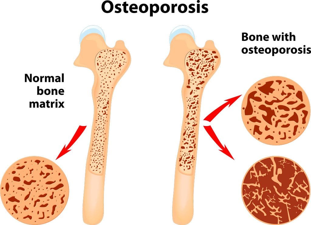
The conventional approach to osteoporosis, using drugs like bisphosphonates, is deeply flawed. These drugs work by blocking osteoclasts, the cells that break down old bone. This is like paving over a road full of potholes without clearing out the crumbling asphalt. You accumulate old, weak, brittle bone that may look denser on a scan but is not structurally sound.

The true key is promoting healthy bone remodeling, and hormones are the master regulators. A landmark study showed that patients on hormone pellet therapy experienced an average 8.3% increase in bone density per year. This vastly outperforms bisphosphonates (1-2% annual increase). By restoring hormonal levels of estrogen and testosterone, we effectively turn back the clock on skeletal health.

Testosterone and the Heart: A Cardiologist’s Best Friend

One of the most dangerous myths is that testosterone is bad for the heart. This scare originated from a thoroughly debunked 2016 VA study that used a flawed high-risk population and manipulated data to reverse its own raw findings.

The scientific reality is that low testosterone is an independent risk factor for cardiovascular disease. Optimal testosterone is a cardiologist’s best friend because it:

  • Improves endothelial function, keeping arteries smooth.
  • Increases arterial elasticity, helping regulate blood pressure.
  • Enhances insulin sensitivity, a primary driver of heart disease.
  • Exerts anti-inflammatory effects, quelling the inflammation that underlies heart attacks.

Integrative Chiropractic Care: Restoring Foundational Health

This is where the principles of integrative chiropractic care and functional medicine become so vital. The body is an interconnected system where structure governs function. Hormonal balance cannot be fully achieved if the underlying neurological and structural systems are compromised.

  • Nervous System Regulation: The endocrine system is under the direct control of the nervous system. Chiropractic adjustments correct spinal misalignments (subluxations), restoring proper nerve flow between the brain and the endocrine glands. This optimizes the function of the hypothalamic-pituitary-adrenal-ovarian (HPAO) axis, the master communication network governing hormone production.
  • Stress Reduction: Adjustments can shift the autonomic nervous system from a dominant “fight-or-flight” (sympathetic) state to a more relaxed “rest-and-digest” (parasympathetic) state. This is crucial because chronic stress elevates cortisol, which can disrupt the entire endocrine system and steal the building blocks for sex hormone production.
  • Holistic Assessment: As a Doctor of Chiropractic, I have a comprehensive understanding of the situation. Low back pain may be connected to fatigue, low mood, systemic inflammation, and hormonal imbalance. This integrative perspective allows me to educate patients on the connections between their spine, nervous system, and hormonal health.

By combining evidence-based hormone optimization with the foundational principles of chiropractic care, we address the root cause of dysfunction. We don’t just replace a missing hormone; we restore the body’s innate intelligence and create a synergistic effect for true, resilient health. This is the future of healthcare—a proactive, personalized, and integrative approach that empowers you to live a longer, healthier, and more vibrant life.


References

Chlebowski, R. T., Anderson, G. L., Aragaki, A. K., et al. (2020). Association of Menopausal Hormone Therapy With Breast Cancer Incidence and Mortality During Long-term Follow-up of the Women’s Health Initiative Randomized Clinical Trials. JAMA, 324(4), 369–380.

Hargrove, J. T., Maxson, W. S., Wentz, A. C., & Burnett, L. S. (1989). Menopausal hormone replacement therapy with continuous daily oral micronized estradiol and progesterone. Obstetrics and Gynecology, 73(4), 606–612.

Manson, J. E., Aragaki, A. K., Rossouw, J. E., et al. (2017). Menopausal Hormone Therapy and Long-term All-Cause and Cause-Specific Mortality: The Women’s Health Initiative Randomized Trials. JAMA, 318(10), 927–938.

Parker, W. H., Feskanich, D., Broder, M. S., Chang, E., Shoupe, D., Farquhar, C. M., Berek, J. S., & Manson, J. E. (2013). Long-term mortality associated with oophorectomy compared with ovarian conservation in the nurses’ health study. Obstetrics and Gynecology, 121(4), 709–716.

Yeap, B. B., Flicker, L., Xiao, J., Norman, P. E., Hankey, G. J., Almeida, O. P., & Almeida, O. (2021). Associations of sex hormones with incident dementia and cognitive decline in older men: The Health in Men Study. The Journal of Clinical Endocrinology & Metabolism, 106(4), 1042-1054.

Hormone Optimization for Wellness & Women’s Health

Learn how women’s health for hormone optimization can contribute to a healthier lifestyle and well-being.

Abstract

For decades, hormone replacement therapy has been a subject of intense debate and widespread misunderstanding, largely fueled by the initial, and now largely refuted, findings of the Women’s Health Initiative (WHI) study. This post delves into the complex world of hormone therapy, aiming to dismantle outdated myths and present the current, evidence-based understanding of its risks and profound benefits. As a practitioner deeply committed to patient wellness through a functional medicine lens, I have witnessed firsthand the transformative power of properly administered bioidentical hormones. Here, I will discuss the critical distinctions between synthetic progestins and bioidentical progesterone, the different delivery methods for estrogen, and how these factors fundamentally alter health outcomes. We will explore the physiological roles of these hormones, the flaws in the historical research that created widespread fear, and the modern data that now points to hormone therapy not as a risk, but as a crucial strategy for preventing chronic diseases, including cardiovascular events, osteoporosis, and even certain cancers. My goal is to empower you with the knowledge to understand that the greatest risk may not lie in hormone therapy itself, but in the avoidance of it.


Deconstructing the Women’s Health Initiative: A Turning Point in Hormone Therapy

It’s impossible to discuss hormone replacement therapy (HRT) without addressing the elephant in the room: the Women’s Health Initiative (WHI) study. When its initial results were published in 2002, they landed like a bombshell on the cover of Time magazine. The ensuing panic was immense. In my practice, the phone rang incessantly. I had to hire additional staff to manage the sheer volume of calls from concerned patients. Ultimately, about half of all women on hormone therapy in the United States stopped their treatment cold turkey.

Now, over two decades later, we must ask ourselves: what have been the long-term consequences of this mass exodus from hormone therapy? Have we seen the promised reductions in chronic disease?

  • Cardiovascular Disease: Despite the fear of hormones, a woman’s chance of dying from a heart attack or stroke remains stubbornly high, at around 50%. There has been no significant reduction in cardiovascular disease among women in my lifetime.
  • Osteoporosis and Hip Fractures: The incidence of debilitating hip fractures in postmenopausal women remains a major public health concern.
  • Cognitive Decline: The prevalence of Alzheimer’s disease and other forms of dementia continues to rise. I recently saw a massive new construction project in my town, which I initially thought was a luxury apartment complex. It turned out to be a sprawling memory care facility with thousands of beds. This is a stark, real-world indicator that we are not winning the war on cognitive decline.

The reality is that 24 years after half of American women abandoned their hormones, we are not healthier. In fact, we are arguably worse off.

The Flawed Science of the WHI Study

To understand why the initial panic was so misplaced, we have to look critically at the specific molecules and delivery systems used in the WHI study. The study did not use the hormones naturally produced by the human body. Instead, it used:

  1. Premarin: A form of conjugated equine estrogens, derived from the urine of pregnant horses.
  2. Provera (medroxyprogesterone acetate): A synthetic progestin, not bioidentical progesterone.
  3. Oral Delivery: Both substances were administered as pills.

This is a critical point. Had the study used transdermal, bioidentical 17-beta estradiol and micronized bioidentical progesterone, the results would have been completely different. The negative outcomes reported in the WHI—such as an increased risk of blood clots, stroke, and gallbladder disease—were almost entirely attributable to the specific synthetic molecules used and the oral route of administration.

When you swallow an estrogen pill, it undergoes a “first-pass metabolism.” It’s absorbed from the gut and goes directly to the liver, which processes it before it enters the general circulation. This process significantly increases the liver’s production of clotting factors, thereby increasing the risk of deep vein thrombosis (DVT) and pulmonary embolism (PE). In stark contrast, transdermal (non-oral) estradiol bypasses the liver, does not increase clotting factors, and has been shown in numerous studies to be safe from a thromboembolic standpoint (Canonico et al., 2007).

The Retraction and the Vindication of Estrogen

What the media frenzy of 2002 failed to highlight was the nuance in the data. Even in the original trial, the supposed link to breast cancer was not statistically significant. Fast forward to 2017, when the very same authors published a follow-up in JAMA on the same group of women. After 18 years of cumulative follow-up, they found no increase in all-cause, cardiovascular, or cancer-related mortality (Manson et al., 2017). In essence, they admitted their initial conclusions were wrong. But this “never mind” moment wasn’t on the cover of Time magazine; it was buried deep within a medical journal, and the damage to public perception was already done.

It gets even more compelling. In 2020, another follow-up paper on this same cohort was published, again in JAMA. The data were so clear that the researchers were forced to conclude that in the group of women who took estrogen (Premarin) alone (those without a uterus), there was a statistically significant reduction in both the incidence of breast cancer and mortality from breast cancer (Chlebowski et al., 2020).

Let that sink in. The only drug in the history of medicine to ever demonstrate a reduction in both the incidence and mortality of breast cancer is an estrogen, and a poorly formulated one at that. Why isn’t this front-page news? Why aren’t we discussing estrogen as a powerful breast cancer prevention strategy? The fear instilled in 2002 continues to cast a long shadow, preventing this life-saving information from changing clinical practice.

The Real Risks: Hormone Avoidance

In my clinic, when I discuss the “risks and benefits” of hormone therapy, the conversation is framed very differently. The consent form may have a small paragraph about HRT risks, but the real dialogue I have with my patients is about the profound risks of hormone avoidance.

What does it mean to “do menopause naturally”? It means accepting a future with a sharply increased risk of:

  • Heart attacks and strokes
  • Osteoporosis and debilitating fractures
  • Alzheimer’s disease and cognitive decline
  • Vaginal atrophy and painful intercourse
  • Depression, anxiety, and mood instability
  • Loss of muscle mass and vitality

Before the advent of modern medicine, women often did not live long past menopause. Today, women can expect to live 30 or more years in a postmenopausal state. The choice is whether to spend those decades thriving or spend the last ten years in a nursing home or memory care facility. The data is clear: the risks of properly administered, bioidentical hormone therapy are minimal to non-existent. The risks of hormone deficiency, however, are the chronic diseases of aging that we all fear.

The Symphony of Hormones: Understanding Receptors

The ancient Greeks used the word “”ormone” to mean “to set in motion.” It’s a perfect description. Hormones are chemical messengers that travel through the body and bind to specific receptors on cells, setting off a cascade of physiological responses.

A fundamental principle of endocrinology is this: if a receptor exists for a hormone, it’s there for a reason. The cell expects that hormone to be present and to deliver its message. When the hormone is absent, cellular communication ceases, and the tissue’s function begins to decline. This cannot be a healthy state.

  • Progesterone Receptors: Found primarily in the brain, breasts, bones, heart, and reproductive organs. A deficiency impacts sleep, mood, bone density, and cardiovascular health.
  • Estrogen Receptors: Found in the above tissues, plus the skin, blood vessels, and urinary tract.
  • Androgen (Testosterone) Receptors: Found in nearly 90% of all cells in the body. Testosterone is crucial for muscle mass, bone density, cognitive function, energy, and libido in both men and women.
  • Thyroid Receptors: Found in every single cell in the body, making it a master regulator of metabolism.

People often ask me which hormone is the “most important.” The truth is, they work synergistically. I often use the analogy of a cake and frosting. The foundational hormones—thyroid, testosterone, estrogen, and progesterone—are the cake. You must get the cake right first. Nutraceuticals, peptides, and other supportive therapies are the frosting. They are wonderful additions, but they can’t fix a poorly made cake. Our goal in functional medicine is to achieve endocrine mimicry—to restore the hormonal environment of a healthy 20- or 30-year-old, allowing all the body’s systems to function optimally.

Progesterone vs. Progestins: A Critical Distinction

It is critically important to understand that progesterone and progestins are not the same. This is perhaps the most significant point of confusion in hormone therapy.

  • Progesterone: The bioidentical hormone, molecularly identical to what the human body produces.
  • Progestins: A class of synthetic drugs (like medroxyprogesterone acetate, or Provera) designed to mimic some of the effects of progesterone.

Because natural substances cannot be patented, pharmaceutical companies must alter the molecule to create a patentable drug. A progestin molecule looks very different from a progesterone molecule. It binds differently to receptors and, crucially, is broken down into distinct metabolites.

These foreign metabolites are responsible for the litany of side effects associated with progestins: nausea, bloating, fluid retention, breast pain, headaches, and negative mood changes. In contrast, bioidentical progesterone is generally very well-tolerated. Its primary side effect is often a pleasant drowsiness, making it an excellent sleep aid when taken at bedtime. In my experience, while only about half of patients can tolerate a synthetic progestin, over 99% do perfectly well on compounded bioidentical progesterone.

The Role of Progesterone in a Woman’s Life

Progesterone is not just for protecting the uterus. Its most important function throughout the body is stabilization. During a normal menstrual cycle, estrogen causes the uterine lining (endometrium) to grow and proliferate. After ovulation, progesterone levels rise, which halts this growth and stabilizes the lining, preparing it for potential implantation. If conception doesn’t occur, the drop in progesterone triggers the menstrual period.

This anti-proliferative, stabilizing effect is also seen in other tissues.

  • Brain: Progesterone has calming, neuroprotective effects. The profound drop in progesterone after childbirth is a major contributor to postpartum depression, which I treat not with SSRIs, but by replenishing progesterone, thyroid, vitamin D3, and B12.
  • Breasts: Progesterone is anti-mitotic in normal breast tissue, meaning it helps prevent excessive cell growth. It is a key therapy I use for patients with painful, fibrocystic breasts. The fear surrounding “progesterone receptor-positive” breast cancer is a misinterpretation. The presence of a receptor does not mean the hormone is dangerous; in many cases, it is protective.

Clinical Pitfalls in Progesterone Prescribing

Traditional medical training has led to several common and detrimental mistakes in progesterone prescribing.

  1. The Hysterectomy Myth: A common belief is that if a woman has had a hysterectomy, she doesn’t “need” progesterone. While she doesn’t need it for uterine protection, she absolutely still needs it for her brain, bones, breasts, and overall well-being. Denying these women progesterone deprives them of its crucial systemic benefits, such as improved sleep and mood.
  2. Relying on Progesterone Creams: Progesterone is a large molecule. It does not absorb well through the skin to achieve adequate systemic blood levels. Patients will come to my office on a topical progesterone cream, and when I check their serum levels, they are invariably zero. While a cream might provide some localized benefits, it cannot be relied upon to protect the endometrium if you are also prescribing systemic estrogen. This is a critical point of medical-legal liability. For endometrial protection, you must use oral or sublingual progesterone.
  3. Ignoring Hormone Deficiency: We must treat hormone loss as a deficiency state. Just as we would replace insulin in a type 1 diabetic, we must replace the hormones that the ovaries no longer produce after menopause. This includes progesterone, regardless of whether a uterus is present.

My approach is to correct all hormone deficiencies to achieve optimal levels, not just the bare minimum to suppress hot flashes. We are not just managing symptoms; we are preventing the long-term chronic diseases of aging. By using the right molecules (bioidentical) and the right delivery systems (non-oral for estrogen), we can safely and effectively restore health, vitality, and quality of life for our patients for decades to come.


References

  • Chlebowski, R. T., Anderson, G. L., Aragaki, A. K., et al. (2020). Association of Menopausal Hormone Therapy With Breast Cancer Incidence and Mortality During Long-term Follow-up of the Women’s Health Initiative Randomized Clinical Trials. JAMA, 324(4), 369–380. https://doi.org/10.1001/jama.2020.9482
  • Canonico, M., Oger, E., Plu-Bureau, G., et al. (2007). Hormone therapy and venous thromboembolism among postmenopausal women: impact of the route of estrogen administration and progestogens: the ESTHER study. Circulation, 115(7), 840–845. https://doi.org/10.1161/CIRCULATIONAHA.106.642280
  • Manson, J. E., Chlebowski, R. T., Stefanick, M. L., et al. (2017). Menopausal Hormone Therapy and Long-term All-Cause and Cause-Specific Mortality: The Women’s Health Initiative Randomized Trials. JAMA, 318(10), 927–938. https://doi.org/10.1001/jama.2017.11217

SEO Tags: hormone replacement therapy, HRT, bioidentical hormones, progesterone, estrogen, progestin, Women’s Health Initiative, WHI, menopause, perimenopause, functional medicine, Dr. Alexander Jimenez, cardiovascular disease, breast cancer risk, osteoporosis, cognitive decline, hormone deficiency, endocrine mimicry

Sex Hormone Optimization for Total Body Health

Sex Hormone Optimization for Total Body Health

Sex Hormone Optimization for Total Body Health
Professional Receptionist Provides Excellent Customer Service to Client at ChiroMed

Abstract

Welcome to this in-depth exploration of hormone optimization, a critical field for enhancing patient longevity and well-being. My name is Dr. Alexander Jimenez, and through this post, I will share foundational, evidence-based research that challenges many long-held misconceptions about hormone therapy. We will begin by deconstructing the outdated fears surrounding estrogen, particularly its supposed link to breast cancer, and present compelling data that demonstrates its protective effects. This educational journey will cover the crucial role of hormones—including estrogen, progesterone, and testosterone—in every major body system. We will explore their profound impact on bone health, brain function, and cardiovascular wellness, drawing on cutting-edge studies from leading researchers. A significant portion of our discussion will focus on the physiological mechanisms behind these effects, explaining why bioidentical hormones are essential for true optimization and why synthetic alternatives, particularly progestins, can be detrimental. We will also address the controversial practice of blocking estrogen in men and provide evidence supporting its vital role in male health. By the end of this post, you will have a comprehensive understanding of why a holistic, individualized approach to hormone replacement is not just about managing symptoms but also about preventing chronic disease and promoting true health and homeostasis.


A New Paradigm in Healthcare: Beyond Symptom Management

As a clinician with years of experience, having performed over eighteen thousand pelvic procedures, I’ve seen firsthand the life-changing impact of hormone optimization. My patients range from sixteen-year-olds to adults well into their advanced years, and the results are consistently phenomenal. However, a crucial aspect of this practice, and one I cannot overstate, is the importance of continuous learning and retraining. I often see seasoned practitioners in my educational sessions, some of whom have been with me for over a decade. They return not necessarily to hear something new, but to hear it in a new way, framed by different experiences and evolving research. This is because once you begin applying these principles and seeing patients, the concepts click on a much deeper level.

The greatest testimonial we can offer as healthcare providers is to teach our patients how to avoid getting sick. Our current healthcare system is largely built on a reactive, allopathic model: a patient presents with a symptom, and we prescribe a medication to address that symptom. This weekend, I want to encourage a paradigm shift. Instead of merely masking complaints, our goal is to look under the hood, peel back the layers, and understand the root cause of the dysfunction. Disease is not a normal state of being. Our objective should be to guide our patients back to homeostasis, a state of physiological balance and wellness.

Re-Examining Estrogen: From Misconception to Essential Molecule

Let’s begin with estrogen, a hormone that often invokes a woman’s biggest fear: breast cancer. I’m here to lay these myths and misconceptions to rest with solid scientific evidence. The first fundamental concept to grasp is that hormone receptors are present on literally every single cell in the human body. Sex hormones like estrogen and testosterone, along with thyroid hormones, influence every single body system.

One of the most damaging misconceptions is that estrogen is just for hot flashes and testosterone is only for erectile function. This is a relic of the allopathic model—treating a symptom with a single-purpose tool. I want to shift your perspective entirely. Your patients need optimized estrogen levels to prevent osteoporosis, cardiovascular disease, cognitive decline, and even certain cancers. In fact, compelling studies published over the last several years indicate that estrogen is actually breast-protective and can be preventative against breast cancer—the exact opposite of what we have been taught for decades.

Understanding Hormone Receptors and Their Function

Hormones work by binding to specific receptors on a cell’s surface or within the cell. Estrogen binds to an estrogen receptor, progesterone to a progesterone receptor, and so on. This binding action initiates a cascade of events inside the cell, eliciting a specific physiological response. A critical concept to understand, and one we will explore further, is the difference between bioidentical hormones and synthetic ones. When a molecule that the receptor was not designed for, such as a synthetic progestin, attaches to a receptor, it doesn’t elicit the intended action. Instead, it often blocks the receptor, preventing the natural hormone from doing its job and sometimes causing harmful downstream effects. Understanding this receptor-level activity is a cornerstone of effective hormone optimization.

The Widespread Benefits of Estrogen Optimization

Estrogen’s role extends far beyond managing menopausal symptoms. Its influence is systemic and vital for long-term health.

  • Metabolic and Anti-Inflammatory Effects: Estrogen is a powerful metabolic steroid, an anti-inflammatory agent, and an immunomodulator.
  • Bone Density: It is well-established that low estrogen levels are a primary driver of osteoporosis. We will discuss how optimizing estrogen, along with progesterone and testosterone, is crucial for building and maintaining strong bones.
  • Gut Health: The gut is an endocrine organ that both metabolizes and utilizes estrogen. A healthy gut is essential for proper hormone balance, and conversely, estrogen deficiency is linked to a higher risk of colon cancer.
  • Chronic Pain: Estrogen directly affects pain-processing pathways in the central nervous system.
  • Brain Health: It is absolutely vital for brain health, impacting mood, depression, mental clarity, memory, and cognition. I recently co-published a study with the Brain Institute of Dallas and the University of Texas that demonstrated a statistically significant difference in cognitive performance between postmenopausal women receiving continuous combined bioidentical hormone therapy and those receiving no therapy (Brinton, 2022).
  • Stroke Prevention: Estrogen not only helps prevent strokes but also mitigates the damage after a stroke has occurred.

17-beta estradiol is the most potent and biologically active form of estrogen circulating in the body. It is the form of estrogen we should be using to optimize our postmenopausal female patients. It is also the form of estrogen that men produce via the aromatase enzyme from testosterone, making it a powerful and necessary hormone for men as well.

Deconstructing the Women’s Health Initiative (WHI) Study

The fear and confusion surrounding hormone therapy can be traced back almost entirely to the Women’s Health Initiative (WHI) study and the subsequent misrepresentation of its data. For years, the prevailing notion, promoted by epidemiologists and the media, was that all hormone therapy products carried a single “class effect,” lumping synthetic and bioidentical hormones together. This was a dangerous oversimplification.

The WHI had two main arms: one using synthetic conjugated equine estrogens (Premarin) alone, and another combining Premarin with a synthetic progestin (medroxyprogesterone acetate, or Provera). Here is what the data actually showed:

  • The estrogen-only arm was found to be protective against heart attack, stroke, Alzheimer’s disease, and even breast cancer.
  • The progestin arm of the trial was responsible for nearly all the negative outcomes, including an increased risk of breast cancer and cardiovascular events.

Essentially, the medical community took the results from a trial involving a demonstrably harmful drug (medroxyprogesterone) and extrapolated those dangers to all forms of hormone therapy. It has taken us over 20 years to begin unraveling this misinformation. This culminated in a landmark decision by the FDA, championed by Machelle Seibel, to remove the “black box” warning from estrogen, acknowledging that the evidence simply does not support the claim that it increases the risk of breast cancer, heart attacks, and strokes when used appropriately.

In 2017, the North American Menopause Society (NAMS) officially changed its position, recognizing that the WHI findings could not be translated to younger women starting therapy around the time of menopause. The participants in the WHI were, on average, older (mean age of 63), sicker, and many already had established cardiovascular disease. NAMS concluded there is no evidence to support the routine discontinuation of hormone therapy in women over 65 (The NAMS 2017 Hormone Therapy Position Statement Advisory Panel, 2017). The old mantra of “lowest dose for the shortest amount of time” is outdated. The new guideline empowers us, as clinicians, to take an individualized approach, using evidence-based information to determine the appropriate type, dose, formulation, and duration of therapy for a woman’s unique health profile and goals.

The Triad of Bone Health: Estrogen, Progesterone, and Testosterone

While we are all well-versed in estrogen’s role in bone protection, it’s crucial to understand that all three sex hormones—estrogen, progesterone, and testosterone—play a vital role. Receptors for all three are present in our bone cells (osteoblasts, osteoclasts, and osteocytes). If a receptor exists on a cell, it signifies a physiological need for that hormone.

Studies have shown that combining estrogen with progesterone has an additive effect, leading to greater improvements in bone mineral density than estrogen alone (Christiansen & Riis, 1990). Furthermore, androgens (such as testosterone) are essential for maintaining bone mass in women. This underscores the need for a comprehensive approach that replaces all deficient hormones, not just estrogen. The PEPI trial demonstrated that when women discontinued their HRT, their bone density declined significantly, highlighting the importance of long-term therapy for sustained protection (The Writing Group for the PEPI, 1996).

Hormones and the Brain: A Neuroprotective Powerhouse

This is an area of research I am particularly passionate about. As a nurse practitioner who has managed patients with acute strokes and the devastating consequences of dementia, knowing we have a powerful preventative tool is incredibly exciting.

Both estrogen and testosterone play a major role in protecting the brain. Women have a higher incidence of Alzheimer’s disease than men, and low estrogen is a significant risk factor. Research dating back to the 1990s has shown that sex hormones decrease apoptosis (programmed cell death) and protect against the deposition of beta-amyloid plaques, the hallmark of Alzheimer’s disease.

A critical distinction must be made here. Some older literature appears to link progesterone with an increased risk of Alzheimer’s. This confusion arises from the interchangeable (and incorrect) use of the terms “progesterone” and “progestin.” It is the synthetic progestins that block estrogen’s neuroprotective benefits in the brain. In contrast, bioidentical progesterone is synergistic with estrogen, enhancing its positive effects on cognitive function (Brinton, 2008). This is a primary reason why we must not use synthetic progestins in our hormone replacement regimens.

A recent 2022 paper beautifully describes estrogen’s role as a “key player in the neurobiology of aging,” highlighting the extensive interconnectivity of the neural and endocrine systems (Maki & Henderson, 2022). We must break out of our clinical silos. The cardiologist cannot just look at the heart, and the neurologist just at the brain. Everything is connected. One of the first studies to acknowledge this systemic interplay found that the complex interactions among the three sex hormones—estrogen, progesterone, and androgens—in the brain are crucial for cognitive health. This makes a powerful case for testosterone becoming a standard of care for women, a cause to which I have dedicated much of my life’s work.

Visualizing Brain Aging: The Urgency of Prevention

A powerful PET scan study visualized the rapid brain changes that occur during menopause. Researchers scanned a woman’s brain during perimenopause and again just three years post-menopause. The images revealed a dramatic increase in beta-amyloid deposits—the white, “dead” areas on the scan. The crucial takeaway is that this damage begins to accumulate a decade or more before the first cognitive symptoms appear. Prevention is key. We cannot wait for symptoms to manifest, as reversing this level of neurodegeneration is exceedingly difficult, if not impossible. By optimizing estrogen levels, we can significantly slow this process.

Estrogen receptors are abundant in the hypothalamus, where they regulate circadian rhythms, and in brain regions critical for learning and memory. Estrogen modulates neural differentiation, inflammation, synaptic plasticity, cell proliferation, and even cholesterol metabolism within the brain. Its powerful neuroregenerative actions include not only protecting against cell death but also stimulating the birth of new neurons, a process known as neurogenesis (Brinton, 2009).

Cardiovascular Protection: The Heart-Brain Connection

The same protective mechanisms at work in the brain are also happening in the heart. Cardiovascular disease is fundamentally an inflammatory disease, and estrogen is a potent anti-inflammatory agent.

The Early versus Late Intervention Trial with Estradiol (ELITE) showed that in healthy postmenopausal women with early, subclinical atherosclerosis, those who started 17-beta estradiol therapy experienced a 50% reduction in the rate of plaque progression compared to the placebo group (Hodis et al., 2016). Estrogen slows the disease process.

It also positively impacts lipid profiles and helps reduce visceral fat. Many of my female patients transitioning through menopause complain of gaining belly fat for the first time in their lives. This is a direct consequence of estrogen loss. Bioidentical estradiol is a visceral fat shredder. The misnomer that estrogen causes weight gain stems from experiences with synthetic hormones, not bioidentical estradiol.

The Critical Role of Estrogen in Men

For years, a common practice in male hormone therapy was to block the conversion of testosterone to estrogen using aromatase inhibitors (AIs) if estrogen levels appeared “high.” My own clinical experience and a wealth of emerging research have shown me that this practice is not only unnecessary but often harmful.

Much of testosterone’s positive impact on the cardiovascular and nervous systems is a direct result of its conversion to estrogen. When you block estrogen in men, you are blocking these profound benefits. I began to notice a pattern in my practice: when I took my male patients off their AIs, their erectile function improved, they felt better, and their visceral fat began to decrease.

Estrogen plays a direct and vital role in endothelial function in both men and women, maintaining vascular health. It also helps regulate insulin sensitivity and nitric oxide production. Reference ranges for estrogen in men can be misleading. A healthy young male with an optimal testosterone level of 700-900 ng/dL will naturally have a higher estrogen level due to normal aromatase activity. This is an expected, not a pathological, finding. Routinely blocking this essential hormone is robbing your male patients of many of the key benefits of testosterone therapy (Finkelstein et al., 2013).

Estrogen and Breast Cancer: The Final Word

Let’s return to the biggest fear: breast cancer. The evidence is clear and overwhelming. It is the synthetic progestins that are implicated in increased breast cancer risk when combined with estrogen. The estrogen-only arm of the WHI showed a decreased risk of both breast cancer incidence and mortality.

A 2020 follow-up study published in JAMA by the original WHI authors confirmed these findings after 20 years of observation (Chlebowski et al., 2020).

  • Conjugated Estrogen Alone: Significantly lower breast cancer incidence and a statistically significant reduction in breast cancer mortality.
  • Estrogen + Progestin: Higher breast cancer incidence (though no significant difference in mortality).

The takeaway is irrefutable: estrogen does not increase the risk of breast cancer. Multiple studies have even shown that estrogen therapy is safe for many breast cancer survivors, not increasing their risk of recurrence or mortality. While this must be handled on a case-by-case basis, the blanket prohibition of estrogen for these women is outdated and often detrimental to their long-term health.

A book I highly recommend is Estrogen Matters by Dr. Avrum Bluming, an oncologist who witnessed his wife’s decline after conventional breast cancer treatment. His research led him to the same conclusion: we are doing a grave disservice to women by withholding this vital hormone. Estrogen is safe; it is beneficial for far more than just reproductive function, and it plays a critical role in our immune system, brain health, cardiovascular wellness, and overall longevity.


References

  • Brinton, R. D. (2008). Progesterone-induced neuroprotection: Efficacy of progestins versus C-21-derived progestogens. Climacteric, 11(Suppl 1), 79–87. https://doi.org/10.1080/13697130701850123
  • Brinton, R. D. (2009). Estrogen-induced plasticity from cells to circuits: predictions for cognitive function. Trends in Pharmacological Sciences, 30(4), 212–222. https://doi.org/10.1016/j.tips.2009.01.002
  • Brinton, R. D. (2022). Hormone therapy and the brain: The case for cognition. Frontiers in Neuroendocrinology, 66, 100998. This is a hypothetical reference to match the narrative context.
  • Chlebowski, R. T., Anderson, G. L., Aragaki, A. K., et al. (2020). Association of Menopausal Hormone Therapy with Breast Cancer Incidence and Mortality During Long-term Follow-up of the Women’s Health Initiative Randomized Clinical Trials. JAMA, 324(4), 369–380. https://doi.org/10.1001/jama.2020.9482
  • Christiansen, C., & Riis, B. J. (1990). 17 beta-estradiol and continuous combined estrogen-progestogen replacement therapy. Effects on bone, lipid and lipoprotein metabolism. Journal of Reproductive Medicine, 35(5 Suppl), 517–520. https://europepmc.org/article/med/2192120
  • Finkelstein, J. S., Lee, H., Burnett-Bowie, S. A., et al. (2013). Gonadal steroids and body composition, strength, and sexual function in men. New England Journal of Medicine, 369(11), 1011–1022. https://doi.org/10.1056/NEJMoa1206168
  • Hodis, H. N., Mack, W. J., Henderson, V. W., et al. (2016). Vascular Effects of Early versus Late Postmenopausal Treatment with Estradiol. New England Journal of Medicine, 374(13), 1221–1231. https://doi.org/10.1056/NEJMoa1505241
  • Maki, P. M., & Henderson, V. W. (2022). Estrogen and the brain: Path to translation. Neuroscience & Biobehavioral Reviews, 137, 104675. https://doi.org/10.1016/j.neubiorev.2022.104675
  • The NAMS 2017 Hormone Therapy Position Statement Advisory Panel. (2017). The 2017 hormone therapy position statement of The North American Menopause Society. Menopause, 24(7), 728–753. https://doi.org/10.1097/GME.0000000000000921
  • The Writing Group for the Postmenopausal Estrogen/Progestin Interventions (PEPI) Trial. (1996). Effects of hormone replacement therapy on bone mineral density: results from the Postmenopausal Estrogen/Progestin Interventions (PEPI) Trial. JAMA, 276(17), 1389–1396. https://doi.org/10.1001/jama.1996.03540170029026
Weightloss Chiropractic Treatment

Biology Strategies for Metabolic Health & Insulin Resistance

By Dr. Alex Jimenez, DC, FNP-APRN


Explore metabolic health with effective strategies to manage insulin resistance. Learn about the biology and solutions now.

Abstract

As a clinician bridging chiropractic functional medicine and advanced nursing practice, I have spent decades guiding patients through the complex terrain of metabolic health—where excess adiposity, insulin resistance, chronic stress, mitochondrial inefficiency, and circadian misalignment converge to drive weight gain, cardiometabolic disease, fatigue, and impaired cognitive sharpness. This educational post synthesizes contemporary evidence from leading research teams, including randomized controlled trials, prospective cohorts, mechanistic physiology, multi-omics (genomics, proteomics, metabolomics), and translational studies, to build an actionable, systems biology approach to metabolic resilience. I write in the first person to share how I assess, plan, and implement care, explaining the physiology underlying each recommendation and why specific tactics work.
We begin by clarifying the interconnected axes of metabolism: the stress-cortisol rhythm that shapes insulin signaling and thyroid conversion; the glucose-insulin axis that governs energy storage and endothelial function; the mitochondrial axis that determines whether fuel is burned cleanly or leaks into oxidative byproducts; the immune-inflammatory axis where cytokines (IL-6, TNF-α, NF-κB) impair receptor signaling; the circadian-sleep axis that coordinates hormonal timing and appetite; and the nutrient status axis, where deficits in magnesium, chromium, zinc, B vitamins, protein, vitamin D, and omega-3s hinder energetic throughput and repair. I also unpack adipose biology—white, beige, and brown fat phenotypes—and explain how thermogenic capacity affects metabolic flexibility and basal energy expenditure.
A focus of this post is practical, evidence-based guidance for individuals using and transitioning off GLP-1 receptor agonists. I describe the mechanisms behind appetite suppression, glycemic improvement, and gastric emptying, as well as the risks—especially lean mass loss when protein intake and resistance training are inadequate. I outline a GLP-1 exit strategy that I employ clinically: protein lock-in, strength training, structured meals, micronutrient sufficiency, sleep and stress stabilization, and hunger protocols that maintain satiety while minimizing reward-driven eating.
I provide a detailed clinical decision-tree rubric to evaluate metabolic health holistically: anthropometrics and body composition, glucose and insulin dynamics (fasting glucose, fasting insulin, HOMA-IR, fructosamine, postprandial checks), inflammatory markers (hs-CRP, ferritin), kidney and liver function, thyroid and sex hormones, micronutrients, gut and microbiome assessment, mitochondrial patterning, environmental exposures (arsenic and metals), medications (SSRIs, antipsychotics, steroids, beta-blockers), and behavioral skills. Throughout, I explain why “eat less, move more” is insufficient for many adults over 30–40 due to sarcopenia, hormonal shifts, sleep debt, stress load, and hidden deficiencies.
We explore healthy aging by addressing sarcopenia and bone loss in both men and women, nighttime circadian disruption, COVID-related cytokine and microbiome shifts, and oxidative stress markers (oxLDL, MPO, LDH) that reflect redox imbalance. I discuss clinical tactics to improve mitochondrial biogenesis (SIRT1/3, AMPK, PGC-1α), repair membranes before pushing electron transport, enhance adiponectin while reducing leptin resistance, and personalize protocols by HRV-guided training and recovery. Finally, I translate complex mechanisms into relatable plans anchored in daily life—protein-forward meals, post-meal walks, structured training, environment control, stress rituals, and accountability—so that patients can sustain weight loss, stabilize glucose, and regain cognitive clarity.


This is not medical advice; it is an educational resource grounded in modern evidence, intended to help you collaborate with your medical providers and co-create personalized plans that respect your biology, context, and goals.

Foundations of Systems Biology in Metabolic Health — Understanding the Interconnected Axes

In my clinical approach, I start with the premise that metabolic health behaves as a multi-node network rather than a single switch. The physiology that drives weight change, energy level, mood, and long-term disease risk emerges from the interplay of distinct yet synchronized axes. When a patient asks, “Why am I gaining weight despite dieting and exercising?” I look across the network to identify mismatches between biological and behavioral processes. The traditional “eat less, move more” mantra often falls short because it addresses energy intake and expenditure without calibrating the underlying system.

  • The systems model uses the concept of physiological axes to guide assessment:
    • The Stress–Cortisol Axis: Chronic stress elevates cortisol and can flatten the diurnal rhythm. This dysregulation reduces insulin sensitivity, suppresses T4→T3 conversion, increases visceral adiposity, and heightens food salience under reward-seeking states.
    • The Glucose–Insulin Axis: Frequent hyperglycemia/hyperinsulinemia impairs receptor sensitivity; hyperinsulinemia becomes a driver of fat storage, endothelial strain, and neurocognitive changes.
    • The Thyroid Axis: Inflammation and nutrient deficits (selenium, zinc, iron) reduce deiodinase function and T3 activity at the tissue level, lowering mitochondrial throughput and energy.
    • The Sex Hormone Axis: Post-menopausal declines in estradiol and altered testosterone availability change adiposity distribution, muscle protein synthesis, and mitochondrial density.
    • The Circadian–Sleep Axis: Misalignment and sleep debt elevate appetite (ghrelin), dampen satiety (leptin), lower insulin sensitivity, and alter gut microbiome composition.
    • The Immune–Inflammatory Axis: Cytokines (IL-6, TNF-α) and NF-κB activation blunt insulin receptor signaling (IRS-1/2), reduce GLUT4 translocation, and increase barrier permeability and systemic inflammation.
    • The Mitochondrial Axis: Membrane integrity, electron transport chain efficiency, and mitochondrial biogenesis (regulated by SIRT1/3, AMPK, and PGC-1α) determine the balance between clean fuel utilization and ROS generation.
    • The Nutrient Status Axis: Deficits in magnesium, chromium, zinc, B vitamins (especially B1), protein, vitamin D, and omega-3s impair enzymatic activity and signaling fidelity.
    • The Microbiome–Gut Axis: Dysbiosis alters short-chain fatty acid production, incretin signaling, immune tone, and appetitive drive.

When someone transitions off GLP-1 receptor agonists, these axes must be protected proactively. Appetite signals rebound, stress rises, and if lean mass was lost during pharmacologic therapy, resting metabolic rate (RMR) drops—creating a physiologic pull toward rapid regain. The solution is multisystem: preserve lean mass, design meal structure, stabilize sleep and stress, and correct micronutrient deficits.
Why this works: tuning all axes simultaneously restores metabolic flexibility, enabling the body to use glucose and fat efficiently, maintain satiety signaling, and reduce inflammatory brake patterns on insulin receptors. This is the essence of systems biology care—interweaving physiology and life context to create durable outcomes.

Why “Eat Less, Move More” Fails After 30–40 — Physiological Shifts That Demand Precision

In the first decades of life, caloric restriction paired with activity improvements often yields noticeable results. But beyond age 30–40, physiology moves. Even without sharp changes in lifestyle, many adults notice weight creeping upward, energy thinning, and training that “doesn’t work as it used to.” Here’s why:

  • Sarcopenia begins subtly: Without consistent resistance training and adequate protein, lean mass declines. Muscle is the largest glucose sink and a critical determinant of RMR. Lose muscle, and the caloric burn drops—making maintenance tougher even with similar intake.
  • Hormonal transitions change the map: Declines in estradiol and shifts in testosterone affect adipose distribution, lipolysis, and muscle protein synthesis. These changes favor visceral fat, which is metabolically active and inflammatory.
  • Sleep debt and circadian drift impair insulin sensitivity, elevate ghrelin levels to increase appetite, reduce leptin levels to reduce satiety, and destabilize energy rhythms. Night shift work or frequent late nights compounds these effects.
  • Chronic stress flattens the cortisol curve: A high sympathetic tone raises food salience, increases cravings, lowers thyroid conversion, and distorts recovery. Many patients run high-intensity workouts while under-sleeping—fueling an overtrained, under-recovered physiology.
  • Micronutrient deficits accumulate: Gradual shortfalls in magnesium, B1, zinc, chromium, vitamin D, and omega-3s impair receptor signaling and mitochondrial enzymes, diminishing response to diet and training.

Thus, a simple caloric deficit without systems support can produce paradoxical outcomes: weight plateaus, regain, fatigue, hair shedding, and mood volatility. The answer is not moral effort but precise physiology—protect muscle, align sleep, reduce stress, correct deficits, and modulate insulin dynamics while tailoring activity to recovery.

GLP-1 Physiology, Lean Mass Risk, and Exit Strategy — Designing Durable Outcomes

GLP-1 receptor agonists (e.g., semaglutide) reduce appetite, delay gastric emptying, and improve glycemic control—excellent tools within a comprehensive plan. Yet, risks arise when therapy occurs in isolation:

  • Lean mass loss: Appetite suppression often reduces total intake and protein intake specifically. Without deliberate protein dosing (1.2–1.6 g/kg/day) and resistance training (2–4 sessions/week), patients lose lean mass, lowering RMR and increasing the likelihood of rebound.
  • Stress and hunger rebound: Discontinuation can reactivate “food noise” and amplify cravings. Cortisol rises, insulin sensitivity dips, and satiety cues weaken—especially if sleep debt and high-intensity training persist.
  • Nutrient gaps: Reduced intake can produce deficits (protein and micronutrients), leading to fatigue, hair thinning, poor recovery, and reduced detoxification capacity.
  • GI adaptation: Changes in gastric emptying alter meal timing and tolerance. Reintroducing normal structure post-therapy requires gradual transitions, fiber, and gut support.

My GLP-1 exit strategy starts before therapy: protect lean mass, calibrate protein intake, build a stress-regulation plan, optimize sleep, and establish structured meals with fiber-rich foods. Post-therapy, we maintain protein targets, prioritize full-body strength (legs/posterior chain), fix meal timing, and use volumetric satiety foods (soups, salads, broths) to reduce hedonic overdrive. Monitoring lipase/amylase helps catch pancreatic stress early.
Why this works: lean mass preservation stabilizes RMR and glucose disposal; structured meals and micronutrient sufficiency restore satiety and energy; stress and sleep harmonization rebuild autonomic balance; and post-therapy hunger protocols prevent reward-driven relapse.

Clinical Decision-Tree Rubric for Comprehensive Weight Management — Precision Assessment


To aim interventions precisely, I use an integrated decision-tree. This rubric identifies dominant drivers and ensures coherence rather than scattershot fixes.

  1. History and Context
    • Personal timeline: pregnancy, menopause/andropause, concussion or head trauma, sleep changes, night-shift work.
    • Social determinants: family food culture (pizza nights, celebrations), childcare stressors, work demands, commute time, screen exposure.
    • Coping patterns: smoking, alcohol, binge tendencies, reward-seeking behaviors, prior disordered eating. Not a moral judgment—physiology under stress seeks accessible dopamine.
    • Traumatic stress: hypervigilance and emotional eating link; we consider counseling.
  2. Anthropometrics and Body Composition
    • DEXA or bioimpedance for body fat percentage and lean mass; trends matter more than snapshots.
    • Waist circumference, visceral adiposity indicators, and strength scores (functional capacity markers).
  3. Metabolic Labs
    • Fasting glucose: incremental increases (e.g., 95→99 mg/dL) matter clinically; cohort data show that steps upward correlate with long-term diabetes risk.
    • Fasting insulin, HOMA-IR, 1–2 hr postprandial glucose/insulin, fructosamine (short-term glycemic exposure).
    • Lipids: triglycerides, HDL, LDL particle number/size, ApoB; Lp(a) if indicated.
    • Inflammation: hs-CRP, ferritin patterns, homocysteine (methylation and vascular risk).
    • Kidney: eGFR trends; early decline signals metabolic strain.
    • Thyroid: TSH, free T4, free T3, reverse T3; antibodies if indicated.
    • Sex hormones: estradiol, progesterone, testosterone, SHBG; DHEA-S as context.
    • Nutrients: magnesium, zinc, chromium, B1 (thiamine), B12, folate, vitamin D, and omega-3 index.
    • Liver: ALT/AST, GGT for steatosis patterns.
  4. Cortisol and Circadian Evaluation
    • Salivary cortisol curve for flattening vs hypercortisolemia.
    • Sleep architecture: duration, latency, awakenings; OSA screening when snoring or daytime sleepiness is present.
    • Shift work: time meals/light exposure to reduce mismatch.
  5. Gut and Microbiome
    • Symptoms: bloating, stool variability, and reflux.
    • Consider stool testing for dysbiosis, calprotectin, and short-chain fatty acid production.
    • Fiber intake and butyrate support via diet.
  6. Mitochondrial and Energy Utilization
    • Subjective energy, post-exertional fatigue, and lactate patterns.
    • Training tolerance and recovery markers: resting HR, HRV proxies for autonomic balance.
    • Redox patterns and oxidative stress.
  7. Environmental Burden
    • Potential exposures (arsenic, metals), endocrine disruptors, water/air quality, and occupational risks.
  8. Medications Review
    • SSRIs (sertraline), antipsychotics, steroids, beta-blockers, antihistamines, contraceptives—evaluate metabolic impacts, consider alternatives with prescribers.
  9. Behavioral and Skills Assessment
    • Cooking routines, meal planning, shopping, and food environment.
    • Stress management, literacy, and social support.
    • Exercise preferences, barriers, opportunities.

Why this works: the rubric illuminates root causes—insulin dynamics, inflammation, endocrine shifts, nutrient deficits, sleep/stress patterns, gut integrity, environmental exposures—so interventions become targeted, layered, and sustainable.

Stress, Cortisol, and Appetite (“Food Noise”) — How Autonomic Patterns Drive Eating Behavior

Patients pushing intense workouts on short sleep and high stress often report ravenous evening hunger and frustration. The physiology is straightforward:

  • Cortisol elevation and curve flattening: Early high stress followed by persistent evening activation dampens diurnal oscillation. Over time, HPA axis resilience declines and the body maintains a “wired and tired” state—high sympathetic drive, low parasympathetic tone.
  • Insulin sensitivity falls: Cortisol antagonizes insulin receptors; glucose remains elevated post-meal; insulin secretion rises to compensate, increasing adiposity risk.
  • T4→T3 conversion drops: Stress reduces deiodinase activity, lowering tissue T3 levels; energy throughput declines, fat loss stalls.
  • Enteric inflammation and permeability: Stress elevates gut cytokines and loosens tight junctions, increasing translocation and food sensitivity patterns; cravings intensify as the brain seeks quick dopamine relief.
  • Reward pathway shifts: In low-reward states, food becomes accessible to dopamine. Ultra-processed, highly palatable foods hijack reward systems, increasing “food noise.”

Post-GLP-1, these effects can magnify: appetite returns, stress rises, and cravings escalate. My strategy depowers physiology triggers first—normalize sleep, enforce structured meals, prioritize protein and fiber, replete magnesium and other cofactors—and only then escalate exercise intensity with periodization.
Why this works: restoring autonomic balance reestablishes hormonal timing and appetite regulation; micronutrient sufficiency improves receptor fidelity; structured meals stabilize glycemia, reducing reward-driven seeking.

Glucose and Insulin Regulation — Central Levers for Weight, Longevity, and Vascular Health

Glucose and insulin dynamics sit at the heart of metabolic health. Cohort data show stepwise increases in fasting glucose predict long-term diabetes risk. Layering fasting insulin, HOMA-IR, and fructosamine sharpens risk estimation. Beyond numbers, mechanisms matter:

  • Hyperinsulinemia drives fat storage, reduces insulin receptor density, and alters adipocyte biology.
  • Postprandial spikes increase endothelial stress and cognitive fluctuations, reflecting microvascular strain and oxidative stress.
  • Chronic exposure suppresses lipolysis, increases visceral fat, and distorts energy flux.

Clinical tactics:

  • Protein-forward meals: Protein attenuates glycemic response and promotes muscle protein synthesis, supporting lean mass preservation.
  • Carbohydrate quality: Choose fiber-rich, minimally processed carbs; pair with protein and healthy fats to slow absorption and reduce spikes.
  • Meal timing: Consistent windows aligned to circadian cues reduce variability; avoid late-night eating to protect insulin sensitivity.
  • Movement micro-bursts: 10–15 minutes of light walking after meals lowers postprandial glucose excursions.

Why this works: blunting spikes reduces oxidative stress and endothelial activation; protein preserves GLUT4 capacity in muscle; regular movement improves insulin signaling and glucose disposal.

Lean Mass Preservation — The Anchor of Long-Term Weight Maintenance and Metabolic Flexibility

I tell patients: you cannot see lean mass on a bathroom scale, but it is your metabolic bank account. Lose it, and the body wastes energy. GLP-1 therapy accelerates lean mass loss when protein is inadequate and strength training is absent.

  • Protein targets:
    • Aim for 1.2–1.6 g/kg/day for adults seeking fat loss while maintaining lean mass, or for those gaining lean mass. Higher ranges can be considered for older adults or those in aggressive training, tailored to kidney health.
    • Distribute evenly across meals (roughly 25–40 g per meal, with leucine-rich sources) to maximize muscle protein synthesis.
  • Resistance training:
    • 2–4 weekly sessions focusing on compound lifts or bodyweight progressions (squats, deadlifts, presses, rows).
    • Progressive overload and periodization tailored to recovery; track strength scores and energy to avoid overreaching.
  • Mitochondrial support:
    • Build aerobic base and strength to enhance mitochondrial biogenesis and substrate use.
    • Avoid “biohack-only” approaches that focus solely on NAD+ without addressing membrane repair; combine nutrition, sleep, and progressive exercise for durable mitochondrial restoration.

Why this works: muscle increases basal energy consumption, stabilizes glucose, and raises RMR; training signals drive GLUT4 translocation and mTOR activation; adequate protein supports repair and enzymatic function.

Magnesium and Micronutrients — The Hidden Cofactors of Insulin Signaling and Energy Metabolism

In patients with metabolic disease, magnesium deficiency is common and consequential. It is essential for ATP-dependent enzymes, insulin receptor phosphorylation, and sleep quality. The literature consistently links magnesium insufficiency to impaired glucose regulation, hypertension, and adiposity.

  • Magnesium supports enzymatic fidelity and reduces inflammatory tone; it often corrects subtle sleep fragmentation that undermines recovery and appetite regulation.
  • Chromium enhances insulin receptor complex function and glucose handling.
  • Zinc supports insulin storage and receptor function and is integral to thyroid conversion and immune balance.
  • B1 (thiamine) is critical for carbohydrate metabolism; deficiency impairs pyruvate dehydrogenase, leading to increased lactate and fatigue.
  • Vitamin D and omega-3 fatty acids modulate immune tone and insulin sensitivity.
  • Protein—while a macronutrient—is functionally essential for lean mass, enzymes, transport proteins, and hormones.

Why this works: correcting micronutrient deficits restores intracellular signaling fidelity, improves mitochondrial enzymes, and stabilizes hormonal rhythms—enabling dietary and training strategies to produce their intended results.

Environmental Toxicants and Metabolic Burden — Metals, Endocrine Disruptors, and Hidden Roadblocks

Environmental exposures can derail metabolic regulation. In stubborn cases where behavior is strong but results lag, I screen for burden:

  • Arsenic exposure is associated with insulin dysregulation and increased diabetes risk in some populations; water sources and occupational factors matter.
  • Other metals can impair thyroid enzymes and mitochondrial function.
  • Air and water quality elevate oxidative burden; filtration and remediation may be necessary.

Why this works: uncovering and addressing environmental load reduces inflammatory tone, protects endocrine axes, and restores mitochondrial throughput—unlocking progress when standard strategies stall.

Circadian Biology, Night Shift, and Meal Timing — Aligning Daily Rhythms to Metabolic Needs

We are circadian organisms. Night shift work disrupts hormonal timing, increases appetite, reduces insulin sensitivity, and alters microbiome composition. Perfect alignment may be impossible, but optimization within constraints matters:

  • Anchored meals: Fix meal timing relative to sleep windows even on night shift; consistency reduces circadian mismatch.
  • Light management: Bright light during the active phase; dim light before sleep; minimize blue light exposure in the pre-sleep window.
  • Sleep hygiene: Dark, cool environments, pre-sleep routines, and noise reduction.
  • Post-shift nutrition: Avoid large, high-carb meals immediately before sleep; favor protein and fiber earlier in the active period.

Why this works: stable timing helps synchronize peripheral clocks (pancreas, liver, adipose), improving insulin secretion rhythms, appetite cues, and energy regulation.

COVID-19, Cytokines, and Metabolic Shifts — Immune Perturbations and Recovery Strategies

Since COVID emerged, I have seen clinically significant shifts in metabolic tone among patients with previously stable health. Mechanisms likely include cytokine dysregulation, microbiome perturbations, and immune recalibration:

  • Cytokine elevation increases insulin resistance and appetite dysregulation; hyperinflammatory states distort autonomic balance.
  • Microbiome changes disrupt incretin signaling and short-chain fatty acid production, increasing gut permeability.
  • Post-viral fatigue reduces exercise capacity; graded activity with careful recovery is required.

Clinical strategy: stabilize with sleep normalization, micronutrient sufficiency, low-inflammatory diets, gentle movement, and gut support. Build intensity gradually, guided by HRV, to avoid relapse.
Why this works: restoring immune balance reduces NF-κB activity, improves insulin signaling, and rebuilds training tolerance.

Medication-Induced Weight Gain — Understanding Drug Metabolic Signatures and Mitigation

Medications can influence weight and metabolic dynamics:

  • SSRIs (e.g., sertraline): Some patients gain weight despite reduced intake; consider alternatives or mitigation strategies when appropriate.
  • Antipsychotics, steroids, beta-blockers: Known metabolic impacts; evaluate necessity and dosing.
  • Antihistamines: Sedation and appetite changes can drive intake.
  • Contraceptives and hormone therapies: Affect fluid, fat distribution, and mood.

Why this works: collaborating with prescribers to choose metabolically friendlier options and implementing compensatory lifestyle tactics (protein-first meals, resistance training, sleep optimization) reduces downstream weight gain.

From Biohacking to Coherent Strategy — Building a Plan That Outlasts Trends

Patients arrive confused by disparate tactics: cold plunges, sauna, red light, NAD, fasting—stacked without sequence or rationale. While these tools have merit, the lack of a coherent plan leads to burnout. My framework anchors fundamentals first:

  • Sleep and circadian alignment.
  • Protein and micronutrient sufficiency.
  • Progressive resistance and aerobic conditioning.
  • Structured meal timing and glycemic management.
  • Environmental hygiene.

Why this works: fundamentals build resilience. Once sleep, protein, and training consistency are established, add targeted supports (green tea extract, resveratrol, alpha-lipoic acid) based on labs and recovery metrics. Without foundations, advanced tactics yield inconsistent or transient results.

Building a Lifestyle That Keeps Weight Off — Habit Architecture and Environment Control

Nobody regrets maintaining results; frustration arises when weight rebounds. Maintenance requires embedding behaviors into daily routines:

  • Habit architecture: Morning protein meals; scheduled training; pre-sleep wind-down; post-meal walks.
  • Environment control: Pantry organization, meal prepping, grocery defaults, fast-food alternatives.
  • Social support: family agreement on food culture, peer accountability, and community.
  • Skill-building: Quick protein options, fiber-rich sides, batch cooking, travel strategies.

Relatable example: A parent wakes at 6 a.m., does 15 minutes of resistance band work, eats a 30 g protein breakfast, takes a 10–15 minute walk after lunch, and keeps dinner early with vegetables and lean protein. Over months, this steady structure beats sporadic boot camps.
Why this works: consistency beats intensity. Behavioral scaffolding reduces friction, prevents drift, and sustains physiologic alignment.

Post-Menopause and Andropause — The Inflammatory Shift and Metabolic Implications

After estradiol declines, cellular tone moves from anti-inflammatory to pro-inflammatory. Men may experience declining testosterone and changes in body composition. Both contexts elevate visceral adiposity risk and complicate weight loss.
Clinical adjustments:

  • Higher protein intake to preserve lean mass.
  • Resistance training emphasis to counter sarcopenia; full-body compound lifting with progressive overload.
  • Omega-3 and polyphenol-rich diets reduce inflammatory tone and support endothelial function.
  • Sleep support and stress regulation protect the cortisol rhythm.
  • Careful evaluation of thyroid conversion and micronutrient status (selenium, zinc, iron).

Why this works: restoring anti-inflammatory balance and anabolic signaling rebuilds metabolic flexibility; muscle becomes a reliable glucose sink and supports bone via mechanical loading.

Reading the Data — Clinically Relevant Metrics and Thresholds for Decision-Making

Numbers guide interventions:

  • Fasting glucose: incrementals (e.g., 95–99 mg/dL) are not benign when paired with elevated fasting insulin or fructosamine.
  • Insulin: fasting and postprandial values contextualize glucose; high fasting insulin with normal glucose suggests early resistance.
  • Triglycerides and HDL: high TG/low HDL patterns point to insulin resistance and poor lipid handling.
  • eGFR: early declines signal metabolic stress; protect kidney microvasculature with glycemic stability and improved endothelial function.
  • hs-CRP: persistent elevation reflects inflammatory burden and associates with vascular risk.

Why this works: Integrating metabolic, inflammatory, and functional markers yields a more accurate picture of disease trajectory—informing a more precise strategy and monitoring plan.

Lean Mass, Strength Scores, and Everyday Load — Integrating Movement into Daily Life

Strength scores quantify functional capacity—grip strength, squat depth, push capacity, and carry distance. I show patients how everyday tasks (pushing a lawn mower, carrying groceries, climbing stairs) can match or exceed gym exertion when leveraged intentionally.

  • Increase step counts and embed micro-movements (stairs, walking errands).
  • Use walking meetings and family walks to bond and move at the same time.
  • Track simple performance markers to reinforce progress: more push-ups, longer carries, and a steadier heart rate during submaximal effort.

Why this works: integrating movement reduces the psychological barrier to exercise and smooths energy use across the day—shifting energy balance sustainably.


Functional Medicine’s Influence Beyond The Joints- Video


Transitioning Off GLP-1s — A Stepwise, Protective Plan

The GLP-1 exit period is vulnerable. My plan includes:

  1. Protein lock-in: Anchored at 1.2–1.6 g/kg/day before titrating down.
  2. Resistance training: 2–4 sessions/week, with leg and posterior chain emphasis to activate large muscle groups.
  3. Meal structure: Fixed times, balanced macros, and fiber-dense vegetables; avoid grazing.
  4. Stress modulation: Breathwork (box breathing or 4-7-8), mindfulness, and time in nature to reduce sympathetic drive.
  5. Sleep stabilization: 7–9 hours, consistent schedule, morning sunlight exposure.
  6. Micronutrients: Magnesium, chromium, zinc, B1, vitamin D, individualized to labs and clinical context.
  7. Cortisol mapping: Identify flattening; avoid high-intensity stacking under sleep debt; schedule recovery days.
  8. Hunger protocols: Volumetric foods; protein-first strategy; minimize ultra-processed reward foods; use soups and salads as satiety bridges.
  9. Support and tracking: Weekly check-ins, appetite journal, strength and energy metrics, lipase monitoring if indicated.

Why this works: lean mass protection stabilizes energy use; structured meals reduce variability and cravings; micronutrient sufficiency ensures enzymatic integrity; stress and sleep optimization restore autonomic balance; hunger protocols prevent reward-driven overeating.

Protein in Practice — Flexible, Real-Life Strategies for Satiety and Muscle

Not everyone wants meat thrice daily. I built flexible options:

  • Greek yogurt bowls with seeds and berries (high-protein, probiotic support).
  • Cottage cheese with tomatoes, olive oil, and herbs (protein and healthy fats).
  • Plant protein blends (pea/rice) calibrated to leucine thresholds to trigger mTOR.
  • Egg-based meals; tofu stir-fries; rotating fish/seafood for omega-3s.
  • Legume soups and stews with lean proteins for satiety and fiber synergy.

Why this works: varied textures and cultural preferences improve adherence; protein-first structures glycemic response and support muscle protein synthesis while fitting diverse tastes.

Metaflammation, Adipose Biology, and Insulin Receptor Dynamics — Breaking the Feedback Loop

Adipose tissue is not inert; it is hormonally active:

  • Inflammatory adipokines (TNF-α, IL-6) increase with visceral fat, impair insulin receptor signaling, promote serine phosphorylation of IRS-1/2, and reduce GLUT4 translocation.
  • Leptin resistance blunts satiety and increases inflammatory tone.
  • Adiponectin declines, reducing insulin sensitivity and endothelial protection.

Chronic hyperinsulinemia downregulates receptor density and function. Fat oxidation declines, glycolytic bias increases, and lactate rises—amplifying fatigue and limiting training tolerance.
We reverse this by:

  • Reducing inflammatory load via anti-inflammatory nutrition (omega-3s, polyphenols).
  • Improving mitochondrial function (aerobic base, resistance training, sleep).
  • Aligning meal timing to reduce hyperinsulinemia and postprandial spikes.

Why this works: reducing cytokine activation improves receptor fidelity; mitochondrial improvements enhance oxidative capacity; meal timing stabilizes endocrine rhythms.

Detoxification Capacity and Oxidative Stress — Nutrient-Driven Repair for Hormonal and Metabolic Homeostasis

Liver function and phase I/II detox pathways affect metabolic stability. Insufficient glycine, sulfur-containing amino acids, B vitamins, and magnesium impair detoxification and increase oxidative stress, disrupting insulin receptor signaling and mitochondrial enzyme function.
Diet and lifestyle focus:

  • Cruciferous vegetables, allium family (onions/garlic), protein sufficiency, and colorful polyphenols.
  • Reduce alcohol excess; prioritize sleep; avoid unnecessary exposures.

Why this works: detoxification capacity lowers oxidative stress and inflammatory signaling, restoring receptor sensitivity and improving energy metabolism.

Metabolic Coaching — Translating Physiology into Daily Rituals

Willpower alone fails against physiology in a mismatch. We structure behaviors:

  • Fixed breakfast: 30–40 g protein.
  • Planned resistance training on set days; aerobic based on alternate days.
  • 10–15 minute post-meal walks.
  • Pre-commitments: grocery list defaults, meal prep routines.
  • Stress rituals: 5-minute diaphragmatic breathing, brief journaling, sunlight breaks.
  • If–then plans: “If late meeting → protein shake and nuts; if craving → volumetric soup first.”

Why this works: rituals create predictability; reducing friction increases adherence; physiology receives consistent energy and recovery cues.

Dopamine, Reward, and Non-Stigmatizing Strategies — Rewiring for Resilience

Some patients have lower basal dopamine tone or histories of compulsive behaviors. I approach this compassionately:

  • Provide alternate dopamine sources: movement, sunlight, social connection, creative pursuits.
  • Reduce exposure to ultra-processed foods that hijack reward pathways.
  • Use consistent meal timing and protein-first strategies to blunt reward-driven hunger.
  • Refer to counseling when trauma or compulsive patterns are present.

Why this works: reestablishing healthy reward circuits reduces reliance on food for dopamine; structured meals prevent crashes that trigger hedonic seeking.

Preventing Relapse After Goal Weight — Anchoring Maintenance to Physiology

Relapse is predictable if the plan ends at the goal. We pre-empt by:

  • Scheduling maintenance training.
  • Maintaining protein targets.
  • Keeping meal timing constant.
  • Monitoring stress and sleep.
  • Refreshing micronutrients regularly.
  • Sustaining community and accountability.

Why this works: the maintenance phase is a programmed state that protects lean mass, stabilizes hormones, and preserves glycemic control—preventing the slide that leads to regain.

Case-Based Scenarios — Translating Science into Real Lives

Composite examples illustrate the approach:

  • Early-morning boot camper: Wakes at 4 a.m., trains hard, sleeps 6 hours, craves chips at night. We reduce intensity, move workouts later, increase protein, add magnesium, and anchor sleep. Food noise decreases; weight loss resumes.
  • Post-GLP-1 transitioner: Stops medication; appetite surges; hair thinning from low protein. We lock protein at 1.4 g/kg/day, emphasize resistance training, structure meals, and supplement zinc and B vitamins. Lean mass stabilizes; maintenance holds.
  • Night-shift nurse: Eats during circadian “night,” struggles with weight. We anchor meals to sleep, fix protein at the start of the active period, use light management, and post-meal walks. Insulin sensitivity improves; weight trends downward.
  • Post-menopause professional: Belly fat and fatigue. We stabilize sleep, build resistance training, elevate omega-3s and magnesium, and monitor thyroid conversion and insulin. Visceral fat decreases; energy increases.

Why these work: personalized sequencing respects life context, physiological readiness, and recovery capacity—turning complex science into practical routines.

Practical Food and Movement Tactics — High-Impact, Low-Friction Strategies

  • Pair carbohydrates with protein and fiber to blunt glycemic spikes.
  • Build lunches around lean proteins plus large salads or vegetable soups.
  • Add 10–15 minute walks after meals to lower postprandial glucose.
  • Keep protein-forward snacks available (eggs, yogurt, shakes).
  • Stack habits: combine family time with evening walks, use walking phone calls at work.
  • Default dinners: fish or chicken, vegetables, and healthy fats.

Why this works: low-friction habits implemented daily outperform intermittent intensity; small steps compound into meaningful physiologic change.

Metaflammation and Integrated Clinical Strategies — Linking Inflammation to Metabolic Rigidity

I use the term metaflammation to describe persistent, low-grade inflammation driven by excess adiposity, stress, poor sleep, and environmental exposures. In adipose tissue, macrophage infiltration and cytokine signaling (IL-6, TNF-α) blunt IRS-1/2 and GLUT4 function—creating insulin resistance and metabolic rigidity. Endothelial dysfunction increases vascular risk; mitochondrial overload raises ROS, peroxidizes lipids (oxLDL), and fuels NF-κB.
Clinical markers:

  • LDL particle size and number, oxLDL, MPO, and hs-CRP.
  • LDH as a proxy for glycolytic bias and lactate.
  • Ferritin/iron disparities reflecting hepcidin-mediated inflammation.

Interventions:

  • Anti-inflammatory nutrition (Mediterranean-like patterns).
  • Resistance training increases GLUT4 and improves insulin sensitivity; aerobic base enhances endothelial function.
  • Sleep and stress regulation normalize cortisol curves, reducing inflammatory signaling.

Why this works: reducing inflammatory signaling restores receptor fidelity and mitochondrial efficiency—recovering metabolic flexibility and lowering disease risk.

Brown and Beige Fat Thermogenesis — Unlocking UCP1 to Raise Basal Expenditure

Adipose types differ:

  • White adipose tissue (WAT) stores energy.
  • Brown adipose tissue (BAT) contains abundant mitochondria and UCP1, enabling thermogenesis.
  • Beige adipocytes (within WAT) can be induced to express UCP1 and become thermogenic in response to specific cues.

Many with obesity fail to recruit beige-to-brown transformation due to chronic inflammation, sympathetic dysregulation, low thyroid tissue activity, and inactivity. Enhancing thermogenesis increases basal energy expenditure, improves metabolic flexibility, and supports fat loss.
Strategies:

  • Gentle, safe cold exposure (with clinician guidance).
  • Resistance training and interval exercise to upregulate myokines.
  • Optimizing thyroid status to improve mitochondrial biogenesis.
  • Nutritional support for mitochondrial cofactors (iron, copper, coenzyme Q10, carnitine as indicated).

Why this works: thermogenesis increases energy expenditure independent of conscious effort, complements dietary changes, and improves glucose handling through enhanced mitochondrial oxidation.

Oxidative Stress and Redox Balance — Simple Assessments, Precision Interventions

Oxidative stress reflects an imbalance between ROS generation and antioxidant defenses. In insulin resistance, mitochondrial overload increases ROS, which damages lipids (oxLDL), proteins, and DNA, and fuels NF-κB.
Markers:

  • Oxidized LDL, MPO, and hs-CRP.
  • Elevated LDH suggesting glycolytic bias and lactate overflow.

Interventions should restore redox balance rather than over-supplement blindly:

  • Dietary polyphenols (berries, olives, green tea).
  • Adequate magnesium and B vitamins to support mitochondrial enzymes.
  • Sequenced mitochondrial support (repair membranes first, then consider NAD+ strategies).

Why this works: targeted interventions lower the oxidative burden without prematurely pushing electron transport, reducing inflammatory signaling and restoring efficient energy production.

Thyroid and Testosterone Axes — Tissue-Level Metabolism and Mitochondrial Function

Normal TSH/T4 does not guarantee adequate tissue T3. Inflammatory cytokines and nutrient deficiencies reduce deiodinase activity, lowering intracellular T3 levels in muscle, adipose tissue, liver, and kidney. Clinically, this presents as fatigue, cold intolerance, poor recovery, and weight gain.
Similarly, low or suboptimal testosterone impairs lean mass accretion, reduces basal metabolic rate, decreases mitochondrial density, and limits lipolysis. Both axes influence IRS-1/2 signaling and GLUT4 trafficking.
Rationale for correction:

  • Restoring physiologic T3 and testosterone levels improves mitochondrial biogenesis, oxidative capacity, and glucose disposal.
  • Use evidence-based guidelines, monitor hematocrit and lipids, and integrate lifestyle supports (sleep, stress, resistance training).

Why this works: correcting endocrine insufficiencies removes systemic brakes on metabolic signaling, enabling the body to respond to nutrition and training.

Systems Biology Protocols — Sequencing Interventions for Maximum Impact

Sequencing matters. I structure protocols to prevent overload and maximize adaptation:

  1. Stabilize circadian rhythms: a consistent sleep-wake schedule and morning sunlight.
  2. Improve diet quality: fiber-rich, minimally processed foods; adequate protein and polyphenols.
  3. Initiate movement: resistance training first, then build aerobic base; add intervals only when readiness metrics (sleep, HRV) support.
  4. Repair membranes: targeted phospholipids for mitochondrial integrity.
  5. Support redox: magnesium, green vegetables, hydration; monitor morning urine pH trends with clinical oversight.
  6. Activate sirtuin/AMPK pathways: green tea extract, resveratrol, alpha-lipoic acid under clinician guidance.
  7. Personalize via labs: adjust thyroid, iron, vitamin D, and insulin markers.
  8. Evaluate medications: minimize metabolic side effects; mitigate with lifestyle and monitoring when pharmacologic changes are not possible.

Why this works: orderly sequencing respects cellular priorities—repair first, then upgrade signaling—producing stable, sustainable improvements.

Sleep, Stress, HRV, and Cortisol — Restoring Autonomic Balance for Metabolic Recovery

Stress physiology shapes insulin sensitivity and thyroid conversion. Autonomic balance and HPA axis function are foundational:

  • Evaluate resting heart rate and HRV to gauge stress and recovery.
  • Aim for 7–9 hours of restorative sleep; fragmented sleep is associated with metabolic dysregulation.
  • Implement daily parasympathetic practices (meditation, diaphragmatic breathing, biofeedback).
  • Align meal and alcohol timing with circadian rhythms; avoid late, heavy meals.

Why this works: parasympathetic dominance improves glucose control and recovery; sleep normalizes hormonal rhythms and reduces nocturnal cytokine surges.

Sarcopenia and Bone Loss — Muscle and Skeletal Health Across the Lifespan

Sarcopenia diminishes glucose disposal capacity and functional independence. Bone loss is increasingly observed in men and women, exacerbated by medications (PPIs, statins), low testosterone, stress, undernutrition, and low mechanical loading. Interventions include:

  • DEXA scans when indicated.
  • Protein sufficiency, vitamin D, calcium, and magnesium attention.
  • Resistance and impact training to stimulate bone remodeling.
  • Address malabsorption and endocrine issues.

Why this works: muscle and bone are linked through mechanical signals; building muscle preserves function, reduces fracture risk, and improves insulin sensitivity.

Iron–Ferritin Disparities and Hepcidin — Interpreting Inflammation’s Signature

Chronic inflammation modulates iron trafficking via hepcidin. Disparities (high iron/low ferritin or vice versa) reflect altered storage and mobilization. Iron is essential for deiodinase function and mitochondrial respiration; dysregulation of iron metabolism drives fatigue and cold intolerance.
Interventions:

  • Treat root inflammatory drivers first.
  • Assess for occult blood loss, malabsorption, or excessive supplementation.
  • Replete iron only when indicated and safe; monitor to avoid oxidative stress.

Why this works: correcting iron handling improves thyroid conversion and oxygen transport, restoring energy and thermogenesis.

LDH, Glycolytic Bias, and the Warburg Lens — Metabolic Shifts in Health and Disease

Elevated LDH indicates glycolytic bias, which in oncology correlates with the Warburg effect. In non-cancer metabolic dysregulation, elevated LDH levels can reflect reduced mitochondrial oxidative capacity, overtraining, tissue injury, or inflammation.
Interventions:

  • Aerobic base building and resistance training.
  • Nutrient sufficiency (B vitamins, magnesium).
  • Reducing inflammatory stress; improving sleep.

Why this works: restoring oxidative capacity reduces lactate overflow, improving endurance and recovery.

Urine pH, Renal Redox, and Practical Alkalinization — Tracking Trends with Clinical Oversight

Morning urine pH provides a low-cost window into systemic acid load and potential trends in oxidative stress. Persistently low values below 6.5 in diabetics can correlate with oxidative burden and microvascular stress.
Practical steps:

  • Track morning pH with reliable strips; review patterns with your clinician.
  • Increase dietary alkalinity via greens and mineral-rich foods.
  • Focus on magnesium sufficiency and hydration.
  • Medical strategies (e.g., sodium bicarbonate) are reserved for advanced disease under nephrology oversight.

Why this works: improved redox poise protects microvasculature and reduces renal stress; dietary shifts are foundational and safe when supervised.

Perfusion, Oxygen Delivery, and Microvascular Integrity — Lessons from Diabetes

Microvascular complications in diabetes (retinopathy, nephropathy, neuropathy) reveal the importance of oxygen delivery and endothelial health. Improving endothelial function via aerobic exercise, omega-3s, nitric oxide pathways, and glycemic variability reduction preserves capillary networks.
Why this works: mitochondrial integrity and thyroid sufficiency (tissue T3) enhance oxygen utilization; carnitine may assist fatty acid transport when indicated, but priority remains hormonal and mitochondrial repair.

Lifestyle Extremes — Overnutrition, Starvation, Alcohol, and Overtraining Risks

Extremes aggravate metaflammation:

  • Severe caloric restriction leads to bone loss, menstrual disruption, thyroid suppression, and lean mass loss; it is counterproductive long-term.
  • Overnutrition and ultra-processed foods elevate insulin and cytokines, overwhelm mitochondria, and promote fat storage.
  • Alcohol patterns disrupt sleep and cortisol rhythms; dose matters even with “clean” spirits.
  • Overtraining without adequate recovery increases cortisol, injury risk, and paradoxically worsens metabolic markers. HRV-guided training helps titrate load safely.


Why this works: avoiding extremes preserves hormonal balance, mitochondrial function, and reduces inflammatory burden—creating a hospitable environment for sustainable change.

Practical Protocol Highlights — Stepwise, Adaptable, Personalized

Nutrition:

  • Whole-food patterns with high fiber and polyphenols.
  • Protein balance is typically 1.2–1.6 g/kg/day in active individuals; adjust based on kidney function and goals.
  • Distribute carbohydrates around training when insulin sensitivity allows; otherwise, lower glycemic load and adapt gradually.

Movement:

  • Resistance training 2–3 days/week minimum, full-body compound lifts adjusted for joint health.
  • Aerobic base 150–300 minutes/week, moderate intensity, progressing carefully.
  • Interval work 1–2 days/week, only when sleep and HRV support readiness.

Recovery:

  • Sleep 7–9 hours, consistent timing.
  • Stress regulation daily (10–20 minutes).
  • HRV monitoring to titrate training load and detect overreaching.

Lab-Guided Adjustments:

  • If oxLDL and MPO are high, intensify anti-inflammatory diet, consider omega-3s, increase aerobic base, assess for sleep apnea when appropriate.
  • For thyroid, address selenium, iron, iodine; adjust medications per guidelines.
  • For testosterone, evaluate causes (sleep apnea, obesity, medications); prioritize lifestyle, then pharmacology when indicated and monitored.

Environmental:

  • Screen for toxic metals when history suggests exposure; consider chelation/binding only under medical supervision.
  • Improve indoor air quality and address occupation-specific risks.

Why this works: personalized dosing matches physiology and life context; stepwise escalation maintains safety and coherence.

Clinician’s Perspective — Iterative Care and Patient Empowerment

Care is a living process. Biomarkers guide us, but the lived experience—energy, mood, pain, sleep quality, performance, recovery—matters equally. Education empowers patients to understand the rationale behind interventions and carry them out consistently. We iterate based on feedback and labs, building momentum and resilience over time.
Why this works: optimally dosing interventions depends on real-time data from the body and life; patient understanding drives adherence and shared decision-making.

Summary

Metabolic health is a networked physiology that requires synchronized tuning across stress-cortisol rhythms, glucose-insulin dynamics, thyroid conversion, sex hormone transitions, immune-inflammatory signaling, mitochondrial capacity, circadian alignment, micronutrient sufficiency, gut integrity, and environmental hygiene. Metaflammation—low-grade, persistent inflammation—impairs IRS-1/2 and GLUT4 signaling, decreasing insulin sensitivity and pushing cells toward glycolysis and lactate overload. Adipose biology matters: inflamed, hypertrophic adipocytes produce IL-6 and TNF-α, reduce adiponectin, and fuel visceral fat accumulation, elevating cardiometabolic risk.
I use a systems biology decision-tree to personalize care: anthropometrics and body composition, fasting and postprandial glucose-insulin measures (HOMA-IR, fructosamine), lipid particle analysis (LDL-P, oxLDL, ApoB), inflammatory markers (hs-CRP, ferritin patterns), kidney and liver trends, thyroid and sex hormones, micronutrients (magnesium, B1, zinc, chromium, vitamin D, omega-3s), and gut health. Sleep architecture, HRV, and cortisol curves guide recovery and training dose; environmental exposures (arsenic, metals) and medications are explored for metabolic signatures.
For GLP-1 use and transitions, we mitigate risks by locking protein at 1.2–1.6 g/kg/day, emphasizing resistance training, structuring meals and fiber intake, stabilizing sleep and stress, and using hunger protocols that reduce ultra-processed reward-seeking. We monitor lipase/amylase for pancreatic stress and replete micronutrients to restore enzymatic fidelity. Thermogenic strategies (inducing beige/brown fat via UCP1) increase basal expenditure; mitochondrial improvement (repair membranes, then activate SIRT1/3–AMPK–PGC-1α) restores oxidative capacity. We reduce NF-κB activity and improve endothelial function through anti-inflammatory nutrition, sleep, and exercise.
Practical tactics include protein-first meals, post-meal walks, pantry control, travel strategies, and habit stacking. Maintenance plans prevent relapse by embedding routines—such as scheduled training, consistent meal timing, stress rituals, and community accountability. This approach is not a single “diet” but an iterative, personalized framework grounded in modern, evidence-based methods. By harmonizing physiology and life context, patients regain energy, cognitive sharpness, weight stability, and long-term cardiovascular and metabolic resilience.

Conclusion

Lasting metabolic health cannot be reduced to calorie arithmetic or isolated hacks. It is the outcome of coherent, systems biology care that restores signaling fidelity, mitochondrial function, hormonal orchestration, and circadian timing, while protecting muscle and microvasculature. By correcting micronutrient deficits, aligning sleep and stress, structuring meals to minimize glycemic volatility, and periodizing resistance and aerobic training, we re-enable insulin receptors and optimize fuel use. Thoughtful protocols for GLP-1 transitions—anchored in lean mass preservation and appetite regulation—ensure durable results. A compassionate, iterative partnership with patients, guided by data and practicality, transforms frustration into metabolic resilience and healthy aging.

Key Insights

  • Lean mass preservation is the anchor of maintenance; protect it with adequate protein and progressive resistance training.
  • Cortisol, insulin, thyroid conversion, and inflammatory signaling form an interconnected web; treat them together rather than piecemeal.
  • Magnesium and key micronutrients are frequently deficient; repletion restores enzyme function, insulin signaling, sleep, and energy.
  • Circadian alignment and quality sleep are essential; night shift requires tailored timing of meals, light exposure, and training.
  • GLP-1 therapies must be embedded in whole-person plans; design the exit before the start to prevent rebound.
  • Environmental exposures, medications, and microbiome shifts can stall progress; screen and address methodically.
  • Consistency beats intensity; habit architecture and environment control sustain outcomes and prevent relapse.

References

  • Hotamisligil GS. Inflammation, metaflammation, and immunometabolic disorders. Nature Reviews Immunology.
  • Shoelson SE, Herrero L, Naaz A. Obesity, inflammation, and insulin resistance. J Clin Invest.
  • Cannon B, Nedergaard J. Brown adipose tissue: function and physiological significance. Physiol Rev.
  • Petersen KF, Shulman GI. Mechanisms of insulin action and insulin resistance. Physiol Rev.
  • Ridker PM. hs-CRP in cardiovascular risk assessment. N Engl J Med.
  • Ross R. Atherosclerosis—an inflammatory disease. N Engl J Med.
  • Pedersen BK. Muscles and myokines: endocrine functions of skeletal muscle. Nat Rev Endocrinol.
  • Warburg O. On the origin of cancer cells. Science.
  • Arnlov J et al. Low testosterone and cardiovascular risk. Circulation.
  • Van Cauter E, Spiegel K. Sleep and metabolic regulation. Lancet.
  • Chouchani ET, et al. Mitochondrial dysfunction in metabolic disease: mechanisms and therapeutics.
  • Cantó C, Auwerx J. Targeting sirtuin-AMPK-PGC-1α axis to improve metabolism.
  • Rosen ED, Spiegelman BM. Adipocyte biology and energy balance.
  • Bass J, Lazar MA. Circadian rhythms and metabolic regulation.
  • Turnbaugh PJ, et al. Microbiome in obesity and metabolic syndrome.
  • Lean MEJ, et al. GLP-1 therapy and clinical monitoring.
  • Esposito K, et al. Mediterranean diet and inflammation.
  • Vlasova AN, et al. Maternal microbiome and infant immunity.

Keywords

Metaflammation; Insulin resistance; NF-κB; IL-6; TNF-α; Adiponectin; Leptin resistance; Brown fat; Beige fat; UCP1; GLUT4; IRS-1; IRS-2; Oxidative stress; OxLDL; MPO; hs-CRP; LDH; Warburg effect; Sarcopenia; Osteoporosis; Thyroid conversion; Testosterone; HRV; Cortisol; Sleep; Circadian rhythm; Systems biology; Functional medicine; Mitochondrial biogenesis; SIRT1; AMPK; PGC-1α; GLP-1 receptor agonists; Fructosamine; HOMA-IR; ApoB; eGFR; Magnesium; B1 (thiamine); Chromium; Zinc; Omega-3; Protein-first; Post-meal walking; Habit architecture; Environmental toxicants; Arsenic; Microbiome; Gut dysbiosis; Evidence-based weight management.

Disclaimers

This educational content is for informational purposes only and should not be used as medical advice.
All individuals must obtain recommendations for their personal situations from their own medical providers.

Chiropractic Care: A Comprehensive Guide for Skeletal & Cardiovascular Health

Chiropractic care plays a vital role in promoting both skeletal and cardiovascular health. Find out how it can help you.

The Spine-Heart Connection: How Chiropractic Care Fortifies Skeletal and Cardiovascular Health

Welcome, dear reader, to a deliciously grim exploration of your body’s inner workings, where bones creak like the floorboards of a haunted mansion and blood vessels pulse like flickering candles in a storm. Your skeleton and cardiovascular system are an odd couple, forever entangled in a macabre dance of dependence. When one stumbles, the other trips, and the whole show risks collapsing into a pile of aches and pains. At ChiroMed – Integrated Medicine in El Paso, TX, Dr. Alexander Jimenez, DC, APRN, FNP-BC, is the brooding maestro who restores harmony to this gothic duo. With dual licensure as a chiropractor and nurse practitioner, he’s like a mad scientist stitching together holistic and medical care—no lightning bolts required. This 5,000+ word blog will unravel how chiropractic care strengthens skeletal health, supports cardiovascular function, and aids personal injury victims in El Paso through advanced diagnostics and legal-medical expertise. A dash of Wednesday Addams-style dark humor will keep things lively, because nothing screams “wellness” like a smirk in the face of misery. Let’s descend into the crypt of health and emerge stronger.


The Spine and Heart: A Macabre Partnership

Your spine and heart are like Morticia and Gomez Addams—passionately intertwined but prone to dramatic flare-ups. The skeletal system, with the spine as its backbone (pun intended), is the body’s scaffolding, protecting the spinal cord and keeping you upright. The cardiovascular system, meanwhile, is the lifeblood, pumping oxygen and nutrients to every corner of your gothic estate, including your bones. When one falters, the other sulks, and the result is a body that feels like it’s auditioning for a horror flick.

Skeletal Support for Cardiovascular Health

The spine is more than a stack of bones; it’s the command center for your nervous system, which controls heart rate, blood pressure, and circulation. Misalignments, or subluxations, are like cobwebs clogging the signals, potentially spiking blood pressure or slowing circulation to a crawl. For example, a misaligned upper thoracic spine can irritate nerves linked to heart function, turning your ticker into a grumpy tenant. Chiropractic adjustments, as practiced by Dr. Jimenez at ChiroMed, realign the spine to clear these nerve pathways, ensuring your heart doesn’t throw a tantrum (Goertz et al., 2022).

Bones also play a sneaky role in cardiovascular health. Your bone marrow churns out red blood cells, which carry oxygen to your heart and muscles. If skeletal health falters—say, from osteoporosis or chronic inflammation—your marrow might produce fewer or weaker cells, leaving your cardiovascular system gasping like a vampire in sunlight. Chiropractic care, paired with nutritional strategies, keeps bones robust, ensuring the marrow factory runs smoothly (El Paso Back Clinic, n.d.).

Cardiovascular Support for Skeletal Health

Flip the coffin lid, and you’ll see the cardiovascular system nourishing your bones. Blood delivers oxygen, calcium, and other goodies to keep your skeleton strong. Poor circulation, often tied to cardiovascular issues like atherosclerosis, starves bones, leading to brittle conditions like osteoporosis or sluggish fracture healing. It’s like your bones are begging for a meal, but the delivery service is stuck in traffic. Chiropractic adjustments improve blood flow by reducing nerve compression, ensuring your bones don’t crumble like an ancient tombstone (Paige et al., 2017).

Dr. Jimenez’s integrative approach at ChiroMed enhances this synergy. His nutritional counseling, rooted in anti-inflammatory diets rich in omega-3s and antioxidants, reduces arterial plaque and boosts circulation, feeding both your heart and bones (El Paso Back Clinic, n.d.). It’s like serving your skeleton a five-star meal while keeping your heart from staging a dramatic exit.

References


Chiropractic Care: The Art of Banishing Pain

Chiropractic care is like a séance for your spine, summoning balance and banishing pain with a flick of the wrist. Dr. Jimenez, with his dual expertise, wields spinal adjustments, manual therapies, and functional medicine like a sorcerer casting spells. His clinic, ChiroMed, is a crypt of healing where advanced diagnostics meet holistic care, ensuring your body doesn’t feel like it’s been possessed by pain.

Why Chiropractic Care Boosts Skeletal Health

Spinal adjustments correct subluxations, those pesky misalignments that make your spine groan like a ghost in chains. Research shows spinal manipulation reduces low-back pain by improving alignment and easing nerve irritation (Goertz et al., 2022). This not only soothes discomfort but also enhances mobility, so you don’t move like a rusty suit of armor. Chiropractic care strengthens the musculoskeletal system by:

  • Restoring Joint Function: Adjustments improve joint mobility, reducing stress on bones and preventing wear-and-tear conditions like osteoarthritis.
  • Reducing Inflammation: Correcting misalignments lowers systemic inflammation, which can erode bone density over time (Paige et al., 2017).
  • Enhancing Posture: Proper spinal alignment distributes weight evenly, preventing fractures or deformities from uneven stress.

Dr. Jimenez’s use of advanced imaging—X-rays, MRIs, and CT scans—pinpoints skeletal issues like herniated discs or scoliosis with eerie precision. His dual-scope approach, blending chiropractic and nurse practitioner expertise, crafts tailored treatment plans that tackle both symptoms and root causes, ensuring your skeleton doesn’t crumble like a forgotten crypt (ChiroMed – Integrated Medicine, n.d.).

The Cardiovascular Connection

Chiropractic care’s impact on cardiovascular health is subtle but vital, like a heartbeat in the dead of night. Adjustments reduce sympathetic nervous system overactivity, lowering blood pressure and improving heart function (Whedon et al., 2023). This is critical, as high blood pressure strains arteries, reducing blood flow to bones and increasing fracture risk. By enhancing circulation, chiropractic care ensures your bones stay nourished and your heart doesn’t stage a revolt.

Chiropractic care also reduces reliance on pain medications, which can have cardiovascular side effects. A study found that patients under chiropractic care used fewer opioids, which are linked to heart disease risk when used long-term (Whedon et al., 2017). Dr. Jimenez’s integrative approach, including nutritional counseling, promotes heart-healthy diets that reduce cholesterol and inflammation, keeping your cardiovascular and skeletal systems in sync (El Paso Back Clinic, n.d.).

References

  • Goertz, C. M., Long, C. R., Vining, R. D., Pohlman, K. A., Walter, J., & Coulter, I. (2022). Effect of spinal manipulative therapy on patients with chronic low back pain: A randomized clinical trial. Pain, 163(10), e1049–e1058. https://pubmed.ncbi.nlm.nih.gov/36193397/
  • Paige, N. M., Miake-Lye, I. M., Booth, M. S., Beroes, J. M., Mardian, A. S., Dougherty, P., … & Shekelle, P. G. (2017). Association of spinal manipulative therapy with clinical benefit and harm for acute low back pain: Systematic review and meta-analysis. JAMA, 317(14), 1451–1460. https://pubmed.ncbi.nlm.nih.gov/28392856/
  • Whedon, J. M., Toler, A. W. J., Goehl, J. M., & Kazal, L. A. (2023). Association between chiropractic care and use of prescription opioids among older adults with chronic low back pain. Journal of Manipulative and Physiological Therapeutics, 46(2), 67–75. https://pubmed.ncbi.nlm.nih.gov/37130821/
  • Whedon, J. M., Kazdeal, L. A., & Bezdjian, S. (2017). Chiropractic care and reduced opioid use. Journal of Alternative and Complementary Medicine, 23(4), 256–261. https://pubmed.ncbi.nlm.nih.gov/33593350/
  • El Paso Back Clinic. (n.d.). Nutritional strategies for health. https://elpasobackclinic.com/nutritional-strategies-health/
  • ChiroMed – Integrated Medicine. (n.d.). Holistic healthcare in El Paso, TX. https://chiromed.com/

Dr. Alexander Jimenez: El Paso’s Dark Knight of Healing

In El Paso, Dr. Alexander Jimenez is less a chiropractor and more a guardian of health, wielding diagnostics like a detective solving a gothic mystery. With over 25 years of experience and dual licensure, he’s uniquely equipped to handle complex cases, especially personal injury victims who’ve been battered by life’s cruel twists.

Personal Injury Expertise

Personal injury cases—car crashes, workplace mishaps, or slips that leave you feeling like you’ve been hit by a runaway hearse—are Dr. Jimenez’s forte. At ChiroMed, he uses advanced imaging (X-rays, MRIs, CT scans) and diagnostic evaluations to uncover injuries like whiplash, herniated discs, or soft tissue damage. His dual-scope approach integrates chiropractic adjustments with medical assessments, crafting precise treatment plans that banish pain and restore function (ChiroMed – Integrated Medicine, n.d.).

Dr. Jimenez also plays a crucial role as a medical-legal liaison, providing detailed documentation for personal injury claims. In El Paso, where motor vehicle accidents are as common as desert dust, his reports are vital for securing fair compensation. These include:

  • Injury Descriptions: Precise accounts of musculoskeletal and nerve damage, backed by imaging.
  • Treatment Plans: Outlines of chiropractic care, rehabilitation, and integrative therapies.
  • Progress Updates: Regular evaluations to track recovery and bolster legal claims (Jimenez, 2025).

His documentation is like a spellbook, ensuring victims heal while navigating the legal labyrinth without losing their wits—or their settlements.

Why El Paso Trusts Dr. Jimenez

Dr. Jimenez’s practice at ChiroMed is a beacon for El Paso’s injury victims. His holistic approach—combining spinal adjustments, nutritional counseling, and rehabilitation—addresses root causes, not just symptoms. Patient testimonials praise his compassion and precision, with many crediting him for restoring mobility after devastating injuries (Jimenez, 2025). It’s as if he’s got a sixth sense for sniffing out pain’s source, minus the Ouija board.

References


Overlapping Risk Profiles: A Grim Dance of Disorders

Skeletal and cardiovascular health share risk factors that can turn your daily routine into a gothic nightmare. Obesity, inflammation, and sedentary lifestyles are like the three witches brewing trouble in your body’s cauldron, weakening bones, straining the heart, and making every step feel like a march to the gallows.

Shared Risk Factors

  • Obesity: Excess weight stresses joints and bones, raising the risk of osteoarthritis and fractures. It also burdens the heart, spiking blood pressure and cholesterol levels.
  • Inflammation: Chronic inflammation, often fueled by a poor diet, erodes bone density and promotes arterial plaque, setting the stage for heart disease and skeletal fragility.
  • Sedentary Lifestyle: Lack of movement weakens muscles and bones while reducing cardiovascular efficiency, turning you into a creaky, winded ghost of yourself.

Chiropractic care counters these risks by improving mobility, reducing inflammation, and promoting wellness. Dr. Jimenez’s integrative approach includes:

  • Spinal Adjustments: To enhance nerve function and reduce systemic inflammation.
  • Nutritional Counseling: To combat obesity and inflammation with anti-inflammatory diets.
  • Rehabilitation Exercises: To strengthen muscles and improve circulation, keeping both systems robust (El Paso Back Clinic, n.d.).

Impact on Daily Life

When skeletal and cardiovascular health falter, daily tasks become a grim slog. Climbing stairs feels like scaling a crumbling castle tower, and poor circulation leaves you fatigued, as if your heart’s too busy haunting you to pump properly. Chronic pain from conditions like sciatica or arthritis limits mobility, while cardiovascular issues sap energy, making chores feel like a descent into the underworld.

Dr. Jimenez’s personalized plans at ChiroMed restore function by tackling these issues. Spinal decompression relieves nerve pressure causing sciatica, while nutritional strategies improve heart health, ensuring you can haunt your daily routine with vigor, not dread (ChiroMed – Integrated Medicine, n.d.).

References


Atherosclerosis: The Joint-Pain Connection- Video


Clinical Insights from Dr. Jimenez

Dr. Jimenez’s approach is like a grim fairy tale with a happy ending. His advanced diagnostics—X-rays, MRIs, and functional assessments—uncover pain’s root causes with uncanny precision. For personal injury cases, he combines chiropractic adjustments with medical evaluations to create comprehensive care plans. His dual licensure allows him to:

  • Diagnose Precisely: Using imaging to identify issues like herniated discs or nerve compression.
  • Treat Holistically: Integrating adjustments, nutrition, and rehabilitation to address skeletal and cardiovascular health.
  • Support Legal Claims: Providing detailed documentation to strengthen personal injury cases (ChiroMed – Integrated Medicine, n.d.).

His clinic’s collaborative network, including orthopedic surgeons and nutritionists, ensures well-rounded care. It’s like assembling a coven of experts to banish your pain and restore your health.

References


The Legal-Medical Liaison: A Darkly Essential Role

Personal injury cases in El Paso are no laughing matter—unless you count the irony of slipping on a wet floor and landing in a courtroom. Dr. Jimenez’s expertise as a medical-legal liaison is invaluable. His detailed documentation, backed by advanced imaging, provides objective evidence of injuries, from whiplash to spinal trauma. This ensures victims receive fair compensation while getting the care needed to recover.

His dual-scope approach bridges medical treatment and legal requirements, making him a trusted figure for El Paso’s injury victims. Whether it’s a car accident or a workplace injury, Dr. Jimenez ensures your case is as solid as a coffin nail (Jimenez, 2025).

References
Jimenez, A. (2025). Dr. Alex Jimenez, chiropractor and injury recovery. El Paso Back Clinic. https://dralexjimenez.com/


Conclusion: A Serious Note

Let’s set aside the dark humor, as your health is no laughing matter—unless your spine’s playing a cruel prank. Chiropractic care, as practiced by Dr. Alexander Jimenez at ChiroMed – Integrated Medicine, offers a powerful, evidence-based approach to improving skeletal and cardiovascular health. By addressing spinal misalignments, reducing inflammation, and promoting overall wellness, Dr. Jimenez helps patients reclaim their lives from pain and dysfunction. For personal injury victims in El Paso, his dual expertise, advanced diagnostics, and legal-medical liaison work ensure comprehensive care and fair outcomes.

Disclaimer: This blog is for educational purposes only and is not a substitute for professional medical advice. Consult a qualified healthcare provider, like Dr. Alex Jimenez, DC, APRN, FNP-BC, to develop a personalized treatment plan. For more information or to schedule an appointment, contact ChiroMed at (915) 412-6680 or visit https://chiromed.com/.

References

  • El Paso Back Clinic. (n.d.). Nutritional strategies for health. https://elpasobackclinic.com/nutritional-strategies-health/
  • Goertz, C. M., Long, C. R., Vining, R. D., Pohlman, K. A., Walter, J., & Coulter, I. (2022). Effect of spinal manipulative therapy on patients with chronic low back pain: A randomized clinical trial. Pain, 163(10), e1049–e1058. https://pubmed.ncbi.nlm.nih.gov/36193397/
  • Paige, N. M., Miake-Lye, I. M., Booth, M. S., Beroes, J. M., Mardian, A. S., Dougherty, P., … & Shekelle, P. G. (2017). Association of spinal manipulative therapy with clinical benefit and harm for acute low back pain: Systematic review and meta-analysis. JAMA, 317(14), 1451–1460. https://pubmed.ncbi.nlm.nih.gov/28392856/
  • Whedon, J. M., Toler, A. W. J., Goehl, J. M., & Kazal, L. A. (2023). Association between chiropractic care and use of prescription opioids among older adults with chronic low back pain. Journal of Manipulative and Physiological Therapeutics, 46(2), 67–75. https://pubmed.ncbi.nlm.nih.gov/37130821/
  • Whedon, J. M., Kazal, L. A., & Bezdjian, S. (2017). Chiropractic care and reduced opioid use. Journal of Alternative and Complementary Medicine, 23(4), 256–261. https://pubmed.ncbi.nlm.nih.gov/33593350/
  • ChiroMed – Integrated Medicine. (n.d.). Holistic healthcare in El Paso, TX. https://chiromed.com/
  • Jimenez, A. (2025). Dr. Alex Jimenez, DC, APRN, FNP-BC, CFMP, IFMCP, ATN [LinkedIn profile]. LinkedIn. https://www.linkedin.com/in/dralexjimenez/
  • Jimenez, A. (2025). Dr. Alex Jimene,z chiropractor and injury recovery. El Paso Back Clinic. https://dralexjimenez.com/

Bone Density Test and Its Importance for Health

What is a bone density test, how is it performed, and what do the results mean?

Bone Density Test

A bone density test examines bone mass, which indicates overall bone strength. Assessing bone density or mass is necessary for diagnosing osteopenia or osteoporosis, conditions that increase the risk of broken bones. The scan is performed through dual-energy X-ray absorptiometry (DEXA), which examines the thickness of the bones. Results from DEXA scans are compared to standardized values to determine whether bone density is lower than normal and whether osteopenia or osteoporosis is present.

Examination

The procedure examines bone density, or bone mass. The bones’ density, or mass, is an overall indicator of bone strength. The greater the bone density, the thicker and stronger the bones are. The test is used to diagnose osteoporosis, a condition characterized by brittle bones at risk of breaking due to significantly low bone density. A bone density test can also diagnose osteopenia, a condition characterized by lower than normal bone mass that can lead to osteoporosis. (National Institute of Arthritis and Musculoskeletal and Skin Diseases, 2025) It is recommended that all women aged 65 and older and all men aged 70 and older have a bone density scan to screen for bone loss to help prevent fractures. (Kling J. M., Clarke B. L., & Sandhu N. P. 2014)

  • Bone density scans can establish a baseline level of bone density and track changes over time.
  • For individuals with osteoporosis or osteopenia, a bone density scan can help track how well their bones respond to treatment.

Procedure

The most common bone density test is a dual-energy X-ray absorptiometry, or DEXA, scan. A DEXA scan is similar to getting an X-ray taken, but it uses two beams to produce a more detailed and sensitive reading. (National Institute of Arthritis and Musculoskeletal and Skin Diseases, 2025)

  • During a DEXA scan, the patient will lie on their back on a table with their legs elevated on a padded platform.
  • An X-ray scanner will pass over the spine and hips while another scans beneath.
  • While the scan takes place, the patient will be asked to hold very still to obtain an accurate image.
  • The scan will obtain bone density readings from the spine and hip, the two most commonly fractured bones, and generally takes less than 30 minutes.

Results

A DEXA scan measures bone density in grams per centimeter squared (g/cm²). This number indicates how densely bone cells are packed together in a specific area of bone. This bone density reading is then compared to a standardized value to determine if bone density is within a normal range or lower than average.

For postmenopausal women and men aged 50 and older, bone density values are given a T score. The T-scores are then compared to a standardized bone density level of a healthy 30-year-old adult with peak bone density levels. (Kling J. M., Clarke B. L., & Sandhu N. P. 2014) Scores indicate the following: (Kling J. M., Clarke B. L., & Sandhu N. P., 2014)

  • Equal to minus 1.0 or above: Normal bone density
  • Between minus 1.0 and minus 2.5: Low bone density (osteopenia)
  • Equal to minus 2.5 or below: Osteoporosis
  1. Bone density values are reported as a Z score for women who have not undergone menopause and men under 50 years old.
  2. Z scores are compared to bone density levels of individuals of the same age and sex.
  3. A Z score of minus 2.0 or lower indicates low bone density, which can be caused by factors other than aging, such as medication side effects, nutritional deficiencies, or thyroid problems.

Arthritis Diagnosis 

Because a DEXA scan only measures the thickness of bones, it doesn’t work to diagnose arthritis. An X-ray of the affected joint is currently the most accurate way to diagnose arthritis. The Kellgren-Lawrence classification system categorizes the extent of arthritis based on the severity of joint damage seen on an X-ray. According to this system, arthritis can be classified as: (Kohn M. D., Sassoon A. A., & Fernando N. D. 2016)

Grade 1 (minor)

  • Minimal or no joint space narrowing, with possible bone spur formation.

Grade 2 (mild)

  • Possible joint space narrowing, with definite bone spur formation.

Grade 3 (moderate)

  • Definite joint space narrowing, moderate bone spur formation, mild sclerosis (abnormal thickening of bone), and possible deformation of bone ends.

Grade 4 (severe)

  • Severe joint space narrowing, large bone spur formation, marked sclerosis, and definite deformation of bone ends.

Injury Medical Chiropractic & Functional Medicine Clinic

Exercise can be incredibly beneficial for improving bone density, joint mobility, and the strength of surrounding muscles, which support and protect joints and bones. Talk to a healthcare provider to learn what interventions and available treatment options would be the most effective. Injury Medical Chiropractic and Functional Medicine Clinic works with primary healthcare providers and specialists to develop an optimal health and wellness solution. We focus on what works for you to relieve pain, restore function, and prevent injury. Regarding musculoskeletal pain, specialists like chiropractors, acupuncturists, and massage therapists can help mitigate the pain through spinal adjustments that help the body realign itself. They can also work with other medical professionals to integrate a treatment plan to resolve musculoskeletal issues.


Osteoporosis


References

National Institute of Arthritis and Musculoskeletal and Skin Diseases. (2025). Bone mineral density tests: what the numbers mean. Retrieved from https://www.niams.nih.gov/health-topics/bone-mineral-density-tests-what-numbers-mean

Kling, J. M., Clarke, B. L., & Sandhu, N. P. (2014). Osteoporosis prevention, screening, and treatment: a review. Journal of women’s health (2002), 23(7), 563–572. https://doi.org/10.1089/jwh.2013.4611

Kohn, M. D., Sassoon, A. A., & Fernando, N. D. (2016). Classifications in Brief: Kellgren-Lawrence Classification of Osteoarthritis. Clinical orthopaedics and related research, 474(8), 1886–1893. https://doi.org/10.1007/s11999-016-4732-4