Chiropractic Care Solutions for Knee Pain & Ligament Injuries
Manage your knee pain & ligament injuries with chiropractic care while regaining your strength and mobility safely and effectively.
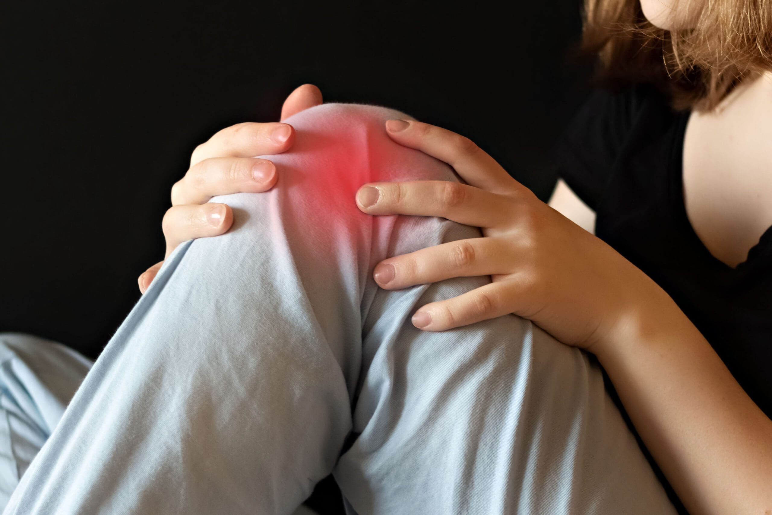
Understanding Knee Pain and Ligament Injuries: The Role of Chiropractic Care
Knee pain is a common complaint that can sideline even the most active individuals, whether you’re an athlete, a weekend warrior, or someone just trying to navigate the daily grind without wincing. Ligament injuries in the knee, such as sprains or tears, are often the culprits behind this discomfort, turning simple movements like walking or climbing stairs into a grim test of endurance. Fortunately, chiropractic care offers a holistic, non-invasive approach to managing knee pain, addressing not just the symptoms but the underlying causes. At ChiroMed – Integrated Medicine in El Paso, TX, Dr. Alexander Jimenez, DC, APRN, FNP-BC, combines advanced diagnostic techniques with integrative treatment protocols to help patients reclaim their mobility and quality of life.
This comprehensive guide explores the musculoskeletal system’s role in knee ligament injuries, environmental factors contributing to knee pain, and the clinical rationale for chiropractic care’s effectiveness. We’ll also highlight Dr. Jimenez’s expertise in personal injury cases, emphasizing his role as a liaison between medical care and legal documentation. With a touch of dark humor to keep things light, this post aims to inform and engage readers while providing actionable insights into managing knee pain.
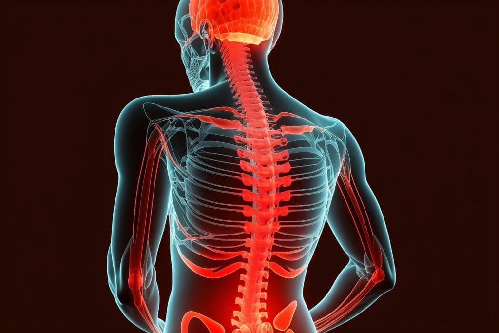
The Musculoskeletal System and Knee Ligaments: The Body’s Structural Framework
The knee is a marvel of engineering, but like any complex machinery, it’s prone to breakdowns—especially when you least expect it, like during a spirited game of tag with your dog or an ill-fated attempt to “jump” that puddle. The knee joint is one of the largest and most intricate in the human body, connecting the femur (thigh bone) to the tibia (shin bone) and stabilized by a network of ligaments, muscles, and tendons.
Key Ligaments of the Knee
The knee relies on four primary ligaments to maintain stability and facilitate movement:
- Anterior Cruciate Ligament (ACL): Prevents the tibia from sliding forward relative to the femur. It’s commonly injured in sports involving sudden stops or pivots, like basketball or soccer.
- Posterior Cruciate Ligament (PCL): Keeps the tibia from sliding backward. PCL injuries often occur in high-impact scenarios, such as car accidents.
- Medial Collateral Ligament (MCL): Stabilizes the inner knee, resisting forces that push the knee inward. MCL sprains are common in contact sports.
- Lateral Collateral Ligament (LCL): Stabilizes the outer knee, countering outward forces. LCL injuries are less common but can occur with lateral impacts.
These ligaments work in concert with muscles like the quadriceps and hamstrings to ensure smooth, controlled motion. However, when a ligament is stretched or torn—whether due to a misstep, a tackle, or just bad luck—the entire system can go haywire, leading to pain, swelling, and instability.
The Role of the Musculoskeletal System
The musculoskeletal system is like the body’s scaffolding, providing structure and support while allowing movement. Ligaments, as tough, fibrous bands, anchor bones to bones, ensuring the knee remains stable during activities like running, jumping, or dodging that rogue shopping cart in the parking lot. Muscles surrounding the knee, such as the quadriceps, hamstrings, and calf muscles, absorb shock and reduce stress on ligaments. However, imbalances in muscle strength, poor biomechanics, or external forces can overload these ligaments, leading to injury.
For example, weak hamstrings relative to the quadriceps can increase stress on the ACL, making it more susceptible to tears (El Paso Chiropractor Blog, 2016). Similarly, improper footwear or uneven surfaces can disrupt the knee’s alignment, causing undue strain on ligaments. Think of it like trying to balance a wobbly table—if one leg is shorter, the whole thing tips, and in this case, your knee pays the price.
Chiropractic Connection
Chiropractic care focuses on restoring proper alignment and function to the musculoskeletal system. By addressing misalignments in the spine, pelvis, or lower extremities, chiropractors can reduce compensatory stress on the knee joint. Techniques like spinal adjustments, soft tissue therapy, and corrective exercises help improve biomechanics, strengthen supporting muscles, and promote healing in injured ligaments (El Paso Chiropractor Blog, 2016). It’s like recalibrating a misaligned machine—suddenly, everything runs smoother, and the risk of further damage drops.
References
- El Paso Chiropractor Blog. (2016, May). Reducing pre- and post-workout knee pain. Retrieved from https://www.elpasochiropractorblog.com/2016/05/reducing-pre-and-post-workout-knee-pain.html
Environmental Factors Contributing to Knee Pain and Ligament Injuries
Knee pain doesn’t always stem from a dramatic injury—sometimes, it’s the little things in your environment that gang up on you like a pack of mischievous gremlins. Environmental factors, from the surfaces you walk on to the shoes you wear, can significantly contribute to knee pain and ligament injuries.
Common Environmental Culprits
- Uneven Surfaces: Walking or running on uneven terrain, like cracked sidewalks or rocky trails, can destabilize the knee, increasing the risk of ligament sprains. Imagine your knee as a tightrope walker—one wrong step, and it’s a painful tumble.
- Improper Footwear: Shoes without proper arch support or cushioning can throw off your gait, placing extra stress on the knee’s ligaments. High heels are particularly notorious, turning your walk into a high-stakes balancing act (Jimenez, n.d.).
- Repetitive Stress: Jobs or activities requiring prolonged standing, squatting, or kneeling—like construction work or gardening—can wear down the knee’s supporting structures over time, leading to microtears in ligaments.
- Weather and Climate: Cold weather can stiffen muscles and joints, reducing flexibility and increasing injury risk. Conversely, hot, humid conditions can lead to dehydration, weakening muscles and making ligaments more vulnerable.
- Sports and Physical Activity: High-impact sports like football or running on hard surfaces can overload the knee, especially without proper warm-ups or conditioning. It’s like asking your knee to run a marathon without training—disaster awaits.
Clinical Insights from Dr. Jimenez
Dr. Alexander Jimenez, a board-certified chiropractor and family nurse practitioner, emphasizes the importance of addressing environmental factors in knee pain management. At ChiroMed, he conducts thorough biomechanical assessments to identify how external factors, like poor posture or improper footwear, contribute to ligament stress (ChiroMed, n.d.). By recommending custom orthotics, corrective exercises, or lifestyle modifications, Dr. Jimenez helps patients mitigate these risks, reducing the likelihood of recurrent injuries.
For instance, runners with knee pain may benefit from switching to shoes with better shock absorption or incorporating pre-workout stretches to enhance muscle flexibility (El Paso Chiropractor Blog, 2016). Dr. Jimenez’s integrative approach ensures that patients not only recover but also prevent future issues by adapting their environment.
References
- ChiroMed. (n.d.). ChiroMed – Integrated Medicine. Retrieved from https://chiromed.com/
- El Paso Chiropractor Blog. (2016, May). Reducing pre- and post-workout knee pain. Retrieved from https://www.elpasochiropractorblog.com/2016/05/reducing-pre-and-post-workout-knee-pain.html
- Jimenez, A. (n.d.). LinkedIn profile. Retrieved from https://www.linkedin.com/in/dralexjimenez/
Why Chiropractic Care Helps with Knee Pain and Ligament Injuries
Chiropractic care might seem like an odd choice for knee pain—after all, aren’t chiropractors just for cracking backs? Not quite. The knee doesn’t operate in isolation; it’s part of a kinetic chain that includes the spine, pelvis, and feet. Misalignments or dysfunctions anywhere along this chain can exacerbate knee pain, and chiropractors are uniquely equipped to address these issues holistically.
Clinical Rationale for Chiropractic Care
- Restoring Biomechanical Alignment: Misalignments in the spine or pelvis can alter weight distribution, placing undue stress on the knee. Chiropractic adjustments correct these misalignments, reducing pressure on ligaments and promoting healing (El Paso Chiropractor Blog, 2016).
- Soft Tissue Therapy: Techniques like myofascial release and active release therapy target tight muscles and scar tissue around the knee, improving flexibility and reducing pain. It’s like giving your knee a much-needed massage after it’s been through the wringer.
- Strengthening Supporting Muscles: Chiropractors prescribe exercises to strengthen the quadriceps, hamstrings, and glutes, which stabilize the knee and reduce ligament stress. Stronger muscles act like shock absorbers, sparing your ligaments from taking the brunt of every step (OrthoInfo, n.d.).
- Bracing and Support: In some cases, bracing can provide temporary stability to an injured knee, allowing ligaments to heal without invasive measures. However, the decision to brace or not depends on the injury’s severity and the patient’s activity level (Mayo Clinic Health System, n.d.).
- Reducing Inflammation: Chiropractic care often includes modalities like ultrasound or cold laser therapy to reduce inflammation and promote tissue repair, speeding up recovery (Jimenez, n.d.).
Evidence-Based Support
Research supports the efficacy of chiropractic interventions for knee pain. A systematic review found that post-exercise stretching, often incorporated into chiropractic treatment plans, improves range of motion and reduces delayed onset muscle soreness, which can indirectly support ligament recovery (Witvrouw et al., 2021). Additionally, studies on knee osteoarthritis—a condition often exacerbated by ligament injuries—suggest that bracing can reduce pain and improve function, complementing chiropractic care (Jones et al., 2021).
Dr. Jimenez’s approach at ChiroMed integrates these evidence-based strategies, tailoring treatment to each patient’s needs. For example, a patient with an ACL sprain might receive a combination of adjustments, therapeutic exercises, and bracing recommendations to restore stability and function (ChiroMed, n.d.).
A Dash of Dark Humor
Let’s face it—knee pain can make you feel like you’re auditioning for a role as a limping zombie in a low-budget horror flick. But instead of shuffling through life groaning, chiropractic care offers a way to rewrite the script. Think of Dr. Jimenez as the director who cuts the scene where your knee betrays you mid-step, replacing it with one where you stride confidently, pain-free.
References
- ChiroMed. (n.d.). ChiroMed – Integrated Medicine. Retrieved from https://chiromed.com/
- El Paso Chiropractor Blog. (2016, May). Reducing pre- and post-workout knee pain. Retrieved from https://www.elpasochiropractorblog.com/2016/05/reducing-pre-and-post-workout-knee-pain.html
- Jimenez, A. (n.d.). LinkedIn profile. Retrieved from https://www.linkedin.com/in/dralexjimenez/
- Jones, R. K., Chapman, G. J., Forsythe, L., Parkes, M. J., & Felson, D. T. (2021). Clinical and cost-effectiveness of bracing in symptomatic knee osteoarthritis management: Protocol for a multicentre, primary care, randomised, parallel-group, superiority trial. BMJ Open, 11(3), e048196. https://pubmed.ncbi.nlm.nih.gov/33727270/
- Mayo Clinic Health System. (n.d.). To brace or not to brace: What’s best?. Retrieved from https://www.mayoclinichealthsystem.org/hometown-health/speaking-of-health/to-brace-or-not-to-brace
- OrthoInfo. (n.d.). Knee exercises. Retrieved from https://orthoinfo.aaos.org/en/staying-healthy/knee-exercises/
- Witvrouw, E., Danneels, L., Asselman, P., D’Have, T., & Cambier, D. (2021). The effectiveness of post-exercise stretching in short-term and delayed recovery of strength, range of motion and delayed onset muscle soreness: A systematic review and meta-analysis of randomized controlled trials. Sports Medicine, 51(5), 1053-1065. https://pubmed.ncbi.nlm.nih.gov/33687650/
Personal Injury Cases in El Paso: Dr. Jimenez’s Expertise
In El Paso, personal injury cases—whether from car accidents, workplace incidents, or slip-and-falls—often involve knee injuries, particularly ligament damage. These injuries can be debilitating, affecting victims’ ability to work, exercise, or even perform daily tasks. Dr. Alexander Jimenez stands out as a distinguished practitioner in this field, offering specialized care that bridges medical treatment and legal documentation.
The Role of Chiropractic Care in Personal Injury
Personal injury cases require precise documentation to support legal claims, and Dr. Jimenez excels in this area. His dual expertise as a chiropractor and family nurse practitioner allows him to conduct comprehensive evaluations, including:
- Advanced Imaging: Using X-rays, MRIs, or CT scans to visualize ligament damage and associated injuries, such as meniscal tears or bone bruising (Jimenez, n.d.).
- Diagnostic Evaluations: Tests like McMurray’s test or joint line tenderness assessments help confirm the presence of specific injuries, such as medial meniscus tears, though their accuracy varies (Hegedus et al., 2015).
- Dual-Scope Procedures: Combining chiropractic assessments with medical diagnostics, Dr. Jimenez provides a holistic view of the injury, ensuring no detail is overlooked.
This meticulous approach is critical in personal injury cases, where accurate documentation can make or break a legal claim. Dr. Jimenez acts as a liaison between patients, attorneys, and insurance companies, providing detailed reports that link injuries to the incident, such as a car accident causing a PCL tear due to dashboard impact (Jimenez, n.d.).
Clinical Insights and Case Example
Consider a hypothetical case: Jane, a 30-year-old El Paso resident, sustains an MCL sprain in a rear-end collision. Dr. Jimenez uses advanced imaging to confirm the injury, noting varus-valgus instability indicative of ligament damage (Wijdicks et al., 2017). He designs a treatment plan involving chiropractic adjustments to correct pelvic misalignment, therapeutic exercises to strengthen the quadriceps, and bracing to stabilize the knee during recovery. Simultaneously, he provides a detailed medical report for Jane’s attorney, linking the injury to the accident and justifying the need for ongoing care.
This integrated approach not only aids Jane’s recovery but also strengthens her legal case, ensuring she receives fair compensation for medical expenses and lost wages. Dr. Jimenez’s ability to navigate both medical and legal landscapes makes him a trusted figure in El Paso’s personal injury community.
A Pinch of Dark Humor
Getting rear-ended in El Paso traffic is bad enough, but when your knee decides to join the drama by staging its own injury protest, you’re in for a rough ride. Luckily, Dr. Jimenez is like the superhero your knee didn’t know it needed, swooping in with imaging, adjustments, and a knack for turning medical jargon into legal gold. It’s almost like he’s got a secret superpower for making insurance companies cry uncle.
References
- Hegedus, E. J., Cook, C., Hasselblad, V., Goode, A., & McCrory, D. C. (2015). McMurray’s test and joint line tenderness for medial meniscus tear: Are they accurate? Physical Therapy in Sport, 16(4), 321-326. https://pubmed.ncbi.nlm.nih.gov/26255139/
- Jimenez, A. (n.d.). LinkedIn profile. Retrieved from https://www.linkedin.com/in/dralexjimenez/
- Wijdicks, C. A., Griffith, C. J., Johansen, S., Engebretsen, L., & LaPrade, R. F. (2017). Varus-valgus instability in the anterior cruciate ligament-deficient knee: Effect of posterior tibial load. Journal of Orthopaedic Research, 35(4), 864-870. https://pubmed.ncbi.nlm.nih.gov/27160194/
Knee Pain Rehabilitation- Video
Diagnostic Tools for Knee Ligament Injuries
Diagnosing knee ligament injuries requires precision, as symptoms like pain, swelling, or crepitus (that unsettling grinding sound) can point to multiple issues. Dr. Jimenez employs a range of diagnostic tools to pinpoint the exact nature of the injury, ensuring targeted treatment.
Common Diagnostic Methods
- Physical Examination: Tests like McMurray’s test assess for meniscal or ligament damage, though they’re not foolproof (Hegedus et al., 2015). Joint line tenderness can also indicate MCL or meniscal issues.
- Advanced Imaging: MRI is the gold standard for visualizing ligament tears, while X-rays rule out fractures or bone misalignment. Dr. Jimenez uses these to confirm diagnoses and guide treatment (Jimenez, n.d.).
- Functional Assessments: Evaluating gait, range of motion, and stability helps identify compensatory patterns that exacerbate knee pain. For instance, a limp due to an ACL tear can strain the posterolateral corner of the knee (LaPrade et al., 2016).
- Patient History: Understanding the injury’s context—whether it occurred during a sports mishap or a car accident—helps correlate symptoms with clinical findings.
The Role of Crepitus
Crepitus, that crunchy sound your knee makes when you move, can be a red flag. Research suggests it’s a risk factor for knee osteoarthritis, often linked to ligament instability (Lo et al., 2018). Dr. Jimenez uses this symptom to guide diagnostic imaging and tailor interventions, such as strengthening exercises to stabilize the joint.
Humor in Diagnostics
Ever wonder what your knee is trying to tell you when it sounds like a bowl of Rice Krispies? It’s not auditioning for a cereal commercial—it’s begging for help. Dr. Jimenez’s diagnostic arsenal is like a detective kit for your knee, sniffing out the culprit behind the crunch and putting it on the path to recovery.
References
- Hegedus, E. J., Cook, C., Hasselblad, V., Goode, A., & McCrory, D. C. (2015). McMurray’s test and joint line tenderness for medial meniscus tear: Are they accurate? Physical Therapy in Sport, 16(4), 321-326. https://pubmed.ncbi.nlm.nih.gov/26255139/
- Jimenez, A. (n.d.). LinkedIn profile. Retrieved from https://www.linkedin.com/in/dralexjimenez/
- LaPrade, R. F., Wentorf, F. A., Fritts, H., Gundry, C., & Hightower, C. D. (2016). Posterolateral corner of the knee: Current concepts. The Archives of Bone and Joint Surgery, 4(2), 97-103. https://pubmed.ncbi.nlm.nih.gov/27200384/
- Lo, G. H., Strayhorn, M. T., Driban, J. B., Price, L. L., Eaton, C. B., & McAlindon, T. E. (2018). Subjective crepitus as a risk factor for incident symptomatic knee osteoarthritis: Data from the Osteoarthritis Initiative. Arthritis Care & Research, 70(1), 53-60. https://pubmed.ncbi.nlm.nih.gov/28320054/
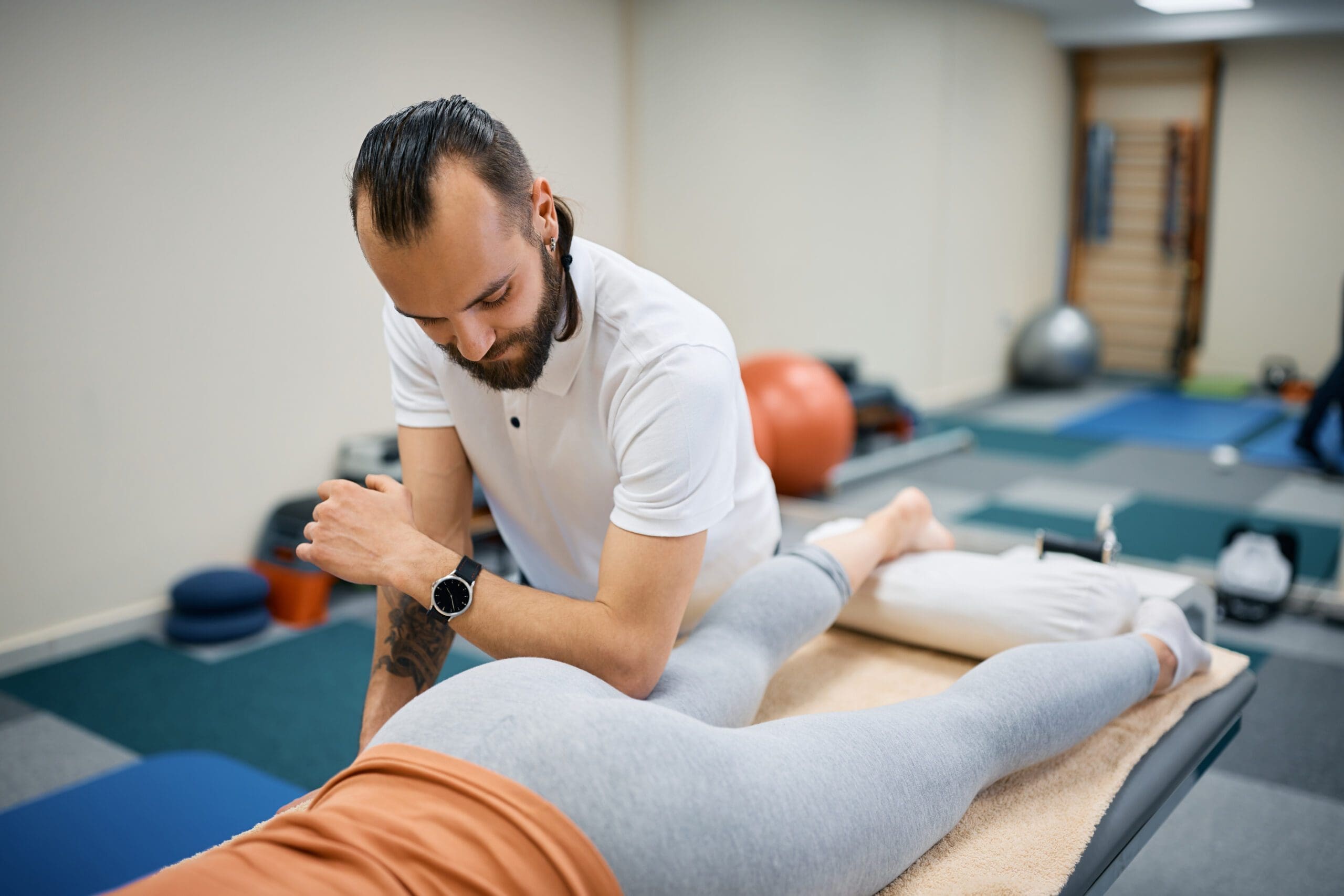
Integrative Treatment Strategies at ChiroMed
At ChiroMed, treatment goes beyond quick fixes, focusing on long-term healing and prevention. Dr. Jimenez combines chiropractic care with complementary therapies to address knee ligament injuries holistically.
Treatment Modalities
- Chiropractic Adjustments: Correcting spinal and pelvic misalignments to reduce knee stress.
- Rehabilitation Exercises: Strengthening and stretching programs to support ligament recovery (OrthoInfo, n.d.).
- Acupuncture: Reducing pain and inflammation through targeted needle placement.
- Nutrition Counseling: Anti-inflammatory diets to support tissue repair.
- Bracing: Providing stability during healing, especially for moderate sprains (Mayo Clinic Health System, n.d.).
Case Study: Athlete Recovery
Take Mike, a high school soccer player with a partial LCL tear. Dr. Jimenez used MRI to confirm the injury, followed by adjustments to correct hip misalignment, exercises to strengthen the glutes, and acupuncture to manage pain. Within weeks, Mike was back on the field, no longer hobbling like a pirate with a peg leg.
Humor in Healing
Recovery can feel like an eternity, especially when your knee seems determined to remind you of every bad decision you’ve ever made. But with ChiroMed’s integrative approach, it’s less like trudging through a swamp and more like a guided tour to Pain-Free Land, with Dr. Jimenez as your trusty guide.
References
- ChiroMed. (n.d.). ChiroMed – Integrated Medicine. Retrieved from https://chiromed.com/
- Mayo Clinic Health System. (n.d.). To brace or not to brace: What’s best?. Retrieved from https://www.mayoclinichealthsystem.org/hometown-health/speaking-of-health/to-brace-or-not-to-brace
- OrthoInfo. (n.d.). Knee exercises. Retrieved from https://orthoinfo.aaos.org/en/staying-healthy/knee-exercises/
Conclusion: A Serious Note on Knee Pain Management
Knee pain and ligament injuries can significantly impact your quality of life, but with the right care, recovery is within reach. Chiropractic care, as practiced by Dr. Alexander Jimenez at ChiroMed – Integrated Medicine in El Paso, TX, offers a holistic, evidence-based approach to managing knee pain. By addressing biomechanical imbalances, strengthening supporting muscles, and using advanced diagnostics, Dr. Jimenez helps patients achieve lasting relief and return to their active lifestyles. His expertise in personal injury cases further ensures that victims receive comprehensive care and legal support, bridging the gap between health and justice.
Disclaimer: This blog post is for informational purposes only and should not be taken as medical advice. Always consult a qualified healthcare professional, such as Dr. Alexander Jimenez, for a personalized diagnosis and treatment plan. The information provided is intended to educate and inform, not to replace professional medical guidance.
References
- ChiroMed. (n.d.). ChiroMed – Integrated Medicine. Retrieved from https://chiromed.com/
- El Paso Chiropractor Blog. (2016, May). Reducing pre- and post-workout knee pain. Retrieved from https://www.elpasochiropractorblog.com/2016/05/reducing-pre-and-post-workout-knee-pain.html
- Hegedus, E. J., Cook, C., Hasselblad, V., Goode, A., & McCrory, D. C. (2015). McMurray’s test and joint line tenderness for medial meniscus tear: Are they accurate? Physical Therapy in Sport, 16(4), 321-326. https://pubmed.ncbi.nlm.nih.gov/26255139/
- Jimenez, A. (n.d.). LinkedIn profile. Retrieved from https://www.linkedin.com/in/dralexjimenez/
- Jones, R. K., Chapman, G. J., Forsythe, L., Parkes, M. J., & Felson, D. T. (2021). Clinical and cost-effectiveness of bracing in symptomatic knee osteoarthritis management: Protocol for a multicentre, primary care, randomised, parallel-group, superiority trial. BMJ Open, 11(3), e048196. https://pubmed.ncbi.nlm.nih.gov/33727270/
- LaPrade, R. F., Wentorf, F. A., Fritts, H., Gundry, C., & Hightower, C. D. (2016). Posterolateral corner of the knee: Current concepts. The Archives of Bone and Joint Surgery, 4(2), 97-103. https://pubmed.ncbi.nlm.nih.gov/27200384/
- Lo, G. H., Strayhorn, M. T., Driban, J. B., Price, L. L., Eaton, C. B., & McAlindon, T. E. (2018). Subjective crepitus as a risk factor for incident symptomatic knee osteoarthritis: Data from the Osteoarthritis Initiative. Arthritis Care & Research, 70(1), 53-60. https://pubmed.ncbi.nlm.nih.gov/28320054/
- Mayo Clinic Health System. (n.d.). To brace or not to brace: What’s best?. Retrieved from https://www.mayoclinichealthsystem.org/hometown-health/speaking-of-health/to-brace-or-not-to-brace
- OrthoInfo. (n.d.). Knee exercises. Retrieved from https://orthoinfo.aaos.org/en/staying-healthy/knee-exercises/
- Wijdicks, C. A., Griffith, C. J., Johansen, S., Engebretsen, L., & LaPrade, R. F. (2017). Varus-valgus instability in the anterior cruciate ligament-deficient knee: Effect of posterior tibial load. Journal of Orthopaedic Research, 35(4), 864-870. https://pubmed.ncbi.nlm.nih.gov/27160194/
- Witvrouw, E., Danneels, L., Asselman, P., D’Have, T., & Cambier, D. (2021). The effectiveness of post-exercise stretching in short-term and delayed recovery of strength, range of motion and delayed onset muscle soreness: A systematic review and meta-analysis of randomized controlled trials. Sports Medicine, 51(5), 1053-1065. https://pubmed.ncbi.nlm.nih.gov/33687650/