Healing Your Spine from the Inside Out
Integrating Chiropractic Care with Regenerative Support
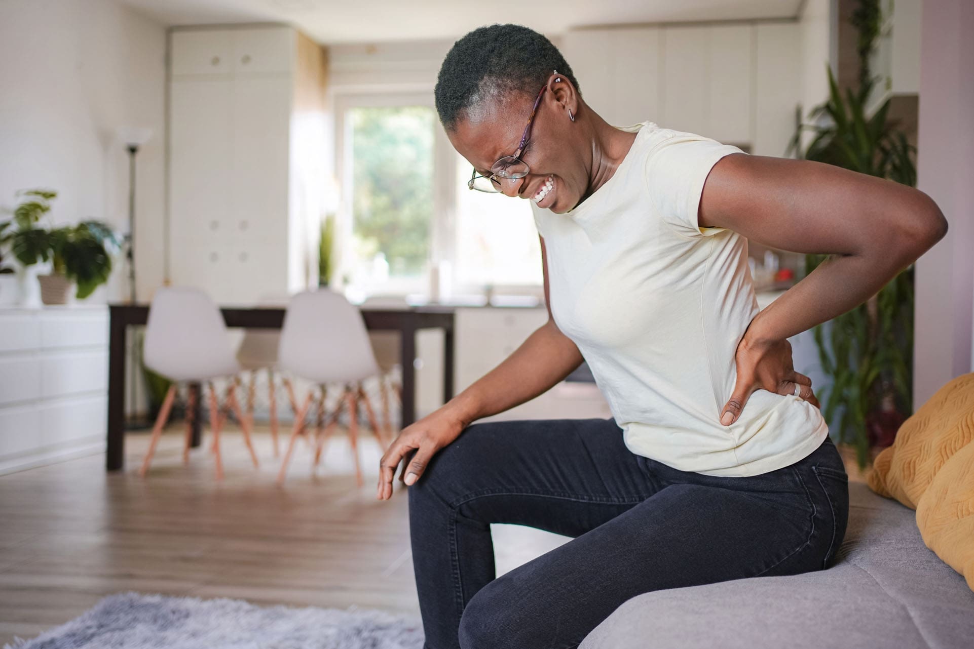
Chronic back or neck pain can slow you down and make simple daily tasks feel hard. Many people try rest, medicine, or basic therapy, yet the pain keeps coming back. Surgery is sometimes suggested, but it often means long recovery times and possible risks. At ChiroMed – Integrated Medicine in El Paso, Texas, there is a better way. Combining chiropractic adjustments with regenerative soft-tissue support repairs the spine from the inside out. Adjustments fix the structure and alignment. Treatments that heal soft tissues stop chronic pain and help rebuild strength without surgery.
This whole-person approach looks at the complete picture. It fits perfectly with ChiroMed’s mission of integrated, holistic healthcare that addresses root causes so you can live life more fully.
Your Spine Is Like a House
Think of your spine like the wooden frame of a house. The bones (vertebrae) are the main beams. The discs, ligaments, tendons, and muscles are the wood, pipes, and supports that hold everything steady.
If the frame leans or shifts, the whole house feels off. Chiropractic adjustments gently move the bones back into better position. This straightens the frame and takes pressure off nerves.
But what if the wood is rotting or the pipes are broken? Straightening the frame alone will not last. The house will lean again. The same thing happens with the spine. If the soft tissues stay damaged or inflamed, pain and weakness return even after good adjustments.
Regenerative therapies, laser support, and shockwave act like a skilled repair crew. They go into damaged areas, reduce swelling, and help new, healthy tissue grow. Massage, soft-tissue work, and spinal decompression then remove daily stress so the repairs hold strong over time. This comprehensive plan delivers lasting results for many people with ongoing spinal problems.
Chiropractic Care: Straightening the Structural Frame
Chiropractic care is the foundation of spine health at ChiroMed. A skilled provider uses gentle, hands-on adjustments or instrument-assisted methods to correct misalignments. These shifts can come from poor posture, old injuries, car accidents, work strain, or years of sitting and bending.
Adjustments help in clear, practical ways:
- They restore normal movement in stiff joints.
- They reduce pressure on nerves that cause pain, numbness, or tingling.
- They improve blood flow and support the body’s natural healing ability.
- They help improve posture so everyday activities feel easier.
Chiropractic care offers a non-invasive solution for chronic pain. It helps many people avoid surgery and long-term medication use. Regular adjustments can also enhance range of motion and correct postural issues for a healthier spine.
Reliable health sources confirm that chiropractic adjustments aim to correct alignment problems, ease pain, and support the body’s natural ability to heal itself. Many people seek this care for low back pain, neck pain, and headaches.
When the frame is straight, the rest of the spine can heal better. At ChiroMed, the results improve even more when adjustments are paired with therapies that fix the soft tissues around the bones.
Regenerative and Soft-Tissue Support: Calling in the Repair Crew
Regenerative approaches use the body’s own materials or advanced tools to heal damaged areas. They target the “rotting wood” parts — inflamed discs, strained ligaments, irritated nerves, and weak tendons. These treatments send growth signals that tell cells to repair and rebuild.
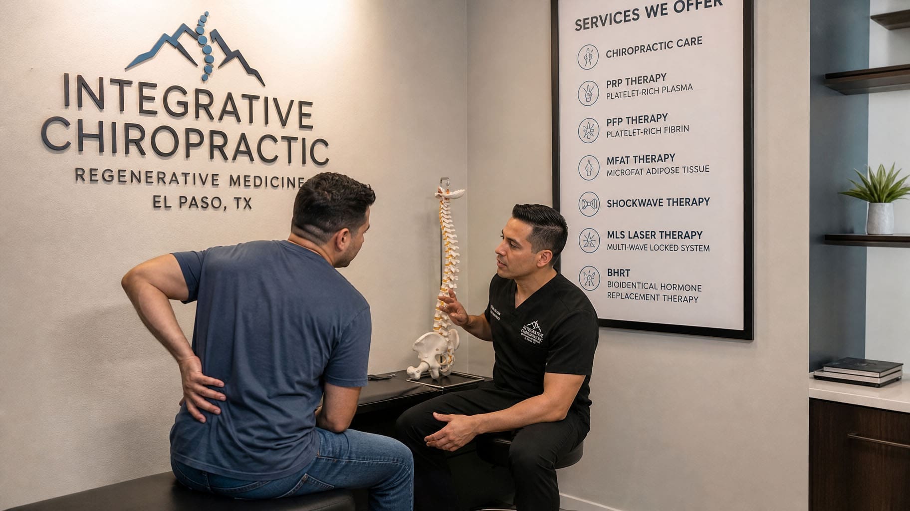
Common supportive options that work well with chiropractic include:
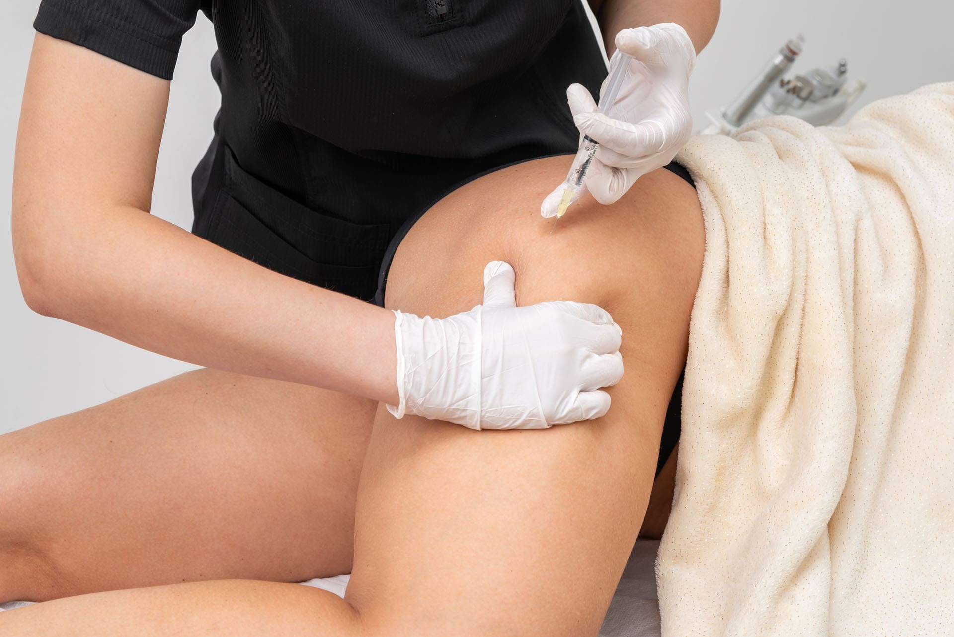
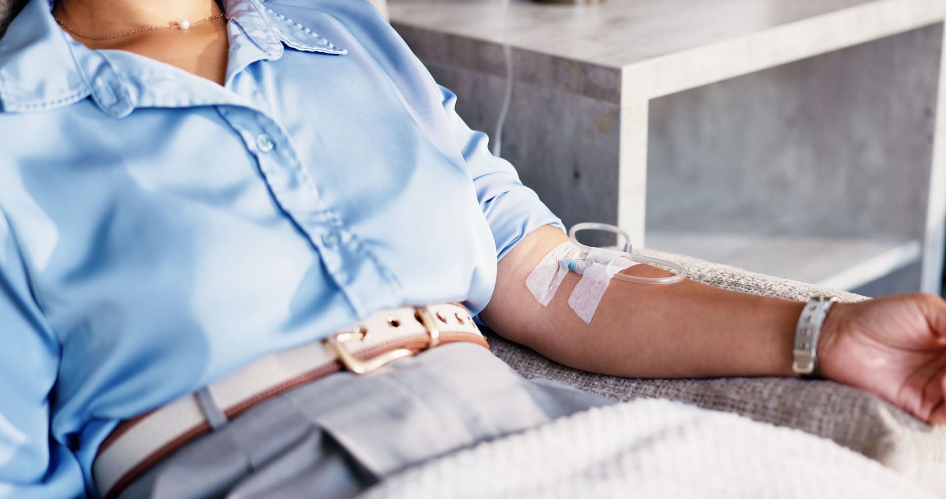
- PRP-style regenerative injections: Concentrated healing factors from your own blood are placed near injured tissues. They calm inflammation and encourage stronger tissue growth.
- Shockwave therapy: Sound waves reach deep into sore muscles and tendons. They increase blood flow, break up scar tissue, and restart the body’s repair process.
- Laser support: Special light energy gives cells more power to reduce swelling and speed healing at the deepest level.
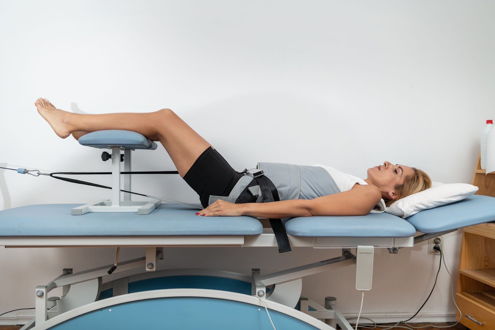
- Spinal decompression: Gentle stretching creates space between vertebrae. This takes pressure off bulging discs, lets nutrient-rich fluid flow in, and helps discs rehydrate and repair.
- Soft-tissue work and massage: These relieve daily stress, improve circulation, and prevent new tissue from tightening again.
These methods help stop the cycle of chronic pain. They reduce swelling, support new tissue growth, and strengthen the areas that hold the spine in place. Many patients notice less pain and improved mobility when these treatments are part of a complete plan.
How Everything Works Together for Lasting Results
The real power comes when chiropractic and regenerative soft-tissue care team up. Adjustments create the right alignment so new tissue can form correctly. Regenerative and soft-tissue support create a healing environment within the tissues, so repairs last longer.
Here is how a typical integrated plan at ChiroMed usually flows:
- A careful exam, history, and imaging find the exact problems.
- Chiropractic adjustments straighten the frame and improve motion.
- Regenerative injections, shockwave therapy, or laser therapy target the damaged soft tissues and nerves.
- Spinal decompression and soft-tissue care (including massage and rehab exercises) protect the healing areas from daily stress.
- Nutrition counseling, acupuncture, and functional medicine support reduce inflammation and help the whole body recover.
One clear clinical insight is that when regenerative support and chiropractic care are combined, results often last longer. The injections or advanced therapies create a better healing environment inside the tissues. The adjustments keep the joints moving correctly so new tissue forms properly and does not get stressed again.
A combined approach using chiropractic care, spinal decompression, regenerative support, and therapies like shockwave often works better. These treatments create both mechanical and biological conditions that help the body heal and maintain better alignment.
Patients often report meaningful drops in pain, better strength, and the ability to return to work, sports, or family activities. The goal is not just short-term relief. It is lasting repair that helps people avoid surgery and strong medications.
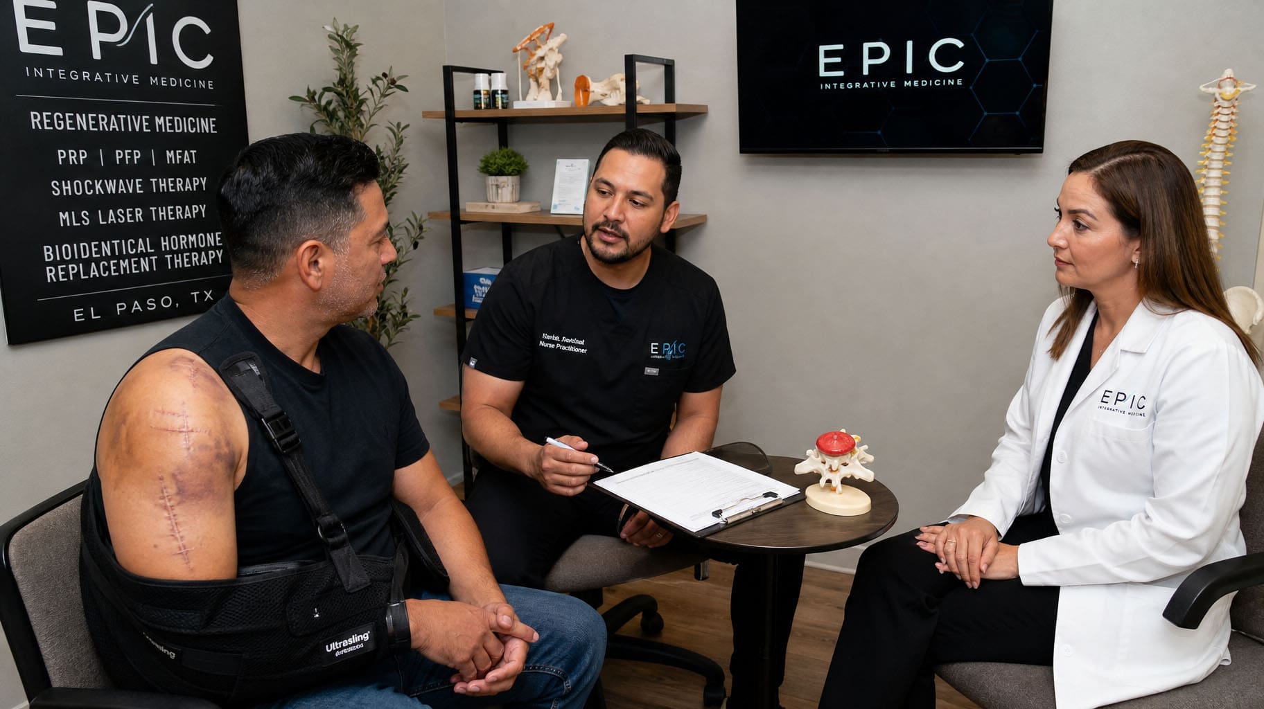
Expert Multidisciplinary Care at ChiroMed in El Paso
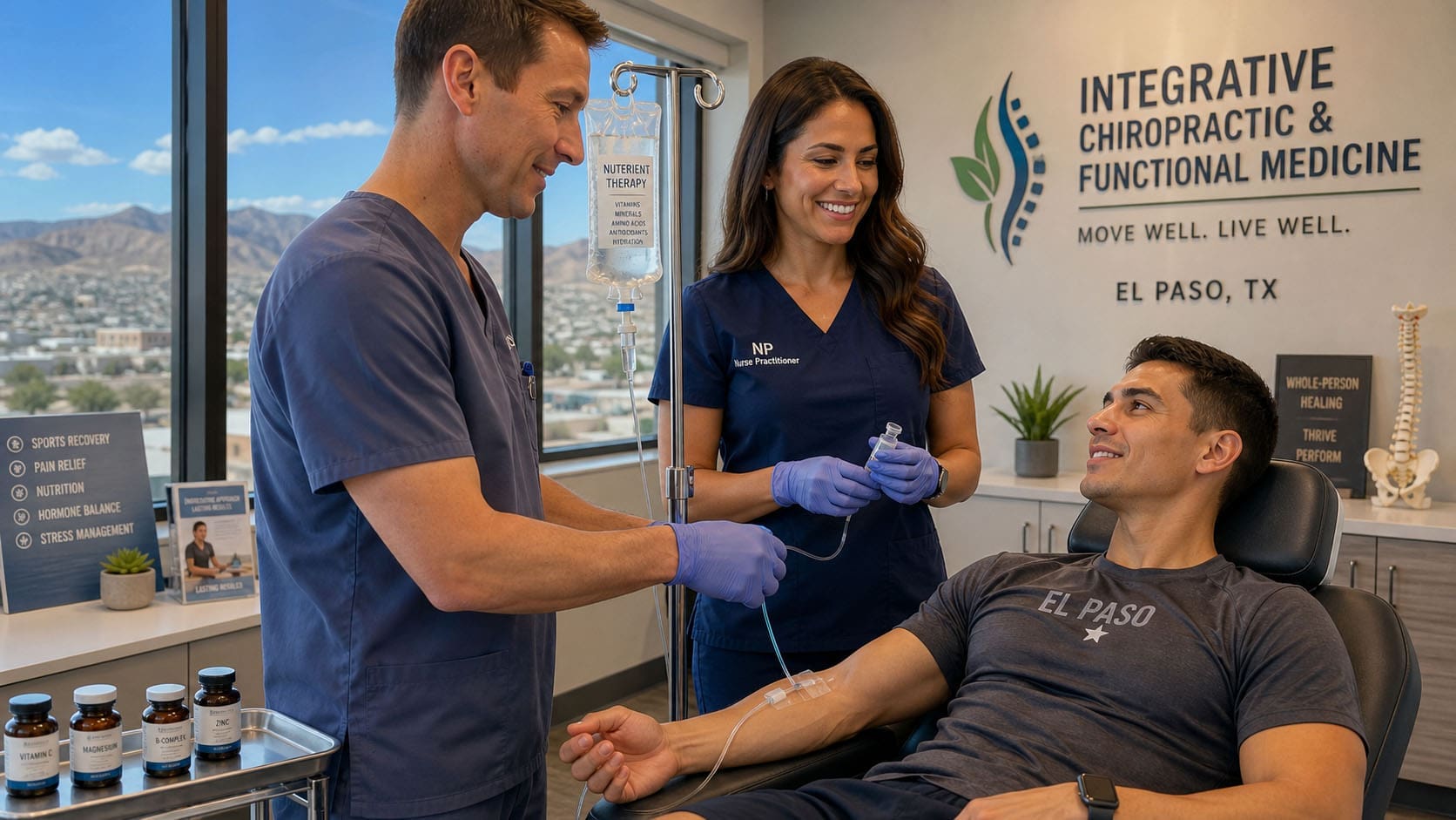
ChiroMed – Integrated Medicine brings this complete approach under one roof at 11860 Vista Del Sol Dr Suite 105, El Paso, TX 79936. The clinic has served the community since 1996 with a goal-oriented, honest, and personalized style of care.
Dr. Alexander Jimenez, DC, APRN, FNP-BC, CFMP, IFMCP, ATN, CCST, serves as Clinical Director. He is a multi-state-licensed Doctor of Chiropractic and a board-certified Family Nurse Practitioner. His clinical observations show that patients with old injuries, car-accident damage, sciatica, posture problems, and chronic spine pain improve when care targets both tissue repair and nervous-system function. He sees people regain mobility, reduce chronic pain, and return to daily life through personalized, non-invasive plans that combine adjustments, soft-tissue healing, rehabilitation, nutrition, and functional medicine support.
Working alongside him is Dr. Maria Guadalupe Cardenas, MD. She is a board-certified internal medicine physician (NPI #1164426749, Texas MD License #J2933) with over 40 years of experience. She serves as Medical Director and Collaborative Physician. In this multidisciplinary setup — common in integrative and injury care clinics — the MD provides medical oversight, ensures procedural safety, reviews complex health factors, and adds an internal medicine perspective. Dr. Jimenez delivers the hands-on chiropractic and regenerative soft-tissue care. Together with nurse practitioner services, rehabilitation, nutrition counseling, acupuncture, and naturopathic approaches, the team creates one coordinated plan.
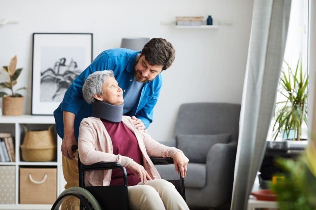
This collaboration means patients receive complete care. The structural work (chiropractic), the biological repair (regenerative and soft-tissue support), and the medical guidance all support each other. It is especially helpful for people with personal injuries, sciatica, chronic back pain, or posture problems that have not fully healed with other approaches. The clinic also works closely with patients’ other healthcare providers to ensure seamless care.
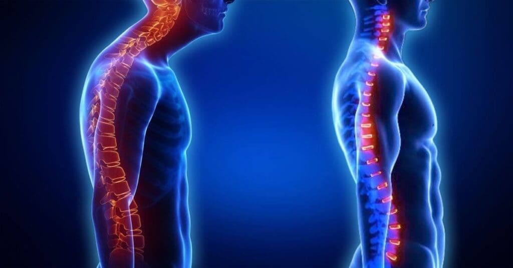
Breaking the Pain Cycle and Rebuilding Strength
Poor posture or old injuries often create a cycle: misalignment stresses soft tissues, tissues become inflamed or torn, pain limits movement, and weakness worsens. The integrated approach at ChiroMed breaks this cycle at every level.
Chiropractic restores alignment and motion. Regenerative and soft-tissue therapies heal and strengthen the supporting structures. Decompression, massage, and rehab exercises protect the repairs from daily stress. Nutrition and lifestyle support help the whole body recover. Over time, many people feel less pain, stand taller, move more freely, and enjoy activities again.
This path focuses on root causes instead of just masking symptoms. It supports the body’s natural ability to heal while giving it the right tools and environment to succeed. ChiroMed’s comfortable, fitness-center-like setting and licensed therapists make the journey supportive and goal-focused.
If ongoing spine pain is limiting your life, learning more about this integrated approach may open new options. Many people in El Paso have found real relief and lasting improvement through careful, combined care that repairs the spine from the inside out. Contact ChiroMed – Integrated Medicine today to schedule an evaluation and start your journey toward better strength and freedom from chronic pain.
References
MedlinePlus. (n.d.). Chiropractic. https://medlineplus.gov/chiropractic.html
El Paso Back Clinic. (n.d.). Regenerative therapies combined with chiropractic for pain relief. https://elpasobackclinic.com/regenerative-therapies-combined-with-chiropractic-for-pain-relief/
Personal Injury Doctor Group. (2026). Chiropractic and regenerative therapies for structural support. https://personalinjurydoctorgroup.com/2026/06/30/chiropractic-and-regenerative-therapies-for-structural-support/
Sleppy Chiropractic. (n.d.). Beyond the adjustment: How decompression, shockwave therapy, and laser treatment work together. https://www.sleppy.net/beyond-the-adjustment-how-decompression-shockwave-therapy-and-laser-treatment-work-together/
ChiroMed – Integrated Medicine. (n.d.). Chiropractor El Paso, TX. https://chiromed.com/services/chiropractor-el-paso-tx/
ChiroMed – Integrated Medicine. (n.d.). Home. https://chiromed.com/
Dr. Alexander Jimenez. (n.d.). Injury specialists. https://dralexjimenez.com/