Chiropractic Prevents Future Injuries in Athletes
How ChiroMed’s Integrative Chiropractic Care in El Paso, TX, Prevents Future Injuries in Athletes Using Functional Movement Assessments
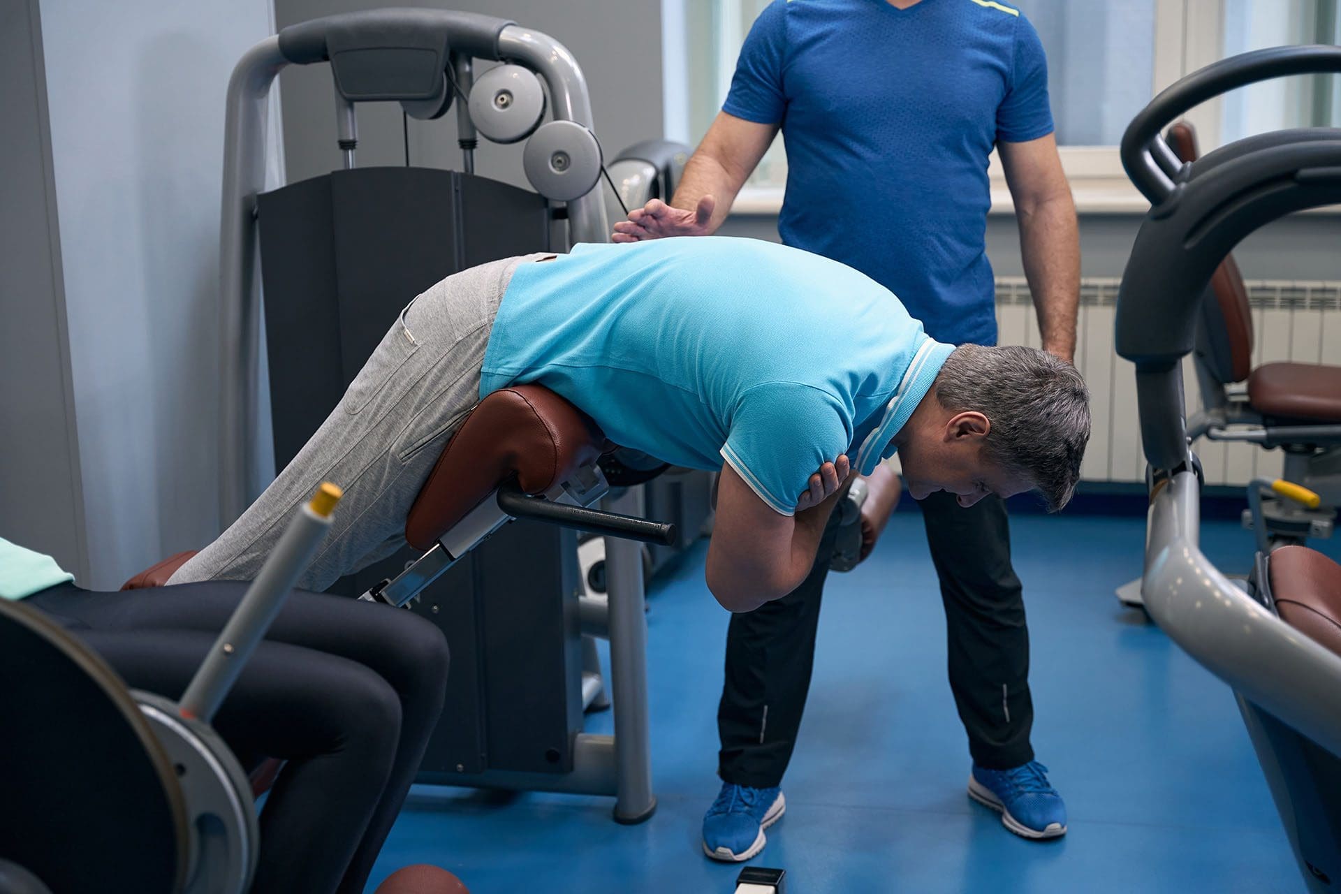
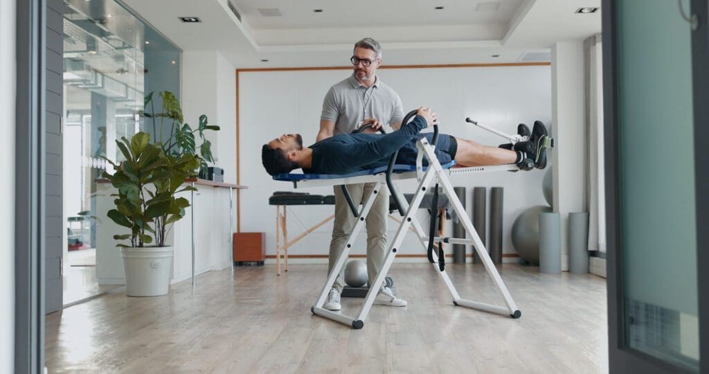
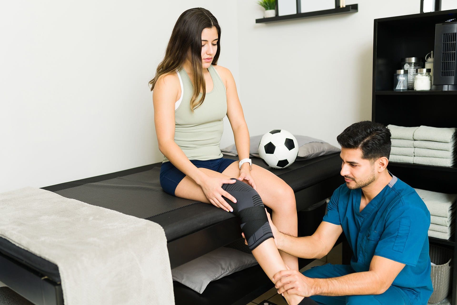
Athletes in El Paso, TX, often train hard to reach their goals, but small body issues can build up and cause injuries that sideline them. At ChiroMed – Integrated Medicine Holistic Healthcare, located at 11860 Vista Del Sol, Suite 128, athletes can get ahead of these problems with functional movement assessments. This approach spots hidden imbalances like tight muscles, weak spots, or stiff joints before pain hits. Our team, led by Dr. Alexander Jimenez, DC, APRN, FNP-BC, uses adjustments, soft-tissue therapies, and targeted exercises to address these issues, boost nervous system function, improve body mechanics, and prevent overuse injuries.
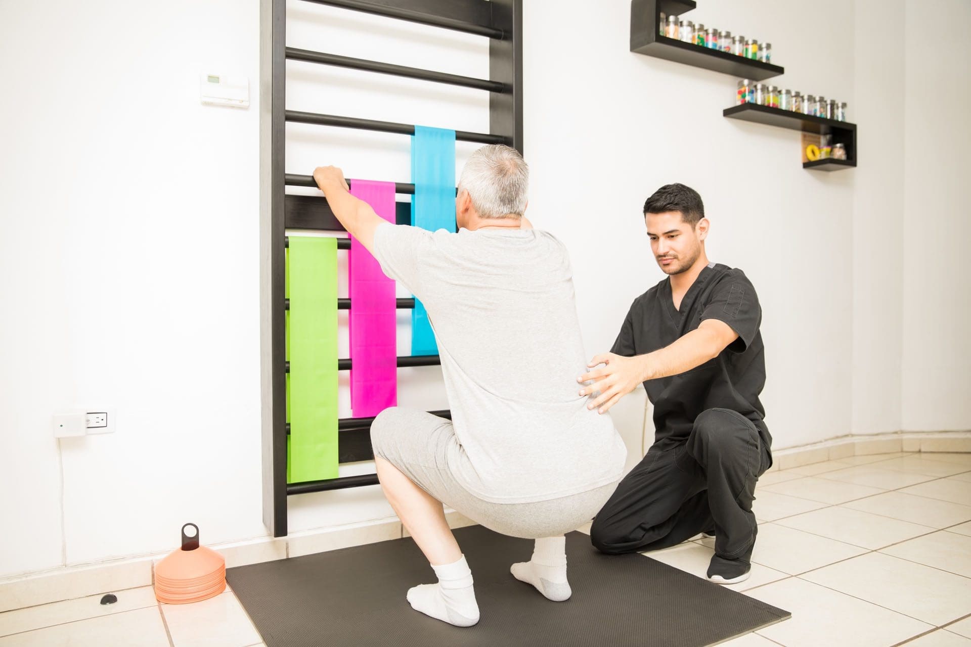
Functional movement assessments at ChiroMed check how your body moves during sports and daily tasks. These tests assess balance, flexibility, strength, and coordination. Simple actions such as squats, steps, or arm reaches help reveal where movements are off. Even without pain, these assessments find subclinical imbalances—minor problems that could turn into big injuries later. Our El Paso experts use these tools to create a plan tailored to each athlete’s needs.
- Detecting uneven posture or weight shifts that strain one side
- Finding limited motion in key areas like the hips or the shoulders
- Spotting weak muscles in the core or legs that affect stability
- Noting tight spots that throw off joint alignment
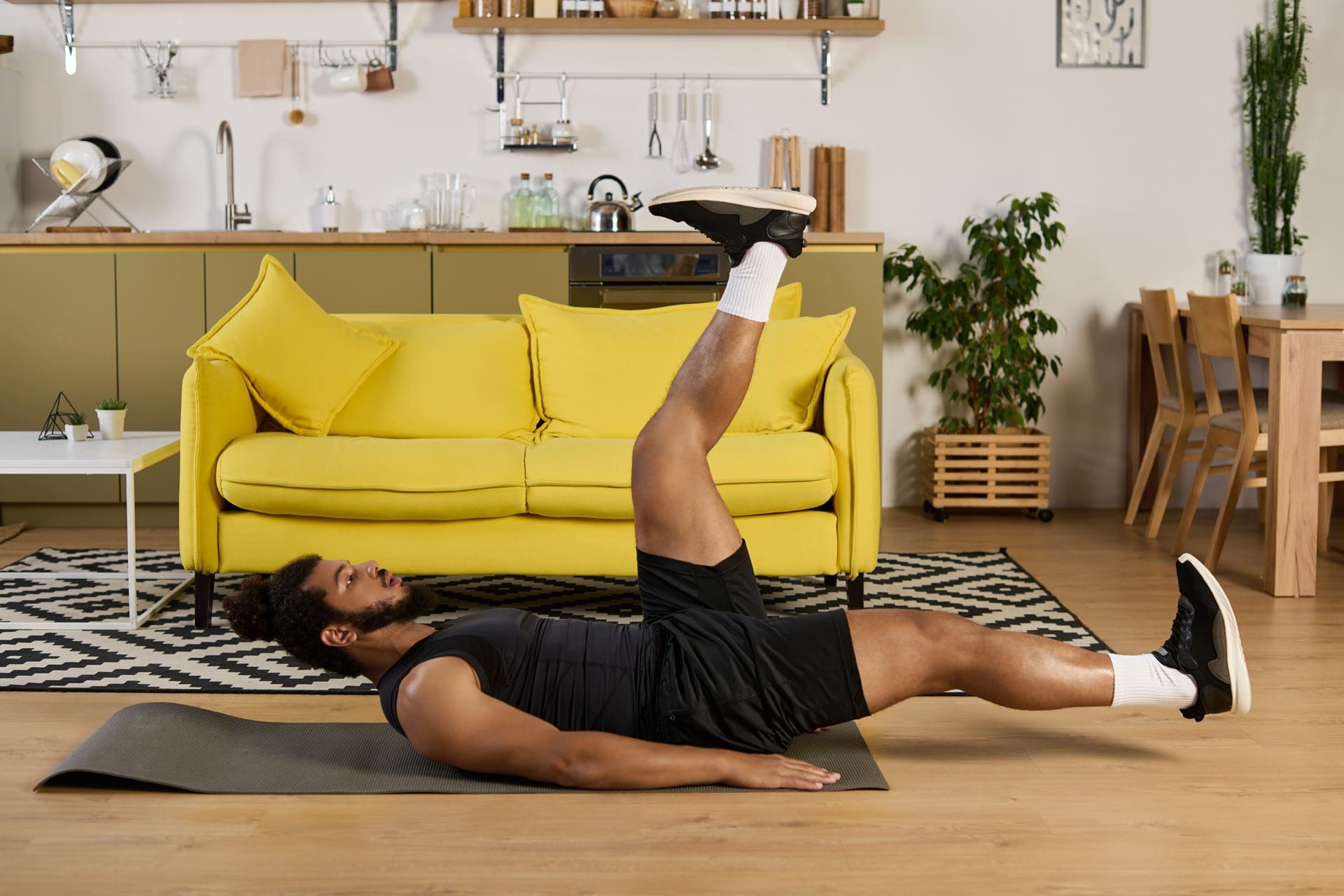
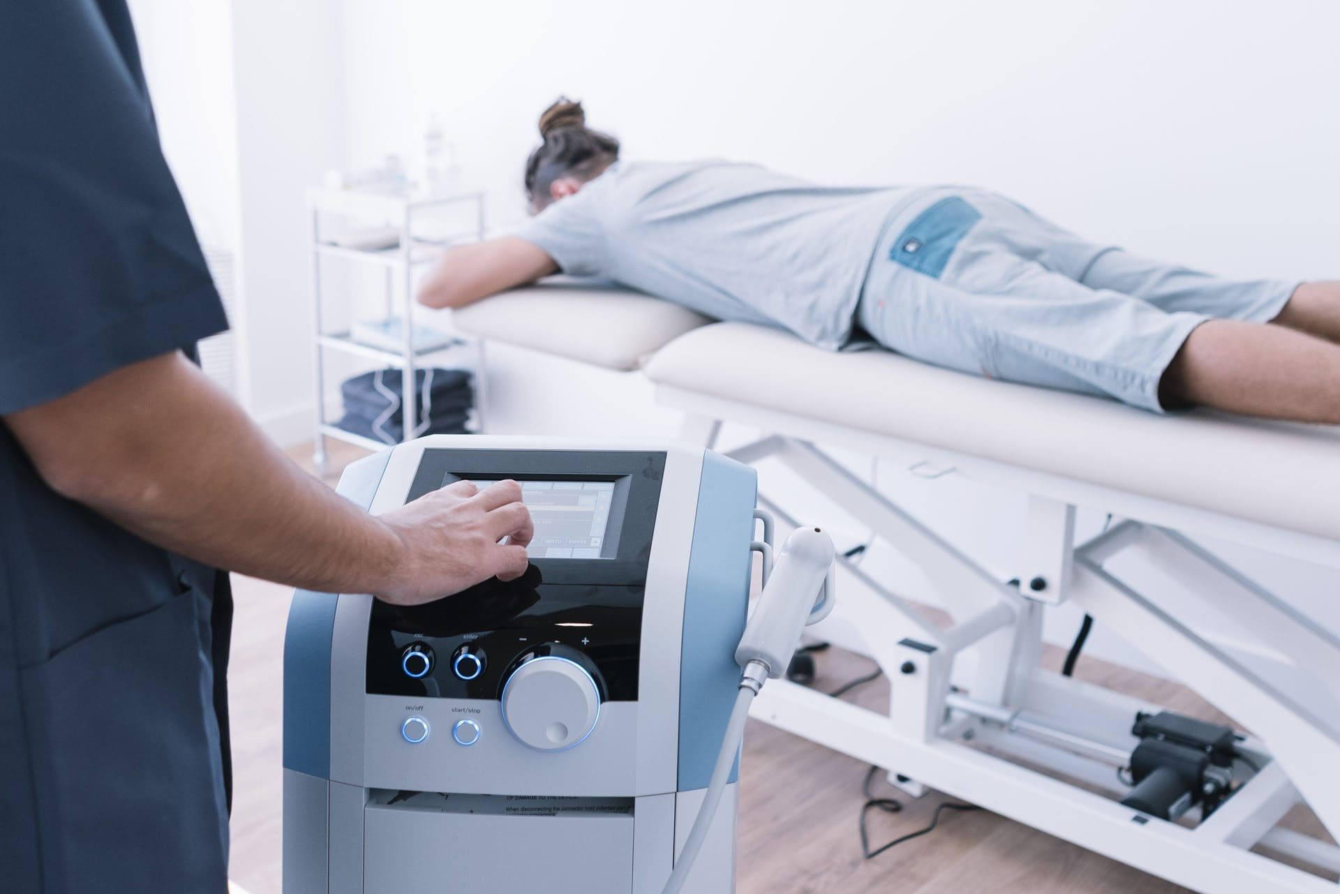
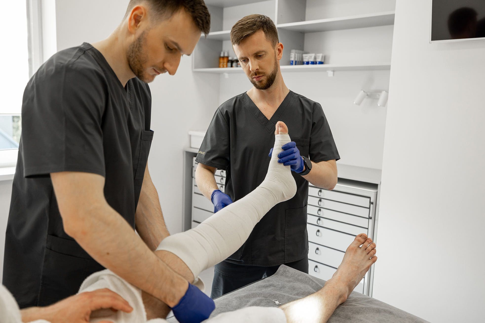
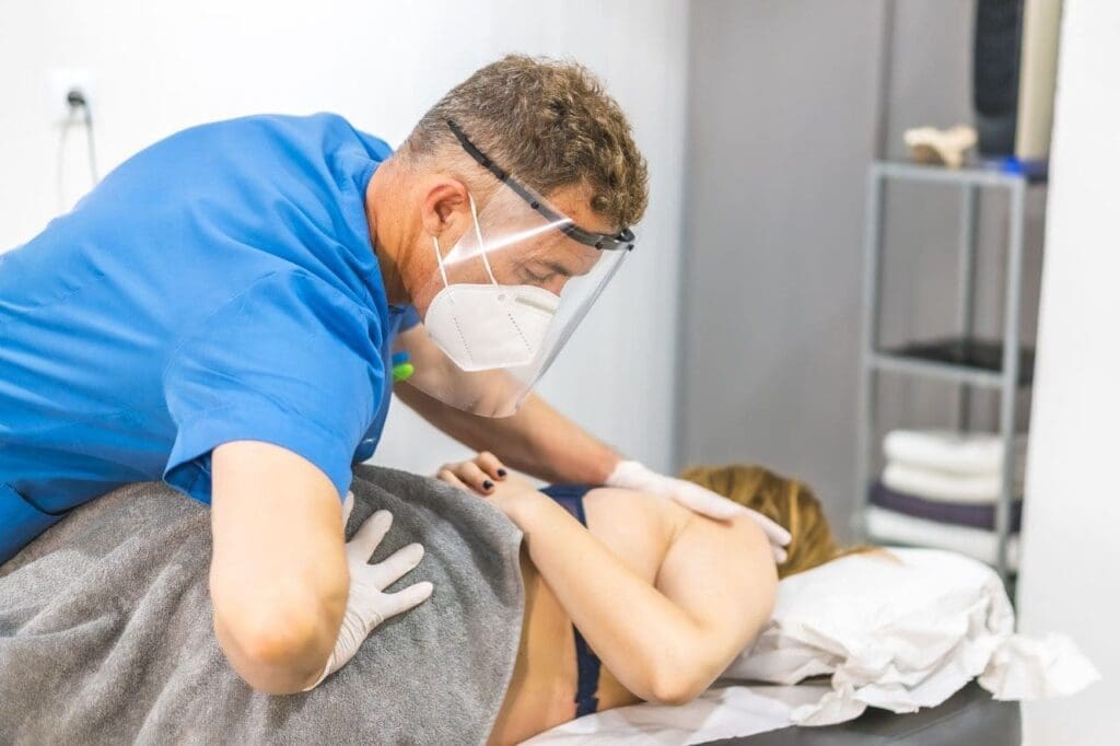
ChiroMed’s integrative chiropractic care blends spinal adjustments, soft-tissue work, and corrective exercises to address these findings. Adjustments gently realign joints to ease nerve pressure and improve signals throughout the body. Soft tissue therapies, such as massage or specialized tools, release tension and help heal old scars. Corrective exercises then strengthen weak areas and teach better movement habits. This holistic method enhances nerve function, refines biomechanics, and prevents the body from compensating in ways that lead to injuries.
The nervous system is key to muscle control. Misalignments can disrupt signals, causing poor coordination or delayed responses. At ChiroMed in El Paso, TX, our adjustments clear these blocks, helping muscles work together smoothly. Better biomechanics allow joints to move freely without extra wear, protecting areas like knees, backs, and ankles from stress.
Compensation patterns develop when the body overuses strong areas to cover for weak ones. For instance, runners with pelvic tilts might strain their knees or shins. Swimmers or lifters with poor shoulder form could overload muscles and cause tears. ChiroMed’s team corrects these early to keep athletes training safely.
Common subclinical imbalances we identify at ChiroMed through functional movement assessments include:
- Tension in the back or legs that limits bending or twisting
- Weak hip muscles that make the body unstable during runs or jumps
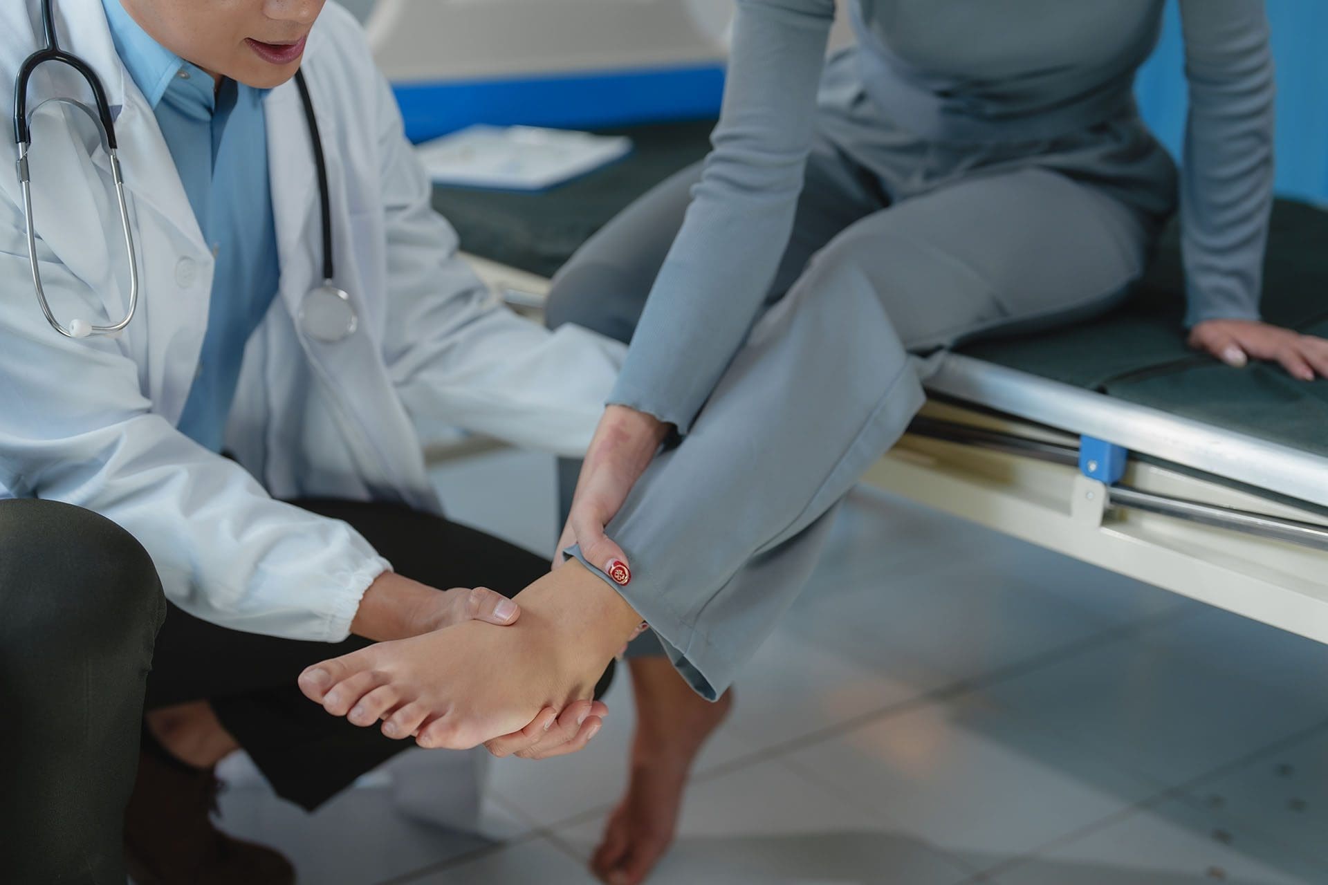
- Stiff ankles that alter how you walk or land
- Uneven arm mobility that impacts throws or lifts
- Shaky core strength leading to back strain in heavy activities
Addressing these at our El Paso clinic lowers injury risks and supports steady training. Regular sessions also mean faster fixes for any small setbacks, reducing time away from sports.
Our process starts with a full review of your history and a hands-on exam. We observe your movements and note any uneven patterns. From there, we build a custom plan. Adjustments fix spinal alignment, soft tissue work eases restrictions, and exercises reinforce strength. Over time, this improves your balance, flexibility, speed, and endurance.
Key benefits of ChiroMed’s functional movement assessments and integrative care:
- Lower risks of twists, pulls, inflammation, or breaks
- Greater joint range and muscle stretch for top performance
- Quicker reflexes from improved nerve signals
- Less swelling and faster bounce-back after sessions
- Extended active years by avoiding long-term wear
Runners visiting ChiroMed often have pelvic imbalances that affect their gait. Our adjustments and core-strengthening exercises level things out, making runs smoother and safer. Lifters with tight shoulders might arch their backs wrongly, risking strain—our therapies and drills fix that before issues arise.
Contact sports players benefit from our spinal checks to better absorb hits. Swimmers get help with shoulder flow to avoid overuse. Even casual gym-goers in El Paso see better stamina and less aching. ChiroMed’s methods suit all levels by proactively targeting root problems.
Dr. Alexander Jimenez, DC, APRN, FNP-BC, leads ChiroMed with his expertise in chiropractic, family nursing, and functional medicine. His clinical observations show that combining assessments with adjustments, nutrition, and rehab effectively treats sports injuries, nerve pain, and body imbalances. At ChiroMed, Dr. Jimenez focuses on non-invasive care that reduces inflammation, restores motion, and prevents reoccurrences. His El Paso practice integrates these elements to boost performance, ease chronic discomfort, and promote overall health.
This interconnected approach views the body as a whole system. A restriction in one spot can ripple through others. ChiroMed breaks these chains by realigning and teaching lasting habits.
Additional advantages athletes experience at ChiroMed include:
- Straighter posture for everyday and athletic tasks
- Sharper body sense for secure movements and changes
- Reduced tiredness in extended workouts
- Stronger output from smooth mechanics
- Mental sharpness from fewer distractions
Our preventive care in El Paso saves athletes from costly fixes or lost events. Many report feeling more balanced, powerful, and assured after our programs.
To sustain gains, we recommend ongoing visits based on your sport and load. High-intensity athletes might come weekly, while others come monthly. Home exercises keep progress going.
We also educate on warm-ups, stretches, and sport-specific form. Nutrition tips help repair and reduce swelling. Teaming with trainers or therapists rounds out support.
ChiroMed also offers nurse practitioner services, naturopathy, and rehab for full holistic care. Whether you’re a pro or a hobbyist in El Paso, our team helps you move better and stay injury-free.
Injuries can derail plans, but ChiroMed’s approach catches them early. We blend modern assessments with proven therapies to optimize your body.
Athletes trust us for personalized plans that fit their lives. From initial scan to follow-ups, we guide you toward peak health.
In conclusion, at ChiroMed – Integrated Medicine Holistic Healthcare in El Paso, TX, functional movement assessments help spot subclinical imbalances before pain starts. Our integrative chiropractic care—with adjustments, soft-tissue work, and exercises—boosts nerve function, optimizes biomechanics, and halts harmful patterns. Runners fix pelvic tilts, lifters correct techniques, and all athletes train uninterrupted. Contact us at (915) 850-0900 to prevent injuries and enhance your game.
References
Prevention of Sports Injuries Rhythm of Life Chiropractic. (n.d.).
Sports Injury Chiropractor: Ultimate Guide 2025 Stanlick Chiropractic. (2025).
Unlocking Athletic Potential: The Chiropractic Advantage AnySpine. (2024, October 1).
Functional Movement Assessments Joint Pain Relief Springfield MO 417 Spine. (n.d.).
The Athlete’s Guide to Preventative Chiropractic Care The KC Chiro. (2024, March 17).
Sports Injuries Treated With Chiropractic Care Advanced Spine & Posture. (n.d.).
Integrating Chiropractic Care with Sports Medicine Dallas Accident and Injury Rehab. (n.d.).
Chiropractic Care for Athletes: Enhancing Performance and Preventing Injuries Hilltop Integrated Healthcare. (n.d.).
Dr. Alexander Jimenez Clinical Insights Jimenez, A. (n.d.).
Dr. Alexander Jimenez LinkedIn Profile Jimenez, A. (n.d.).