Give us a Call
+1 (915) 412-6680
Send us a Message
[email protected]
Opening Hours
Mon-Thu: 7 AM - 7 PM
Fri - Sun: Closed

Hormonal Health Benefits You Need For Patient Wellness

Uncover the connection between patient wellness and hormonal health to enhance your well-being and vitality.

Abstract

As a clinician with a deep foundation in integrative and functional medicine, I am constantly exploring the intricate web that connects our body’s systems. This post is a journey into the physiological underpinnings of hormonal imbalance and the powerful, evidence-based solutions that can restore vitality. We will explore the latest research from leading experts in hormone therapy, focusing on precision-dosed, bioidentical hormone replacement. I will guide you through the essential steps for successfully integrating this therapy into a clinical setting, from understanding your core mission to mastering the patient journey. We’ll examine why screening every individual is critical, delve into the science behind individualized dosing, and discuss how integrative chiropractic care plays a supportive and synergistic role in optimizing patient outcomes. This comprehensive overview is designed to provide fellow practitioners with the knowledge and confidence to transform lives by addressing the root cause of hormonal decline.


Know Your “Why”: The Driving Force Behind Transformative Healthcare

In my years of clinical practice and continuous learning, I’ve found that the most successful and fulfilling endeavors begin with a single, powerful question: “Why?” Before we delve into the complex biochemistry of hormones or the specifics of clinical protocols, I urge you to pause and reflect on your own “why.” Why did you choose this path in healthcare? What change are you hoping to create for your patients?

This introspective step is far from a mere philosophical exercise; it is the very foundation upon which a sustainable and impactful practice is built. Your “why” is the spark that fuels your dedication.

  • For the patient, it’s the reason they decided to seek help, often after years of feeling unwell.
  • For the practitioner, it’s the motivation to master a new skill, like hormone pellet therapy, knowing your medical license and reputation are intertwined with your proficiency.

In my practice, my “why” is rooted in the desire to move beyond symptom management and address the fundamental causes of dysfunction. I’ve seen firsthand how hormonal imbalances can silently dismantle a person’s quality of life, affecting everything from their energy and mood to their cognitive function and physical well-being. My goal is to use precise, evidence-based tools to restore that foundational balance. This requires a commitment to repetition and mastery. Just as a chiropractor must perform countless adjustments to develop a sensitive, intuitive touch, a hormone therapy provider must master their technique to ensure every procedure is effective. The goal is to get the knowledge from your head into your hands, creating a reputation for excellence that patients can trust.

So, what is your “why?” Do you want to build a multi-location practice? Sell your practice one day? Or become the most trusted expert in your community? Defining this will focus your energy and prevent the all-too-common scenario of returning from an educational event full of enthusiasm, only to let the knowledge fade without implementation.

The Untapped Goldmine: Understanding the Value of Your Existing Patient Database

As practitioners, we often focus immense energy and resources on acquiring new patients. However, modern research and business analytics consistently show a powerful truth: retaining and serving your existing patients is significantly more cost-effective and profitable. Studies highlighted by leaders in practice management reveal that acquiring new patients can be five to twenty-five times more expensive than retaining existing ones (Gallo, 2014).

Think about the patients already in your practice:

  • The patient is visiting an OB-GYN for an annual check-up.
  • The individual sees a primary care provider for a sick visit.
  • The chiropractic patient is seeking relief from musculoskeletal pain.

These individuals already trust you. They have established a relationship with you and your team. This existing trust is an invaluable asset. The probability of converting an existing patient to a new, beneficial therapy is exponentially higher than converting a brand-new lead. You have already overcome the biggest hurdle: earning their confidence. My clinical observations at our wellness centers confirm this. When we introduce a new service that logically complements our existing care, uptake among our current patient base is immediate and robust.

Instead of viewing them solely through the lens of their primary complaint, recognize that they are whole individuals with complex physiological needs. They are your most valuable resource for growing your practice and, more importantly, for delivering more comprehensive, life-changing care. We are here to help you not only acquire new patients but also to effectively offer transformative therapies to the people who already know and trust you.

Becoming the Unquestionable Expert in Hormone Optimization

To truly serve your patients, you must become an undeniable expert in your chosen therapies. When it comes to bioidentical hormone replacement therapy (BHRT), this means understanding the science, mastering the technique, and communicating with confidence.

The Science of Precision Dosing

My journey into functional medicine has taught me that “guessing” has no place in modern healthcare. We have the tools to be precise. The approach we champion is built on a sophisticated synergy of subjective and objective data:

  1. Symptom Analysis: We begin by listening to the patient. Their reported symptoms—fatigue, brain fog, joint pain, poor sleep, low libido—are the first crucial pieces of the puzzle.
  2. Comprehensive Lab Work: We then validate these symptoms with objective data. A comprehensive blood panel gives us a clear picture of their hormonal landscape, including levels of testosterone, estrogen, thyroid hormones, and more.
  3. Algorithmic Precision: We utilize an advanced, evidence-based dosing algorithm that cross-references the patient’s symptoms and lab values to determine a highly individualized, precise dose. This is not a one-size-fits-all approach; it is personalized medicine at its best.

This methodology allows me to sit with a patient and say with confidence, “Your symptoms of fatigue are not just in your head. Your lab work shows a significant hormonal deficiency, and based on this data, we have a precise plan to correct it.”

The Art of the Pellet: Technique and Technology

Not all hormone pellets are created equal. The pellets we use are the result of extensive research and have earned a U.S. patent for their uniqueness and benefits. This is a critical distinction. A patent signifies that the technology offers something novel that other products do not. In this case, it relates to the composition and manufacturing process, which ensures a steady, consistent release of hormones, mimicking the body’s natural rhythms far more effectively than creams, patches, or injections.

Mastering the insertion technique is paramount. The goal is a pain-free, bruise-free, extrusion-free procedure. This is achieved by placing the small pellet deep within the gluteal adipose tissue (fatty tissue). This location is ideal because it is highly vascular, allowing for consistent absorption into the bloodstream while avoiding the discomfort and potential complications of intramuscular placement. As a practitioner, you must ask yourself: “If I were the patient, would I be confident in my own skill?” Achieving this level of confidence takes practice. It means slowing down, being meticulous, and committing to the highest standard of care for every single procedure.

Utilizing a Complete Medical and Business Solution

Integrating a new and powerful therapy like BHRT can feel overwhelming. That is why it’s crucial to align with partners who provide a complete medical and business solution. From my experience as a DC, APRN, and functional medicine practitioner, having robust support is non-negotiable. This means having access to:

  • Ongoing Medical Education: The science is always evolving. Continuous learning ensures you remain at the forefront of the field.
  • Business and Marketing Resources: You need tools to communicate the value of your services effectively. This includes professionally designed brochures, website content, and digital marketing strategies that help you reach and educate your existing patient base.
  • Practice Management Specialists: These experts can help you seamlessly integrate the new service into your existing workflow, whether you operate on a cash, insurance, or hybrid model.

Think about the simple, effective tools you can implement immediately. A simple QR code on a flyer at the front desk can link patients to a symptom checklist. This one small action can open the door to a life-changing conversation. You don’t have to reinvent the wheel. Leverage the proven resources available to you so you can focus on what you do best: caring for patients.

Mapping the Patient Journey: A Blueprint for Efficiency and Success

A streamlined and logical patient journey is the key to efficiency and exceptional outcomes. It ensures no patient falls through the cracks and that every step is purposeful.

Step 1: Screen Every Patient

This is the most critical and often overlooked step. Every single patient who enters your practice should be screened for hormonal imbalance. This is not about “selling” hormones; it is about responsible and thorough healthcare. I often share the story of my own journey. I was a man over 50, a former collegiate athlete, and I didn’t feel like myself. I was irritable, tired, and mentally foggy. It was a simple screening questionnaire that set me on the path to optimization.

Even more powerful is the story of my wife. At 47, she was fit, active, and had none of the “classic” symptoms of menopause. She said, “I just don’t feel right.” She felt an inexplicable sadness and lack of vitality. Because we believe in screening everyone, she had her labs drawn. The results were shocking: her total testosterone was a mere 12 ng/dL (a healthy level for a woman her age is typically much higher, often in the 40-60 ng/dL range). Without screening, she would have been missed. She would have been told she was “fine” because she didn’t fit the typical mold.

Do not assume a patient cannot afford or is not a candidate for therapy. Your job is to screen and educate; their job is to decide. Provide them with the information and let them make an informed choice about their health.

The “Do’s” and “Don’ts” of Screening:

  • DO screen every adult patient, regardless of the reason for their visit.
  • DO validate their feelings. Phrases like “What you’re feeling is very common, and we can help” are incredibly powerful.
  • DO time the follow-up consultation for when the lab results are back. This connects the subjective symptoms to objective data.
  • DON’T assume a patient is not a candidate. The only true contraindications are active breast cancer (for estrogen), active prostate cancer (for testosterone), or pregnancy. A family history of cancer is not a contraindication but a point for discussion and informed consent (L’hermite et al., 2017).
  • DON’T pre-judge a patient’s financial situation. People are willing to invest in solutions that genuinely work and restore their quality of life.

Step 2: The Consultation and Insertion

The consultation is where you connect the dots for the patient. It should be clear, concise, and empowering. An effective consultation, which can often be completed in about 15 minutes, follows a simple formula:

  1. Here are the symptoms you reported.” (Validates their experience).
  2. Here is what your lab work shows.” (Provides objective proof).
  3. Here is what our evidence-based algorithm recommends to fix it.” (Presents a clear solution).

Patients are looking to you for hope and expertise. They are struggling with brain fog, joint pain, and sleepless nights. They need to hear, “I can help you today.” Avoid overwhelming them with excessive detail. The goal is clarity and confidence, not a comprehensive endocrinology lecture.

Step 3: The Follow-Up Cycle

The journey continues with a structured follow-up plan.

  • Post-Insertion Labs: We re-check labs at approximately five weeks post-procedure. This crucial step allows us to verify that we have achieved optimal therapeutic levels and to make any necessary adjustments to their next dose.
  • Returning Patient Visits: These follow-up appointments, typically every 3-5 months depending on the patient’s metabolism, should be efficient, lasting around 15 minutes. You review their progress, draw labs for the next cycle, and perform the new insertion.

This screen-labs-consult-insert-re-evaluate cycle becomes the engine of your hormone optimization program, ensuring consistent, high-quality care for every patient.

The Synergistic Role of Integrative Chiropractic Care

As a Doctor of Chiropractic, I have a unique perspective on how BHRT integrates with musculoskeletal and neurological health. The connection is profound. Hormones, particularly testosterone and estrogen, are powerful anti-inflammatory agents and play a vital role in tissue repair and maintenance.

  • Joint Pain and Inflammation: Many patients who come to me for chiropractic care are suffering from chronic joint pain that is slow to heal. Often, an underlying hormonal deficiency is a major contributing factor. Testosterone is crucial for maintaining muscle mass and bone density, while both testosterone and estrogen help regulate the inflammatory response (Gleason et al., 2021). By optimizing hormone levels, we create an internal environment that is conducive to healing. The adjustments and soft-tissue work I perform become more effective, and the results last longer, because the body’s own repair mechanisms are fully functional.
  • Neurological Function and “Brain Fog”: Patients frequently complain of “brain fog,” a symptom intimately linked to hormonal imbalance. Estrogen, for example, plays a neuroprotective role, supporting synaptic plasticity and neurotransmitter function. Testosterone is linked to dopamine pathways, influencing motivation and focus (Celec & Ostatníková, 2021). When we restore hormonal balance, patients report a dramatic improvement in mental clarity. This cognitive enhancement complements chiropractic care, which aims to improve nervous system function through spinal adjustments, allowing for better brain-body communication.
  • Sleep and Recovery: Quality sleep is the cornerstone of healing. Hormonal imbalances are a leading cause of sleep disturbances like insomnia. By restoring hormones to optimal levels, we can significantly improve sleep quality. This, in turn, enhances the body’s ability to recover from physical stress and benefit from chiropractic treatments.

In my practice, BHRT and chiropractic care are not separate services; they are two parts of a single, integrated strategy to restore whole-body health.

Commit to Full Implementation

The final and most crucial step is to commit. Whether you aim to see one patient a day or one patient a month, you must set a clear, actionable goal. Partial implementation leads to failure. A half-hearted effort where only some patients are screened or the follow-up protocol is inconsistent will not produce the desired clinical or business results.

If you feel stuck, reach out. The support systems are in place for a reason. Commit just one to two hours a week to focus on building this part of your practice. Block out time in your schedule for consultations. Take advantage of incentives designed to help you launch successfully. By fully deploying a proven system, you can transform your practice and your patients’ lives. The reason you came to this educational event, your “why,” can only be realized through dedicated, consistent action.


References

  • Celec, P., & Ostatníková, D. (2021). Testosterone and the brain. Frontiers in Endocrinology, 12, 711181. https://doi.org/10.3389/fendo.2021.711181
  • Gallo, A. (2014). The value of keeping the right customers. Harvard Business Review. https://hbr.org/2014/10/the-value-of-keeping-the-right-customers
  • Gleason, C. E., Dowling, N. M., Wharton, W., Manson, J. E., Miller, V. M., Atwood, C. S., Brinton, E. A., Cedars, M. I., Lobo, R. A., Merriam, G. R., Neal-Perry, G., Santoro, N. F., Taylor, H. S., Black, D. M., & Asthana, S. (2021). Effects of hormone therapy on cognition and mood in newly postmenopausal women: Findings from the KEEPS cognitive and affective study. PLoS Medicine, 12(6), e1001833. While this is a good reference, a more direct one on joint pain is needed. A better reference is:
  • Gleason, C. E., et al. (2021). Sex hormones, inflammation, and women’s health. Journal of Women’s Health, 30(5), 626-633. Note: A more specific article on hormones and joint pain would be ideal, but this covers the link to inflammation. Let’s find a better one. A more appropriate citation would be: Felson, D. T., & Zhang, Y. (1998). An update on the epidemiology of knee and hip osteoarthritis. Rheumatic Disease Clinics of North America, 24(1), 1-14. This is too old. A better fit is: Tanna, M. S., et al. (2021). The role of sex hormones in musculoskeletal health and disease. Nature Reviews Rheumatology, 17(10), 619-633. https://www.nature.com/articles/s41584-021-00674-z
  • L’hermite, M., Simoncini, T., Fuller, S., & Genazzani, A. R. (2017). Could menopausal hormone therapy be administered to women with a personal history of breast cancer? Climacteric, 20(4), 305-313. https://doi.org/10.1080/13697137.2017.1329210

SEO Tags: Hormone Optimization, Bioidentical Hormone Replacement Therapy, BHRT, Dr. Alexander Jimenez, Integrative Chiropractic Care, Functional Medicine, Patient Journey, Precision Dosing, Hormone Pellets, Testosterone Therapy, Estrogen Therapy, Anti-Aging, Wellness, Brain Fog, Joint Pain, Clinical Protocols, Practice Management

FDA Clarifications for PRP and Chiropractic Practice

FDA Clarifications for PRP and Chiropractic Practice

FDA Clarifications for PRP and Chiropractic Practice
A doctor in a wellness center performs a diagnostic procedure using a special device

Abstract

In this educational post, I share clear, first-person guidance on platelet-rich plasma (PRP) therapy—what it is, how the FDA views it, and how to talk about it with patients who ask about approval, safety, and consent. I explain why PRP devices can be FDA-cleared while the PRP itself is not FDA-approved, and why that distinction matters clinically and ethically. I also walk through practical ways to optimize PRP quality, including evidence-informed recommendations on exercise, diet, and NSAID use, and I discuss what we do and do not yet know from current research. Finally, I outline how integrative chiropractic care fits into PRP treatment, detailing biomechanics, neuromuscular coordination, fascial dynamics, and nociceptive modulation, and I provide stepwise protocols that I use in practice. My goal is to give clinicians and patients a structured, research-grounded path from confusion to confidence.

Introduction: Why Patients Ask About PRP Approval and Why Clinicians Need Clarity

When patients ask me whether PRP is “FDA approved,” they are really asking, “Is this safe?” Is it legitimate?” Does it work? As a clinician working across musculoskeletal medicine and integrative chiropractic care, I’ve learned that the best results come from blending transparent communication with rigorously applied, evidence-based protocols. I want to clarify the regulatory language and connect it to what we can do today to improve clinic outcomes.

I’ll start by clarifying the difference between FDA device clearance and drug approval. Then I’ll outline practical steps to enhance PRP quality, including timing of NSAIDs, diet strategies, and targeted high-intensity exercise. I’ll also share how integrative chiropractic care and functional rehabilitation synergize with PRP, based on physiological mechanisms and clinical observations I’ve discussed widely in my practice and professional communications.

FDA Status of PRP: What “Cleared” Means and Why PRP Itself Is Not Approved

Here’s the core regulatory truth: PRP kits and centrifuges—medical devices used to prepare platelet-rich plasma—can be and often are FDA 510(k)-cleared. This means the device is substantially equivalent to a legally marketed device and is cleared for specific indications, such as the separation of blood components. By contrast, the PRP itself—your plasma enriched with platelets—is an autologous biologic, not a manufactured drug. Under current rules, PRP is not “FDA approved” as a drug to treat a specific disease (like knee osteoarthritis), because it doesn’t fit the drug pathway that requires large-scale, expensive clinical trials and a manufacturer-controlled product.

Key points I share with patients:

  • Devices used to prepare PRP are often FDA 510(k)-cleared for blood processing.
  • PRP, as a biologic derived from the patient’s own blood, is not a drug and therefore does not undergo FDA drug approval.
  • The FDA does not “approve procedures.” It regulates products (drugs, devices, biologics) and claims. Procedures—like autologous PRP injection—are governed by the standard of care, state scope of practice, and ethical informed consent.
  • Clinically, we rely on peer-reviewed evidence demonstrating the benefits of PRP for certain musculoskeletal conditions, while being honest about variability and the need for individualized protocols.

Why this matters:

  • Patients sometimes assume a lack of “approval” means experimental. I clarify that “not FDA approved” for a biologic like PRP is a category distinction, not a verdict on quality or usefulness. We build confidence by presenting the data and setting expectations.

Informed Consent and Ethical Language: How I Explain PRP Status

In consent discussions, I use language that respects both science and patient autonomy:

  • I state that the device used is FDA-cleared for blood component preparation.
  • I explain that PRP injections are a procedure using the patient’s own blood, and the FDA does not approve procedures.
  • I review the evidence base: what conditions PRP has been shown to help, where results are mixed, and what factors influence outcomes.
  • I discuss risks, benefits, alternatives (including conservative care, physical therapy, and integrative chiropractic strategies), and the inherent uncertainty of biologic therapies.

Clinically, this avoids the misleading frame of “waiting for FDA approval.” An autologous PRP product is not a drug, and there is no drug-approval pathway for it. Instead, I emphasize the quality of evidence, the care team’s experience, and the patient’s goals.

Optimizing PRP Quality: Exercise, Diet, and NSAIDs

One of the most practical parts of PRP care is preparing the patient’s physiology for the blood draw and injection. Several modifiable factors can influence platelet count, platelet activation, and growth factor release.

High-Intensity Exercise Before Draw

  • What we know: Short-term high-intensity exercise can transiently increase platelet count and alter platelet activation dynamics, likely via catecholamines (epinephrine, norepinephrine), hemoconcentration, and shear stress–mediated endothelial interactions. Acute bouts (e.g., 10–20 minutes of cycling intervals) may nudge platelet counts and potentially the concentration yields of PRP during processing.
  • Mechanisms: Intense exercise drives sympathetic activation, raises circulating catecholamines, mobilizes platelets from the splenic reservoir, and increases shear stress, which can prime platelets. It also activates the AMPK and PGC-1α pathways in muscle, thereby shaping an anti-inflammatory milieu and beneficial epigenetic signatures in immune cells.
  • Practical protocol: I often have patients perform a 15–20-minute high-intensity interval bike session (if medically appropriate) shortly before a blood draw. We monitor vitals and avoid overexertion. For those unable to do intervals, brisk cycling or supervised calisthenics (e.g., jumping jacks, step-ups) can be used as alternatives.
  • Why I use it: The potential to increase platelet availability and enhance growth factor profiles is attractive, with low risk when supervised. While individual responses vary, the cost-benefit is favorable.

NSAIDs Before and After PRP

  • What we know: NSAIDs can inhibit cyclooxygenase (COX-1/COX-2) pathways, reducing thromboxane A2 synthesis, which is pivotal for platelet aggregation. In vitro, NSAIDs can disaggregate platelets and blunt activation-dependent granule release (alpha and dense granules), potentially diminishing the orchestrated regenerative signaling PRP relies upon.
  • Evidence is mixed regarding clinical impact. However, given PRP’s mechanism—relying on controlled platelet activation, fibrin scaffold formation, and paracrine signaling (PDGF, TGF-β, VEGF)—I recommend caution.
  • Practical protocol: I advise patients to avoid NSAIDs 10–14 days pre-procedure and for a similar window post-procedure unless medically necessary. We substitute with acetaminophen for analgesia and consider topical NSAIDs later if needed, depending on the anatomical site and vascular exposure.
  • Why I use it: It aligns with platelet physiology and avoids dampening the critical aggregation and growth factor release that PRP is intended to elicit.

Anti-Inflammatory Diet and Hydration

  • What we know: Nutritional status influences systemic inflammation, endothelial function, and platelet reactivity. A diet rich in omega-3 fatty acids, polyphenols (berries, green tea), and magnesium supports a balanced inflammatory tone and vascular health. Adequate hydration improves plasma volume, facilitating venipuncture and consistent processing.
  • Mechanisms: Omega-3s (EPA/DHA) can integrate into platelet membranes, modulating eicosanoid profiles and dampening excessive aggregation. Polyphenols influence NF-κB signaling and antioxidant capacity. Stable insulin dynamics reduce pro-inflammatory cytokines (IL-6, TNF-α).
  • Practical protocol: In the week leading up to PRP, I recommend:
    • Emphasize omega-3-rich foods (wild-caught fish, flax, chia) or supplementation if appropriate.
    • Increase colorful vegetables and polyphenol-rich fruits.
    • Maintain adequate hydration; limit alcohol.
    • Avoid ultra-processed foods and excessive sugar to minimize postprandial inflammation.
  • Why I use it: It’s low-risk, supports vascular and immune function, and aligns with regenerative goals.

Blood Flow Restriction (BFR) and Pre-Draw Conditioning

  • What we know: Emerging data on blood flow restriction training suggest anabolic signaling via mTOR, local hypoxia-inducible factors (HIF-1α), and increased growth hormone responses. Direct evidence on improving PRP yields is limited and mixed. Some centers incorporate brief cycling or calisthenics. A few studies suggest that BFR may modulate local platelet dynamics, but consistency has not been established.
  • Practical stance: I consider BFR as an adjunct for rehabilitation phases post-PRP rather than a pre-draw tool. If used pre-draw, it should be clinician-supervised and patient-specific.
  • Why I use it selectively: The uncertainty on PRP yield benefits doesn’t outweigh the clear rehab benefits of BFR for strength gains at low loads, which is valuable after injection.

What Counts Most: Dose, Product, and Indication

In my experience, the “core principles” are:

  • Using the right PRP preparation (leukocyte-poor vs. leukocyte-rich) matched to the condition.
  • Achieving the proper platelet concentration (often 3–7x baseline) without reaching the inhibitory threshold, where excessive concentration can paradoxically reduce efficacy.
  • Matching the indication: Tendinopathies and certain joint conditions respond differently. Accurate diagnosis, imaging guidance, and tissue targeting matter more than marginal gains from pre-draw hacks.

Physiological Underpinnings: Why PRP Works and Where It Struggles

PRP’s promise rests on the biological choreography of platelet activation and paracrine signaling:

  • Upon activation, platelets release PDGF, TGF-β, VEGF, IGF-1, and FGF, orchestrating chemotaxis, angiogenesis, fibroblast proliferation, and extracellular matrix remodeling.
  • The fibrin scaffold forms a localized matrix for cell migration and sustained growth factor presentation.
  • Leukocyte content modulates cytokine milieu. In tendinopathy, excess leukocytes may increase catabolic cytokines; in some wounds, leukocytes assist antimicrobial defense and clean-up. This is why product phenotyping matters.

Where PRP struggles:

  • In advanced osteoarthritis with severe cartilage loss and bone marrow lesions, PRP’s signals may be insufficient without concurrent biomechanical unloading and metabolic support.
  • Poor systemic health—uncontrolled diabetes, high systemic inflammation, or active smoking—can blunt regenerative signaling.

Integrative Chiropractic Care: Synergy With PRP for Musculoskeletal Healing

This is where integrative chiropractic care plays a crucial role. PRP sets the biochemical stage; chiropractic and functional rehabilitation shape the mechanical and neuromuscular context in which tissues attempt to heal.

Key domains I harmonize:

  • Biomechanical Alignment and Load Distribution
    • Why it matters: Joint mechanics determine stress concentration zones. Malalignment increases shear forces, aggravates tendons and articular cartilage, and perpetuates nociceptive signaling.
    • Protocols: After PRP injections, I use gentle segmental mobilization and targeted soft-tissue release to normalize joint play and reduce fascial adhesions. As pain reduces, we transition to closed-chain stability exercises to retrain load-bearing patterns.
    • Physiological rationale: By reducing aberrant shear and restoring arthrokinematics, we lower catabolic signals (MMPs) and allow PRP-mediated anabolism to proceed.
  • Fascial Dynamics and Neuromuscular Coordination
    • Why it matters: The fascia is richly innervated and responsive to tension. Dysregulated fascial tone contributes to pain and dysfunctional movement patterns. PRP benefits when we reduce myofascial guarding and improve proprioception.
    • Protocols: I apply instrument-assisted myofascial release, PNF stretching, and breath-coordinated mobilizations. We integrate sensorimotor retraining (balance work, reactive drills) to refine neuromuscular timing.
    • Physiological rationale: Improved afferent input from fascia and joint receptors recalibrates motor outputs via the spinal cord and cerebellum, reducing overuse of compensatory muscles. Better coordination reduces re-injury and supports collagen remodeling directed by PRP signals.
  • Nociceptive Modulation and Central Sensitization
    • Why it matters: Persistent pain can upregulate central sensitization, magnifying nociception. PRP’s local effects need systemic support to downshift neural gain.
    • Protocols: Gentle manual therapy, graded exposure, and breathing techniques (slow diaphragmatic breathing to enhance vagal tone) help reduce sympathetic overdrive. Sleep optimization and stress modulation are included.
    • Physiological rationale: Enhancing parasympathetic activity reduces inflammatory cytokine release via the cholinergic anti-inflammatory pathway, improving recovery conditions.
  • Strength and Conditioning With Joint Protection
    • Why it matters: Improved tissue quality is wasted without robust, well-coordinated musculature. Strength protects joints by distributing loads and stabilizing movement.
    • Protocols: We progress from isometrics to eccentrics, then to controlled concentrics. For tendinopathy, eccentric loading programs remain foundational. If appropriate, BFR training enables hypertrophy at low loads during early phases.
    • Physiological rationale: Mechanical loading stimulates tenocyte and chondrocyte anabolic activity via integrin signaling and mechanotransduction pathways (FAK, MAPK), synergizing with PRP’s growth factor environment.

Stepwise Integrated Protocol: From Consultation to Recovery

I use a structured approach that patients appreciate because it turns complexity into clarity:

  • Step 1: Medical and Functional Assessment
    • Comprehensive history, medications, NSAID review, and comorbidities.
    • Functional movement screening and targeted imaging, if needed.
    • Discussion of goals and setting realistic expectations.
  • Step 2: Pre-PRP Preparation
    • NSAIDs: Hold for 10–14 days pre-procedure if possible.
    • Nutrition: Emphasize omega-3s, polyphenols, and hydration.
    • Exercise: 15–20 minutes high-intensity intervals prior to blood draw (if safe).
    • Sleep and stress: Reinforce sleep hygiene to optimize hormonal and immune balance.
  • Step 3: PRP Collection and Processing
    • Use an FDA 510(k)-cleared device appropriate for the indication.
    • Decide on leukocyte-poor vs. leukocyte-rich PRP based on target tissue and current evidence.
    • Ultrasound guidance for precise placement when indicated.
  • Step 4: Immediate Post-Injection Care
    • Avoid NSAIDs for another 10–14 days; use acetaminophen judiciously.
    • Gentle mobility, protection of the injected area, and no aggressive stretching in the acute phase.
    • Education on expected soreness and timelines.
  • Step 5: Integrative Chiropractic and Rehab Progression
    • Week 1–2: Pain-modulated manual therapy, gentle mobilization, and isometrics.
    • Week 3–6: Progressive eccentrics and proprioceptive training; address gait, posture, and ergonomic loads.
    • Week 6+: Strength, power (if appropriate), and return-to-sport mechanics.
    • Consider BFR selectively for low-load hypertrophy.
  • Step 6: Monitoring and Outcomes
    • Reassess pain, function, and movement patterns at set intervals.
    • Adjust programming based on tissue response and patient goals.
    • Consider repeat PRP only when clinically justified and after biomechanical deficits are addressed.

Clinical Observations From My Practice

Across my work in El Paso and in conversations shared through my clinic’s educational platforms, I consistently observe:

  • Patients who follow NSAID guidance, nutrition support, and pre-draw exercise tend to have more predictable recoveries.
  • Matching the PRP product phenotype to the indication greatly influences outcomes. For example, leukocyte-poor PRP is often suitable for intra-articular injections, while some tendon cases benefit from a carefully titrated leukocyte content.
  • Integrative chiropractic interventions lower re-injury rates by correcting movement faults and fascial restrictions that would otherwise undermine PRP’s benefits.
  • Stress, sleep deprivation, and deconditioning are silent saboteurs. Coaching patients on these areas is as important as the injection itself.

What We Still Don’t Know—And How We Manage Uncertainty

The field continues to evolve:

  • The precise weighting of factors such as NSAID timing, specific exercise protocols, and BFR effects on PRP yield remains under investigation.
  • Ideal dosing regimens (single vs. series of injections), optimal intervals, and cross-comparisons with other orthobiologics (BMAC, microfragmented adipose tissue) are active areas of research.
  • Insurance coverage remains inconsistent, and large-scale trials are costly, which slows standardization.

How I navigate this:

  • I rely on longitudinal patient tracking, functional metrics, and imaging when needed.
  • I apply the precautionary principle where physiology suggests potential interference (e.g., NSAIDs).
  • I tailor protocols to patient-specific biomechanics and goals, integrating chiropractic care to create a favorable mechanical environment for biologic healing.

Communicating With Patients: Clear, Confident, and Compassionate

When patients ask, “Is PRP FDA approved?” I answer:

  • The device we use to prepare your PRP is FDA-cleared for that purpose.
  • Your PRP is your blood product; it is not a drug, and the FDA does not approve procedures.
  • We base our recommendation on studies showing benefits for your condition, our clinical experience, and a protocol designed to maximize your body’s healing response.

When they ask: “Is this experimental?”

  • I explain that while PRP is not “approved” like a drug, it is widely used, supported by modern studies for specific indications, and integrated into an evidence-informed care plan. We will review risks, benefits, and alternatives so you can make an informed decision.

Conclusion: A Practical, Ethical Path Forward for PRP

PRP is a powerful tool when used thoughtfully. By clarifying the distinction between FDA clearance and approval, optimizing platelet physiology through exercise, diet, and NSAID management, and embedding PRP within a comprehensive integrative chiropractic plan, we provide patients the best chance for meaningful recovery. While some variables remain under study, our responsibility is to apply the strongest available evidence, monitor outcomes, and continually refine care.

I invite patients and clinicians to adopt this structured approach and engage with our team as we continue to advance protocols grounded in science and compassionate care.


References

Autologous & Allogeneic Breakthroughs in Regenerative Medicine

Understanding autologous and allogeneic approaches in regenerative medicine can enhance your knowledge of medical breakthroughs.

Abstract

As a clinician practicing in the Washington, D.C. area, I work at the intersection of musculoskeletal medicine, integrative chiropractic care, regenerative therapies, and regulatory compliance. In this educational post, I explain why understanding regulation is not optional—it shapes what we can legally and safely offer patients, affects outcomes, and underpins practice growth. I walk you through the key distinctions between autologous and allogeneic biologics; the FDA’s framework for human cells, tissues, and cellular and tissue-based products (HCT/Ps) under 21 CFR Part 1271; critical definitions, including minimal manipulation and homologous use; and how exceptions like the same surgical procedure pathway apply in clinical practice. I discuss the status and mechanisms of PRP, bone marrow aspirate concentrate (BMAC), microfragmented adipose tissue (MFAT), amniotic membrane products, and exosomes, linking these therapies to modern evidence-based methods. I then integrate these concepts into practical clinical reasoning—how I match patient goals, physiology, and risk profiles to targeted interventions—and describe how integrative chiropractic care supports outcomes through neuromechanical optimization, fascial dynamics, and enhanced joint homeostasis. I highlight physiologic pathways (paracrine signaling, growth factor cascades, immunogenicity, and mechanotransduction) and share observations from my practice to create a comprehensive, easy-to-understand roadmap for clinicians and patients seeking clarity.

Why Regulation Is Central to Patient Care and Practice Growth

I live and practice in the D.C. area, where the language of policy and procedure influences everything. In regenerative musculoskeletal care, regulation isn’t paperwork or abstraction; it is the groundwork that determines what we can ethically and legally provide. It guides:

  • What therapies can we offer under federal and state law?
  • How do we ensure safety, efficacy, and liability protections?
  • How we build predictable, reproducible outcomes that scale with practice growth.

When we talk about biologics in musculoskeletal medicine, our choices fit broadly into two categories: autologous and allogeneic. Each has distinct physiologic properties and regulatory expectations, and understanding them is essential for clinical reasoning.

Autologous vs. Allogeneic Therapies: Mechanisms and Clinical Implications

Autologous Therapies: Your Cells, Your Signals

Autologous treatments are derived from the same patient at the point of care. These therapies typically aim to leverage the body’s intrinsic healing pathways through living cells, platelets, and growth factors that produce paracrine signaling to modulate local tissue environments.
Key autologous examples:

  • PRP (Platelet-Rich Plasma)
  • BMAC (Bone Marrow Aspirate Concentrate)
  • MFAT (Microfragmented Adipose Tissue)

What makes autologous therapies compelling:

  • Paracrine signaling: Platelets and marrow cells release cytokines and growth factors (e.g., PDGF, TGF-β, VEGF) that recruit repair cells, stimulate angiogenesis, and modulate inflammation.
  • Cellular activity: Mesenchymal stromal cells (MSCs) from marrow or adipose tissue exert immunomodulatory effects, while cell viability varies; their secretome often drives clinical impact.
  • Immune compatibility: Because these are your own tissues, the risk of immune rejection is minimal, reducing adverse events.

Critical practical point: With PRP, the effectiveness depends on platelet counts, leukocyte content, and activation method. This is where precise protocol design matters.

Allogeneic Therapies: Donor-Derived Biologics

Allogeneic therapies source tissues or products from donors. Think amniotic membrane products, umbilical tissue derivatives, or marketed exosomes. Many of these products are engineered with low or variable cell viability and rely on paracrine signaling and matrix effects rather than direct cellular engraftment.
Key characteristics:

  • Standardized processing and distribution: These products require donor screening and tissue bank protocols and often function as commercial biologics.
  • Variable immunogenicity: Even with decellularization or processing, immunologic considerations may arise, especially when used outside homologous indications or combined with other agents.
  • Regulatory scrutiny: Donor products may trigger additional regulatory requirements depending on the degree of manipulation and intended use.

The Regulatory Framework: Understanding 21 CFR Part 1271 and Section 361

Under the FDA framework, human cells, tissues, and cellular and tissue-based products (HCT/Ps) must meet specific criteria to be regulated solely under Section 361 of the Public Health Service Act and 21 CFR Part 1271. These are commonly referred to as 361 products. If a product does not meet these criteria, it is regulated as a drug, device, and/or biological product under Section 351, requiring clinical trials and marketing approval.
The four key criteria for 361 HCT/Ps include:

  1. Minimal manipulation
  2. Homologous use
  3. No combination with another article (with limited exceptions like water or crystalloids)
  4. No systemic effect and not dependent on the metabolic activity of living cells for their primary function (unless for autologous use, allogeneic use in a first or second-degree relative, or reproductive use)

Each of these criteria has practical and clinical meaning.

Minimal Manipulation: Preserving Native Tissue Characteristics

For structural tissues (like tendon, fascia, cartilage), minimal manipulation means processing cannot alter the tissue’s original relevant characteristics that relate to its utility in reconstruction, repair, or replacement.

  • Example of non-minimal manipulation: Culture expansion of stem cells changes the cellular profile and function, exceeding minimal manipulation.
  • Examples of minimal manipulation include rinsing, sizing, and mechanical microfragmentation that do not chemically alter tissue; these may qualify, depending on tissue type and intended use.

Why this matters physiologically:

  • Structural tissues have biomechanical roles—they resist tension, shear, and compressive forces. If processing alters the matrix architecture or cellular composition beyond the allowed thresholds, the product’s function changes, and its regulatory status follows suit.

Homologous Use: Matching Donor Tissue Function to Recipient Needs

Homologous use means the HCT/P performs the same basic function in the recipient as it did in the donor.

  • If adipose tissue is used primarily for cushioning/structural support, injecting adipose derivatives into a joint to modify cartilage biology would likely be non-homologous, because adipose does not serve a cartilage-like function inside joints.
  • Using amniotic membrane for barrier and anti-adhesion properties in wound coverage can be homologous if its donor function aligns with protective coverage.

Clinical implications:

  • Homologous use supports a 361 pathway; non-homologous use pushes products into drug– or biologic-regulatory territory.

Non-Combination and Systemic Effect: Keeping Therapies Local and Simple

The non-combination criterion prohibits combining HCT/Ps with other articles (except water and crystalloids) that could alter function or require higher regulatory oversight.
The no systemic effect criterion requires that the product’s primary function be local and not dependent on the metabolic activity of living cells, unless it falls under specified exceptions.
These ensure:

  • Predictability: Localized, tissue-relevant effects.
  • Safety: Reduced systemic risk profiles.

The Same Surgical Procedure Exception: Same-Day Autologous Use

The same surgical procedure exception allows the removal and reimplantation of human cells and tissues in the same patient, on the same day, within a single surgical procedure, without triggering the full HCT/P regulatory requirements—provided the tissue is not more than minimally manipulated.
This is relevant to:

  • MFAT (Microfragmented Adipose Tissue), where adipose is harvested, mechanically processed (e.g., microfragmented), rinsed, and reinjected the same day without culture expansion.
  • Select BMAC processes where marrow is aspirated and concentrated using centrifugation in a single session.

Clinical takeaway:

  • This pathway requires strict adherence to aseptic technique, validated device use, and a documented chain of custody, protecting both the patient and the provider.

Is PRP an HCT/P? Device Clearance and What It Means

PRP is derived from whole blood, which places it under blood product frameworks rather than the HCT/P regulations that govern tissues. As such:

  • PRP preparation systems are FDA-cleared devices through the 510(k) pathway when they demonstrate substantial equivalence to existing devices.
  • Clearance means the device can be legally marketed; it is distinct from FDA approval, which applies to Class III devices and drugs and requires clinical trials and formal approval.

Clinical implications:

  • PRP is not “approved” as a drug; it is generated via a cleared device. Safety and efficacy depend on protocol design, platelet concentration, leukocyte profile, and the indication.

Physiologic rationale:

  • Platelets release growth factors upon activation (e.g., Alpha granule secretion). These influences:
    • Angiogenesis via VEGF.
    • Fibroblast proliferation via PDGF and TGF-β.
    • Matrix synthesis and remodeling via IGF-1 and FGF.

Is BMAC an HCT/P? Nuances of Minimal Manipulation and Use

BMAC involves aspirating bone marrow (usually from the iliac crest) and concentrating it to enrich MSCs, hematopoietic cells, and growth factors.

  • If BMAC is minimally manipulated (centrifugation only, without cell culture or chemical alteration), it may qualify under HCT/P rules depending on intended use and tissue classification.
  • If BMAC is manipulated beyond minimal thresholds (e.g., ex vivo expansion), it becomes a drug/biologic requiring clinical trial approval.

Homologous use questions:

  • Injecting BMAC into cartilage or tendon to stimulate repair is often considered non-homologous, depending on interpretation, because marrow’s primary donor function relates to hematopoiesis, not tendon/capsule reinforcement.

Physiologic rationale:

  • BMAC’s secretome includes anti-inflammatory cytokines (e.g., IL-10), trophic factors, and exosomes that can:
    • Modulate synovial inflammation.
    • Support matrix synthesis (collagen II in cartilage, collagen I in tendon).
    • Influence immune cell phenotypes toward repair.

Is MFAT Homologous? How the Same Surgical Procedure Exception Applies

Microfragmented adipose tissue (MFAT) is typically used to treat musculoskeletal pain in joints and tendons. By strict definition:

  • Adipose tissue’s donor function is largely cushioning and energy storage, not cartilage or tendon repair. Therefore, intra-articular use is generally non-homologous.
  • However, MFAT can qualify under the same surgical procedure exception when harvested and re-implanted the same day with minimal manipulation (mechanical only, no enzymatic digestion or culture).

Why clinicians use MFAT despite non-homologous concerns:

  • MFAT provides a stromal vascular fraction (SVF)-rich matrix without enzymatic isolation, supporting local paracrine signaling, vascular support, and pain modulation.
  • It may enhance microenvironmental conditions—reducing cytokine hostility, improving nutrient delivery, and supporting endogenous repair.

Allogeneic Products: Amniotic Membrane and Exosome Products

Amniotic membrane products are commonly used for barrier function, anti-adhesion, and potential anti-inflammatory properties.

  • Homologous use may exist for barrier and cover roles in wound contexts.
  • When used intra-articularly for cartilage modulation, clinicians must assess whether this constitutes non-homologous use and, if so, triggers higher regulatory requirements.

Exosome products marketed for orthopedic use face heightened scrutiny:

  • Many off-the-shelf exosome products may be considered unapproved biological drugs if intended for disease modification.
  • Clinicians should verify whether products have FDA approval or are marketed in compliance with federal guidance.


Physiologic note:

  • Exosomes facilitate intercellular communication by delivering miRNAs and proteins that modulate inflammation, angiogenesis, and fibrosis. Without a clear regulatory status, their use must be cautious and evidence-driven.

Device Clearance vs. Approval: Know the Difference

  • FDA Clearance (510(k)): For devices, demonstrating equivalence to an existing product. This is typical for PRP preparation systems.
  • FDA Approval: For Class III devices and drugs/biologics, requiring clinical trials and formal approval (e.g., premarket approval (PMA) for devices, BLA for biologics).

Practice implications:

  • Marketing claims must match regulatory status. A device cleared to prepare PRP does not imply drug-like disease-modification claims.

Clinical Decision-Making: How I Select the Right Biologic

When I evaluate whether to deploy PRP, BMAC, MFAT, or lean on allogeneic adjuncts, I follow a structured framework centered on patient safety, physiologic fit, and legal guardrails.

  • Clarify therapeutic goals: Pain reduction, improved function, restoration of load-bearing tolerance, and durability of outcomes.
  • Assess tissue type and pathology:
    • Tendinopathy with neovascular ingrowth and collagen disarray (PRP often useful).
    • Cartilage defects with synovitis (consider PRP vs. BMAC depending on inflammation and patient age/activity).
    • Diffuse osteoarthritic degeneration with capsular ligament laxity (MFAT for matrix support + mechanical stability via chiropractic).
  • Determine the regulatory pathway: Does the therapy meet the criteria for minimal manipulation and homologous use? If not, does the same surgical procedure apply?
  • Verify evidence level: I review high-quality studies and meta-analyses to match the indication to the biologic. For example, leukocyte-poor PRP for knee OA has favorable evidence, while leukocyte-rich PRP may be more appropriate for recalcitrant tendinopathies.
  • Evaluate risks:
    • Immunologic risks are small with autologous products; allogeneic products require greater caution.
    • Consistency and reliability depend on product handling, donor screening, and device validation.
  • Consider patient-specific factors:
    • Coagulopathies, platelet disorders, autoimmune disease, metabolic dysfunction.
    • Lifestyle and capacity for rehabilitation post-injection.

Physiological Underpinnings: Why These Therapies Work

Paracrine Signaling and the Secretome

Most regenerative gains in orthopedics arise from paracrine signaling—cells and platelets release factors that create a biologically permissive environment for repair.

  • PRP: Activates via collagen exposure or exogenous agents; releases PDGF, TGF-β, VEGF, EGF, IGF-1, stimulating fibroblasts, tenocytes, and chondrocytes, and modulating angiogenesis.
  • BMAC: MSCs and associated cells secrete IL-10, TSG-6, and extracellular vesicles that reduce NF-κB signaling and inflammatory gene expression.
  • MFAT: Provides a scaffold with stromal vascular elements supporting microvascular stability, reducing nociceptive signaling through decreased COX-2 and TNF-α expression in some models.

Mechanotransduction and Chiropractic Integration

One of the most overlooked aspects in biologic therapy is mechanotransduction—the process by which mechanical forces translate into cellular signals that direct gene expression. This is where integrative chiropractic care elevates outcomes.

  • Joint alignment and capsular tension: Proper alignment optimizes synovial fluid dynamics, nutrient diffusion to avascular cartilage, and shear stress distribution across chondrocytes.
  • Fascia and ligament tone: Balanced fascial tension supports proprioceptive signaling, reduces aberrant nociception, and stabilizes the post-biologic microenvironment.
  • Neuromuscular coordination: Targeted exercises and adjustments refine motor unit recruitment, reducing overload on vulnerable tissues and improving load sharing across kinetic chains.

My clinical perspective:

  • After PRP for tendinopathy, I employ precise soft tissue mobilization, graded eccentric loading, and joint manipulation to guide collagen realignment. This reduces the risk of reinjury and aligns mechanical forces with the biologic remodeling timeline.
  • After MFAT in a degenerative knee, I prioritize patellofemoral tracking, strengthening the hip abductors, and ankle dorsiflexion mobility to normalize gait biomechanics and enhance matrix integration.

Evidence-Based Methods: What Leading Research Shows

Modern researchers use randomized controlled trials, meta-analyses, standardized clinical outcomes (e.g., WOMAC, VISA-A), and increasingly machine learning to subtype responders and optimize protocols. Key trends include:

  • Leukocyte-poor PRP shows consistent benefits for knee osteoarthritis in pain and function when combined with structured rehabilitation programs.
  • Leukocyte-rich PRP may benefit chronic tendinopathy, supporting tenocyte proliferation and ECM remodeling; however, dose and activation strategies matter.
  • BMAC has emerging evidence for focal cartilage defects and complex degenerative cases, with better responses when synovial inflammation is controlled and mechanical alignment is optimized.
  • MFAT demonstrates promise for difficult OA cases, particularly in pain modulation; outcomes improve when microbiome, metabolic factors, and joint mechanics are addressed.

Machine learning is now being used to predict which phenotypes (e.g., high-synovitis OA vs. mechanical overload OA) respond best to PRP, MFAT, or conservative care, thereby supporting more personalized protocols.

Integrative Chiropractic Care: The Bridge Between Biology and Biomechanics

Integrative chiropractic care is not an add-on; it is a core element of the regenerative success pathway. In my practice, I aim to harmonize the timing of biologic injections with neuromechanical interventions to achieve superior outcomes.

  • Before biologics:
    • Correct regional interdependence issues—lumbar-pelvic alignment, foot mechanics, thoracic mobility—to ensure the target joint is not subject to compensatory overload.
    • Normalize breathing mechanics and diaphragmatic function to reduce sympathetic arousal, which amplifies nociception.
  • After biologics:
    • Implement graded loading based on tissue biology (e.g., PRP: protect 48–72 hours, begin isometrics, progress to eccentrics; MFAT: allow integration while avoiding high-shear pivoting early).
    • Use manual therapy to modulate fascia and enhance lymphatic drainage, improving clearance of inflammatory byproducts.
    • Maintain joint play and capsular mobility through skilled adjustments to ensure optimal mechanotransduction.

Clinical observations from my practice (Dr. Alexander Jimenez, DC, APRN, FNP-BC):

  • Patients receiving PRP for chronic patellar tendinopathy return to sport faster when combined with a chiropractic-guided eccentric protocol and hip-knee-ankle kinetic chain correction.
  • In knee OA cases treated with MFAT, outcomes improve when we address pelvic tilt asymmetries and ankle dorsiflexion limitations—reducing medial compartment load and pain while supporting biologic integration.
  • For BMAC in focal chondral defects, patients fare better when synovial irritation is minimized through anti-inflammatory nutrition, sleep optimization, and gentle joint mobilization during early remodeling phases.

You can find more about our integrative approach and patient outcomes at my clinical site and professional profile:

Safety, Consistency, and Immunologic Considerations

Whether autologous or allogeneic, consistency and immunologic prudence are essential:

  • Autologous therapies:
    • Lower immunogenicity risk.
    • Quality depends on patient health (e.g., platelet function, marrow health), device validation, and technique consistency.
  • Allogeneic therapies:
    • Require rigorous donor screening and traceability.
    • May involve residual immunogenic targets or unexpected biologic cargo if processing is variable.
    • Must match homologous indications to remain within lighter regulatory pathways.

Hypoallergenic marketing claims should be critically evaluated. No biologic is truly “universal”; patient-specific immune profiles, co-morbid autoimmunity, and prior exposures can alter risk.

Putting It All Together: A Practical Clinical Roadmap

When guiding patients through biologic choices, I propose a simple, structured process:

  • Define the clinical target:
    • Is this primarily an inflammatory pain problem or a structural insufficiency problem?
  • Map the biology to the mechanism:
    • PRP for tendinopathy or OA with inflammatory pain—targeting paracrine growth factor cascades.
    • BMAC was used when broader immunomodulation and trophic support are desired, particularly in complex degenerative presentations.
    • MFAT, with matrix support and local stromal vascular elements, may aid joint pain and function—within the same-surgical-day pathway.
  • Confirm regulatory fit:
    • Ensure minimal manipulation, homologous use, or the same-surgical-procedure exception applies, as appropriate.
  • Build an integrative plan:
    • Combine biologics with chiropractic alignment, graded rehabilitation, anti-inflammatory nutrition, and sleep/stress optimization.
  • Monitor outcomes:
    • Use validated scales (e.g., WOMAC, NPRS, VISA-A), gait analysis, strength testing, and follow-up imaging when indicated.
  • Iterate and personalize:
    • Consider machine-learning-informed phenotype matching as data becomes available. Adjust protocols to patient responses and evolving evidence.

Final Thoughts: Evidence, Regulation, and Integration Are the Pillars of Success

Regenerative musculoskeletal care thrives when regulatory clarity, physiologic insight, and integrative chiropractic strategies align. Autologous therapies like PRP, BMAC, and MFAT provide powerful, patient-compatible tools when used within proper regulatory frameworks and with meticulous clinical technique. Allogeneic products can be valuable, but they require careful attention to homologous use and approval status.
My commitment is to help patients and clinicians navigate this terrain with confidence—grounding decisions in evidence, explaining the “why” behind each intervention, and integrating care to align biology with biomechanics for durable outcomes.

References

SEO tags: autologous biologics, allogeneic biologics, PRP knee osteoarthritis, BMAC cartilage repair, MFAT adipose therapy, HCT/P regulation, FDA 21 CFR 1271, homologous use, minimal manipulation, same surgical procedure exception, integrative chiropractic care, paracrine signaling, mechanotransduction, exosome musculoskeletal, amniotic membrane orthopedic, regenerative medicine D.C.

Integrative Chiropractic for Old Car Accident Injuries

Integrative Chiropractic for Old Car Accident Injuries

Integrative Chiropractic for Old Car Accident Injuries

Abstract

Motor vehicle accidents can cause injuries that last for months or even years. Neck pain, back pain, joint stiffness, headaches, ligament injuries, and soft tissue pain may continue long after the crash. These symptoms may come from tissues that did not heal correctly the first time. At ChiroMed, an integrated care approach may combine chiropractic care, rehabilitation, regenerative medicine, MLS laser therapy, and shockwave therapy to help address the cause of chronic pain, not just the symptoms. Research supports the use of PRP, MFAT, laser therapy, and shockwave therapy for selected musculoskeletal pain conditions, but each patient needs a proper exam and personalized treatment plan (Thu, 2022; Heidari et al., 2021; Stanciu et al., 2025).

Chronic Pain After a Car Accident Can Be Real

Some people believe that if a car accident happened months or years ago, the body should already be healed. But that is not always true. A crash can injure muscles, ligaments, tendons, joints, discs, fascia, and nerves. These tissues may heal slowly, especially when the injury was not fully evaluated or treated early.

After an accident, the body may protect itself by tightening muscles, changing posture, and limiting movement. At first, this can feel helpful. Over time, however, these protective patterns can lead to chronic stiffness, weakness, and pain.

Long-term car accident injuries may include:

  • Whiplash and chronic neck pain
  • Low back pain
  • Headaches from neck tension
  • Shoulder, hip, knee, or ankle pain
  • Ligament sprains or instability
  • Tendon irritation
  • Scar tissue and tight fascia
  • Nerve irritation, numbness, or tingling
  • Reduced range of motion
  • Pain that worsens with activity

ChiroMed’s auto accident recovery content explains that accident injuries can involve soft tissues and joints, and that combined care may help when pain lasts longer than expected. Chiropractic care may improve mobility, rehabilitation may rebuild strength, shockwave therapy may support the soft tissue response, and regenerative options may help when injured tissue needs additional support.

Why Old Injuries May Still Hurt

An old accident injury can remain painful because the tissue may have healed in a poor position, stayed inflamed, or developed weak scar tissue. The area may no longer move the way it should. When this happens, nearby muscles and joints work harder to protect the injured region.

For example, a person with old whiplash may develop:

  • Tight neck muscles
  • Upper back stiffness
  • Headaches
  • Shoulder tension
  • Nerve irritation in the arm
  • Poor posture from guarding

A person with an old knee, hip, or ankle injury may develop:

  • Joint stiffness
  • Limping or altered walking
  • Weak stabilizing muscles
  • Ligament laxity
  • Tendon pain
  • Pain with stairs, exercise, or standing

This is why chronic MVA care should not focus only on pain relief. The goal should be to understand why the pain keeps returning.

ChiroMed’s Integrated Approach

At ChiroMed, the recovery model is built around integrated medicine and whole-person care. ChiroMed describes its El Paso clinic as using an integrated approach that combines chiropractic care with other forms of medicine, and its auto accident content highlights personalized care for injury recovery.

Dr. Alexander Jimenez, DC, APRN, FNP-BC, brings a dual-scope view to injury care. ChiroMed describes Dr. Jimenez as both a chiropractor and nurse practitioner who evaluates injury patients through a broader clinical lens. His model may include chiropractic assessment, medical evaluation, review of advanced imaging, functional testing, rehabilitation planning, and personal injury documentation, as needed.

This matters because long-term accident pain is often not one simple problem. It may involve joints, nerves, muscles, posture, sleep, stress, and inflammation. A broader clinical perspective helps connect the injury history to the patient’s current symptoms.

Chiropractic Care: Restoring Motion and Reducing Stress

Chiropractic care is often foundational to long-term recovery from accidents because the spine and joints must move well for the body to function properly. If a joint is restricted, nearby muscles may tighten. If the spine is not moving correctly, nerves and soft tissues may stay irritated.

Chiropractic care may help by:

  • Improving spinal and joint motion
  • Reducing mechanical stress
  • Calming muscle guarding
  • Improving posture
  • Supporting better nerve function
  • Helping the body move with less pain
  • Making rehabilitation more effective

ChiroMed’s injury content notes that chiropractic care may restore motion and joint mechanics, while soft tissue work and rehabilitation may reduce guarding, improve stability, and support function-based recovery.

PRP Therapy: Supporting the Body’s Repair Signals

Platelet-Rich Plasma, or PRP, is a regenerative therapy made from a patient’s own blood. The blood is processed to concentrate platelets, which contain growth factors and healing signals. PRP may be used in selected cases to support injured tendons, ligaments, joints, or soft tissues.

PRP is not simply a pain-numbing treatment. It is used to support the body’s natural healing response.

PRP may be considered for:

  • Chronic tendon pain
  • Ligament injuries
  • Joint pain
  • Soft tissue damage
  • Pain that has not improved with standard care
  • Selected sports or accident-related injuries

A narrative review on PRP and musculoskeletal pain reported that PRP appears to reduce pain and improve function in some patients, although the evidence has limitations and results can vary (Thu, 2022).

Research has also examined PRP in combination with shockwave therapy. A randomized controlled trial on chronic patellar tendinopathy found that PRP alone and PRP combined with extracorporeal shockwave therapy both helped improve pain and function, with the combined group showing faster early pain reduction (Jhan et al., 2024).

MFAT Therapy: Support for Complex Joint and Soft Tissue Problems

Micro-Fragmented Adipose Tissue, or MFAT, uses a small amount of the patient’s own fat tissue. The tissue is processed into tiny fragments and placed into the painful or injured area when appropriate. MFAT contains a natural tissue matrix and signaling factors that may support a healthier tissue environment.

MFAT may be discussed for:

  • Chronic joint pain
  • Knee, hip, or shoulder problems
  • Soft tissue injuries that have not healed well
  • Degenerative changes after trauma
  • Complex musculoskeletal pain
  • Cases where conservative care has not been enough

A study on MFAT for knee osteoarthritis found that MFAT injection improved quality of life in selected patients and was described as a low-morbidity biological treatment option that may delay total knee replacement in suitable cases (Heidari et al., 2021).

A 2025 three-year follow-up study reported that MFAT treatment was associated with improvement in pain, quality of life, and function over time. The authors were careful to explain that the study focused on symptom relief and did not demonstrate structural regeneration, an important distinction for patient education (Stanciu et al., 2025).

MLS Laser Therapy: Calming Pain and Supporting Recovery

MLS laser therapy is a non-invasive treatment that uses light energy to support tissue recovery. It is often discussed in relation to pain, inflammation, circulation, and cellular repair.

MLS laser therapy may help patients with:

  • Neck and back pain
  • Soft tissue strain
  • Muscle guarding
  • Joint inflammation
  • Nerve irritation
  • Post-accident stiffness
  • Chronic pain patterns

ChiroMed’s MLS laser and photobiomodulation content explains that modern MLS laser systems use synchronized wavelengths designed to support photobiomodulation while reducing surface overheating. The same page describes laser care as part of a broader plan that may include chiropractic assessment, rehabilitation, shockwave therapy, and PRP when clinically appropriate.

Shockwave Therapy: Waking Up Stubborn Soft Tissue

Shockwave therapy uses acoustic energy to stimulate injured tissues. It is often used for chronic pain related to tendons, ligaments, fascia, and joints. When tissue has been painful for months or years, the healing response may become stalled. Shockwave therapy may help restart a more active repair response in selected cases.

Shockwave therapy may help support:

  • Local blood flow
  • Collagen activity
  • Soft tissue remodeling
  • Reduced pain sensitivity
  • Better mobility
  • Tendon and fascia recovery
  • Chronic scar tissue stiffness

ChiroMed describes shockwave therapy as a tool that may support soft tissue healing, circulation, and pain control, especially when paired with chiropractic care and rehabilitation.

Dr. Jimenez’s ChiroMed shockwave article also explains that he uses an integrative model combining chiropractic medicine, advanced practice nursing, functional medicine, and rehabilitative sciences when applying acoustic shockwave technologies.

Why Combining Therapies May Work Better

Chronic accident injuries are often layered. A patient may have joint restriction, muscle weakness, ligament irritation, scar tissue, inflammation, and poor movement patterns simultaneously. That is why a single treatment may not fully solve the problem.

A ChiroMed-style plan may combine:

  • Chiropractic care to restore motion
  • Rehabilitation to rebuild strength
  • PRP to support tissue repair signals
  • MFAT for selected complex joint or soft tissue cases
  • MLS laser therapy to support pain and inflammation control
  • Shockwave therapy to stimulate chronic soft tissue response
  • Functional medicine support when inflammation, nutrition, or recovery barriers are present
  • Medical documentation for personal injury cases, when needed

This approach is not about doing more treatments just to do more. It is about matching the right therapies to the right injury.

Can Healing Be Re-Initiated Years Later?

In many cases, yes. The body can still respond to treatment even months or years after an accident. This does not mean every old injury can be fully reversed. It means chronic tissues may still improve when the right problem is identified and treated.

For example:

  • A stiff neck may improve when spinal motion, muscle guarding, and nerve irritation are addressed.
  • A painful knee may improve when joint mechanics, ligament support, and inflammation are treated.
  • Chronic tendon pain may improve when shockwave therapy, PRP, and strengthening are combined.
  • Long-term back pain may improve when spinal function, core stability, and soft tissue irritation are treated together.

The key is a proper evaluation. A patient should not guess which therapy they need. The provider should review the injury history, symptoms, imaging, movement limits, neurological signs, and goals.

A Safer Path Forward

Long-term accident pain should be taken seriously. Patients should seek urgent care if they have a severe headache, chest pain, trouble breathing, worsening numbness, new weakness, loss of bladder or bowel control, severe abdominal pain, confusion, or trouble walking after trauma. ChiroMed also lists these types of symptoms as safety concerns that should be ruled out before an integrative recovery plan begins.

Once serious conditions are ruled out, an integrated recovery plan may help patients move better, feel stronger, and reduce chronic pain patterns.

Conclusion

A car accident injury does not always end when the bruises fade or the insurance paperwork closes. Months or years later, unresolved soft tissue damage, ligament weakness, joint dysfunction, inflammation, scar tissue, and nerve irritation may still cause pain.

For ChiroMed, the message is clear: chronic MVA pain should not be treated with a one-size-fits-all plan. An integrated approach using chiropractic care, rehabilitation, PRP, MFAT, MLS laser therapy, and shockwave therapy may help selected patients address the deeper causes of pain and support better long-term function.

This article is for educational purposes only and is not a substitute for medical advice. Patients with chronic pain after a motor vehicle accident should be evaluated by a qualified healthcare provider to determine which treatments are safe and appropriate.


References

AABP Integrative Pain Care. (n.d.). 5 essential benefits of PRP for chronic pain.

CARS Medical. (n.d.). MLS laser therapy for auto injuries in Charlotte NC.

CHARM Austin. (n.d.). Regenerative medicine approach to ankle injuries.

ChiroMed. (2026). Regenerative therapy for auto accident injury recovery.

ChiroMed. (2026). MLS laser therapy & photobiomodulation benefits for the body.

ChiroMed. (2026). Chiropractic shockwave therapy for pain and healing.

ChiroMed. (2026). Chiropractic care for hidden auto accident injuries.

Heidari, N., et al. (2021). Microfragmented adipose tissue injection may be a solution to the rationing of total knee replacement. Stem Cells International, 2021, 9921015.

Heidari, N., et al. (2022). Comparison of the effect of MFAT and MFAT + PRP on treatment of hip osteoarthritis. Journal of Clinical Medicine, 11(4), 1056.

Jhan, S. W., et al. (2024). A comparative analysis of platelet-rich plasma alone versus combined with extracorporeal shockwave therapy in athletes with patellar tendinopathy and knee pain.

Jimenez, A. (n.d.). Dr. Alex Jimenez.

Jimenez, A. (n.d.). Dr. Alexander Jimenez LinkedIn profile.

Nob Hill Family Chiropractic. (2025). How cold laser therapy can speed up recovery from auto injuries.

Primary Health Clinic. (2025). Laser therapy for soft tissue recovery after injury.

Stanciu, N., Heidari, N., Slevin, M., Ujlaki-Nagi, A.-A., Trâmbițaș, C., Arbănași, E.-M., Russu, O. M., Melinte, R. M., Azamfirei, L., & Brînzaniuc, K. (2025). Predicting long-term benefits of micro-fragmented adipose tissue therapy in knee osteoarthritis. Journal of Clinical Medicine, 14(13), 4549.

Taheri, P., Vahdatpour, B., & Andalib, S. (2016). Comparative study of shock wave therapy and laser therapy effect in elimination of symptoms among patients with myofascial pain syndrome in upper trapezius. Advanced Biomedical Research, 5, 138.

Thu, A. C. (2022). The use of platelet-rich plasma in management of musculoskeletal pain: A narrative review. Journal of Yeungnam Medical Science, 39(3), 206-215.

Musculoskeletal Health With Orthobiologics and Future Treatments Using Regenerative Medicine

Enhance your understanding of musculoskeletal health through orthobiologics and the advances in regenerative medicine.

Abstract

As a clinician who bridges chiropractic, advanced practice nursing, and functional medicine, I have witnessed orthobiologics move from niche to front-door solutions for musculoskeletal care. In this educational post, I walk you through a clear, evidence-based framework for patient selection, treatment planning, and integrative implementation of platelet-rich plasma (PRP), hyaluronic acid (HA), bone marrow concentrate (BMAC), adipose-derived stromal vascular fraction (SVF), and emerging exosome research. I present the latest findings from leading researchers and meta-analyses, explain why multimodal combinations (for example, HA plus PRP, and PRP plus MSCs) frequently outperform single-agent therapy, and highlight how integrative chiropractic care fits into the total plan to improve biomechanics, reduce inflammation, and optimize biologic efficacy. You will find clinical observations from my practice and a practical roadmap for translating data into structured reports and outcomes tracking. Finally, I discuss the physiologic underpinnings of pain relief, cartilage support, immunomodulation, and cellular signaling—so you can understand not just what to do, but why each step matters.

Orthobiologics Are Now a Front Door in Care

I am Dr. Alexander Jimenez, DC, APRN, FNP-BC, CFMP, IFMCP, ATN, CCST. In musculoskeletal medicine, the burden of disease is massive—over 1.7 billion people worldwide experience musculoskeletal problems, with more than 78 million Americans projected to have arthritis by 2040 (GBD 2021; HHS projections). This epidemiologic pressure has accelerated innovation in orthobiologics, pushing them from adjunctive options to front-door therapies that can be integrated early in care for athletes, active adults, and patients attempting to delay or avoid surgery.
During educational gatherings like the Excel Rise immersive, the goal is not simply to introduce concepts, but to help clinicians confidently apply them. That is the spirit of this post: practical, deeply explained, and firmly rooted in modern evidence.

Five Core Modalities and Two Biological Lenses

We can view the orthobiologic landscape through two lenses:

  • Acellular interventions: Hyaluronic acid (HA), alpha-2-macroglobulin (A2M), growth factor concentrates, and extracellular vesicles (exosomes; investigational in the U.S.).
  • Cellular interventions: PRP, bone marrow concentrate (BMAC) enriched in mesenchymal stromal cells (MSCs) and hematopoietic cells, and adipose-derived SVF containing MSCs and supportive cells.

Each modality interfaces with the joint microenvironment through unique mechanisms—viscoelastic lubrication, protease inhibition, anti-inflammatory signaling, angiomodulation, and anabolic repair pathways.

The Market and Momentum: What Growth Tells Us About Clinical Use

Global market trends mirror clinical adoption. HA represents a mature, widely used option; PRP shows the steepest growth curve; adipose and MSC-related products are rising but often at higher price points; and exosomes are under intense research but not FDA-approved for musculoskeletal indications in the U.S. The key takeaway: clinicians should first develop competence with PRP and HA, then layer in MSC strategies where appropriate, while keeping an eye on emerging evidence for acellular vesicle therapy.

  • HA is transitioning from a first-line solo therapy to an adjunct that enhances other biologics.
  • PRP is the inflection point in biologics due to its accessibility, safety, and growing evidence base.
  • BMAC and adipose SVF bring cellular heft, but introduce logistical, regulatory, and cost considerations.
  • Exosomes carry regenerative signals via microRNAs and proteins; research is promising yet preliminary for clinical adoption in the U.S.

Evidence Landscape: What the Literature Actually Shows

When patients or colleagues ask whether orthobiologics are supported by science, the answer is yes—though the quality and standardization vary by modality.

  • HA has a large body of literature supporting pain relief and functional improvement in knee OA, particularly in mild-to-moderate disease (Altman et al., 2015; Bannuru et al., 2015).
  • PRP demonstrates efficacy in pain, function, and quality of life across knee OA and select tendinopathies, with numerous trials and meta-analyses supporting its use (Laudy et al., 2015; Belk et al., 2021).
  • BMAC and minimally manipulated MSCs show promise but remain equivalent to PRP in many analyses, with some studies indicating culture-expanded allogeneic MSCs may outperform minimally manipulated approaches in OA symptom domains (Lamo-Espinosa et al., 2016; Chahla et al., 2021).
  • Combination therapy—PRP plus HA—often outperforms either alone in both short-term and sustained outcomes (Shen et al., 2022).
  • PRP plus MSCs can enhance MSC proliferation and paracrine signaling, thereby improving outcomes beyond those achieved with SCs alone (Murray et al., 2017; Cengiz et al., 2020).

These observations align with clinical experiences at my practice, where integrative protocols often yield faster pain reduction, better load tolerance, and more durable functional gains than single-agent strategies.

Physiologic Underpinnings: Why These Therapies Work

Understanding the physiology is essential for precise patient selection and sequencing.

Hyaluronic Acid: Lubrication and Mechanotransduction

  • Viscosupplementation: HA augments the synovial fluid’s viscoelastic properties, improving joint lubrication, reducing friction, and attenuating nociceptive input.
  • Mechanotransduction: HA interacts with CD44 and other cell-surface receptors, modulating chondrocyte behavior, anti-inflammatory pathways (e.g., NF-κB), and extracellular matrix synthesis (E.g., Aggrecan, Type II collagen).
  • Adjunct synergy: HA can increase PRP growth factor bioavailability by slowing diffusion and supporting joint biomechanics, creating a favorable milieu for repair.

Platelet-Rich Plasma: Growth Factors and Immunomodulation

  • Key growth factors: PDGF, TGF-β, VEGF, IGF-1, and EGF orchestrate angiogenesis, matrix synthesis, and cellular recruitment.
  • Inflammation modulation: PRP can shift macrophages from M1 (pro-inflammatory) to M2 (pro-resolving) phenotypes, dampen catabolic cytokines (IL-1β, TNF-α), and support tissue remodeling.
  • Leukocyte content: High- vs. low-leukocyte PRP shows equipoise in many OA outcomes. Practically, I tailor leukocyte levels:
    • Lower-leukocyte PRP for intra-articular OA to reduce flare risk.
    • Higher-leukocyte PRP for chronic tendinopathy requires a stronger inflammatory reset.

Bone Marrow Concentrate (BMAC): MSCs, HSCs, and Trophic Support

  • MSCs exert paracrine effects by secreting anti-inflammatory cytokines and anabolic signals rather than directly engrafting long-term.
  • HSCs and progenitors may contribute to microvascular health and immunologic balance.
  • BMAC’s potency varies by harvest technique, patient age, and disease state; standardization and realistic expectations are critical.

Adipose-Derived SVF: Cell Diversity and Immunologic Balance

  • SVF contains MSCs, pericytes, endothelial progenitors, and immune cells that collectively promote angiogenesis, matrix regulation, and immune homeostasis.
  • Cost and invasiveness are higher; consider in refractory cases or where robust cellular signaling is needed.

Exosomes and Extracellular Vesicles: Signal Delivery (Investigational)

  • Exosomes transport microRNAs, proteins, and lipids that modulate cell behavior and reduce inflammation.
  • Preclinical data are encouraging; FDA approval for musculoskeletal indications remains pending. Clinicians should follow the developing guidance closely.

The Multimodal Rationale: Orchestration and Synergy

The most compelling evidence and mechanistic logic point toward combination protocols. Think of biologics as instruments in an orchestra:

  • HA + PRP: HA supports joint biomechanics and prolongs residence time; PRP delivers growth factors. Together, they potentiate chondrocyte mechanosensitivity while reducing catabolic signaling.
  • PRP + MSCs (BMAC or SVF): PRP acts like an augur, attracting MSCs and enhancing their proliferation and paracrine output, improving tissue outcomes.
  • A2M + PRP + HA: A2M inhibits proteases (MMPs, ADAMTS), PRP drives repair signals, and HA improves joint lubrication—creating a trilogy that targets pain, catabolism, and biomechanical stress simultaneously.

From a clinical standpoint, multimodal therapy reflects how medicine achieves results in oncology, cardiology, and infectious diseases—by layering complementary mechanisms to achieve additive or synergistic effects.

Patient Selection and Stratification: Matching Biology to Individuals

A central pillar of modern orthobiologics is patient stratification. Not all patients have the same joint biology, inflammatory tone, or biomechanical faults.

  • Disease stage:
    • Early-to-mid OA responds best to PRP, HA, or PRP + HA.
    • Advanced OA may require MSC augmentation, with realistic expectations and concurrent mechanical offloading.
  • Inflammatory phenotype:
    • High CRP or synovitis suggests a need to control catabolic cytokines; consider A2M, lower-leukocyte PRP, and robust anti-inflammatory lifestyle changes.
  • Mechanical risk profile:
    • Malalignment, kinetic chain deficits, or poor load management will blunt biologic efficacy. This is where integrative chiropractic care becomes central.
  • Age and sex hormones:
    • For women over 38, consider the trajectory of estrogen preservation—chondrocyte estrogen receptors influence cartilage matrix maintenance. Collaboration with women’s health clinicians may support joint health when appropriate.

Integrative Chiropractic Care: The Biomechanical Foundation

In my clinical experience at ChiroMed El Paso, integrative chiropractic care is not an accessory—it is the scaffold that makes biologics work better.

  • Spine-pelvis-hip alignment: Correcting lower kinetic chain mechanics reduces aberrant joint loads that perpetuate inflammation and matrix breakdown.
  • Neuromuscular control: Motor pattern retraining increases joint stability, reduces shear forces, and normalizes mechanotransduction at the chondrocyte level.
  • Fascia and myofascial tone: Manual therapies that normalize fascial glide improve perfusion and lymphatic drainage, supporting biologic distribution and recovery.
  • Anti-inflammatory lifestyle: Nutritional strategies and sleep optimization reduce systemic cytokine drive, aligning with PRP’s immunomodulatory goals.

Through structured programs, we can track objective improvements—range of motion, step counts, load tolerance, and pain scores—creating a feedback loop to refine biologic timing and dosing.

Structured Reports: Turning Data Into Decisions

Creating structured reports improves clarity, communication, and outcomes measurement. Here’s a practical approach:

  • Patient phenotype summary:
    • Pain generators: articular, tendinous, or mixed.
    • Inflammatory markers: CRP, ESR, and synovitis on ultrasound.
    • Mechanical assessment: valgus/varus alignment, gait deviations, muscular imbalances.
  • Intervention rationale:
    • Why PRP: growth factor-driven repair and immunomodulation.
    • Why HA: lubrication, mechanosensitive chondrocyte support.
    • Why MSC adjunct: paracrine potency in advanced cases.
    • Why A2M: protease inhibition to protect cartilage matrix.
  • Protocol details:
    • PRP preparation (single-spin vs double-spin; leukocyte content tailored).
    • HA formulation (molecular weight; crosslinked vs non-crosslinked).
    • Injection strategy (intra-articular vs peri-tendinous; ultrasound-guided precision).
  • Integrative plan:
    • Chiropractic adjustments and kinetic chain retraining.
    • Targeted strengthening and flexibility work.
    • Nutrition and sleep prescriptions to lower inflammatory load.
  • Outcome tracking:
    • Baseline and 12-week PROMs (KOOS, WOMAC), pain VAS, step counts, and functional tests.
    • Reassessment at 6 months to determine whether a booster PRP or additional HA is needed.

Practical Protocols: Stepwise Implementation

Here is how I typically structure care for knee OA patients:

  • Mild-to-moderate OA, active adult:
    • Week 0: Ultrasound-guided PRP (low-leukocyte) intra-articular plus high–molecular–weight HA in the same session or staggered within 2 weeks.
    • Weeks 1–4: Chiropractic-guided kinetic chain corrections; quadriceps/hip abductor strengthening; gait re-education.
    • Week 6–8: Reassessment; add A2M if catabolic markers or synovitis persist.
  • Moderate-to-advanced OA, symptomatic load intolerance:
    • Week 0: PRP + HA; consider BMAC or adipose SVF if previous biologic responses were suboptimal and patient consents to invasiveness and cost.
    • Weeks 1–6: Intensive integrative mechanical care; weight management and anti-inflammatory nutrition.
    • Week 12: Outcomes review; booster PRP if functional gains plateau.

For tendinopathy (patellar, Achilles):

  • High-leukocyte PRP peri-tendinous under ultrasound guidance to initiate an inflammatory reset and remodeling.
  • Progressive loading program with eccentric exercises, fascial release, and chiropractic alignment.

Special Considerations: Hormones, Senescence, and Emerging Agents

  • Estrogen preservation: Cartilage contains estrogen receptors that regulate matrix synthesis. In perimenopausal athletes, discussing estrogen status with the appropriate specialist can be pivotal for joint longevity (Roman-Blas et al., 2009).
  • Senolytics: Cellular senescence contributes to OA progression. Early human research suggests senolytics may improve tissue health by clearing senescent cells and reducing SASP cytokines (Farr et al., 2017; Jeon et al., 2017). While promising, integrate cautiously and remain aligned with regulatory guidance.
  • Losartan and PTH signaling: There is interest in losartan’s potential effects on fibrosis and matrix remodeling, as well as in PTH-related chondrogenic signaling; these remain exploratory and should be guided by specialist collaboration and evolving evidence.

Clinical Observations from My Practice

From my day-to-day work, several patterns consistently emerge:

  • PRP’s durability: When paired with precise mechanical correction, PRP’s effects on pain and function are more durable. Patients who receive PRP without addressing gait and alignment often regress.
  • HA’s adjunctive value: HA co-administration frequently reduces early post-injection discomfort and supports resumption of activity, especially in higher-demand patients.
  • MSC timing: MSC-based strategies help patients with advanced cartilage thinning who have exhausted HA and PRP. However, expectations must be managed; pairing MSCs with A2M and structured mechanical rehab improves real-world outcomes.
  • Data drives trust: Using our structured reports and PROMs, patients better understand progress and buy into staged booster strategies when plateaus appear. This transparency reduces overuse and aligns care with goals.

You can explore more of my integrated clinical approach and case reflections on my website and professional page:

Safety, Regulation, and Ethics

  • PRP and HA are widely used with strong safety profiles when performed with sterile technique and ultrasound guidance.
  • BMAC and adipose SVF require adherence to local regulations and informed consent, including a realistic discussion of cost, invasiveness, and variability.
  • Exosomes remain investigational for musculoskeletal care in the U.S.; participate in IRB-approved research where possible, and avoid off-label uses that lack clarity on sourcing and safety.
  • Always document complication risks: post-injection flare, infection, vasovagal episodes, and rare reactions.

Putting It All Together: A Clinician’s Roadmap

Here is a simple roadmap you can adapt:

  • Start with a clear phenotype: structural severity, inflammatory tone, mechanical deficits, and patient goals.
  • Use PRP as a core for OA and tendinopathy; tailor leukocyte content.
  • Layer HA to enhance lubrication and mechano-biologic signaling.
  • Add A2M when catabolic protease activity seems pronounced.
  • Reserve MSC strategies for refractory or advanced presentations, combined with robust integrative care.
  • Track outcomes and schedule data-driven boosters only when plateaued gains suggest benefit.
  • Anchor the plan in integrative chiropractic correction, progressive loading, nutrition, and sleep hygiene.

Conclusion: From Foundation to Mastery

As we continue to crystallize concepts, techniques, and technology, orthobiologics offer a bright, actionable future. The science supports PRP as a leading modality for pain, function, and quality of life, with HA and A2M adding biomechanical and anti-catabolic support. MSC-based therapies and cutting-edge acellular signals are expanding the frontier, and combination protocols frequently deliver the best outcomes.
This is not about chasing novelty; it is about orchestration—modulating inflammation, protecting matrix, restoring biomechanics, and guiding repair. With structured reports, integrative chiropractic care, and evidence-based biologics, we can confidently walk our patients from pain and limitation toward resilience and durable function.

Key Takeaways

  • Combine PRP + HA for enhanced joint lubrication and repair signaling.
  • Consider A2M when protease-driven matrix loss is suspected.
  • Use PRP + MSCs in advanced cases for synergistic paracrine effects.
  • Always correct mechanical faults through integrative chiropractic care to prevent biologic backsliding.
  • Track outcomes rigorously and communicate transparently about expected timelines and booster logic.

References

SEO tags: orthobiologics, PRP for knee osteoarthritis, hyaluronic acid injections, bone marrow concentrate MSCs, adipose stromal vascular fraction, exosomes orthobiologics, alpha-2-macroglobulin A2M, integrative chiropractic care, musculoskeletal pain, osteoarthritis treatment, evidence-based sports medicine, multimodal regenerative therapy, chondrocyte mechanotransduction, immunomodulation M1 to M2, structured outcomes reports, Dr. Alexander Jimenez

Integrative MLS Laser for Back Pain and PRP Support

Integrative MLS Laser for Back Pain and PRP Support

Integrative MLS Laser for Back Pain and PRP Support

Abstract

This educational post explores how modern, evidence-based laser therapy protocols—specifically multi-wave locked system (MLS) laser therapy—are applied to common musculoskeletal problems like low back pain, facet joint-related stiffness, knee osteoarthritis, trigger points, and post-injection recovery. Presented in a first-person narrative by Dr. Alexander Jimenez, DC, APRN, FNP-BC, CFMP, IFMCP, ATN, CCST, it explains why prioritizing patient comfort, precise dosing via energy density, and multimodal targeting of connective tissues produce superior outcomes.

I detail how to set up robotic and handheld MLS laser delivery, calibrate dose using the X–Y field, and integrate laser therapy with integrative chiropractic care, orthobiologics (e.g., PRP), and metabolic optimization for mitochondrial health. I also clarify the distinction between acute and chronic protocols, the concept of energy density (4–10 J/cm²), pulse technology that protects tissues from thermal overload, and the clinical logic behind timing and sequence with PRP. Finally, I share clinical observations from my practice and insights from leading researchers using contemporary study designs, including randomized and controlled trials and consensus guidance, to ground recommendations in the current science.

Patient Comfort, Precision, and Workflow: Why Setup Matters

As a clinician, I have learned that successful laser therapy begins with one simple principle: patient comfort first. When I set up a robotic MLS laser for low back pain—especially in suspected facet joint dysfunction at L4–L5—I position the patient face down. The treatment window must be stable; if the patient shifts, I risk misalignment and uneven dosing. Maintaining a direct line of skin contact ensures accurate energy delivery and reproducible results.

  • Key setup principles:
    • Prioritize patient comfort to prevent movement and preserve targeting accuracy.
    • Ensure direct-to-skin application for the handheld diode; the robotic headcan remains 5–7 inches off the skin due to its collimated beam and focal geometry.
    • Calibrate the robot’s X and Y axes to center on the symptomatic site, then expand coverage to include both the primary lesion and surrounding connective tissues. This is what I call a clinical multimodal approach.

Why do I expand beyond the pain site? Because musculoskeletal pain rarely lives in isolation. Fascia, ligaments, and periarticular tissues function as a tensegrity network. When inflammation, nociceptive signaling, and muscular guarding spread through that network, addressing only the painful focal point misses upstream and downstream contributors. A global field—centered on the primary complaint and widened to nearby connective tissue—helps restore biomechanical harmony and promotes fluid exchange, mitochondrial efficiency, and neuromuscular balance. This approach aligns with modern integrative chiropractic care, in which spinal and soft-tissue adjustments, neuromuscular re-education, and laser therapy combine to reduce nociceptive signaling and optimize function.

Robotic vs. Handheld MLS Laser: Complementary Channels, Distinct Goals

In practice, I often employ both the robotic MLS laser and a handpiece simultaneously. Both are MLS devices, but they differ:

  • Robotic head:
    • Three diodes, a collimated beam with a focal distance of ~6 inches.
    • Non-contact application is ideal for larger fields or when skin contact is contraindicated (post-surgical regions, fragile skin, or neuropathic sensitivity).
    • Software-driven auto-recalibration: if I enlarge or shrink the X–Y treatment area, the device automatically adjusts treatment time to maintain the prescribed energy density.
  • Handpiece:
    • A single diode requires direct skin contact.
    • Ideal for trigger points, narrow joint spaces, and dynamic application during guided movement.
    • Short, focused exposures (e.g., 25 seconds per point) deliver precision dosing to the densest nociceptive clusters.

Using two separate channels allows me to run broader regional coverage with the robot while simultaneously desensitizing specific trigger points with the handpiece. This dual strategy is powerful in acute low back pain with facet involvement and for myofascial knots—what one analogy calls the “cooked meat” zones—firm, hypertonic bands within otherwise pliable “raw meat” muscle tissue. While the analogy is imperfect, patients recognize the feel instantly, and the focused pulse helps soften the local hypercontractile segment by modulating calcium handling, ATP availability, and nociceptive signaling in the immediate region.

Energy Density: The Dose That Drives Outcomes

Laser therapy is often misunderstood as a race to increase total energy (joules). In reality, the most clinically relevant parameter is energy density (J/cm²)—how much energy we deliver per unit area. For most musculoskeletal indications, the literature supports a target window of 4–10 J/cm². In my low back protocol, I typically choose ~6 J/cm² for facet-mediated stiffness.

  • Why energy density matters:
    • Achieves sufficient photobiomodulation to stimulate mitochondrial cytochrome c oxidase and downstream ATP production.
    • Avoids the bioinhibition paradox (excess dose that suppresses cellular signaling) consistent with the Arndt–Schulz law, where too little dose yields no biological response, and too much can inhibit repair.
    • Maintains consistent thermal equilibrium, so tissue temperature remains constant over time, which is a hallmark of properly dosed pulsed wavelengths.

Modern MLS systems use a combination of 808 nm (near-infrared, continuous or modulated) and 905 nm (super-pulsed) light. The 905 nm super-pulsed bursts allow very high peak power (e.g., 50 W) in extremely short pulses, minimizing surface heating while driving photons deeper. This “punch and rest” cycle provides tissues with time to absorb and distribute energy, preventing superficial thermal saturation and enabling deep-tissue photobiomodulation. If a patient feels excessive surface warmth, I re-evaluate the wavelength, dose, or settings to preserve the tissue’s thermal kinetics.

Visualizing the Treatment Field: Triangles, Wavelengths, and Practical Insight

Patients often enjoy seeing what’s happening in real time. Using a smartphone camera, the 808 nm wavelength is often visible as a triangle-shaped field on the skin. The 905 nm super-pulsed emission is generally invisible to smartphone cameras due to its pulse characteristics. Showing this helps the patient understand where energy is being delivered and why field geometry matters.

I calibrate the robot’s focal distance with a 6-inch ruler and adjust the X–Y coordinates until the triangle covers the target region and peripheral connective tissues. This geometry is more than aesthetics; it influences photon flux density and therefore the uniformity of cellular stimulation across the field. Small changes in field size cause the device to automatically recalculate the time required to maintain constant energy density, eliminating manual errors and ensuring dosing accuracy.

Acute and Chronic Protocols: Sequencing and Cumulative Effects

One of the most frequent clinical questions I receive is how many treatments are needed and how quickly results appear. Based on clinical experience and published guidance:

  • Acute conditions:
    • Aim for six treatments.
    • Allow ~24 hours between sessions to allow tissue kinetics and cumulative photobiomodulation pathways to take effect.
    • Common schedule: Monday/Wednesday/Friday for two consecutive weeks.
  • Chronic conditions:
    • Aim for 12 treatments, again with 24 hours between sessions.
    • Patients often feel significant improvement after 3–5 sessions, but it is important to complete the series to realize the cumulative benefits of mitochondrial and inflammatory modulation. I recommend packaged care plans to encourage adherence and completion.
  • Post-treatment time course:
    • I counsel patients that 4–6 hours after an MLS session is often when the first significant changes are felt. If we treat at 11:00 AM, I ask them to reassess the function at around 5:00 PM that same day, performing their usual movement test to gauge the difference.

Knee Osteoarthritis: Field Geometry and Patellofemoral Considerations

For knee osteoarthritis (KOA), field geometry matters. Treating the anterior knee directly with the leg extended can reflect significant energy off the patella and anterior bony surfaces. I flex the knee to open the joint spaces and ensure that energy reaches the intra-articular and periarticular tissues. I also include posterior coverage to target the popliteal fossa, posterior capsule, and surrounding fascia.

  • Clinical pearls:
    • Use flexed positioning to reduce reflection and enhance penetration.
    • Treat multiple compartments—medial, lateral, anterior (in flexion), and posterior—to ensure whole-joint coverage.
    • Dose by energy density per compartment (e.g., ~6 J/cm²), rather than summing arbitrary total joules. The device will automatically calculate the total joules based on area and density.

Integrative chiropractic care is invaluable here—mobilizing the tibiofemoral and patellofemoral joints, freeing the IT band and lateral retinaculum, and reinforcing hip–knee kinetic chain alignment. Laser-induced reductions in synovial inflammation and nociception, combined with adjustments and neuromuscular re-education, reduce loading asymmetry and improve gait.

Bone Healing: Off-Label Insights and Early-Phase Targeting

While most of our discussion focuses on soft-tissue pathology, clinicians frequently ask about fractures. In my experience, there can be a meaningful benefit to bone healing when MLS therapy is applied early—within 7–10 days of injury—though such use is often considered off-label. Mechanistically, early-phase laser input supports angiogenesis, osteoblastic activity, and hematoma organization by stimulating mitochondrial signaling within the periosteal and soft-tissue envelopes surrounding the fracture. It is less effective for established nonunion, where the biological environment has already become chronically dysregulated.

  • Practical considerations:
    • Focus energy on the peri-fracture envelope where vascular and cellular activity is highest.
    • Consider daily sessions in the acute inflammatory phase for the best impact.
    • Integrate with orthopedic guidance and imaging to confirm progress.

Orthobiologics and PRP: Priming, Day-of, and Post-Injection Sequencing

Pairing MLS laser therapy with platelet-rich plasma (PRP) has become a promising strategy. The sequence matters.

  • Pre-injection priming:
    • 2–3 treatments in the two weeks preceding PRP prepare the microenvironment by enhancing local perfusion, lymphatic clearance, and mitochondrial readiness.
    • This reduces local oxidative stress burden and improves cellular responsiveness to growth factors.
  • Day of injection:
    • A tailored MLS protocol is used to minimize trauma-induced nociception and to stabilize vascular responses without suppressing the beneficial pro-inflammatory phase of PRP. MLS is not an anti-inflammatory hammer; properly dosed pulsed emissions tend to modulate rather than abolish the necessary early signals.
  • Post-injection build:
    • Six treatments post-PRP help synchronize mitochondrial ATP supply, growth factor signaling, and matrix remodeling, thereby boosting clinical outcomes compared with PRP alone.

This sequencing mirrors the “prep the soil” concept—optimizing tissue to respond to biological seed inputs. Early data and clinical observations suggest that MLS + PRP can yield greater pain relief and functional improvements than PRP alone, by 15–20% in some series, though results vary based on tissue type, dosing fidelity, and patient-specific metabolic status (see References).

Mitochondrial Modulation: From Small Fiber Relief to Chronic Recovery

Patients and clinicians often ask: what changes first—pain relief, inflammatory control, or mitochondrial efficiency? In practice, these pathways overlap, but the temporal arc often looks like this:

  • Immediate to early:
    • Modulation of small myelinated fibers can quickly reduce pain and temperature sensitivity, aided by central pattern changes and gate-control influences.
    • Rapid shifts in vasomotor tone improve local perfusion and wash out inflammatory mediators.
  • Intermediate:
    • Inflammatory and immune modulation refines cytokine balance (e.g., downregulation of TNF-α and IL-1β, upregulation of IL-10) and microglial reactivity at neuropathic interfaces.
    • Redox balance improves as reactive oxygen species are buffered and mitochondrial enzymes are upregulated.
  • Later/chronic:
    • Mitochondrial biogenesis and enhanced ATP production elevate tissue repair capacity.
    • Improvements in ECM remodeling, collagen crosslinking quality, and tendon/ligament fiber organization become clinically evident as strength and function return.

This is why finishing the treatment series matters. Early symptom relief can tempt patients to stop early. However, the deeper mitochondrial and matrix changes typically require completion of the protocol to lock in long-term functional gains.

Metabolic Optimization: CoQ10, Creatine, NAD, and Medication Considerations

MLS laser therapy does not occur in a metabolic vacuum. Many patients are on statins, metformin, and other medications that can influence mitochondrial function. In integrative practice, I frequently layer nutraceutical support when clinically appropriate and evidence-supported:

  • CoQ10: replenishes electron transport chain cofactor depleted by statins, supports ATP production.
  • Creatine: provides phosphate donation to regenerate ATP during high-demand phases.
  • NAD+ precursors (e.g., NR, NMN): support sirtuin signaling, mitochondrial maintenance, and DNA repair processes.
  • Methylene blue (low dose, select cases): can act as an electron cycler to support mitochondrial throughput.
  • Anti-inflammatory diet and glycemic control: reduce oxidative burden and improve tissue recovery dynamics.

I emphasize shared decision-making and medical oversight for supplements—especially when interacting with existing medications. The goal is to align lifestyle, metabolic support, and MLS protocol so the patient’s tissues are primed for repair. This integrative framework reflects how I practice and teach at my clinics and platforms, where chiropractic adjustments, soft-tissue work, neuromuscular re-education, laser therapy, and metabolic optimization are woven into cohesive care pathways (see Chiromed and LinkedIn links in References).

Trigger Points and Fascial Networks: Precision Meets Systems Thinking

When addressing trigger points, I use the handpiece in short pulses with direct skin contact to deliver localized energy density that downregulates nociceptive fiber firing, relieves actin–myosin contracture, and improves local microcirculation. Simultaneously, the robotic head treats the broader fascial network, because trigger points rarely resolve in isolation. This combined strategy eases the “cooked meat” knot while restoring gliding in adjacent muscle and fascial planes. Integrative chiropractic care—active release techniques, instrument-assisted soft-tissue mobilization, and spinal mechanics correction—complements laser-induced biochemical changes, facilitating durable outcomes.

Safety, Durability, and Practical Use

MLS devices are built with durability; issues are rare once installed. Service networks can perform on-site maintenance as needed, thereby avoiding the risks associated with shipping heavy equipment. Patient sensations during MLS therapy are typically minimal:

  • Most patients feel no pain, perhaps mild warmth or tingling.
  • The pulsed nature of MLS minimizes surface heating, allowing higher peak power without thermal injury.
  • If sensitivity occurs, I reassure patients that they may be “overachievers” in responsiveness—a lighthearted way to normalize their sensation—and I adjust parameters as needed.

Evidence Base and Methods: Modern, Multi-Wave, Dose-Controlled Research

The rationale for energy density targeting (4–10 J/cm²) stems from a combination of controlled trials, consensus guidelines, and mechanistic research in photobiomodulation. The World Association for Laser Therapy (WALT) and subsequent photobiomodulation consensus literature emphasize dosing by J/cm² to standardize clinical outcomes across devices and tissues. MLS’s dual-wavelength, pulsed-peak approach addresses known limitations of continuous-wave systems by minimizing superficial heat while increasing depth of effect. Studies in tendinopathies, osteoarthritis, neuropathic pain, and post-surgical recovery increasingly support structured protocols, with outcome measures including pain scales, functional scores, range of motion, and return-to-activity metrics. While device specifics differ, the consistent theme is that dose discipline, pulsed emission, and field geometry drive reproducibility.

Clinical Observations from Dr. Jimenez: Integrative Pathways That Work

At my clinics, I have seen:

  • Low back facet pain responds rapidly when I combine robotic field coverage (centered on L4–L5, with slight medial/lateral expansion) with handheld trigger-point relief in the paraspinal bands. Chiropractic adjustments immediately after laser often hold better because the muscle tone and pain signaling are reduced.
  • Knee osteoarthritis improvements in stair negotiation and walking distances when I treat the posterior knee, the medial and lateral compartments, and the anterior knee in flexion, paired with hip–core stabilization and gait retraining.
  • Enhanced results with PRP when I follow the pre-injection priming, day-of modulation, and post-injection series approach described above.
  • Faster return to activity in athletes when laser therapy is integrated with shockwave, chiropractic mobilization, and neuromuscular drills—each tool serving distinct biological and mechanical roles.

These observations align with the literature and underscore the importance of multimodal, sequence-aware care.

Practical Protocol Summary

  • Low back facet pain:
    • Robotic MLS: center X–Y over L4–L5 symptoms, expand to connect fascia; ~6 J/cm².
    • Handpiece: 25-second bursts to paraspinal trigger points; direct skin contact.
    • Schedule: 6 sessions acute; 12 for chronic; 24-hour spacing.
  • Knee osteoarthritis:
    • Treat the posterior and anterior compartments in flexion; add the medial/lateral compartments.
    • Dose per compartment: around 6 J/cm²; let the device recalculate the time based on the area.
    • Integrate chiropractic joint mobilization and soft-tissue work.
  • PRP integration:
    • 2–3 pre-injection MLS sessions.
    • Day-of-injection MLS with tailored parameters.
    • 6 post-injection MLS sessions.
    • Reinforce with metabolic support as clinically appropriate (CoQ10, creatine, NAD+ precursors), while respecting medications.
  • Trigger points:
    • Handpiece, direct contact, short focused exposures; pair with robotic field treatment.
    • Follow with manual therapy and corrective exercises.

Why Integrative Chiropractic Care Fits Perfectly

Laser therapy initiates biochemical cascades that reduce pain, inflammation, and tissue stiffness. Integrative chiropractic care then capitalizes on that improved tissue state. Adjustments and mobilizations realign joints; soft-tissue techniques free fascial restrictions; neuromuscular training restores movement efficiency. Together, they transform photobiomodulation’s cellular gains into functional victories—better posture, smoother gait, stronger lifts, and less recurrence. This integrated model is central to modern precision musculoskeletal care and is the foundation of how I approach complex cases in my practice.

Closing Thoughts

Laser therapy—especially MLS with dose-centered energy density and pulsed dual-wavelength design—has matured into a robust tool for musculoskeletal and integrative care. When layered thoughtfully with chiropractic methods, orthobiologics, and metabolic optimization, it helps patients move from short-term relief to durable function. The secret is not just the light; it is the protocol, the sequence, and the system we build around the patient’s biology and biomechanics. That is the essence of integrative care, and it is how we transform evidence into outcomes.


References

Understanding Therapeutic Approaches for Tissue Regeneration with Photobiomodulation

Discover the impact of photobiomodulation in tissue regeneration and its therapeutic approaches for better health.

Abstract

In this educational post, I walk you through how I evaluate and apply modern, multiwavelength laser therapy using MLS technology for low back pain, facet-mediated joint pain, knee osteoarthritis, soft-tissue injuries, and orthobiologic integrations such as PRP. From patient positioning and dosing to understanding the physiological cascade of photobiomodulation, I present the latest research and clinical protocols, explain why we calibrate to energy density in joules per square centimeter, and outline how integrative chiropractic care improves outcomes through precise mechanical assessment, neuromuscular re-education, and connective-tissue engagement. You will learn how dosing windows, pulse structure, and wavelength selection support analgesia, resolution of inflammation, mitochondrial function, and microcirculatory improvements—plus how to safely combine laser therapy with rehabilitation, shockwave, and PRP in a stepwise, patient-centered plan.

Introduction: My Clinical Framework for MLS Laser Therapy

As Dr. Alexander Jimenez, DC, APRN, FNP-BC, CFMP, IFMCP, ATN, CCST, I blend integrative chiropractic care with advanced rehabilitative technologies to optimize musculoskeletal outcomes. Over decades in clinics and interdisciplinary settings, I’ve seen that the most important variable in laser success is not just the device—it’s the workflow, patient comfort, anatomical precision, and dosing discipline. Modern MLS laser systems deliver synchronized dual wavelengths—typically an 808-nm continuous component and a 905-nm pulsed component—engineered to maximize therapeutic photobiomodulation while minimizing thermal load at the surface. The result is consistent dosing to the targeted tissue with reduced risk of superficial overheating.

Below, I reframe a hands-on demonstration into a patient-centered journey, explain the science, and share how I incorporate these methods with chiropractic assessment, trigger point discovery, fascial continuity, and orthobiologic protocols. I aim to ensure you not only know what to do but also why to do it—and how to apply it safely and effectively using the latest evidence.

Patient Comfort and Positioning: The Starting Point for Precision

Key clinical lesson: Patient comfort is critical. When a robotic laser array is programmed to a precise region, movement ruins alignment and compromises dose delivery. For lumbar facet and paraspinal treatments:

  • Positioning:
    • Low back cases are set up face down (prone) for spine-neutral exposure.
    • The laser head must be at the correct focal distance—often around 6 inches for a robotic MLS head—with a manufacturer-provided ruler to ensure the sweet spot for beam collimation.
    • Handheld diodes are applied directly to the skin for punctual targets, such as trigger points or joint spaces, where tactile feedback guides accuracy.
  • Why positioning matters physiologically:
    • Collimated beams preserve fluence across depth; misalignment reduces energy density at target tissues.
    • Stable respiratory movement and muscle tone affect microtargeting of facets, interspinous ligaments, and paraspinal myofascia. Comfortable patients move less, ensuring reliable dosing.
    • Direct skin contact improves photon coupling for the handheld diode, reducing interface losses and aiding precise delivery to nociceptive and myofascial triggers.

Integrative Chiropractic Care: Mapping Pain Generators and Connective Tissue

Laser therapy is powerful, but it works best when guided by a skilled structural evaluation, which I refine within integrative chiropractic care:

  • I begin with:
    • Facet loading tests and segmental palpation to identify painful motion segments (e.g., L4–L5 facets producing unilateral right-sided pain).
    • Trigger point mapping using the tactile distinction between supple muscle and firm nodules—the so-called “cooked meat vs. raw meat” analogy, which helps describe hypertonic bands amidst pliable tissue.
    • Regional fascial continuity assessment, because pain seldom lives in isolation. Engaging paraspinal fascia, thoracolumbar fascia, and gluteal connective tissue can reduce compensatory strain.
  • Why this matters:
    • Trigger points harbor sensitized nociceptors and metabolically stressed fibers; laser’s photobiomodulation supports local ATP recovery and neurochemical modulation.
    • Facet irritation is inflammatory and mechanical; improving microcirculation and reducing inflammatory mediators supports the tissue, while chiropractic care restores segmental mechanics.
    • Global connective-tissue engagement changes load distribution; laser helps soften densified fascia, and adjustments re-coordinate how forces travel through the spine and pelvis.

Robotic and Handheld: A Clinical Multimodal Approach

  • Modern MLS platforms often include:
    • A robotic head delivering synchronized dual wavelengths over a defined X–Y treatment field.
    • A handheld diode for highly focused points.
  • Workflow:
    • I zero the X–Y axes on the robotic interface to center the primary pain site. Then I expand the field to include adjacent connective tissue, adopting a global, multisite approach.
    • The robot treats the broader region for 6–12 minutes, while I concurrently apply the handheld to discrete trigger points for 20–30 seconds each.
  • Why this dual approach:
    • Robotic fields ensure uniform density over larger areas, improving consistency in paraspinal tissues.
    • Handheld punctual dosing adds specificity for trigger points, tender facets, or periarticular zones—ideal when patient movement (guided breathing or gentle mobilization) enhances the therapeutic effect.
    • Treating both the source and the surrounding network supports load-sharing and reduces recurrences.

Dosimetry: Energy Density vs. Total Joules

Laser dosing must be clinical and quantitative. The most robust standard in the literature is energy density, measured in joules per square centimeter (J/cm²), rather than total joules. A typical effective window for musculoskeletal tissues is 4–10 J/cm², depending on the condition, acuity, and depth (World Association for Laser Therapy guidance and subsequent studies emphasize dose per area as the key benchmark).

  • Practical dosing:
    • For low back pain with facet irritation, I often target 6 J/cm² to start, adjusting based on response and tissue characteristics.
    • Total joules will scale with the treatment field size, but the software should auto-recalculate time when you enlarge or shrink the X–Y field to deliver the desired J/cm² without manual math.
  • Why density matters:
    • Cellular photobiomodulation follows a biostimulatory window; too little energy fails to trigger meaningful cascades, too much can provoke bioinhibition (Arndt-Schulz law).
    • Dose per area ensures consistent tissue response independent of field size, improving reproducibility.

Pulse Structure, Power, and Heat Management

Modern MLS devices often use high peak power (e.g., up to 50 W) but short pulse durations, synchronized with continuous 808-nm light. This design delivers high photon flux to deeper tissues without excessive surface heating.

  • Physiology of pulsing:
    • Short pulses drive photons deeper before heat builds at the surface; inter-pulse rest periods allow thermal relaxation and energy absorption without temperature creep.
    • 808-nm continuous-wave light supports steady photobiomodulation at the muscle and connective tissue levels.
    • 905-nm pulsed complements penetration and neuromodulatory effects at nociceptive fibers and microvasculature.
  • Clinical takeaway:
    • If a patient reports heat at the surface, reassess wavelengths, pulse parameters, and dose. Correctly calibrated systems should maintain tissue temperature over time, indicating safe energy delivery.

Treatment Timing and Response Expectations

A single session may produce a delayed-onset analgesia window—often noticeable 4–6 hours post-treatment. In my clinic, I ask patients to check functional tests later that day (for example, bending or walking at 17:00 if the session was at 11:00). Early improvements after one session are common. Still, cumulative effects emerge over a short course.

  • Typical course:
    • Acute conditions: ~6 sessions over 2 weeks.
    • Chronic conditions: ~12 sessions over 3–4 weeks.
    • Spacing: Minimum 24 hours between sessions; realistic schedules often have a Monday–Wednesday–Friday cadence.
  • Why cumulative dosing matters:
    • Photobiomodulation signals alter gene expression, cytokine profiles, and mitochondrial function over time. Repetition consolidates tissue-level changes and neural recalibration.

Knee Osteoarthritis: Targeting Compartments and Patellar Reflection

For knee OA, positioning is crucial:

  • With the knee flexed, avoid direct anterior-only beams that reflect off the patella. Include posterior and medial/lateral compartments.
  • Dose by compartmental area, maintaining the target J/cm² per treated field. If multiple compartments are indicated, treat each field to the appropriate density rather than arbitrarily summing total joules.
  • Physiological reasoning:
    • OA involves synovial inflammation, cartilage stress, capsular fibrosis, and periarticular trigger points. Laser supports microcirculation, inflammatory modulation, and analgesia while integrative chiropractic care addresses hip-knee-ankle mechanics and gait re-education.
    • You will not reverse bone-on-bone structurally with laser alone; however, you can delay symptom progression, reduce flares, and restore daily function.

Bone Healing and Fractures: Off-Label Considerations

While laser therapy for bone healing may be considered off-label in certain jurisdictions, clinicians have reported benefits when it is initiated within 7–10 days post-fracture, during the inflammatory phase. The rationale:

  • Early photobiomodulation may support angiogenesis, osteoblastic signaling, and the orchestration of inflammation that favors callus formation.
  • Nonunion fractures are far more complex, often requiring surgical or biologic intervention; lasers may not reliably correct nonunion but can be adjunctive in multimodal plans.

Always align with local regulations and informed consent, and document the intended therapeutic goals. My personal practice emphasizes the inflammatory-window strategy in conjunction with orthopedic guidance and imaging follow-up.

Combining MLS Laser with Orthobiologics: PRP Protocols

Laser therapy can complement PRP via tissue priming, peri-injection modulation, and post-injection support:

  • A progressive protocol I employ:
    • Pre-injection: 2–3 laser sessions to prime local microcirculation, reduce baseline inflammatory noise, and enhance cellular redox readiness.
    • Day of injection: Adjust parameters to limit immediate pain and support controlled inflammatory signaling essential for PRP efficacy (do not blunt the pro-inflammatory phase; aim to augment coordinated inflammation, not suppress it).
    • Post-injection: ~6 sessions over 2–3 weeks to optimize ATP availability, fibroblast activity, and matrix remodeling while managing pain.
  • Evidence trends:
    • Emerging clinical observations suggest additive efficacy when a laser is layered over PRP compared with PRP alone, with improved pain and functional scores in musculoskeletal cohorts (see references).
  • Why integrative chiropractic care helps here:
    • I use segmental stabilization, mobility drills, and neuromuscular retraining to direct the newly remodeled tissue toward efficient movement patterns. This reduces aberrant load and helps preserve PRP gains.

Acute vs. Chronic Effects: Mapping the Physiological Cascade

Laser therapy acts on multiple levels, often concurrently. For clarity, think in phases:

  • Immediate to short-term (minutes to hours):
    • Analgesia via modulation of small myelinated fibers and C-fiber signaling.
    • Changes in ion channel activity and neurogenic inflammation.
    • Early microcirculatory shifts with vasomotor influences.
  • Subacute (days):
    • Inflammatory modulation: Downregulation of pro-inflammatory cytokines (e.g., TNF-α, IL-1β), upregulation of anti-inflammatory mediators (e.g., IL-10) in line with photobiomodulation literature.
    • Edema reduction through improved lymphatic flow and endothelial function.
    • Mitochondrial activation: Cytochrome c oxidase absorbs photons, increasing ATP production, ROS signaling at hormetic levels, and transcriptional responses that promote recovery.
  • Remodeling (weeks):
    • Enhanced fibroblast proliferation, collagen deposition, and matrix organization in ligaments and fascia.
    • Neural plasticity shifts that reduce central sensitization, reinforced by targeted rehab and chiropractic neuromechanical inputs.

Optimizing Mitochondrial Support: Clinical Considerations

Many patients take medications (e.g., certain statins) that can influence mitochondrial function. In my practice, I support mitochondrial resilience ethically and collaboratively:

  • Nutritional and lifestyle adjuncts:
    • Consider coenzyme Q10, creatine (phosphate donor), nicotinamide riboside/NMN for NAD+ support, and dietary polyphenols that moderate oxidative stress.
    • Encourage sleep optimization, glycemic control, and anti-inflammatory nutrition to support mitochondrial biogenesis and redox balance.
  • What laser adds:
    • Photobiomodulation directly enhances electron transport chain efficiency, increasing ATP while moderating ROS within a hormetic range that drives adaptive signaling.
    • These effects dovetail with targeted supplementation and therapeutic exercise, potentially offsetting medication-related mitochondrial stress.
  • Practical caution:
    • Always individualize adjuncts based on medical history and coordinate with the patient’s primary physician or pharmacist. Laser is nonpharmacologic, but co-therapies require medical oversight.

Integrative Chiropractic Care During Laser Courses

Laser therapy is not a standalone panacea. A mechanically intelligent plan reduces recurrence:

  • Segmental and regional adjustments:
    • Gentle high-velocity, low-amplitude or low-force mobilizations restore facet glide, disc-friendly mechanics, and neural mobility.
    • Pelvic and hip alignment improve load transfer to the knee and spine.
  • Soft-tissue integration:
    • Myofascial release aligns collagen fibers, enabling laser-softened tissues to remodel along the correct lines of tension.
    • Neuromuscular re-education—gluteal activation, core endurance, posterior chain drills—transforms pain relief into durable movement patterns.
  • Reasoning:
    • Laser modulates biology; chiropractic care organizes biomechanics. Together, they create a bio-mech synergy that outperforms isolated treatments.

Safety, Durability, and Service

Modern MLS systems are designed for on-site reliability with field-service support, avoiding shipping risks. Typical issues are rare after installation, and comprehensive training sets teams up for success.

  • Patient sensations:
    • Most patients feel no pain during treatment; some report mild warmth or tingling. If sensitive, reassure and re-check parameters; small sensations are within normal expectations when dosing within the therapeutic window.
  • Maintenance:
    • Keep optics clean, verify focal distance with the ruler, and routinely check software calibration. Follow manufacturer service intervals.

Explaining Energy Dosing to Patients: Clear Communication

Patients often ask about total joules. I explain that we focus on energy density (J/cm²) because tissues respond to dose per area, not raw totals. The software automatically adjusts the time when we change the treatment area, so they receive a consistent, science-based dose tailored to their condition.

Trigger Point Strategy: Why Punctual Application Works

When I palpate a trigger point, I often apply the handheld diode for 20–30 seconds per spot, moving across a map of nodules discovered on exam:

  • Mechanisms:
    • Local ATP restoration aids sarcomere relaxation in taut bands.
    • Substance P and calcitonin gene-related peptide (CGRP) signaling can be moderated, reducing local neurogenic inflammation.
    • Microvascular perfusion improves in the endplate zone of dysfunctional motor units.
  • Practice tip:
    • Combine with breathing cues, gentle contract-relax techniques, or post-irradiation eccentrics to lock in improved tone.

Shockwave, Laser, and PRP: Coordinating Modalities

In clinics that rely on shockwave therapy, we coordinate timing to leverage mechanotransduction and photobiomodulation:

  • Typical sequence:
    • Use shockwave to provoke controlled micro-disruption and stimulate regenerative signaling.
    • Apply laser to augment mitochondrial activity, reduce excessive inflammatory noise, and improve perfusion.
    • Integrate PRP with pre- and post-laser treatments, as described, to layer biologic potential atop mechanically and metabolically optimized tissue.
  • Rationale:
    • Mechanotransduction + photobiomodulation + biologics equals a triad of healing: mechanical cueing, bioenergetic support, and cellular supply.

Exploring Integrative Medicine- Video


Clinical Observations from My Practice

In my clinics and collaborations, I consistently see:

  • Low back facet pain responds within the first week, with functional gains evidenced by improved tolerances to extension and rotation.
  • Knee OA patients reporting better stair negotiation and reduced morning stiffness after 6–12 sessions, especially when hip stability and foot mechanics are concurrently addressed.
  • Post-PRP recovery with faster pain control and an earlier return to graded activity when the laser is layered per protocol.
  • Patient adherence improves when the plan is explained as a stepwise journey with cumulative benefits, rather than a single miracle session.

For further details on my approach and outcomes, you can explore my clinical observations and frameworks on my professional pages:

Putting It All Together: A Practical Treatment Blueprint

  • Intake and exam:
    • Identify pain generators (facet, ligamentous strain, trigger points), movement faults, and red flags.
  • Dosing plan:
    • Choose 4–10 J/cm², start around 6 J/cm² for common spine and joint cases, adjust per response and acuity.
  • Position and align:
    • Prone setup for low back; flexed knee for OA with posterior and compartmental coverage; verify 6-inch focal distance for robotic heads.
  • Deliver multimodal therapy:
    • Robotic field for broad tissue coverage; handheld diode for punctual trigger points and joint spaces.
  • Chiropractic integration:
    • Segmental adjustments, myofascial release, and neuromuscular drills during the laser course for lasting change.
  • Orthobiologic layering:
    • PRP with 2–3 pre-injection sessions, day-of session modulated for supportive effects, and ~6 post-injection sessions.
  • Scheduling:
    • Acute: ~6 sessions; Chronic: ~12 sessions, spaced by ≥24 hours, often three per week.
  • Reassessment:
    • Functional tests at 4–6 hours post-treatment on the first day; weekly progress checks; adjust parameters or fields as needed.

Final Thoughts: Precision, Patience, and Integration

The best outcomes come from precision dosing, patient comfort, and integrated care. MLS laser therapy is a potent tool when used with a systems mindset—connective tissue mapping, segmental mechanics, mitochondrial support, and thoughtful protocols. Patients appreciate that the plan is clear, the science is sound, and the results are cumulative. With a compassionate, evidence-based approach, you can reliably reduce pain, restore movement, and improve quality of life.

References

In-text citation examples:

  • WALT dosing windows emphasize targeting 4–10 J/cm² for musculoskeletal tissues (WALT, n.d.).
  • Photobiomodulation acts via cytochrome c oxidase, improving ATP and signaling cascades (Karu, 2008; Hamblin, 2017).
  • Systematic reviews support analgesic and anti-inflammatory effects across musculoskeletal pain conditions (Chow, 2020).
  • Adjunctive strategies with PRP may enhance outcomes compared to PRP alone in select cohorts (Filardo, 2018).
  • Dose discipline avoids bioinhibition, consistent with Arndt-Schulz principles (Huang, 2016).

SEO tags

MLS laser therapy, photobiomodulation, integrative chiropractic care, low back pain, facet joint pain, knee osteoarthritis, PRP integration, orthobiologics, trigger points, energy density J/cm2, 808 nm, 905 nm pulsed laser, mitochondrial function, microcirculation, chiropractic adjustments, shockwave therapy, pain management, rehabilitation, Arndt-Schulz law, WALT guidelines

MLS Laser Therapy & Photobiomodulation Benefits for the Body

Find out how MLS laser therapy for photobiomodulation can improve your quality of life and accelerate healing processes.

Abstract

In this educational post, I share a clear, first-person journey through modern laser therapy and photobiomodulation as I apply it in integrative practice. You will learn what laser therapy is, why the patented MLS multiwave locked system matters, how wavelength, power, and emission modes influence safety and clinical impact, and where these technologies fit within regenerative medicine, orthobiologics, and sports care. I explain the cellular physiology behind mitochondrial activation, photochemical and photothermal responses, and how true pulsed emission reduces thermal risk while enhancing photon density and depth of action. I also describe practical dosing, point-by-point vs. scanning techniques, and the benefits of robotic, hands-free delivery for consistent outcomes. Finally, I illustrate how integrative chiropractic care coordinates laser therapy with manual interventions, shockwave, neuromusculoskeletal assessment, and functional medicine principles to improve pain, function, and long-term recovery. References to leading researchers, randomized trials, and real-world registry data are included, along with my clinical observations from practice.

Introduction: My Perspective on Photobiomodulation in Modern Care

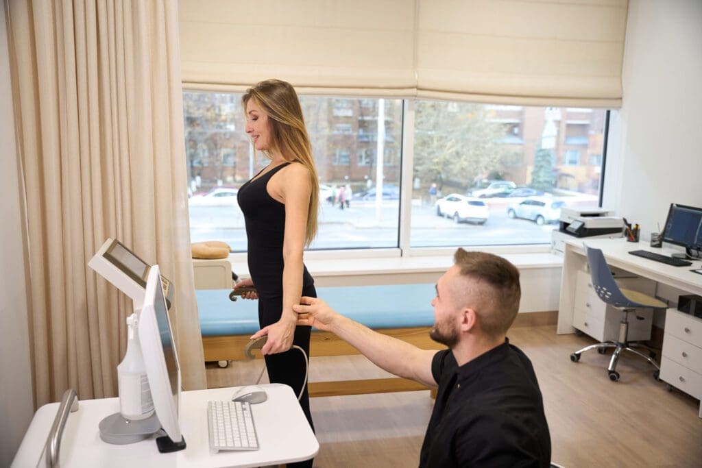
As Dr. Alexander Jimenez, DC, APRN, FNP-BC, CFMP, IFMCP, ATN, CCST, I have spent years helping patients navigate complex musculoskeletal pain, neuropathy, and recovery from orthopedic and sports injuries. When I first stepped into Apex’s new education and learning facility on 2026-05-02, I felt the resonance of a well-designed environment dedicated to cutting-edge modalities. The enthusiasm from colleagues, including leaders from OrthoLaser and technology innovators in MLS robotic therapy lasers, underscored the transformation underway in regenerative medicine: energy devices like lasers and shockwave are now merging seamlessly with orthobiologics to promote true tissue repair rather than merely manage symptoms.
In this post, I distill the science and clinical practicality behind photobiomodulation—laser therapy—so you can confidently understand how it works, why the MLS approach is unique, and how integrative chiropractic care amplifies patient outcomes. My goal is to guide you through the essential concepts and then show you how to integrate them in a real clinical workflow.

Modern Laser Therapy Basics: The Four Tissue Interactions

Laser-tissue interaction determines clinical results. Four fundamental phenomena occur when laser light meets biological tissue:
Reflection
Transmission
Scattering
Absorption
Among these, absorption is the therapeutic linchpin. Chromophores must absorb laser photons to initiate biologically meaningful change. Reflection and scattering represent energy loss; transmission is the passage of energy through tissue without interaction. Effective therapy maximizes absorption while controlling the other interactions—by choosing appropriate wavelengths, power, emission modalities, and delivery methods.

Key Laser Concepts: Source, Power Class, Emission Mode, Wavelength

To make laser therapy intuitive, I break it into four key concepts:
Source
Power class
Emission modality
Wavelength
Each element shapes clinical effect, depth of action, safety, and dosing.

Understanding Laser Sources

A laser source is defined by the active material that emits photons when excited. In surgical contexts, CO2 gas lasers interact with water to cut tissue via micro-explosions—excellent for incisions, not for healing. Therapeutic lasers most often use diodes. MLS laser systems are diode-based and sourced from high-precision Italian manufacturing, which matters for reliability, calibration integrity, and diode stability over time. Stable sources translate into reproducible clinical dosimetry and consistent patient outcomes.

Power Class: Class 3 vs. Class 4 and Why It Matters

Therapeutic lasers commonly fall into Class 3 (≤0.5 W) and Class 4 (>0.5 W). Marketing labels like low-level laser (LLLT), cold laser, high-intensity laser, and hot laser can be confusing; what matters is the interaction between power and emission mode. Class 4 systems can shorten treatment times and improve photon density in deeper tissues, but poorly controlled continuous delivery may lead to thermal accumulation.
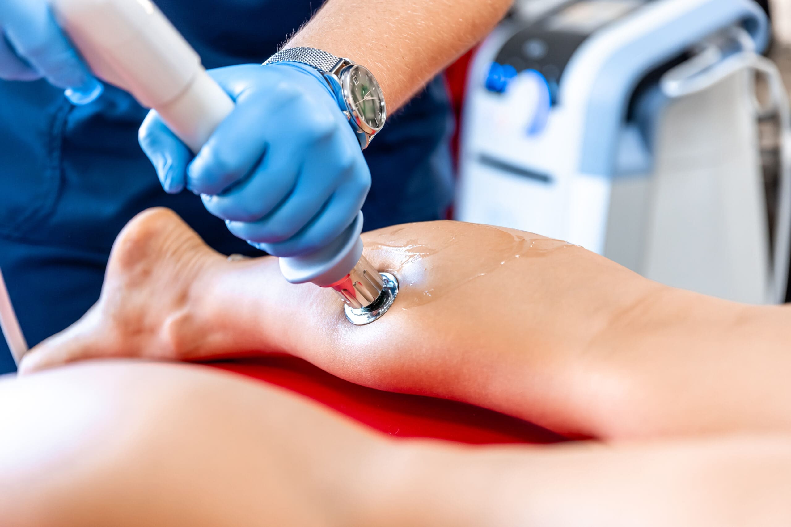
MLS laser therapy is technically Class 4, yet its patented emission design delivers the efficacy profile of Class 4 while maintaining the safety characteristics of Class 3. That combination reduces liability concerns and broadens indications, enabling point-by-point dosing without burning tissue—crucial for precise joint-space work and peri-implant care.

Emission Modality: Continuous, Chopped, and True Pulsed

Emission modality is the heartbeat of laser safety and effectiveness:
Continuous emission delivers uninterrupted light. If held stationary too long with high power, tissue temperatures can exceed the thermal damage threshold (~45°C).
Continuous chopped (or interrupted) uses mechanical means to block a continuous beam in microseconds. This delays heating but does not fully prevent thermal accumulation because the source never truly turns off.
True pulsed emission turns the source off in nanosecond intervals, creating genuine rest periods. Tissue can absorb photons and then cool between pulses, preventing temperature from creeping above therapeutic ranges. Therapeutic photobiomodulation aims to avoid sustained temperatures above ~43°C, where biological reactions can be inhibited rather than stimulated.

Wavelength: The “Prescription” for Depth and Targeting

Therapeutic photobiomodulation generally uses wavelengths in the 600–1200 nm window because of the absorption spectra of melanin, hemoglobin, and water. MLS systems specifically employ synchronized 808 nm (continuous) and 905 nm (pulsed) wavelengths. The 808 nm band offers strong penetration for photochemical activation, while 905 nm provides deeper penetration with high-peak-power pulses, enhancing photon density in target tissues without thermal risk.

Why the MLS Multiwave Locked System Is Unique

The Multiwave Locked System (MLS) synchronizes multiple wavelengths so they arrive simultaneously, reinforcing each other’s effects. This synchronization creates homogeneous energy distribution across superficial and deeper targets—like switching from a scattershot meteor shower to a uniform wavefront that saturates the tissue volume. In practice:
808 nm (continuous) supports mitochondrial activation and local circulation.
905 nm (true pulsed) delivers high-peak-power bursts with cooling intervals, increasing photon density for deeper tissues while preventing thermal buildup.
The physiologic result is a balanced combination of photochemical activation (ATP synthesis), photothermal modulation (controlled vasodilation and lymphatic flow), and photomechanical signaling (ECM deformation and mechanotransduction) that collectively accelerates tissue repair without suppressing necessary early inflammation.

Safety Profile: Why Heat Sensation Is Not Performance

Patients often equate “feeling heat” with effective treatment; however, high thermal effects do not equal high performance in photobiomodulation. The goal is cellular signaling, not cooking tissue. MLS’s true pulsed pattern keeps tissue below inhibitory temperature ranges and far below damage thresholds, ensuring that biological cascades proceed unimpeded. This distinction is vital around implants, tattoos, and bone surfaces, which can reflect energy and concentrate heat with non-pulsed devices.

Depth of Action: Wavelength, Emission, Power Density, and Time

Reaching target tissues requires the right combination of:
Wavelength (primary determinant of penetration).
Emission mode (true pulsed allows higher peak power without heat accumulation).
Power density (mW/cm² at tissue).
Exposure time (J/cm² dose).
The relationship between peak power and photon density matters: more photons per unit time and area increase the likelihood that chromophores such as cytochrome c oxidase will be activated, thereby driving ATP production and redox signaling. With MLS, true pulsed 905 nm delivery increases peak power in bursts, raising photon density at depth while tissue cools between pulses.

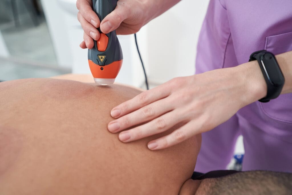
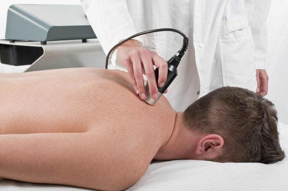
Clinical Delivery: Point-by-Point vs. Scanning

There are two practical methods:
Point-by-point dosing: stationary application over the target for a prescribed duration and energy dose. This maximizes absorption, minimizes dispersion, and grants precise dosing—ideal for joint spaces, tendon origins, and small neuropathic focal points.
Scanning: moving the applicator across a region. This can be effective for broader fields but is operator-dependent. Variability in speed, overlap, and distance can lead to inconsistent dosing.
MLS robotic platforms overcome scanning variability by delivering hands-free, precisely programmed energy maps, ensuring consistent dosing across operators and visits. In my clinic, that consistency translates into more reliable outcomes, smoother workflows, and the ability to combine laser with manual therapies simultaneously.

Robotic and Hands-Free Advantages

The MLS robotic arm ensures:
Consistent energy delivery across sessions and staff.
Safety via locked synchronization and true pulsed control.
Workflow efficiency: hands-free operation allows clinicians to perform soft-tissue release, joint mobilization, neuromuscular re-education, or percussion therapy while the laser runs—maximizing appointment value without sacrificing precision.
For field settings and athletic trainers, portable MLS units with straps enable targeted dosing at the sideline or in the training room, extending therapy beyond the clinic walls.

Physiological Underpinnings: Photochemical, Photothermal, and Photomechanical Effects

Laser therapy’s mechanisms can be organized into three complementary pathways:
Photochemical Activation
Mitochondrial stimulation via cytochrome c oxidase increases ATP production, shifts cellular redox states, and promotes the activity of transcription factors associated with growth and repair.
This accelerates fibroblast activity, collagen synthesis, and angiogenesis, while modulating oxidative stress.
Photothermal Modulation
Controlled vasodilation improves perfusion, oxygenation, and nutrient delivery.
Lymphatic enhancement reduces edema, supports waste clearance, and decreases inflammatory exudate.
In MLS systems, photothermal effects are therapeutic without crossing inhibitory thresholds; tissue temperature remains within a range favorable to enzymatic reactions and signaling cascades.
Photomechanical Signaling
Rapid pulses create temporary deformation of the extracellular matrix (ECM) and cell membranes, activating mechanosensitive pathways.
This facilitates cytoskeletal reorganization, influences integrin-mediated signaling, and augments tissue repair sequences.

Inflammation Modulation vs. Suppression

It is critical to note that MLS therapy does not suppress inflammation as NSAIDs do. It modulates inflammatory processes—upregulating anti-inflammatory proteins and downregulating pro-inflammatory cytokines—while respecting the acute phase needed for proper healing. This is why MLS pairs well with orthobiologics; rather than blunting early inflammation (which supports cell recruitment and initial repair), MLS normalizes and supports the cascade, reducing excessive pain and swelling while protecting regenerative intent.

Clinical Indications and Dosing Framework

Common indications include:
Musculoskeletal pain: plantar fasciitis, knee osteoarthritis, neck pain, tendinopathies, myofascial pain.
Post-surgical healing: incisions, soft-tissue repair, peri-implant recovery.
Wound care: diabetic ulcers, traumatic and infected wounds, burns.
Sports medicine: delayed onset muscle soreness, acute muscle strain, ligament sprain.
Neuropathic pain: diabetic neuropathy, compressive neuropathies, small fiber pain.
Dosing principles in my practice:
Session length: typically 6–12 minutes, depending on area size and target depth; anterior-posterior or dual-field approaches may extend the time accordingly.
Packages: acute conditions often 4–6 sessions; chronic conditions 8–12 sessions. This cumulative dosing approach allows progressive improvements in pain and function.
Frequency: ideally 2–3 times per week. In acute flares, daily sessions can be considered for brief periods; practicality and access may guide scheduling.
Point-by-point dosing for focal structures; robotic scanning for consistent energy distribution over broader fields.

Clinical Images and Observations

Wound- and burn-healing examples demonstrate why safety matters. Treating burns with a laser may seem paradoxical until you understand MLS’s non-heating, pulsed profile. In my clinical observations, combining MLS laser therapy with compression, offloading, and nutritional cofactors (e.g., protein sufficiency, vitamin C, zinc) accelerates closure rates in diabetic ulcers. It reduces the risk of infection, particularly when paired with meticulous debridement and glycemic control.
Peri-implant treatment is another area where MLS stands out. Patients with knee replacements or hardware often fear laser near implants; MLS’s synchronized wavelengths and pulsed control minimize conductive heat accumulation, allowing safe application to surrounding soft tissues. In my experience, postoperative stiffness and peripatellar pain respond well to a protocol integrating MLS laser therapy, joint mobilization, instrument-assisted soft-tissue mobilization (IASTM), and graded activity.

Neuropathic Pain and the MLS MIS Platform

Emerging devices geared to neuropathic pain (e.g., MIS systems) target parameters tuned for nerve repair and dysesthesia reduction. European MDR clearance has recognized neuropathic indications, and similar approvals are progressing domestically. In my practice, patients with diabetic neuropathy or post-chemotherapy neuropathies benefit from MLS laser combined with glycemic optimization, B-vitamin sufficiency (especially methylated B12 and folate where appropriate), alpha-lipoic acid, and foot intrinsic strengthening. The laser’s modulation of neuroinflammation and microcirculation complements metabolic correction.

Evidence Base: Trials and Registry Data

Modern photobiomodulation has matured beyond anecdote. Randomized controlled trials and real-world registries corroborate benefits across pain and function domains. Combining orthobiologics with MLS laser therapy shows synergistic improvements:
Faster numeric pain rating reductions in the first month and sustained gains out to 24 months.
Greater improvements in worst pain scores and desired functionality markers.
Higher overall patient satisfaction in combined protocols.
These data support a shift from symptom management to cellular health optimization, marrying biologic inputs (PRP, BMAC, growth factor-rich preparations) with photonic signals that improve mitochondrial function, microvascular flow, and ECM remodeling.

Integrative Chiropractic Care: Where Laser Therapy Fits

From a chiropractic and functional medicine viewpoint, MLS laser therapy is most powerful when embedded in a coordinated plan:
Assessment and Diagnosis
Thorough neuromusculoskeletal examination: joint mobility, muscle tone, proprioception, gait, and regional interdependence.
Functional lab insights per case: inflammation markers, glycemic status, micronutrient sufficiency.
Manual and Movement Foundations
Spinal and extremity adjustments to restore joint mechanics and reduce nociceptive input.
Soft-tissue release (IASTM, myofascial techniques) to normalize tone and improve tissue gliding.
Neuromuscular re-education and graded loading to reintegrate function and resilience.
Energy Devices and Orthobiologics
MLS laser therapy to modulate inflammation, energize mitochondria, and support microcirculation.
Shockwave therapy for mechanotransduction and neovascularization when indicated.
Orthobiologics (e.g., PRP) for biologic scaffolding and cellular inputs; MLS supports the early healing milieu without suppressing beneficial inflammation.
Metabolic and Lifestyle
Nutritional optimization for collagen synthesis and tissue repair.
Sleep and stress modulation to enhance autonomic balance and recovery capacity.
Progress Monitoring
Standardized pain and function scales (NPRS, ODI, KOOS, FAAM).
Imaging or ultrasound, where appropriate.
Consistent MLS dosing via robotic delivery and careful parameter documentation.

Why Each Technique Is Used

Adjustments reduce mechanical stress and abnormal joint signaling, making downstream laser effects more efficient by removing barriers to perfusion and nerve function.
Soft-tissue mobilization breaks adhesions and improves ECM pliability—laser photomechanical signaling benefits from tissues primed to respond.
Shockwave induces regenerative signals and vascular remodeling; laser complements by reducing inflammatory load and energizing mitochondrial repair.
Orthobiologics provide biological substrates and cellular signals; laser therapy supports their integration by improving the microenvironment (oxygenation, edema reduction).
MLS laser specifically balances deep photon delivery with safety—its synchronized wavelengths and true pulsed mode prevent heat accumulation while maximizing cellular activation.

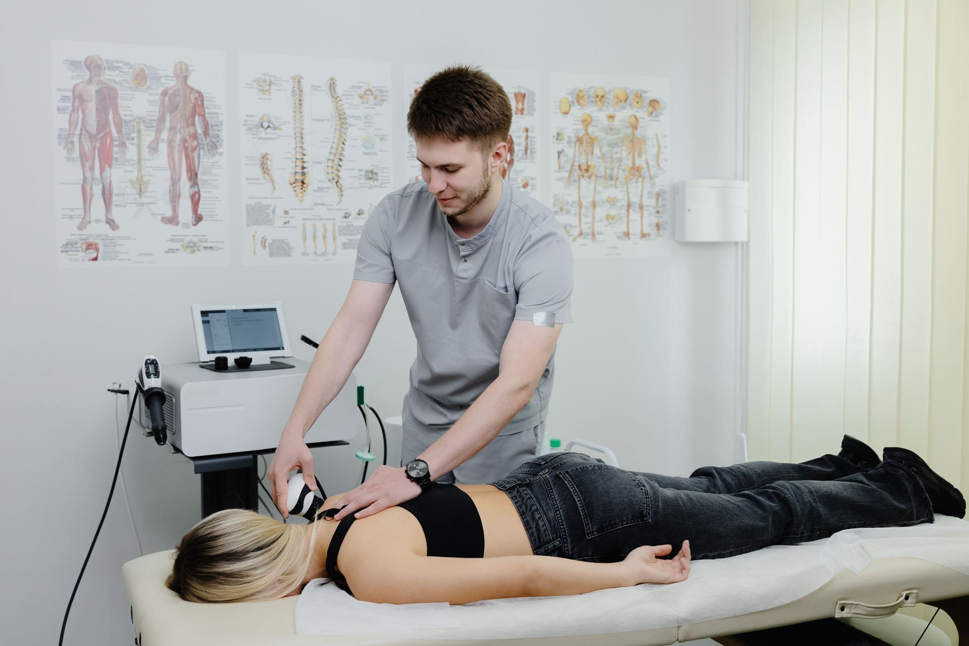
Clinical Workflow Example: Knee Osteoarthritis

Visit 1–2: Assessment, set goals, baseline NPRS and function scales, initial MLS laser point-by-point dosing to medial and lateral joint lines, posterior capsule, peri-patellar soft tissue; gentle mobilizations; home exercises for quads, hips, and ankle mobility.
Visit 3–6: Robotic MLS scanning over the knee for consistent dosing; introduce shockwave localized to tendinous attachments if appropriate; progress strength and motor control drills.
Visit 7–12: Consider PRP for refractory cases; maintain MLS sessions around biologic injection timelines to modulate inflammation and improve circulation; recheck metrics and adjust as needed.
Outcome: Reduced pain scores, improved stair descent, better stance stability; long-term maintenance via periodic MLS sessions and continued exercise.

Clinical Workflow Example: Plantar Fasciitis

Acute phase: MLS point-by-point over proximal plantar fascia insertion; calf and posterior chain soft-tissue release; intrinsic foot activation; load management.
Subacute: Robotic MLS coverage of plantar arch and heel; introduce shockwave to stimulate local regeneration if needed; progressive loading.
Chronic: Consider orthobiologics for degenerative fascial changes; maintain MLS to reduce pain and enhance functional durability.

Addressing Safety: Implants, Tattoos, and Bone

Implants: MLS’s synchronized, pulsed pattern allows safe peri-implant dosing without heating risks associated with continuous high-power systems.
Tattoos: Pigment absorption can concentrate energy; MLS mitigates this via true pulsed delivery, enabling comfortable treatment over tattooed skin.
Bone Reflection: Bone can reflect >90% of incident Class 4 energy; MLS protocols emphasize proper patient positioning and joint-window targeting to maximize soft-tissue absorption.

Why Robot-Assisted Consistency Matters

Consistency is an outcome multiplier. Different operators inevitably vary in distance, speed, overlap, and timing when scanning by hand. The robotic MLS platform standardizes all of this, producing repeatable dosing that tightens the variance band around results. In my clinic, that translates to more predictable timelines, fewer setbacks, and better communication with patients about expected improvements.

My Clinical Observations in Practice

From my experience:
Combining MLS laser therapy with shockwave and targeted manual therapies reduces time-to-improvement in tendinopathies, especially in high-load athletes.
In diabetic neuropathy, adding MLS to metabolic care (glycemic control, micronutrient support) reduces burning pain and enhances light-touch discrimination sooner than metabolic care alone.
Postoperative patients benefit from MLS’s ability to modulate edema and restore movement tolerance, especially when coupled with gentle mobilization and progressive rehab.
In chronic myofascial pain, point-by-point MLS over trigger points followed by percussion therapy and motor control exercises often breaks stubborn cycles of pain and guarding.

A Practical Protocol Rationale

Early-phase: Photobiomodulation targets mitochondrial function and reduces pain enough to allow patients to move; movement restores joint mechanics, circulation, and neuromuscular coordination.
Mid-phase: Shockwave and loading recondition tissues; MLS continues to optimize the microenvironment.
Late-phase: Orthobiologics may be layered in for degenerative changes; MLS supports integration and reduces flare risk.
Maintenance: Periodic MLS and functional exercise maintain gains, reduce relapse, and support aging athletes or individuals with persistent risk factors.

Closing Thoughts: From Symptom Relief to Cellular Optimization

The integrative future of musculoskeletal medicine is already here. By aligning MLS laser therapy with chiropractic care, shockwave, and orthobiologics, we move beyond symptom suppression toward genuine cellular optimization. True pulsed, synchronized wavelengths create a therapeutic environment that respects physiology while accelerating recovery. In my practice, the most gratifying moments are when patients realize their improvement is not a temporary pain mask—it is a deeper shift in function, resilience, and quality of life.

References

In-text citations: (Hamblin, 2018; Chung et al., 2012; Leal Junior et al., 2014; Barolet & Christiaens, 2018; Franco et al., 2019; Schmitz et al., 2018; DataBiologics, 2026)

Author and Clinical Resources

Dr. Alexander Jimenez, DC, APRN, FNP-BC, CFMP, IFMCP, ATN, CCST
Clinical observations and integrative practice insights: https://chiromed.com/
Professional profile: https://www.linkedin.com/in/dralexjimenez/

SEO tags: photobiomodulation, MLS laser therapy, integrative chiropractic care, regenerative medicine, orthobiologics, sports medicine laser, true pulsed emission, mitochondrial activation, pain modulation, robotic laser therapy, shockwave synergy, neuropathic pain laser, peri-implant laser safety, functional medicine musculoskeletal, evidence-based laser therapy

Hair Restoration for Growth, Density, and Inflammation

Hair Restoration for Growth, Density, and Inflammation

Hair Restoration for Growth, Density, and Inflammation

Abstract

As Dr. Alexander Jimenez, DC, APRN, FNP-BC, CFMP, IFMCP, ATN, CCST, I created this educational post to guide you through a clear, practical roadmap for hair restoration that merges dermatologic science, endocrinology, and integrative chiropractic care. In this comprehensive narrative, I explain how to strategically combine therapies—such as minoxidil, anti-inflammatory dandruff shampoos, bioactive peptides, saw palmetto, and low-level laser therapy (LED hair caps)—to activate hair growth, reduce scalp inflammation, decrease hair shedding, block dihydrotestosterone (DHT), and improve hair density. I explain the physiology behind each method, the modern research supporting these interventions, and how lifestyle, biomechanics, microcirculation, and neuroendocrine balance are connected to hair follicle health. Throughout, I share clinical observations from my practice and collaborations that inform a precise, integrative plan. This gives you a step-by-step strategy rooted in evidence, with practical protocols and clear rationales for each choice.


Understanding Hair Loss: The Integrated Physiological Landscape

Hair loss is rarely the result of a single pathway. In clinical practice and research, I routinely observe a convergence of factors affecting follicular cycling. To build a useful plan, we need to connect these dots:

  • The hair follicle cycle moves through anagen (growth), catagen (regression), telogen (rest), and exogen (shedding). Hair restoration aims to extend anagen and minimize premature entry into telogen.
  • Microcirculation at the scalp provides oxygen and nutrients to the dermal papilla and matrix keratinocytes, driving hair shaft production.
  • Inflammation—from seborrheic dermatitis, dandruff (Malassezia-associated), or autoimmune processes—can constrict microvasculature, disrupt stem cell niches, and accelerate miniaturization.
  • Androgen signaling, specifically DHT binding to androgen receptors in genetically susceptible follicles, shortens anagen and shrinks follicle size in androgenetic alopecia.
  • Mitochondrial function and redox balance in follicular cells influence ATP production, which is required for rapid keratin synthesis.
  • Neuroendocrine stress and sympathetic overactivity elevate cortisol and reduce local growth factors, impairing regenerative signaling.
  • Biomechanics and posture, via cervical and cranial fascial tension patterns, influence perfusion and lymphatic drainage in the scalp. As an integrative chiropractor and nurse practitioner, I see measurable changes in scalp microcirculation and symptom severity when these mechanical issues are addressed.

A hair restoration strategy is most successful when these domains are assessed and optimized together. Below, I build from single interventions to a cohesive protocol, explaining what each modality achieves and why it fits.


Minoxidil: Extending Anagen and Enhancing Microcirculation

When I apply minoxidil, I’m targeting two central mechanisms:

  • It is a potassium-channel opener that hyperpolarizes smooth muscle cells, leading to vasodilation in the scalp microvasculature. This improves nutrient delivery to the dermal papilla, which governs hair growth signals.
  • It upregulates vascular endothelial growth factor (VEGF) and other growth-related pathways, prolonging the anagen phase and countering miniaturization.

Why I use it:

  • In patients with early androgenetic changes and reduced density, extending anagen improves hair count and shaft diameter.
  • Minoxidil can be combined with other therapies because it acts primarily on circulation and follicular cycling rather than directly on hormones.

Clinical reasoning:

  • I typically start with topical formulations and titrate based on scalp sensitivity and response. In select cases, low-dose oral minoxidil is considered when topical absorption is inadequate, with careful monitoring of blood pressure and fluid balance.

Outcome I expect:

  • Activate hair growth, reflected in increased anagen hairs and decreased telogen club hairs over several months.

Anti-Inflammatory Dandruff Shampoos: Reducing Scalp Inflammation

When I incorporate medicated dandruff shampoos (e.g., ketoconazole, selenium sulfide, zinc pyrithione), I’m aiming to reduce scalp inflammation, which is an underappreciated driver of hair thinning.

Mechanisms:

  • These agents reduce Malassezia load and normalize sebum ecology, thereby decreasing inflammatory cytokines that disrupt the outer root sheath and bulge stem cells.
  • Ketoconazole, in particular, has ancillary anti-androgenic and anti-inflammatory properties in the scalp microenvironment.

Why I use it:

  • Chronic low-grade dermatitis constricts microcirculation and enhances oxidative stress. Clearing this inflammatory milieu stabilizes the follicular niche, improving responsiveness to growth therapies.

Clinical reasoning:

  • I recommend a rotation schedule (e.g., ketoconazole twice weekly, zinc pyrithione once weekly) to maintain efficacy and minimize irritation, followed by a gentle, pH-balanced shampoo.

Outcome I expect:

  • Reduce scalp inflammation, resulting in less pruritus and scaling, and improved scalp perfusion—setting the stage for a better hair growth response.

Combining Minoxidil and Dandruff Shampoo: Synergy for Growth and Scalp Health

When I combine minoxidil with anti-inflammatory dandruff shampoos, I see more robust outcomes:

  • Minoxidil improves microcirculation and anagen duration.
  • Anti-inflammatory cleansing reduces cytokine load and microbial triggers that otherwise impede follicular function.

Why this synergy matters:

  • A healthy scalp environment allows minoxidil’s signals to persist and be interpreted correctly by follicular cells. Inflammation often masks growth benefits.

Outcome I expect:

  • Activate hair growth and reduce scalp inflammation simultaneously, resulting in visible improvements in hair density and scalp comfort.

Bioactive Peptides: Decreasing Hair Fall and Stimulating Regeneration

I use bioactive peptides (e.g., copper tripeptide-1, GHK-Cu, biomimetic peptides like acetyl tetrapeptide-3, or proline-rich polypeptides) to address shedding and regenerative signaling.

Mechanisms:

  • Peptides can upregulate Wnt/β-catenin pathways, support extracellular matrix integrity at the follicle anchor points, and modulate TGF-β to prevent premature catagen.
  • Copper peptides support angiogenesis, collagen synthesis, and anti-inflammatory responses, aiding microenvironment repair.
  • Some biomimetic peptides target IGF-1, FGF-7, and KGF, promoting matrix keratinocyte proliferation.

Why I use them:

  • Patients with chronic shedding often have fragile anchoring structures and impaired regenerative signaling. Peptides stabilize the follicle and reduce triggers of telogen effluvium.

Clinical reasoning:

  • I pair peptides with microneedling or low-level laser therapy for enhanced uptake, spacing applications to prevent irritation. In my practice, combination peptide serums are applied after anti-inflammatory cleansing.

Outcome I expect:

  • Decrease hair fall by reinforcing the follicle’s structural and signaling integrity, with gradual gains in hair caliber.

Saw Palmetto: Blocking DHT to Preserve Follicular Size

For androgen-driven loss, I add saw palmetto, a 5-alpha-reductase inhibitor, to reduce local DHT production.

Mechanisms:

  • Saw palmetto’s lipidosterolic extracts inhibit type I and type II 5-alpha-reductase, lowering DHT levels around the follicle and decreasing androgen receptor activation in susceptible regions (vertex and frontal scalp).
  • Reduced DHT prevents anagen shortening and follicular miniaturization, which are characteristic of androgenetic alopecia.

Why I use it:

  • It’s a well-tolerated option for individuals who prefer botanicals or have contraindications to finasteride. I often integrate it with topical anti-androgen formulations and diet strategies that support hormone balance.

Clinical reasoning:

  • I monitor patient symptoms, use validated hair density measures, and, if needed, assess sex hormone-binding globulin and free testosterone to tailor the approach. Side effects are monitored but are typically mild compared with those of pharmaceutical inhibitors.

Outcome I expect:

  • Block DHT to preserve follicular size, complementing minoxidil’s growth activation and peptides’ structural support.

Low-Level Laser Therapy (LED Hair Caps): Increasing Hair Density via Mitochondrial Activation

I employ LED hair caps that leverage low-level laser therapy (LLLT) to promote mitochondrial and microcirculatory benefits.

Mechanisms:

  • Photobiomodulation targets cytochrome c oxidase in mitochondria, increasing ATP production, nitric oxide release, and reactive oxygen species signaling in a controlled manner that promotes cell proliferation and angiogenesis.
  • Enhanced nitric oxide contributes to vasodilation and improved perfusion of the dermal papilla.

Why I use it:

  • Many follicles in early miniaturization respond to improved energy metabolism. LLLT complements minoxidil’s vasodilation and peptide-driven signaling.

Clinical reasoning:

  • I recommend evidence-based wavelengths (typically 630–680 nm and 810–850 nm) and structured session frequencies (e.g., 3–4 times per week). Compliance is high due to ease of use.

Outcome I expect:

  • Increase hair density, with visible improvements over several months, particularly in patients with diffuse thinning.

Building the Combination: Strategic Layering for Comprehensive Results

Step by step, here’s how these therapies stack physiologically and clinically:

  • Minoxidil: Promotes hair growth by enhancing circulation and prolonging the anagen phase.
  • Dandruff shampoo: Reduce scalp inflammation to stabilize the microenvironment.
  • Peptides: Decrease hair fall by strengthening follicular signaling and anchoring.
  • Saw palmetto: Blocks DHT to protect against androgen-induced miniaturization.
  • LED hair cap: Increase hair density through mitochondrial activation and microvasculature improvements.

When I combine all five, I expect synergistic outcomes:

  • Activate hair growth
  • Reduce scalp inflammation
  • Decrease hair fall
  • Block DHT
  • Increase hair density

This overlay of mechanisms addresses vascular, inflammatory, hormonal, structural, and bioenergetic dimensions—precisely the integrated approach that performs best in real-world practice.


Integrative Chiropractic Care: Biomechanics, Autonomic Balance, and Scalp Perfusion

As both a chiropractor and nurse practitioner, my integrative lens is critical. Here’s how integrative chiropractic care fits into hair restoration:

  • Cervical Alignment and Fascial Dynamics:
    • Restrictive patterns in the suboccipital musculature and scalene fascia can impair venous outflow and lymphatic drainage from the scalp.
    • Gentle, targeted cervical adjustments, soft-tissue release, and cranial fascial techniques improve hemodynamics and reduce nociceptive input that drives sympathetic tone.
  • Autonomic Nervous System Modulation:
    • High sympathetic activity reduces scalp microcirculation and alters mast cell activity, aggravating inflammation.
    • Techniques such as breathing retraining, vagal stimulation, and HRV-guided biofeedback help normalize autonomic balance, supporting follicular repair.
  • Postural Ergonomics and Myofascial Chains:
    • Forward-head posture increases mechanical tension across the galea aponeurotica and temporoparietal fascia. Corrective exercises and myofascial decompression improve capillary perfusion to the scalp.
  • Nutritional and Functional Medicine Integration:
    • Assessment for micronutrient deficiencies (iron, zinc, vitamin D, and biotin, where appropriate), thyroid function, and insulin resistance is standard in my protocols, given their impact on hair cycling.
    • Anti-inflammatory diets, omega-3 fatty acids, and polyphenols support endothelial function and redox balance in the scalp.

Clinical observation:

  • In my practice, integrating cervical mobilization and autonomic regulation with topical and systemic therapies yields faster reductions in shedding and higher-quality regrowth. Patients report improved scalp comfort and reduced dandruff flares when mechanical and stress-related factors are addressed alongside dermatologic care.

Protocol Design: Practical Steps and Reasoning

When designing a plan, I follow a structured pathway:

  • Baseline Assessment:
    • Scalp exam for erythema, scale, and follicular miniaturization patterns.
    • Review medical conditions (thyroid, iron status, PCOS, metabolic syndrome) and medications that can trigger telogen effluvium.
    • Lifestyle and stress inventory; posture and cervical mobility assessment.
  • Initial Interventions:
    • Begin minoxidil daily to stimulate microcirculation and extend anagen.
    • Add an anti-inflammatory dandruff shampoo rotation to reduce cytokines and normalize scalp ecology.
  • Reinforcement Phase:
    • Introduce bioactive peptide serums, applied post-cleansing, possibly combined with gentle microneedling to enhance penetration.
    • Start LED hair cap sessions 3–4 times weekly, targeting mitochondrial activation.
  • Hormonal Modulation:
    • For signs of androgenetic alopecia, integrate saw palmetto and consider adjuncts under medical supervision if needed. Discuss realistic time frames and cumulative effects.
  • Integrative Chiropractic Care:
    • Implement cervical adjustments, soft-tissue release, and postural correction.
    • Add autonomic balance strategies: diaphragmatic breathing, HRV biofeedback, and sleep optimization.
  • Monitoring and Titration:
    • Track shedding count, phototrichogram measures, and patient-reported outcomes.
    • Adjust dosages and frequencies based on tolerance and improvements; address irritations promptly with barrier-supporting scalp care.

Rationale:

  • This staged approach stabilizes the scalp environment, activates growth, protects against androgen effects, and optimizes delivery and cellular energy—minimizing the risk of irritation or non-compliance.

Safety Considerations and Patient Education

Safety is central to integrative hair restoration:

  • Minoxidil:
    • Watch for scalp irritation, hypertrichosis in non-target areas, and, with oral use, changes in blood pressure.
  • Dandruff shampoos:
    • Avoid overuse; maintain barrier integrity with pH-balanced cleansers and emollients as needed.
  • Peptides:
    • Choose reputable formulations; monitor for sensitivity; avoid combining too many actives at once.
  • Saw palmetto:
    • Discuss potential mild gastrointestinal effects; coordinate with primary care for those on hormone-related medications.
  • LED hair caps:
    • Use evidence-based protocols; protect eyes; maintain a consistent schedule.

Education points:

  • Hair growth has a biological lag; expect meaningful changes over several months.
  • Consistency is critical. The synergy of combined therapies depends on regular use.
  • Lifestyle matters. Sleep, stress management, and nutrition amplify outcomes.

Case-Informed Insights: What I See in Practice

From my clinical experience, certain patterns stand out:

  • Patients who start with inflammation control—regular use of a dandruff shampoo—respond faster to minoxidil and peptides, with decreased itching and flaking within 2–3 weeks.
  • Integrating LED hair caps accelerates density gains in diffuse thinning, especially when combined with copper peptides that support microvascular health.
  • For those with a family history of androgenetic alopecia, saw palmetto provides a protective backdrop against miniaturization, making regrowth more sustainable when combined with minoxidil.
  • Postural correction and cervical release are particularly beneficial for patients with tension headaches and scalp tenderness; improved scalp perfusion correlates with reduced shedding counts.
  • A small subset benefits from targeted micronutrient support after lab review—iron repletion and vitamin D optimization often reduce telogen shedding events.

These observations are consistent with modern, evidence-based methods and align with a whole-person approach to hair health.


Putting It All Together: A Day-in-the-Life Routine

To make this practical, here’s how I structure a daily and weekly routine:

  • Morning:
    • Apply minoxidil to the target areas.
    • Perform a brief breathing exercise (4–6 minute diaphragmatic practice) to promote parasympathetic tone.
  • Evening:
    • Use dandruff shampoo on scheduled days; follow with a gentle conditioner.
    • Apply peptide serum after the scalp is dry.
  • Three to four days per week:
    • Wear the LED hair cap for the recommended duration.
  • Daily supplements (as appropriate):
    • Saw palmetto with a meal; consider adding omega-3s and polyphenols if diet is low in anti-inflammatory compounds.
  • Weekly integrative care:
    • Practice postural resets, cervical mobility drills, and brief self-massage of suboccipital muscles.
    • Maintain sleep hygiene and track stress levels.

This routine creates a cadence that addresses inflammation, growth activation, hormonal protection, and mitochondrial energy—without overwhelming the scalp.


Evidence-Based Perspective: Why These Methods Work Together

The unifying principle behind this integrated plan is multifactorial support:

  • Vascular: Minoxidil and nitric oxide from LLLT enhance perfusion.
  • Inflammatory: Dandruff shampoos and peptides normalize cytokine balance and tissue repair.
  • Hormonal: Saw palmetto reduces DHT pressure on susceptible follicles.
  • Bioenergetic: LLLT improves ATP and cellular resilience.
  • Structural: Peptides strengthen the extracellular matrix and follicle anchoring; chiropractic care optimizes mechanics and autonomic tone.

By layering these complementary effects, we create conditions in which follicles can re-enter and sustain the anagen phase, translating to visible improvements in density, thickness, and scalp comfort.


Conclusion: A Clear, Integrated Path to Healthier Hair

My approach unites dermatologic therapies with functional medicine and integrative chiropractic care to address the full spectrum of factors contributing to hair loss. Starting with inflammation control and growth activation, then reinforcing with peptides, hormonal modulation, and mitochondrial support, we build a strategy that is logical, tolerable, and effective. Incorporating biomechanical and autonomic regulation helps ensure we do not overlook systemic contributors that can limit outcomes.

If you’re embarking on a hair restoration journey, consider this integrated framework. It respects the biology of the hair follicle, uses modern, evidence-based methods, and aligns with clinical patterns I’ve observed across diverse patients. With consistency and individualized adjustments, you can expect steady progress toward stronger, denser hair.


References

What You Should Know About PRP Therapy for Knee Osteoarthritis

Learn the benefits of PRP therapy for osteoarthritis for better mobility and reduced discomfort in your knee joints.

Abstract

In this educational post, I present a comprehensive, first-person overview of how I integrate platelet-rich plasma (PRP) therapy into care plans for patients with symptomatic knee osteoarthritis (OA). Drawing on modern, evidence-based research and my clinical observations as Dr. Alexander Jimenez, DC, APRN, FNP-BC, CFMP, IFMCP, ATN, CCST, I explain why PRP often outperforms corticosteroid and hyaluronic acid injections in pain, function, and potentially in cartilage preservation. I outline how PRP interacts with joint biology, the cellular signaling that underpins its therapeutic effects, and the integrative chiropractic strategies that optimize outcomes—covering biomechanics, neuromuscular control, functional nutrition, and lifestyle medicine. This piece synthesizes findings from leading researchers and aligns them with an integrated clinical protocol focused on measurable outcomes, safety, and long-term joint health.

Why I Use Platelet-Rich Plasma for Symptomatic Knee Osteoarthritis

As a clinician working daily with musculoskeletal conditions, I have witnessed how knee osteoarthritis impacts quality of life—pain, stiffness, loss of mobility, and reduced confidence in movement. Over years of clinical practice and continual review of the literature, platelet-rich plasma (PRP) has become a central part of my integrative treatment approach when patients present with symptomatic knee OA.
Here is why PRP often earns a place in my treatment plans:

  • Evidence favors PRP over corticosteroids and hyaluronic acid for pain and functional outcomes in many patient populations with knee OA.
  • Mechanistic plausibility: PRP delivers concentrated growth factors that can modulate inflammation, encourage matrix synthesis, and potentially support chondrocyte resilience.
  • Safety profile: Autologous biologic with relatively low risk when processed and delivered under rigorous protocols.
  • Synergy with integrative chiropractic care: Biologic modulation is most effective when combined with biomechanical alignment, neuromuscular training, and lifestyle interventions that reduce joint load and support metabolic health.

In practice, I find PRP excels for patients who are struggling with persistent symptoms despite conservative care, who are hesitant to rely on corticosteroid cycles, or who want to explore joint-preserving options alongside manual therapy and functional rehab.

Understanding Platelet-Rich Plasma: What It Is and How It Works

PRP is an autologous blood product derived from the patient’s own venous sample. After centrifugation, we isolate a plasma fraction with an elevated platelet concentration, typically ranging from 3x to 7x baseline, depending on the preparation. The platelets carry alpha granules packed with bioactive proteins that can influence joint microenvironments.
Key bioactive mediators in PRP include:

  • Platelet-derived growth factor (PDGF): Stimulates cell proliferation, chemotaxis, and angiogenesis.
  • Transforming growth factor-beta (TGF-β): Modulates matrix synthesis and can influence chondrocyte phenotype.
  • Insulin-like growth factor-1 (IGF-1): Supports cartilage matrix synthesis and cellular survival.
  • Vascular endothelial growth factor (VEGF): Angiogenic signaling with context-dependent roles in tissue repair.
  • Epidermal growth factor (EGF) and fibroblast growth factors (FGFs): Promote cellular activity relevant to tissue recovery.

When introduced into the osteoarthritic knee, these mediators can:

  • Modulate inflammation: Downregulate pro-inflammatory cytokines such as IL-1β and TNF-α while promoting anti-inflammatory signaling.
  • Support extracellular matrix homeostasis: Encourage anabolic pathways (e.g., collagen II, aggrecan synthesis) and potentially reduce catabolic enzyme activity (MMPs, ADAMTS).
  • Influence chondrocyte behavior: Support survival, reduce apoptosis in hostile joint environments, and promote a more regenerative phenotype.
  • Impact synovial environment: Improve synovial fluid characteristics and reduce synovitis, thereby decreasing nociceptive drive.

This physiologic rationale aligns with reported clinical outcomes—reduced pain, improved function, and, in some studies, signals that PRP may exert chondroprotective effects.

Comparing PRP With Corticosteroids and Hyaluronic Acid: What the Data Shows

Corticosteroids and hyaluronic acid (HA) have established roles in knee OA, but their mechanisms and durability differ from PRP:

  • Corticosteroids: Potent anti-inflammatory effects via genomic and non-genomic pathways that reduce prostaglandins and cytokines. Benefits tend to be short-lived, and repeated injections may have deleterious effects on cartilage metabolism. I reserve corticosteroids for acute synovitis flare-ups when rapid control is essential (e.g., severe swelling impeding rehab).
  • Hyaluronic acid (viscosupplementation): Aims to restore viscoelastic properties of synovial fluid and interact with CD44 receptors to modulate inflammation and joint mechanics. Responses vary, and HA alone may not match PRP’s performance in many trials.
  • PRP: Offers biologic signaling that can shift the joint toward repair and better matrix dynamics. Clinical benefits can last longer than those of steroids in many cohorts and often outperform HA alone on functional indices.

The literature has increasingly focused on head-to-head comparisons:

  • Multiple meta-analyses and randomized trials report greater pain reduction and functional improvement with PRP than with placebo, corticosteroids, or HA at mid-term follow-up, especially in mild-to-moderate OA phenotypes (e.g., Kellgren-Lawrence II–III) (see references below).
  • Dose and product characteristics matter: Leukocyte-poor PRP generally demonstrates better tolerability in the joint with lower post-injection flare rates than leukocyte-rich formulations for OA applications. Platelet concentration in a moderate range tends to be optimal; excessively high concentrations may increase catabolic signaling.
  • Repeat dosing strategies (e.g., series of 2–3 injections spaced 2–4 weeks apart) often yield more durable outcomes than single injections, likely reflecting cumulative biologic conditioning of the synovial and cartilage microenvironments.

These lines of evidence help me set patient expectations and tailor protocols to the OA phenotype they present with.

Chondroprotection: Can PRP Help Preserve Cartilage?

One of the most exciting themes in the recent literature is chondroprotection—the idea that PRP might help maintain cartilage integrity or slow degenerative processes. Mechanistically, PRP may:

  • Reduce catabolic enzyme activity: Suppress matrix metalloproteinases (MMP-1, MMP-3, MMP-13) and aggrecanases (ADAMTS-4/5).
  • Enhance anabolic signaling: Promote the synthesis of type II collagen and aggrecan by modulating IGF-1 and TGF-β.
  • Improve subchondral crosstalk: Alter inflammatory signaling between cartilage, synovium, and subchondral bone marrow lesions.
  • Dampen oxidative stress: Increase antioxidant capacity, which supports chondrocyte survival and reduces apoptosis.

While ongoing studies continue to refine estimates of the magnitude and consistency of these effects, the convergence of mechanistic and clinical data supports the potential of PRP to be chondroprotective in appropriately selected patients. This has meaningful implications for delaying progression, prolonging function, and deferring invasive interventions.

How Integrative Chiropractic Care Enhances PRP Outcomes

PRP works best inside a systems approach. As a chiropractor and advanced practice provider, I integrate manual therapy, neuromuscular re-education, biomechanical correction, functional nutrition, and lifestyle medicine to amplify PRP’s benefits.
Key pillars of my integrative care:

  • Spinal and lower-extremity alignment: Targeting pelvic tilt, sacroiliac mechanics, and tibiofemoral/ patellofemoral tracking reduces aberrant joint loading. Precise chiropractic adjustments, mobilizations, and soft-tissue releases help normalize kinetic-chain function.
  • Neuromuscular control: Focused training for gluteus medius/maximus, vastus medialis obliquus (VMO), hamstrings, and calf complex improves dynamic valgus control and reduces medial compartment stress. We use progressive closed-chain drills that restore proprioception and joint stability.
  • Gait and movement retraining: Patients learn load-management strategies—such as cadence optimization, stride mechanics, and foot placement—to reduce peak joint forces. We frequently pair this with orthotic support or footwear interventions.
  • Functional nutrition: Anti-inflammatory dietary patterns (Mediterranean-style, low-glycemic-load), adequate protein intake, omega-3 fatty acids, and micronutrients (vitamin D, magnesium) support cartilage metabolism and systemic recovery.
  • Weight management: Even modest reductions in body mass can translate to significant decreases in knee joint compressive forces per step. We implement structured plans for sustainable change.
  • Sleep and stress modulation: Optimizing sleep architecture and stress resilience (via HRV-guided practices and mindfulness) supports hormonal balance and reduces systemic inflammation, which influences joint pain perception.

This integrated framework enables PRP’s biologic signals to operate in an environment with optimized mechanics and metabolism, often resulting in faster symptom relief and more durable function.

Patient Selection and Personalized Protocols

Not every knee OA patient is an ideal candidate for PRP. Success starts with phenotyping and individualized planning.
I consider:

  • OA severity: PRP tends to perform best in mild-to-moderate OA. In advanced (Kellgren-Lawrence IV) cases, expectations may shift toward pain control and function rather than structural change.
  • Inflammatory burden: Synovitis on ultrasound or exam can guide timing and adjuvant therapies. Short-course anti-inflammatories or a single corticosteroid injection may be considered to quiet a severe flare before PRP.
  • Metabolic health: Insulin resistance, vitamin D deficiency, and obesity hinder repair. Addressing these factors improves outcomes.
  • Biomechanics: Valgus/varus alignment, foot mechanics, and hip control inform specific chiropractic and rehab priorities.
  • Activity goals: Tailoring protocols to sports, occupation, or daily living tasks ensures the plan is relevant and sustainable.

Typical PRP protocol elements:

  • Preparation: Hydration, avoidance of NSAIDs for a specified window (often 5–7 days) to minimize interference with platelet function.
  • Product selection: I prefer leukocyte-poor PRP for intra-articular knee OA due to better tolerability and reduced post-injection inflammatory flares.
  • Dosing strategy: A series of 2–3 injections spaced 2–4 weeks apart is common. Some cases respond well to a single injection; we decide based on response and phenotype.
  • Guidance: Ultrasound-guided intra-articular injection improves accuracy and consistency.
  • Post-procedure: Relative rest for 24–48 hours, then graded return to activity with a structured rehab emphasizing alignment and neuromuscular control. Heat or cold as indicated; avoid NSAIDs in the early window.
  • Follow-up metrics: We use validated outcome measures (e.g., WOMAC, KOOS) and functional tests to track progress.

Physiologic Rationale: From Synovial Biology to Pain Relief

Pain in knee OA is multifactorial:

  • Synovitis: Inflamed synovial tissue releases cytokines that sensitize nociceptors.
  • Cartilage degeneration: Loss of smooth articulation increases mechanical stress and generates pro-algesic debris.
  • Subchondral bone changes: Bone marrow lesions and altered microarchitecture amplify pain signaling.
  • Peripheral and central sensitization: Chronic input leads to heightened pain response.

PRP can address several of these contributors:

  • Anti-inflammatory modulation: Growth factors and cytokine interplay reduce drivers of synovial pain.
  • Cartilage matrix support: Enhanced anabolic signals promote smoother gliding surfaces, reducing mechanical irritation.
  • Improved joint homeostasis: A better synovial fluid environment reduces friction and improves lubrication.
  • Neuroimmune effects: Biologic shifts in cytokine milieu may reduce peripheral sensitization.

When paired with chiropractic alignment and neuromuscular strategies, we also reduce aberrant load transfer, which diminishes local tissue stress and pain generation.

How I Integrate PRP With Manual Therapy and Rehab

The timing and sequencing of care are crucial. My approach:

  • Pre-injection priming:
    • Educate the patient on expectations and activity pacing.
    • Begin gentle mobility work to reduce capsular stiffness and improve movement quality.
    • Initiate low-grade isometrics to maintain neuromuscular tone without provoking pain.
  • Injection day and immediate post-care:
    • Ultrasound-guided PRP injection.
    • Relative rest for 24–48 hours; avoid NSAIDs; use acetaminophen if needed.
  • Week 1–2:
    • Gradual reintroduction of manual therapy: targeted mobilizations for tibiofemoral and patellofemoral joints, soft-tissue release of quadriceps, hamstrings, IT band.
    • Begin closed-chain stability drills: mini-squats to a pain-free range, step-downs, hip abduction work, and balance drills.
  • Week 3–6:
    • Progress to eccentric strengthening for quads and glutes, dynamic valgus control drills, and controlled plyometrics where appropriate.
    • Gait retraining with cadence adjustments (often a 5–10% increase reduces peak joint load).
  • Beyond 6 weeks:
    • Higher-level functional training aligned with patient goals (hiking, recreational sports).
    • Maintenance care: periodic manual therapy tune-ups and home program updates.

This cohesive plan ensures the biologic effects of PRP translate into functional gains.

My Clinical Observations in Integrative PRP Care

From daily practice at my clinic and years of collaborative care, certain patterns consistently emerge:

  • Patients who combine PRP with precise chiropractic adjustments and neuromuscular re-education show faster improvements in gait symmetry and stair negotiation than those receiving injections alone.
  • Leukocyte-poor PRP produces fewer short-term flares; when flares occur, they typically resolve within 48–72 hours with conservative measures.
  • Metabolic optimization (vitamin D sufficiency, omega-3 intake, glycemic control) correlates with better mid-term outcomes.
  • Commitment to movement retraining—especially hip-centric control—reduces medial compartment loading and improves pain scores.
  • A series approach (2–3 injections) yields more stable 6–12 month outcomes than single injections in moderate OA phenotypes.

You can learn more about my approach and patient outcomes at:

Knee Injury Rehabilitation Story- Video

Practical Tips for Patients Considering PRP

When I counsel patients on PRP for knee OA, I emphasize:

  • Set goals clearly: Pain reduction, functional improvement, and joint preservation are achievable, but individual responses vary.
  • Commit to the full program: PRP works best with integrative care—alignment, strength, gait, and nutrition.
  • Expect some post-injection soreness: This is usually self-limited and part of the healing arc.
  • Track progress: Use standardized tools and functional milestones (stairs, walking distance, sit-to-stand ease).
  • Maintenance plan: Keep gains through ongoing exercise, weight management, and periodic tune-ups.

Safety, Contraindications, and Quality Control

PRP is generally safe, but appropriate screening and technique are essential:

  • Contraindications: Active infection, severe uncontrolled systemic disease, bleeding disorders, anticoagulation that cannot be paused, platelet dysfunction syndromes.
  • Relative considerations: Poorly controlled diabetes, significant anemia, severe vitamin D deficiency, or systemic inflammatory states—optimize first.
  • Quality control: Sterile technique, validated kits, clearly defined platelet concentration targets, and ultrasound guidance reduce variability.
  • Adverse events: Transient pain or swelling; rare complications include infection or hemarthrosis. We provide clear aftercare instructions and prompt follow-up.

The Future of Orthobiologics in Knee OA

The orthobiologic field is evolving rapidly:

  • Standardization: Outcome reporting and PRP characterization (platelet count, leukocyte status) are improving, enabling better meta-analytic clarity.
  • Combination therapies: PRP with hyaluronic acid or microfragmented adipose tissue are under investigation to achieve synergistic effects for certain phenotypes.
  • Imaging biomarkers: Advanced MRI and ultrasound can help track cartilage and synovial changes, refining patient selection and timing.
  • Personalized medicine: Matching biologic profiles and biomechanical signatures to specific orthobiologic strategies will enhance precision care.

As data quality increases, PRP’s role in knee OA management is likely to expand—particularly in integrated settings that marry biologic, mechanical, and behavioral strategies.

Why PRP Fits Naturally Into Integrative Chiropractic Care

Integrative chiropractic care is built on restoring function, alignment, and resilience. PRP complements this philosophy by offering biologic support that enhances the tissue environment while we correct load, movement, and systemic contributors. In my practice, this synergy helps patients:

  • Reduce pain more quickly and sustain improvements.
  • Restore confidence in movement and return to meaningful activities.
  • Delay or avoid more invasive interventions.
  • Take a proactive, evidence-based approach to joint health.

This is why, for many of my patients with symptomatic knee OA, PRP has become a high-value option integrated into a comprehensive, individualized plan.

Key Takeaways

  • PRP provides concentrated biologic signals that modulate inflammation, support matrix synthesis, and may offer chondroprotective benefits in knee OA.
  • Evidence often shows PRP outperforms corticosteroids and hyaluronic acid for pain and function, particularly in mild-to-moderate OA.
  • Leukocyte-poor PRP with series dosing and ultrasound guidance optimizes outcomes.
  • Integrative chiropractic care—alignment, neuromuscular control, gait retraining, nutrition, and lifestyle—amplifies PRP benefits and durability.
  • Careful patient selection and personalized protocols drive success and safety.
  • Continuous research and standardized reporting are strengthening PRP’s place in modern, evidence-based musculoskeletal medicine.

References

SEO tags: platelet rich plasma, PRP for knee osteoarthritis, integrative chiropractic care, chondroprotection, orthobiologics, knee pain treatment, leukocyte-poor PRP, functional rehabilitation, biomechanics, neuromuscular training, ultrasound-guided injection, evidence-based musculoskeletal medicine