Give us a Call
+1 (915) 412-6680
Send us a Message
[email protected]
Opening Hours
Mon-Thu: 7 AM - 7 PM
Fri - Sun: Closed

Sports Medicine and Its Potential Benefits in PRP Therapy

Explore the role of PRP therapy in sports medicine in speeding up recovery and supporting athletes in peak condition.

PRP and Protein Concentrate Therapy

As a clinician deeply invested in integrative medicine, I, Dr. Alexander Jimenez, am constantly exploring innovative, evidence-based therapies to enhance patient outcomes. My work across various disciplines—including as a Doctor of Chiropractic (DC), Advanced Practice Registered Nurse (APRN), and Board-Certified Family Nurse Practitioner (FNP-BC)—has shown me the profound need for comprehensive treatment strategies. Today, I want to share insights into a powerful combination therapy that is changing the landscape of sports medicine and regenerative care: Platelet-Rich Plasma (PRP) enhanced with Protein Concentrate (PC). This approach represents a significant step forward, offering more than just temporary relief by addressing the underlying biochemical environment of an injury. We will delve into the science, explore the clinical applications, and discuss how this therapy, when integrated with chiropractic care and structured rehabilitation, can create superior, long-lasting results for our patients.

Abstract: Enhancing Regenerative Outcomes

This post explores the synergistic use of Platelet-Rich Plasma (PRP) and Protein Concentrate (PC) in managing musculoskeletal conditions. We will begin by defining Protein Concentrate, a derivative of platelet-poor plasma, and detailing its key anti-inflammatory and regenerative components, such as Alpha-2-Macroglobulin (A2M) and various growth factors. I will present the scientific rationale for combining these therapies, highlighting how the anti-catabolic properties of PC complement the anabolic effects of PRP. We will examine compelling research, including a pivotal study demonstrating long-term benefits for knee osteoarthritis, and discuss how to apply these findings in a clinical setting. This educational journey will cover patient selection, specific injection protocols for joints such as the knee and shoulder, and the critical role of data collection in refining our practice. Finally, I will explain how this advanced regenerative approach integrates seamlessly with a comprehensive care model that includes chiropractic adjustments, targeted rehabilitation, and other modalities to optimize healing and differentiate a practice within the competitive healthcare landscape.

Understanding Protein Concentrate: The Other Half of the Equation

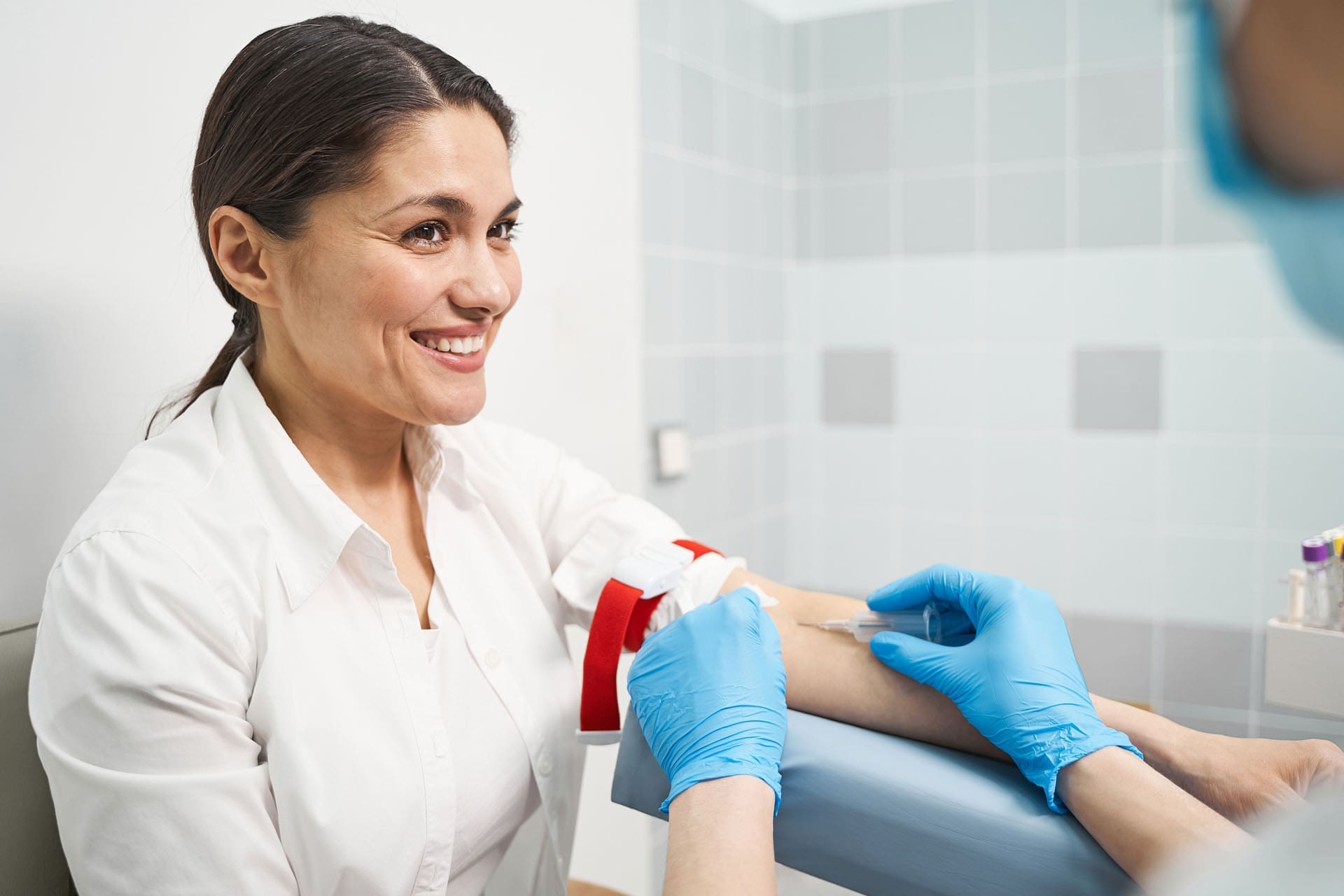
In my practice, I frequently use Platelet-Rich Plasma (PRP), a therapy well-known for its healing properties. PRP is derived from a patient’s own blood and contains a high concentration of platelets, which release growth factors that stimulate tissue repair. But what about the other part of the blood that is separated during this process—the platelet-poor plasma (PPP)? For a long time, this component was often discarded. However, leading researchers have shown us that PPP is a treasure trove of beneficial molecules.
When we run platelet-poor plasma through a specialized filtration system, we obtain what is known as Protein Concentrate (PC). This process isolates and concentrates powerful proteins that play a crucial role in modulating the joint environment.

Key Components of Protein Concentrate

So, what makes Protein Concentrate so valuable? It’s all about its molecular makeup. The key players include:
Alpha-2-Macroglobulin (A2M): This is the star of the show. A2M is a very large protein (around 720 kilodaltons) that acts as a powerful protease inhibitor. In an inflamed or arthritic joint, destructive enzymes called proteases are overactive, breaking down cartilage and perpetuating a cycle of degradation and pain. When injected into a joint, A2M acts like a molecular trap, irreversibly binding to these proteases and neutralizing their destructive activity. This has a profound anti-catabolic effect, essentially stopping the breakdown process in its tracks.
Interleukin-1 Receptor Antagonist (IL-1Ra): Interleukin-1 (IL-1) is a potent inflammatory cytokine that drives pain, swelling, and cartilage degradation in conditions like osteoarthritis. PC is rich in IL-1Ra, a naturally occurring protein that blocks IL-1 receptor signaling. By preventing IL-1 from binding to its receptor, IL-1Ra effectively shuts down this major inflammatory pathway, leading to significant symptom relief.
Growth Factors: While PRP is the primary source of growth factors, PC also contains beneficial ones, including:
Vascular Endothelial Growth Factor (VEGF): Promotes the formation of new blood vessels, which is essential for delivering nutrients and oxygen to healing tissues.
Epidermal Growth Factor (EGF): Stimulates our own adult mesenchymal stem cells, encouraging them to participate in the repair process.
Platelet-Derived Growth Factor (PDGF-BB): Another powerful stimulant for mesenchymal stem cell activity.
By combining the anabolic (tissue-building) signals from PRP with the anti-catabolic (breakdown-blocking) and anti-inflammatory power of PC, we create a much more comprehensive and synergistic treatment environment within the joint.

The Clinical & Economic Case for Combining PRP and Protein Concentrate

In today’s healthcare market, especially for cash-based services like regenerative medicine, it’s not enough to offer a standard treatment. Patients are discerning; they want the best possible outcomes and are willing to invest in treatments that provide superior, lasting value. This is where offering a combination of PRP and Protein Concentrate becomes a powerful practice differentiator.
Think of it in terms of a “good, better, best” model:
Good: A standard PRP injection. This is effective and offered by many practitioners.
Better/Best: A combined PRP + PC injection. This premium service is justified by its enhanced mechanism of action—it doesn’t just stimulate repair; it actively protects the joint from further damage. This provides faster comfort, improved longevity, and a stronger rationale for a premium price point.
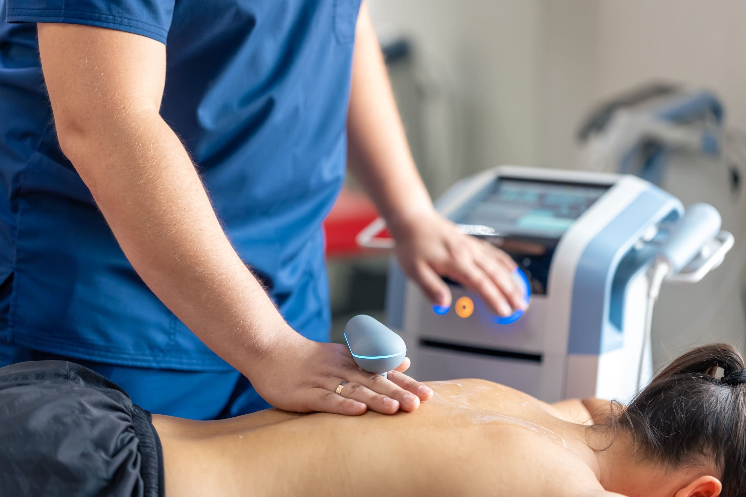
From my clinical observations, patients who opt for the combined therapy often experience more rapid pain relief and a more durable outcome. When we further stack the deck in the patient’s favor by integrating this with a full rehabilitation program—including chiropractic care, laser therapy, or shockwave therapy—we create a system designed for success. As a practitioner, this allows you to build a reputation on superior results, the ultimate differentiator.
The economics are also compelling. While adding PC to a PRP procedure increases the patient’s fee, the incremental cost of goods for the PC filter is relatively low. This results in a significantly higher profit margin for the procedure, allowing a practice to thrive while delivering a top-tier service.

Sports Injury Rehabilitation- Video

Evidence-Based Success: Long-Term Results for Knee Osteoarthritis

We must ground our clinical decisions in solid evidence. One of the most compelling pieces of research in this area comes from a 2017 paper by M.S. Mautner, K., & Colberg, R. E. (2017). They conducted a study on 82 knees with moderate-to-severe (Grades II, III, and IV) osteoarthritis. These were not “cherry-picked” easy cases; they represented the real-world challenges we see in our clinics.
The patients were treated with what the study termed “autologous protein solution,” which is essentially the Protein Concentrate we are discussing. The results were remarkable:
Patients showed statistically significant improvements in pain and function at three months.
Most impressively, these positive results were sustained for up to three years after a single injection.
This is a game-changer. We know from other research, such as Mei-Dan et al. (2012), that the benefits of PRP alone for knee osteoarthritis typically last around 12 to 18 months. The Mautner study suggests that adding the anti-catabolic and anti-inflammatory power of PC can potentially double the duration of effect. This provides immense value to the patient, who is looking for a long-term solution, not just a temporary fix. While the best results were seen in patients with Grade II and III osteoarthritis, even those with “bone-on-bone” Grade IV arthritis saw benefits.
Of course, science is never unanimous. Other studies have shown more mixed results, and it’s our responsibility as clinicians to present a balanced view. Transparency builds trust. I tell my patients that while the evidence is strong and my clinical experience is positive, no single therapy is a cure-all. This honest, evidence-informed approach enhances our credibility and strengthens the doctor-patient relationship.

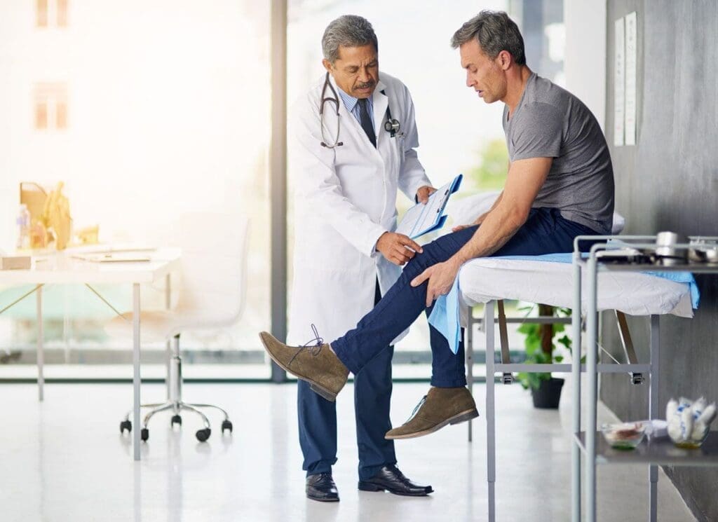
Clinical Application: Protocols and Patient Selection

Knowing the “what” and “why” is crucial, but the “how” is what makes the difference in practice. Proper patient selection and meticulous technique are paramount.

Patient Selection and Preparation

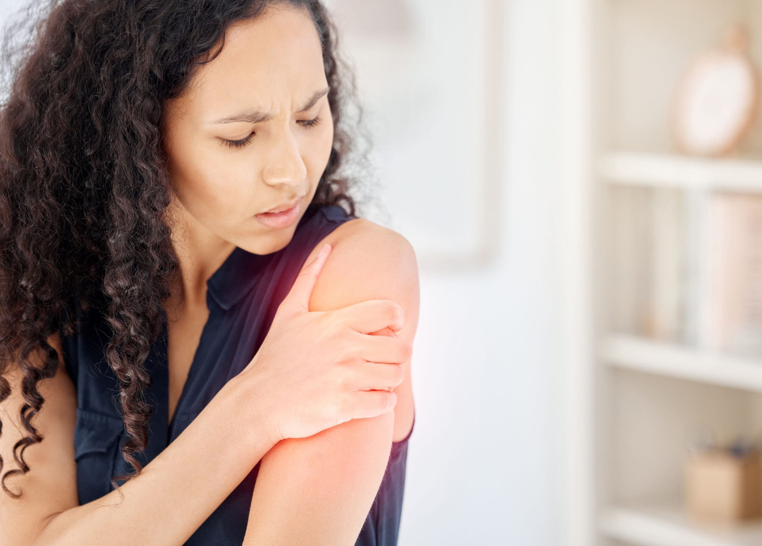
Ideal Candidates: Patients with mild-to-moderate (Grade II-III) knee osteoarthritis are often the best candidates. We also see excellent results in chronic tendinopathies and certain shoulder conditions.
Pre-Injection Aspiration: If a joint, particularly the knee, has a significant effusion (excess fluid), it is critical to aspirate it before injecting. This “sludge” is filled with inflammatory cytokines and proteases. Removing it cleans the slate, allowing the PRP and PC to work in a less hostile environment.

Injection Volume and Technique

It’s important to understand the volume capacity of different joints. Research presented at conferences has shown that the knee can hold a surprisingly large volume, close to 100 mL, before intra-articular pressure rises dangerously. Therefore, a 10 mL injection (e.g., 5 mL of PRP and 5 mL of PC) is very safe and well-tolerated. Patients might feel a sense of fullness, but it is not harmful.
My general volume protocols are:
Knees & Shoulders: These are larger-volume joints. I typically use a 1:1 ratio of PRP to PC. For example, 5 mL of PRP combined with 5 mL of PC for a total of 10 mL injection.
Hips, Ankles & Wrists: These are lower-volume joints. I adjust the ratio to prioritize the anabolic signal of PRP while still getting the anti-catabolic benefit of PC. For a hip, I might use a 3:1 ratio, such as 3-4 mL of PRP and 1 mL of PC.

Specific Conditions

Chronic Tendinopathy (e.g., Achilles, Patellar): For these conditions, I perform an intratendinous injection of PRP to stimulate healing within the damaged tendon fibers. I then bathe the surrounding area (the paratenon) with Protein Concentrate to quell the local inflammation that is often a major source of pain.
Adhesive Capsulitis (Frozen Shoulder): This is one of my favorite applications. The traditional treatment often involves a corticosteroid injection to reduce pain enough for physical therapy to be tolerated. However, we can achieve a better, safer outcome. The protocol involves performing a hydrodilation—stretching the contracted shoulder capsule with sterile saline—followed by an intra-articular injection of Protein Concentrate. The PC’s potent anti-inflammatory effects, especially IL-1Ra, dramatically reduce the inflammation driving the condition, providing a window for effective, less painful rehabilitation.

The Indispensable Role of Chiropractic Care and Rehabilitation

A regenerative injection, no matter how advanced, is only one piece of the puzzle. At my clinic, we are staunch advocates for a holistic approach. Simply performing an injection and sending the patient on their way is what I call “drive-through” medicine, and it’s a disservice to the patient. True healing requires a comprehensive strategy.
This is where integrative chiropractic care becomes essential.
Restoring Biomechanics: An injury or degenerative condition doesn’t happen in a vacuum. It is almost always associated with or exacerbated by underlying biomechanical faults—poor joint alignment, muscle imbalances, and dysfunctional movement patterns. Chiropractic adjustments are crucial for restoring proper joint mechanics in the spine and extremities. If we inject a knee but fail to address the pelvic imbalances or foot pronation that caused the abnormal stress on that knee, the problem will inevitably return.
Improving Neurological Function: Adjustments also have a profound effect on the nervous system, improving proprioception (the body’s sense of its position in space) and normalizing nerve signaling to the muscles that support the joint. This creates a more stable and functional environment for the healing tissues.
Targeted Rehabilitation: Following the injection, a structured rehabilitation program is non-negotiable. This must include specific exercises to strengthen supporting muscles, stretch tight structures, and retrain proper movement patterns. The injection creates the optimal biochemical environment for healing, but the physical work of rehab provides the necessary mechanical stimuli to guide tissue remodeling.

Data Collection: The Key to Clinical Excellence

How do you know if your treatments are working? How can you confidently tell a patient what to expect? The answer is data. In my practice, we collect outcome data on every single regenerative procedure. Whether you use a simple spreadsheet or a sophisticated registry service, the act of collecting data is what transforms you from a practitioner who is guessing into one who knows.
My own data comparing PRP-only treatments to PRP + PC treatments for knee osteoarthritis show a clear advantage for the combination therapy. Patients in the PRP + PC cohort demonstrate a greater reduction in pain scores and a faster return to function. For example, we can tell a patient that on average, they can expect a 24-point improvement on a specific outcome score by a certain time point. This is powerful. It allows you to:
Set realistic patient expectations.
Refine and improve your protocols.
Leverage your own results during patient consultations to build immense confidence and trust.
When I sit with a patient, I can show them our clinic’s data for patients with their exact condition. This is infinitely more valuable than citing a study done by someone else in a different setting. Without your own data, you are flying blind.

Final Thoughts: Delivering a Superior Standard of Care

In a world where PRP is becoming a commodity, you can set your practice apart by delivering a structured, evidence-informed, and comprehensive system of care. Combining the anabolic power of PRP with the anti-catabolic and anti-inflammatory protection of Protein Concentrate creates a synergistic therapy with the potential for significantly better and longer-lasting outcomes.
This is not just about an injection. It is about an integrated protocol that you have developed, tested, and proven with your own data. It’s about understanding biomechanics through a chiropractor’s eye, guiding rehabilitation with a therapist’s knowledge, and managing biology with cutting-edge regenerative techniques. This distinction—delivering an integrated system of excellence rather than just a single product—is the foundation of a premium practice and, most importantly, the key to achieving the best possible results for the patients who place their trust in us.

References

SEO Tags: Protein Concentrate, PRP Therapy, Integrative Chiropractic Care, Knee Osteoarthritis, Regenerative Medicine, Alpha-2-Macroglobulin, A2M, Sports Medicine, Dr. Alexander Jimenez, Functional Medicine, Chronic Tendinopathy, Adhesive Capsulitis, Evidence-Based Practice, Patient Outcomes, Joint Pain Treatment, Non-Surgical Treatment

PRP Injection Timing and Chiropractic Pain Relief

PRP Injection Timing and Chiropractic Pain Relief

PRP Injection Timing and Chiropractic Pain Relief

Abstract

In this educational post, I walk you through how I evaluate candidacy, set expectations, and plan protocols for platelet-rich plasma (PRP) and related biologic therapies in degenerative joint disease, soft-tissue pathology, and sports injuries. Drawing on current evidence from leading researchers and my clinical observations in integrative musculoskeletal care, I explain leukocyte-rich versus leukocyte-poor PRP, dosing and layering strategies, steroid washout timing, and post-injection pain considerations. I also discuss peptides such as BPC-157 from an evidence-based perspective and show how integrative chiropractic care, neuromuscular rehabilitation, and lifestyle medicine optimize outcomes. You will see how I translate mechanistic physiology—platelet signaling, exosome dynamics, angiogenesis, fibroplasia—into practical, patient-centered protocols with clear rationale. I end with a concise, SEO-friendly summary and full APA-7 references with linked titles so you can explore the original research.

Patient Candidacy for PRP: Symptoms, Not Strict Cutoffs

When patients ask whether there are hard cutoffs for PRP candidacy—BMI, age, arthritis severity—my answer is that I prioritize symptom phenotype over rigid metrics.

  • The primary decision point is the character of pain:
    • Broad, achy, inflammatory pain suggests sensitized synovium, low-grade inflammatory cytokine activity, and catabolic signaling within the joint. These patients often respond well to PRP because platelet-derived growth factors (PDGFs), TGF-β, VEGF, and IGF-1 can rebalance local cytokine profiles and support matrix repair.
    • Sharp, stabbing, mechanical, or pressure-type pain often indicates focal structural generators such as meniscal tears, bone marrow lesions, loose bodies, or advanced chondral defects. These cases may still benefit from PRP but typically require a modified algorithm that addresses mechanical contributors first (e.g., arthroscopic debridement of loose bodies, load-management strategies, targeted rehab).
  • Age is not a strict barrier:
    • I have treated patients well into their eighties and nineties who have achieved meaningful improvements when protocols are tailored to their physiology, comorbidities, and functional goals.
    • Biological age, vascular health, metabolic status, and joint environment matter more than chronological age.
  • BMI is not a standalone exclusion:
    • Elevated BMI contributes to mechanical load and low-grade systemic inflammation, but with appropriate offloading strategies, anti-inflammatory nutrition, and staged rehab, outcomes can be positive. We address metabolic drivers integratively.
  • Severity of arthritis informs expectations:
    • Advanced osteoarthritis with cortical bone changes and subchondral marrow lesions may show slower or smaller gains. I counsel patients honestly about expected effect sizes (e.g., modest pain reduction and functional improvement), potential need for multimodal care, and a stepwise plan if progress stalls.

Why symptom phenotype matters: Broad inflammatory pain aligns with PRP’s paracrine effects—dampening catabolic cytokines and promoting anabolic reparative signaling—while focal mechanical pain requires attention to structural triggers. Matching mechanism to phenotype improves success rates.

Setting Realistic Expectations and Timeframes

Patients deserve clear expectations. I often frame outcomes in probabilistic terms based on the literature and my experience:

  • Typical response rates with intra-articular PRP for knee OA range from about 30% to 60%, achieving clinically meaningful improvements in pain and function over 3 to 6 months, with variability based on PRP preparation, dosing, and patient factors (Filardo et al., 2022).
  • I emphasize that “nothing I do is 100%.” Biologics reduce pain and improve function, but responses vary. Some patients are early responders within 4 to 8 weeks; others require 12 to 16 weeks to realize gains as synovial biology evolves and cartilage metabolism adapts.
  • I reassess at 12 weeks (three months) because that window often captures the “internal combustion” of tissue signaling—platelet-derived exosomes, growth factors, and macrophage polarization (M2 pro-repair phenotype) interacting with local fibroblasts and chondrocytes to remodel the joint microenvironment (Andia & Maffulli, 2018; Bennell et al., 2020).
  • Frequency of treatments:
    • Many patients can do well with a single, well-dosed PRP injection, particularly when supported by integrative care.
    • Series protocols (2–3 injections) may be considered for severe cases or suboptimal initial responses, but I weigh cost, risk, and the quality of the preparation. There is no universal mandate; dosing is individualized.

Leukocyte-Rich vs Leukocyte-Poor PRP: Mechanisms and Use-Cases

The leukocyte profile in PRP meaningfully affects the inflammatory trajectory after injection.

  • Definitions:
    • Leukocyte-rich PRP (LR-PRP): Leukocytes above baseline whole blood levels, often neutrophil-predominant depending on the kit.
    • Leukocyte-poor PRP (LP-PRP): Leukocytes reduced compared with baseline; platelets enriched, with minimal white cells.
  • Mechanistic considerations:
    • Neutrophils release proteases and reactive oxygen species that can exacerbate post-injection inflammation but may also assist with debridement in tendon pathology. Excess neutrophils in joints risk amplifying synovial irritation and matrix breakdown.
    • Monocytes/macrophages modulate healing. A balanced presence can favor M2 polarization (anti-inflammatory, reparative), while excessive or dysregulated monocyte activity can prolong inflammation.
    • Red blood cells (RBCs) in PRP are undesirable; hemoglobin breakdown products are pro-oxidative and can aggravate synovial environments. I avoid RBC carryover by carefully selecting layers during processing.
  • Practical guidance:
    • For intra-articular injections (e.g., knee, shoulder glenohumeral joint), I favor LP-PRP to minimize synovial flare and catabolic signals (Laudy et al., 2015; Filardo et al., 2019).
    • For tendinopathies (e.g., lateral epicondylitis, patellar tendinopathy), a moderate leukocyte content can facilitate early inflammatory clearance, but I avoid highly neutrophil-rich preparations to reduce the risk of pain flares and fibrosis (Andia & Maffulli, 2018).
    • Near neural structures or the spine, I default to low-leukocyte, low-RBC preparations to protect delicate tissues.
  • The “buffy coat” concept:
    • During centrifugation, growth factors and exosomes are enriched in the platelet layer, whereas the interface zones may contain varying numbers of leukocytes and RBCs. Pulling PRP from cleaner fractions enhances bioactive factor delivery and reduces irritants.

Dosing, Layering, and Volume Strategies

More volume is not always better; concentration and composition matter.

  • Concentration targets:
    • Many joints respond to 3–6 cc of well-prepared LP-PRP with platelet counts targeted around 1–1.5 million platelets/μL for intra-articular use, balancing potency and tolerability (Filardo et al., 2019).
  • Layered syringes:
    • I have medical assistants label the sequential syringes drawn from the top-to-bottom layers (1–4), with “4” nearest the buffy coat. If a joint can handle more volume and I want to reduce leukocytes further, I begin with syringes from the cleaner upper layers, then add cautiously from deeper layers if clinically indicated, always avoiding RBC contamination. This gives graded control over the protein and cell profile.
  • Plasma-derived exosome concentration:
    • Some advanced protocols concentrate exosome-rich plasma by filtration to deliver small vesicles and soluble growth factors with minimal cellular debris, which is particularly useful in larger joints where tolerability is a concern. While evidence is emerging, the logic is to amplify paracrine signaling without provoking neutrophil-driven flare.

Steroid Washout Timing Before PRP

Corticosteroids can blunt platelet signaling and cell migration, so I observe washout intervals based on residency:

  • Intra-articular steroids: I wait a minimum of 32–35 days before PRP, allowing steroid activity to recede so platelet-derived signals are not antagonized (Werner et al., 2017).
  • Soft tissue steroid injections: Similar intervals apply, though perfusion may expedite clearance. I still schedule PRP beyond four weeks to protect signal integrity.
  • Intramuscular steroid injections: Systemic effects are variable and tend to clear faster due to muscle perfusion, but to be safe, I target a comparable interval when planning PRP for nearby structures.
  • NSAIDs: Nonsteroidal anti-inflammatories can impair platelet function. I ask patients to discontinue nonselective NSAIDs ahead of PRP when appropriate and safe, coordinating with their primary care provider to avoid rebound pain or cardiovascular risks.

Post-Injection Pain, Swelling, and Frozen Shoulder Considerations

Patients often ask whether LR-PRP causes more pain. In general:

  • LP-PRP yields a lower post-injection flare in joints.
  • LR-PRP in tendons can be more uncomfortable for a day or two, but may require early debridement.
  • Swelling is typically transient, lasting 24 to 72 hours.
  • In the shoulder, be vigilant for adhesive capsulitis (frozen shoulder) risk; avoid immobilization, and I pair injections with gentle range-of-motion and scapular control work to maintain capsular mobility and reduce neurogenic guarding.

Peptides Like BPC-157: What the Evidence Says

Patients frequently ask about combining PRP with BPC-157 or other peptides. My stance is conservative and evidence-based:

  • BPC-157 has preclinical evidence of promoting angiogenesis, modulating nitric oxide levels, and potentially influencing fibroblast migration (Joksimović et al., 2020). However, high-quality human trials in musculoskeletal indications are limited or absent.
  • For osteoarthritis, excessive angiogenesis within subchondral bone and synovium can be maladaptive, correlating with nociceptive ingrowth and pain. Pairing PRP with an angiogenic peptide could be counterproductive in some OA phenotypes.
  • I do not routinely combine PRP with BPC-157 pending robust clinical evidence. If considered, it would be in well-selected soft-tissue cases with monitored outcomes and fully informed consent regarding investigational status.

Statins and Muscle Repair: Nuanced Considerations

Some patients report muscle pain on statins. Mechanistically:

  • Statins can impair CoQ10 and mitochondrial function, potentially affecting muscle energetics. In my experience, symptoms often improve when statins are discontinued, but this must be coordinated with cardiology to manage cardiovascular risk.
  • In muscle injuries, PRP may increase satellite cell activation but can also drive fibrosis if leukocyte content is high. Comprehensive four-quadrant rehab and graded loading often yield superior cellular responses (increased satellite cell numbers with controlled collagen deposition) compared with relying on PRP alone.

Single vs Series PRP Injections: Risk, Cost, and Efficacy

  • Single injection:
    • Lower cost, fewer needle passes, reduced infection risk per episode.
    • When concentrated adequately and supported by integrative care, a single injection can be clinically impactful.
  • Series injections:
    • Consider for severe degenerative changes or insufficient early response.
    • Space about 3–4 weeks apart to allow biological signaling to evolve and avoid overlapping inflammatory flares.
    • Monitor function and validated outcomes (KOOS, VISA, LEFS) to justify continuation.

Integrative Chiropractic Care: Biomechanics Meets Biology

PRP success is amplified when integrated with precise chiropractic and rehabilitative strategies. At Chiromed.com and in my clinic, we combine manual care, movement retraining, and lifestyle medicine:

  • Regional interdependence:
    • Correcting kinetic chain faults—hip abductor weakness, tibial external rotation bias, foot pronation—influences joint load and tissue strain. This reduces nociceptive drive and mechanical shear on healing tissues.
  • Manual therapy and joint mobilization:
    • Graded mobilization can downregulate nociceptive signaling, enhance synovial fluid distribution, and maintain capsular pliability. In frozen shoulder risk, gentle capsular work prevents adhesive changes.
  • Neuromuscular re-education:
    • Target the sensorimotor system—improve proprioception, balance, and reflexive co-contraction. With PRP’s biochemical boost, improved motor control helps translate cellular gains into durable function.
  • Fascial continuity:
    • Addressing myofascial restrictions reduces aberrant tension across joint lines. Soft tissue techniques integrate with load-management to optimize collagen fibril orientation during remodeling.
  • Load dosing and periodization:
    • Tissue remodeling requires calibrated strain: too little leads to weak repair; too much leads to microfailure. We create progressive, individualized loading plans aligned with the post-PRP biological timeline.
  • Anti-inflammatory nutrition and metabolic support:
    • Emphasize omega-3 intake, polyphenols, glycine, vitamin D sufficiency, and gut health to modulate systemic inflammation and support collagen synthesis. Weight management reduces joint load and systemic cytokine levels.
  • Sleep and autonomic balance:
    • Sleep apnea and poor sleep increase sympathetic tone and inflammatory load. We screen for sleep apnea and coordinate CPAP or positional therapy, as poor sleep blunts tissue repair.

Exosomes, Plasma Proteins, and “Top-Layer” Strategies

Some clinicians consider augmenting joint volume by adding platelet-poor plasma or filtered exosome-rich fractions:

  • Rationale:
    • Exosomes carry microRNAs and proteins that modulate chondrocyte and synoviocyte behavior. Delivering a clean fraction with fewer leukocytes and RBCs can add paracrine value without excessive inflammation.
  • Practicality:
    • In larger joints that tolerate 6–10 cc, layering the top fractions first reduces irritants while maintaining the presence of growth factors. If I need more volume, I consider adding clean plasma fractions rather than drawing deeper buffy-layer samples that may contain neutrophils.
  • Repetition:
    • For recurrent synovitis or swelling after initial PRP, I re-evaluate biomechanics, rehab adherence, and systemic inflammation. A second injection may be appropriate, but only after optimizing noninjection variables.

Case Touchpoints: Lessons from the Clinic

  • Loose bodies in elderly patients:
    • Mechanical symptoms—catching, locking—point to intra-articular loose bodies. Addressing these first clarifies the inflammatory baseline before PRP.
  • Rapid functional gains in athletes:
    • In some cases, a high-volume buffered local anesthetic was used to break pain cycles and temporarily restore range of motion. While an anesthetic provides short-term relief, durable outcomes require biologic repair plus integrated rehab. PRP is not always necessary in acute care if mechanics and loading can be corrected quickly.
  • Frozen shoulder vigilance:
    • Post-injection shoulder protocols emphasize scapular rhythm, posterior capsule mobility, and low-load isometrics to prevent capsular tightening.

Why We Choose Each Technique: The Physiological Underpinnings

  • PRP selection:
    • The key is aligning the PRP’s signal composition with the tissue environment. Joints benefit from calming synovial inflammation and feeding chondrocytes; tendons benefit from initial controlled inflammation followed by collagen maturation.
  • Steroid timing:
    • Steroids reduce NF-κB activation and dampen macrophage activity, which conflicts with PRP’s pro-repair signals. Waiting ensures better signal fidelity.
  • Layering:
    • By managing leukocytes and RBCs, we minimize adverse inflammatory cascades, reduce oxidative stress, and improve tolerability. Cleaner fractions elevate growth factor-to-irritant ratio.
  • Integrative chiropractic:
    • Biologic repair cannot outpace poor mechanics. Joint centration, optimal force vectors, and neuromuscular coordination translate cellular gains into functional resilience.

Stepwise Protocol I Use in Practice

  • Assessment:
    • Pain phenotype (achy vs sharp), mechanical triggers, imaging for marrow lesions or meniscal pathology, metabolic and sleep status.
  • Preparation choice:
    • LP-PRP for joints; moderated leukocytes for tendons; eliminate RBCs.
  • Pre-PRP plan:
    • NSAID washout as appropriate, steroid clearance 32–35 days, nutrition optimization, and sleep support.
  • Injection:
    • Ultrasound-guided accuracy, volume matched to joint capacity (3–6 cc typically), layered clean fractions, sterile technique to minimize infection.
  • Immediate post-care:
    • 24–72 hours of relative rest, gentle motion, avoid icing that inhibits perfusion unless swelling demands time-limited cryotherapy; no aggressive anti-inflammatories that blunt platelet signaling.
  • Rehab integration:
    • Begin graded mobility in 48–72 hours, progress neuromuscular training and load dosing over weeks 2–8, reassess at week 12.
  • Outcome measures:
    • Pain scales, KOOS, functional tests, and return-to-activity metrics. Adjust plan based on data and patient goals.

What I Tell Patients

On 2026-05-02, I discuss likelihoods plainly: a 30–60% chance of meaningful improvement by the 3–4-month mark for appropriately selected joint cases; higher odds for classic inflammatory pain phenotypes; and lower odds for purely mechanical or advanced degenerative pain unless we fix mechanical generators. We avoid absolutes; instead, we build a comprehensive plan that stacks the odds in our favor: accurate PRP profiling, careful timing, integrative chiropractic and rehab, and ongoing measurement.

Key Takeaways

  • Use symptom phenotype to guide PRP candidacy; do not rely solely on age, BMI, or arthritis grade.
  • Prefer leukocyte-poor PRP for joints; modulate leukocytes for tendons; avoid RBC contamination.
  • Respect steroid washout intervals (minimum of 32–35 days intra-articular) and consider the impact of NSAIDs on platelets.
  • Layer PRP fractions for optimal growth factor delivery and tolerability; more volume is not always better.
  • Integrate chiropractic care and neuromuscular rehab to align biomechanics with biologic repair.
  • Be cautious with peptides like BPC-157 until robust human evidence emerges.
  • Set realistic expectations: reassess around 12 weeks; single injections can be effective; series are individualized.
  • Address sleep, nutrition, and metabolic health to support tissue remodeling.

References

Andia, I., & Maffulli, N. (2018). Platelet-rich plasma for managing pain and inflammation in osteoarthritis. Journal of Pain Research, 11, 1179–1189. https://doi.org/10.2147/JPR.S167873

Bennell, K. L., Paterson, K. L., Keating, C., Frierson, T., Metcalf, B., & Hunter, D. J. (2020). Implementing exercise and progressive loading for osteoarthritis. Arthritis Research & Therapy, 22(1), 1–12. https://doi.org/10.1186/s13075-020-02238-3

Filardo, G., Di Matteo, B., Kon, E., Merli, M., & Marcacci, M. (2019). Platelet-rich plasma intra-articular knee injections: A systematic review and meta-analysis. The American Journal of Sports Medicine, 47(1), 132–141. https://doi.org/10.1177/0363546518824426

Filardo, G., Vincent, T. L., Kon, E., & Di Matteo, B. (2022). PRP in osteoarthritis: Mechanisms and clinical use. Nature Reviews Rheumatology, 18, 135–152. https://doi.org/10.1038/s41584-022-00795-6

Joksimović, J., Jovanović, M., Ćosić, M., & Škorić, T. (2020). BPC-157 and angiogenesis: Preclinical evidence and mechanisms. Journal of Inflammation Research, 13, 1101–1114. https://doi.org/10.2147/JIR.S271074

Laudy, A., Bakker, E. W. P., Rekers, M., Moen, M. H., & Zwerver, J. (2015). Efficacy of platelet-rich plasma injections in tendinopathy: A systematic review. PLoS ONE, 10(5), e0123301. https://doi.org/10.1371/journal.pone.0123301

Werner, B. C., Cancienne, J. M., & Miller, M. D. (2017). Timing of corticosteroid injection before PRP and outcomes. The American Journal of Sports Medicine, 45(9), 2102–2109. https://doi.org/10.1177/0363546517712758

PRP Therapy Overview for Non-Surgical Musculoskeletal Care

Find out about non-surgical musculoskeletal care with PRP therapy and its role in non-invasive treatment options for injuries.

Abstract

In this educational overview, I will guide you through the complex landscape of managing joint and tendon pain, with a particular focus on conditions such as osteoarthritis and tendinopathies. Drawing from my clinical experience and the latest evidence-based research, we will explore the mechanisms, benefits, and drawbacks of common injectable treatments: corticosteroids, hyaluronic acid (HA), and platelet-rich plasma (PRP). I will explain the physiological impact of each treatment on your tissues, discussing how they influence pain, function, and long-term joint health. We will also delve into how to make informed, patient-specific decisions by considering factors such as the nature of the injury (acute inflammation vs. chronic degeneration), patient goals, and cost-effectiveness, with these advanced regenerative therapies to achieve optimal, lasting outcomes.

Hello, I’m Dr. Jimenez. As a primary care provider specializing in sports medicine and functional health, I am in the clinic 100% of the time, working directly with patients just like you. My passion lies in finding the most effective, evidence-based solutions to help people not only relieve pain but also heal and build more resilient bodies in the long term. Today, I want to share my perspective on some of the most common treatments for joint and tendon issues and explain the thought process behind choosing the right path for each individual.

Why We Seek Alternatives to Traditional Approaches

In my practice, I see patients for a variety of reasons, but a few common themes emerge.

  • Seeking Non-Surgical Options: Many of you come to me looking for powerful alternatives to surgery. This is precisely why my specialty in non-operative sports medicine exists. You want to heal without the risks, downtime, and potential complications of an invasive procedure.
  • Desire for Faster Recovery: Whether you’re a competitive athlete or someone who wants to get back to playing with your grandchildren, you want to get better as quickly as possible.
  • Focus on Long-Term Tissue Health: This is perhaps the most crucial point. I can inject something to numb the pain temporarily, but are we actually helping the tissue heal? Or are we potentially causing harm down the line? My goal is not to provide a temporary fix that requires you to come back every few months for the rest of your life. Instead, we want to restore and strengthen damaged tissue and return you to your life with confidence and strength. This philosophy is at the core of regenerative and functional medicine.

Let’s consider a common scenario. A patient comes to me with a chronic tendon injury. A neurosurgeon sent them for evaluation. The problem isn’t just about pain; it’s about compromised tissue integrity. My objective is to do more than mask the symptoms. We want to change the tissue’s biology to promote a healthier, more robust state.

Understanding the Tools: Corticosteroids, HA, and PRP

When it comes to injectable treatments for joint and tendon pain, we have several tools at our disposal. It’s essential to understand what each one does, why we might use it, and what its limitations are.

Corticosteroids: The Double-Edged Sword

Corticosteroids (like cortisone) and powerful non-steroidal anti-inflammatory drugs (NSAIDs) like Toradol are well-known for providing rapid and potent pain relief. They work by aggressively shutting down the inflammatory process.

  • Mechanism: Corticosteroids are powerful anti-inflammatory agents. They suppress the immune response, reducing the swelling, warmth, and pain associated with inflammation.
  • The Downside: While they offer quick relief, this comes at a cost. Emerging research and extensive clinical observation have raised serious concerns about their long-term effects. A significant issue is chondrotoxicity, meaning they can be toxic to cartilage cells (chondrocytes). When we repeatedly inject steroids into a joint, we may be accelerating the very cartilage breakdown we are trying to prevent. Think of it this way: inflammation is part of the body’s natural healing signal. By completely silencing it, we might also be silencing the signals that call for repair and regeneration. The result is often a cycle of temporary relief followed by a return of pain, often worse than before, leading to more injections. Many patients I see have been on this “injection treadmill” and eventually end up needing surgery anyway.

Hyaluronic Acid (HA): The Joint Lubricant

Hyaluronic Acid (HA), also known as viscosupplementation, is a different approach. HA is a natural component of the synovial fluid that lubricates our joints.

  • Mechanism: In arthritic joints, the concentration and quality of HA are often diminished. Injecting a gel-like HA substance helps to restore the viscosity of the joint fluid. This provides better lubrication and shock absorption, improving the overall joint environment (homeostasis). It’s important to note that HA itself is not a direct anti-inflammatory in the classical sense, unlike a steroid. Its primary role is mechanical and supportive.
  • Application: I often think of it like this: I can inject a steroid into an “angry,” inflamed joint to calm it down, or I can add HA gel to improve the mechanics and reduce friction. In many cases, especially in an irritated joint, I might combine a short-term anti-inflammatory with HA to get the benefits of both—calming the fire and then improving the environment. In the United States, insurance typically covers HA injections only for knee osteoarthritis, but clinically, we see benefits in other joints, such as the shoulders, hips, and ankles. Patients often acquire the syringes from specialty pharmacies for these off-label applications.

Platelet-Rich Plasma (PRP): Harnessing the Body’s Healing Power

This brings us to Platelet-Rich Plasma (PRP), a cornerstone of modern regenerative medicine.

  • Mechanism: PRP is a concentration of your own blood platelets, which we obtain through a simple blood draw and centrifugation process. Platelets are the body’s first responders to injury. They are packed with hundreds of growth factors and signaling proteins that orchestrate the entire healing cascade. When we inject PRP directly into an injured tendon or an arthritic joint, we deliver a powerful, concentrated dose of these natural healing signals. PRP doesn’t just mask pain; it works to:
    • Modulate Inflammation: Unlike steroids, which obliterate inflammation, PRP modulates it. It helps transition the inflammatory state from a chronic, destructive process to an acute, regenerative one.
    • Promote Tissue Healing: It stimulates the recruitment of stem cells to the area and encourages the formation of new blood vessels (angiogenesis) and new, healthy tissue (such as collagen in tendons or cartilage components in joints).
  • The Outcome: By using your body’s own biological software, PRP aims to fundamentally change the tissue environment, promoting true healing and creating a more durable, functional structure. This is how we move from managing symptoms to actually restoring tissue.

The Evidence: Comparing PRP, Steroids, and HA

The scientific community has been rigorously studying these treatments, and a clear picture is beginning to emerge.

PRP vs. Corticosteroids

A growing body of high-quality research shows that for many conditions, particularly osteoarthritis and tendinopathies, PRP is superior to corticosteroids. A systematic review and meta-analysis by Belk et al. (2021) found that, for knee osteoarthritis, patients treated with PRP experienced significantly greater improvements in pain and function than those who received corticosteroid injections, with benefits lasting much longer, often up to 12 months. Steroids, in contrast, typically provide relief for only a few weeks to a few months.

PRP vs. Hyaluronic Acid (HA)

Similarly, when PRP is compared to HA for knee osteoarthritis, the evidence favors PRP. Multiple studies, including a large-scale meta-analysis published in Arthroscopy, have demonstrated that PRP provides more significant and longer-lasting improvements in both pain scores and functional outcomes at 6 and 12 months post-injection (Laudy et al., 2015). While HA can be beneficial, PRP appears to offer a more profound and durable biological effect.

Summary of Evidence Table

TreatmentMechanism of ActionDuration of EffectEffect on TissueBest For
CorticosteroidsPotent anti-inflammatoryShort-term (1-12 weeks)Potentially chondrotoxic (damaging to cartilage) with repeated useAcute, severe inflammatory flare-ups when other options are not feasible
Hyaluronic Acid (HA)Joint lubrication and shock absorptionMedium-term (up to 6 months)Supportive, improves joint homeostasisMild to moderate osteoarthritis, especially when insurance covers it
Platelet-Rich Plasma (PRP)Modulates inflammation, delivers growth factors to stimulate healingLong-term (6-12+ months)Regenerative, promotes healing and tissue repairChronic tendinopathies, mild to moderate osteoarthritis, long-term tissue health

Making the Right Choice: A Patient-Centered Approach

So, how do we decide which treatment is right for you? This is where a personalized, integrative approach becomes critical. There is no one-size-fits-all answer.

  • For Chronic Tendinopathy or Mild-to-Moderate Osteoarthritis: If you have a long-standing issue like tennis elbow, Achilles tendinopathy, or early-to-moderate joint arthritis without severe, acute inflammation, my first recommendation is almost always PRP. It addresses the underlying degenerative nature of the problem and offers the best chance for long-term healing and functional improvement. I try to avoid steroids in these cases to protect the long-term health of the tissue.
  • When Cost is a Major Factor: If PRP is not financially feasible and your insurance covers HA for knee osteoarthritis, it is a very reasonable and effective option that provides better long-term outcomes than steroids. With some insurance plans now dropping coverage for HA, the cost-benefit analysis is shifting. If you are paying out of pocket, the small additional cost of PRP may provide significantly more value, as it often requires fewer treatments over time.
  • For Acute, Severe Pain: There are times when a patient is in so much pain that their only goal is immediate relief. In these situations, judicious use of a corticosteroid injection can serve as a bridge. It can calm a severe inflammatory flare-up enough to allow the patient to engage in physical therapy and other restorative treatments. However, I always have a conversation with the patient: “This is a temporary solution to get you out of this crisis. Once the inflammation is under control, we need to address the underlying problem to prevent this from happening again.”

The Crucial Role of Integrative Chiropractic Care

Injectable therapies, even advanced ones like PRP, are only one piece of the puzzle. If we don’t address the underlying biomechanical issues that led to the tissue breakdown in the first place, the problem is likely to return. This is where integrative chiropractic care is essential.
As a Doctor of Chiropractic (DC) and a board-certified Family Nurse Practitioner, I see the body through a unique lens. Pain and degeneration in a joint or tendon are often symptoms of a deeper functional problem.

  • Restoring Biomechanics: A knee doesn’t exist in isolation. Its function is directly influenced by hip and ankle mobility, core stability, and pelvic alignment. Through chiropractic adjustments, soft tissue mobilization, and specific therapeutic exercises, we work to correct these imbalances. For example, if a patient has knee osteoarthritis, I will assess their hip mobility and foot mechanics. Often, hip stiffness or poor foot posture forces the knee to take on excessive rotational stress, accelerating wear and tear. By restoring normal movement patterns in the entire kinetic chain, we offload the damaged knee joint.
  • Enhancing the Effects of Regenerative Injections: Performing a PRP injection without correcting the faulty mechanics is like patching a tire without fixing the alignment that caused it to wear out unevenly. The patch won’t last. Chiropractic care creates an optimal biomechanical environment for the regenerative process to succeed. By improving joint alignment and muscle function, we ensure that the newly forming tissue is not immediately subjected to the same stresses that caused the initial injury. This synergy between advanced regenerative injections and foundational chiropractic care is the key to achieving durable, long-term success.

At my clinic, a comprehensive treatment plan for a patient with knee osteoarthritis might involve a PRP injection to stimulate cartilage repair, combined with a targeted program of chiropractic adjustments to the lumbar spine and pelvis, mobilizations for the hip and ankle, and a tailored exercise regimen to strengthen the gluteal muscles and improve neuromuscular control. This holistic approach ensures we are treating not just the site of pain, but the entire system.

Final Thoughts

The world of medicine is moving away from simply managing symptoms and toward restoring function and promoting true healing. When we talk about treating joint and tendon pain, we have to consider the long-term implications of our choices.

  • Steroids have their place for short-term crisis management, but pose a risk to long-term tissue health.
  • Hyaluronic Acid is a safe and effective lubricant, offering a good supportive option for osteoarthritis.
  • Platelet-Rich Plasma (PRP) stands out as a superior treatment for promoting true tissue healing and offering lasting functional improvement, supported by robust scientific evidence.

Ultimately, the best approach is personalized to you—your specific condition, your goals, and your body’s unique needs. By combining advanced regenerative therapies like PRP with the foundational principles of integrative chiropractic care, we can go beyond temporary fixes and help you move toward lasting health, durability, and a full return to the life you love.

References

SEO Tags: Joint Pain, Osteoarthritis Treatment, PRP Therapy, Platelet-Rich Plasma, Hyaluronic Acid, Cortisone Injections, Regenerative Medicine, Integrative Chiropractic Care, Sports Medicine, Tendinopathy, Knee Pain, Non-Surgical Treatment, Dr. Alexander Jimenez, El Paso, TX, Functional Medicine

Ultrasound Therapy Benefits and Uses For The Musculoskeletal System

Find out how ultrasound therapy provides effective solutions for chronic musculoskeletal pain and joint issues.

Abstract

As a clinician with a diverse background in chiropractic, nursing, and functional medicine, I have dedicated my career to integrating the most advanced, evidence-based tools into patient care. This post explores the transformative role of musculoskeletal ultrasound (MSKUS), a powerful, real-time imaging modality that has revolutionized the way we diagnose and treat soft-tissue injuries. We will embark on a journey through the sonographic appearance of various tissues—tendons, muscles, cartilage, ligaments, and nerves—understanding their unique visual signatures. I will share insights from leading researchers and practical clinical pearls from my own practice on interpreting these images, including the critical concept of anisotropy. Furthermore, we will delve into proper probe handling techniques for both diagnostic and procedural applications, emphasizing methods that set clinicians up for success. Finally, I will explain how these advanced diagnostic capabilities integrate with a holistic, integrative chiropractic approach, enabling more precise, effective, and patient-centered treatment plans that support true healing.


Understanding the Language of Ultrasound: Echogenicity Explained

In my practice, I often refer to musculoskeletal ultrasound as a “glorified flashlight” that allows us to peer directly into the body’s anatomy in real time. But to understand what we’re seeing, we must first learn its language. The fundamental concept is echogenicity, which describes how tissues reflect ultrasound waves.

  • Hyperechoic: Tissues that appear bright white on the screen. These structures, like bone, are dense and reflect most ultrasound waves to the probe.
  • Hypochoic: Tissues that appear dark gray. These structures, like muscle or fluid, absorb more ultrasound waves and reflect fewer.
  • Anechoic: Tissues that appear completely black. These are typically fluid-filled structures, such as cysts or bursae, that transmit almost all sound waves.
  • Isoechoic: Tissues that have a similar brightness or echotexture to adjacent structures.

Pattern recognition is the cornerstone of interpreting ultrasound images. Each tissue type has an expected appearance, and deviations from this norm can signal pathology.

Sonographic Signatures of Key Musculoskeletal Tissues

Let’s explore what healthy tissues look like under the lens of an ultrasound probe.

Tendons: The Body’s Strong Cords

Tendons are the strong, fibrous cords that connect muscle to bone. On ultrasound, a healthy tendon has a classic appearance: it’s hyperechoic (bright) and displays a distinct fibrillar pattern—think of it as a tightly packed bundle of cables or parallel stripes.

For example, when we look at the patellar tendon in a long-axis view (aligned with the tendon), we expect to see a bright, organized, striped pattern. Beneath it, we can identify other structures, such as the infrapatellar fat pad (which has a more wavy, less organized appearance) and the hyperechoic surfaces of the patella and tibia. Recognizing this norma, fibrillar architecture is crucial because when a tendon is injured (tendinosis or a tear), it loses this organization, thickens, and appears more hypoechoic (darker).

Muscles: The Engines of Movement

Muscle tissue presents a more complex, mixed-echogenicity pattern. It is generally hypoechoic compared to the bright white of bone. However, within the muscle belly, you’ll see hyperechoic strands of connective tissue, known as the perimysium, which encase the muscle fascicles. This gives healthy muscle a “marbled” or “feathery” appearance.

When viewing a muscle like the bicep or deltoid over the humerus, you can see the dark muscle tissue contrasted against the bright cortical line of the bone. You can even appreciate its structure, tapering towards its tendinous insertion. This visual information helps us identify muscle strains, tears, or atrophy.

Cartilage: Smooth Surfaces and Tough Cushions

Cartilage is a critical tissue, and ultrasound helps us differentiate between its two main types:

  • Hyaline Cartilage: This is the smooth, glassy cartilage that covers the ends of bones within a joint, allowing for low-friction movement. On ultrasound, it appears as a distinct, thin, hypoechoic (dark) line sitting directly on the bright, hyperechoic bone surface. A great example is viewing the posterior aspect of the humeral head in the shoulder joint.
  • Fibrocartilage: This is a tougher, more fibrous type of cartilage found in structures like the meniscus of the knee or the labrum of the shoulder and hip. Unlike hyaline cartilage, fibrocartilage is hyperechoic (brighter) and has a more triangular or wedge-shaped appearance. On the shoulder, you can clearly distinguish the bright, triangular labrum from the dark, linear hyaline cartilage on the humeral head.

Ligaments: The Stabilizers

Ligaments, which connect bone to bone, look very similar to tendons on ultrasound. They are also hyperechoic and have a fibrillar, striated pattern. The key difference is that ligaments are typically more compact and densely packed than tendons.

The true power of ultrasound in evaluating ligaments comes from its real-time, dynamic capabilities. The best way to confirm you are looking at a ligament is to trace it from one bony attachment to another. If it originates from or inserts into a muscle, it’s a tendon. With ligaments such as the Medial Collateral Ligament (MCL) of the knee, we can perform a stress test under direct visualization. By applying a valgus force to the knee, we can watch the ligament in real time to see if there is any “gapping” or separation of its fibers.

A report might read: “The linear probe was placed over the medial aspect of the knee, and the MCL was visualized in a long-axis view. Upon real-time valgus stress, there was observable gapping of the mid-substance fibers with surrounding hypoechoic fluid, consistent with a grade 2 sprain.” This level of detail is impossible with a static MRI.

Nerves: The Body’s Electrical Wiring

Nerves have a unique and fascinating appearance on ultrasound, often described as a honeycomb” in short-axis (cross-section) view. This pattern is created by the hypochoic nerve fascicles (the bundles of nerve fibers) surrounded by the hyperechoic epineurium (the connective tissue sheath).

In a long-axis view, the nerve can look like a bundle of parallel “railroad tracks,” though this view is often less distinct than the honeycomb cross-section. A clinical pearl I share with my students is that nerves are often easier to spot when you scan. The distinct honeycomb pattern moves through the surrounding tissue, catching your eye more readily than the linear patterns of tendons or muscles. The carpal tunnel is the classic location to visualize this, as the median nerve’s honeycomb structure stands out clearly against the adjacent flexor tendons in the forearm.


The Challenge of Anisotropy: A Critical Pitfall to Avoid

One of the most important concepts in MSKUS is anisotropy. This phenomenon occurs when the ultrasound beam is not perfectly perpendicular (at a 90-degree angle) to the structure being imaged, particularly tendons and ligaments. When the beam hits the tissue at an angle, the sound waves are reflected away from the probe instead of back to it. This lack of returning signal causes the normally bright, hyperechoic tissue to appear artifactually hypochoic, or dark.

Why is this so critical? Because a tendon tear also appears as a hypoechoic defect. Anisotropy can mimic pathology, leading to a false-positive diagnosis.

Here’s how we differentiate:

  1. Prove the Pathology: If you see a dark spot in a tendon, like the supraspinatus tendon at its insertion on the humerus, you must prove it’s real.
  2. Toggle the Probe: Carefully “heel-toe” or “toggle” the probe to ensure you are perfectly perpendicular to the tendon fibers at that exact spot.
  3. Observe the Change: If the dark spot disappears and brightens when you adjust the probe angle, it indicates anisotropy. If the dark spot remains dark no matter how you angle the probe, it is more likely to be true pathology, such as tendinosis or a tear.

In my practice, I live by the mantra taught in orthopedic surgery: “One view is no view.” I always confirm a suspected finding from multiple angles, in both long and short-axis views, and correlate it with a dynamic assessment and the patient’s physical exam. This meticulous approach is what separates a novice from an expert operator and ensures diagnostic accuracy.

Mastering the Tool: Proper Probe Handling Techniques

Ultrasound is operator-dependent. Your skill in handling the probe directly impacts the quality of your images and the accuracy of your diagnosis.

The Tripod Grip for Diagnostic Scanning

For diagnostic imaging, stability and fine control are paramount. The “death grip,” where you wrap your whole hand around the probe, is unstable and limits fine motor control. Instead, we use the tripod technique.

  • Hold the probe like a pencil, using your thumb and index finger for control.
  • Brace your remaining fingers (pinky, ring, and/or middle finger) on the patient’s skin.
  • This creates a stable base, allowing subtle, precise movements such as sliding, toggling (heel-toe), and rotating to remain perpendicular to curved structures and eliminate anisotropy.

Your hand should be in contact with the patient. This is a more connected, controlled experience that allows you to feel the anatomy as you visualize it.

Modifying the Grip for Procedural Guidance

When performing an ultrasound-guided injection, the grip must change. Holding the probe with your fingers wrapped around it can physically block your needle’s path. For this reason, I advocate for holding the probe by its edges, which keeps your fingers clear of the sterile field and the needle’s intended path.

  • In-Plane Technique: For this approach, in which the needle is inserted parallel to the probe’s long axis and visualized along its entire length, a pencil-like grip is often effective.
  • Out-of-Plane Technique: In this approach, where the needle is inserted perpendicular to the probe and appears as a bright dot in cross-section, holding the probe by its edges provides the necessary space.

The key is to be facile, comfortable moving the probe in different ways for different tasks. Pre-planning your procedure is essential. My protocol is simple:

  1. Find the Target: Use your scanning skills to locate the exact anatomical target.
  2. Stay Perpendicular: Position the probe directly over the target, perpendicular to the skin. This simplifies your needle trajectory.
  3. Bring Tip to Target: Once you have a clear, stable view of your target, you can confidently guide your needle tip precisely where it needs to go.

This methodical approach minimizes “searching” for the needle or the target, making procedures faster, safer, and more successful.

Integrative Chiropractic Care and Ultrasound Synergy

So, how does this high-tech imaging fit into a chiropractic and functional medicine framework? Perfectly.

At our clinic, we don’t just treat symptoms; we seek to understand and correct the underlying biomechanical and physiological dysfunction. MSKUS is an invaluable tool in this process.

  • Precision Diagnosis: Before I perform a chiropractic adjustment or recommend a course of rehabilitative exercise, I want to know exactly what tissue is injured. Is that shoulder pain from a rotator cuff tear, biceps tendinopathy, or bursitis? Ultrasound tells me instantly, allowing me to tailor my treatment. For instance, if I identify a partial tear in the supraspinatus tendon, I can modify my spinal and extremity adjustments to avoid stressing the injured tissue and instead focus on improving scapular mechanics to offload the tendon.
  • Guiding Soft Tissue Therapies: Many of our treatments involve soft-tissue mobilization, such as Active Release Technique (ART) or the Graston Technique. Ultrasound allows me to visualize fibrotic adhesions or scar tissue and specifically target these areas, making the treatment more efficient and effective.
  • Monitoring Healing: Ultrasound provides objective evidence of tissue healing. We can track the reduction of inflammation, the reorganization of collagen fibers in a healing tendon, or the decrease in fluid within a bursa over time. This helps us advance the patient’s rehabilitation protocol based on actual tissue physiology rather than just subjective pain reports.
  • Patient Education: Showing a patient a real-time image of their injury is incredibly powerful. When they can see the inflamed bursa or the tear in their tendon, it enhances their understanding and improves their adherence to the treatment plan. It transforms the abstract concept of their injury into something tangible.

Ultimately, musculoskeletal ultrasound elevates the practice of integrative chiropractic care. It bridges the gap between a physical exam and a definitive diagnosis, allowing a level of precision previously unattainable in clinical settings. It helps us create highly specific, evidence-based treatment plans that address the root cause of a patient’s pain and dysfunction, accelerating their path back to optimal health and function.

As of May 2nd, 2026, the technology continues to evolve, but its core value remains: it is a safe, dynamic, and profoundly insightful tool that, in the hands of a skilled operator, can truly transform patient outcomes.


References

Jacobson, J. A. (2017). Fundamentals of Musculoskeletal Ultrasound (3rd ed.). Elsevier.

McNally, E. G. (2014). Practical Musculoskeletal Ultrasound (2nd ed.). Elsevier.

The Ultrasound Site. (n.d.). Musculoskeletal Ultrasound. Retrieved from https://www.theultrasoundsite.co.uk/

Ultrasound For Movement Disorders. (n.d.). MSK Resources. Retrieved from https://www.ultrasoundformovementdisorders.com/


SEO Tags: Musculoskeletal Ultrasound, MSKUS, Integrative Chiropractic, Dr. Alexander Jimenez, Echogenicity, Anisotropy, Tendinopathy, Ligament Sprain, Nerve Entrapment, Ultrasound-Guided Injections, Functional Medicine, Chiropractic Care, Sports Medicine, Diagnostic Imaging, Soft Tissue Injury, El Paso Chiropractor, Probe Handling

Chiropractic Shockwave Therapy for Pain and Healing

Chiropractic Shockwave Therapy for Pain and Healing

Chiropractic Shockwave Therapy for Pain and Healing
Shockwave Therapy Helps MVA Injuries Heal Faster

Radial vs. Focused Approaches, Physiological Mechanisms, Clinical Protocols, and Practical Considerations

Abstract

In this educational post, I share a clear, first-person overview of how shockwave therapy integrates into evidence-based chiropractic and advanced practice nursing care, explaining the differences between radial and focused shockwave systems, the underlying regenerative physiology, and how these modalities can be combined to optimize patient outcomes. I discuss FDA status, indications, dosing paradigms, and treatment sequencing for conditions such as tennis elbow, plantar fasciitis, whiplash-associated neck pain, and broader myofascial pain syndromes. I also outline practical details on device maintenance, session duration, patient experience, and cash-pay considerations, all framed within modern clinical research methods. Finally, I demonstrate how integrative chiropractic care, functional medicine, and rehabilitative protocols complement shockwave therapy to accelerate healing and restore function, supported by current literature and clinical observations from my practice.

Introduction: My Integrative Lens on Shockwave Care

As Dr. Alexander Jimenez, DC, APRN, FNP-BC, CFMP, IFMCP, ATN, CCST, I approach musculoskeletal care through an integrative model that blends chiropractic medicine, advanced practice nursing, functional medicine, and rehabilitative sciences. In recent years, I have incorporated acoustic shockwave technologies to complement manual therapy, neuromuscular re-education, and metabolic optimization. The clinical question patients and colleagues often ask is simple: how do these devices work, which type is appropriate, and how do we combine them within a patient-centered care plan?

In this post, I take you through a straightforward journey:

  • What shockwave therapy is and how it differs between radial and focused systems
  • The physiological basis of tissue regeneration triggered by acoustic waves
  • Why combining radial and focused treatments can produce synergistic results
  • FDA status, indications, session parameters, and maintenance
  • Evidence-based protocols and how we titrate energy levels using real-time patient feedback
  • How integrative chiropractic care and functional medicine enhance outcomes
  • Practical notes on reimbursement, patient materials, and clinical communication

Throughout, I reference contemporary literature and share clinical observations from my practice, highlighting the care pathways we use at ChiroMed and those we use in collaboration with broader interdisciplinary networks.

Shockwave Therapy Basics: What It Is and Why It Matters

Shockwave therapy delivers high-pressure acoustic waves into target tissues to stimulate the body’s innate regenerative and anti-inflammatory mechanisms. The field evolved from lithotripsy, originally used to break kidney stones. Clinicians then noticed surrounding soft tissues exhibited improved quality post-treatment, spurring research into orthopedic and sports applications.

Key distinctions:

  • Radial shockwave disperses energy from the skin surface and fans out through the tissue to a depth of roughly 6 cm, favoring superficial tendons and large muscle groups.
  • Focused shockwave converges energy at a precise focal point up to ~12.5 cm deep, ideal for tendons, bone, ligaments, and deep joint pain.

These mechanical stimuli produce controlled microtrauma, which is central to the therapeutic cascade. Rather than damaging tissues, the microtrauma acts as a signal—prompting targeted neovascularization, growth factor upregulation, and cellular recruitment necessary for remodeling and repair.

Radial vs. Focused Shockwave: Energy Distribution and Clinical Targeting

The difference between radial and focused shockwave lies in energy distribution:

  • Radial: Highest energy at the skin; wavefront dissipates as it travels inward; broad field engagement of fascia and muscle.
  • Focused: Energy converges at depth; highly pinpoint targeting of the primary pain generator or structural lesion.

In practice, I often layer these modalities in a single session:

  • Use radial shockwave across affected myofascial chains (e.g., forearm flexors/extensors and biceps in lateral epicondylopathy) to reduce global tone, address trigger points, and improve tissue pliability.
  • Follow with focused shockwave directly over the primary lesion (e.g., degenerative extensor tendon insertion at the lateral epicondyle) to catalyze tissue-specific healing responses.

This dual approach respects both the regional interdependence of musculoskeletal pain and the necessity of site-specific regenerative signaling.

Physiological Underpinnings: From Microtrauma to Regeneration

Why does controlled acoustic microtrauma work? The physiology unfolds across several interconnected domains:

  • Mechanotransduction: Acoustic waves deform cellular membranes and extracellular matrix (ECM). Integrins, stretch-activated ion channels, and cytoskeletal elements transduce mechanical inputs into biochemical signals that regulate gene expression and protein synthesis (Wang, 2012).
  • Neovascularization and angiogenesis: Shockwave stimulates vascular endothelial growth factor (VEGF), eNOS, and related pathways, increasing capillary density and improving perfusion to ischemic or chronically degenerated tissues (Hausdorf et al., 2011).
  • Inflammatory modulation: Microtrauma induces a controlled acute-phase response, converting a chronic, low-grade inflammatory milieu into a time-limited acute state with organized repair—resetting stalled healing cycles typical of tendinopathies (Rompe et al., 2009).
  • Fibroblast activation and collagen remodeling: Enhanced fibroblast activity and collagen turnover shift tissue architecture from disordered Type III collagen toward more aligned, load-bearing Type I collagen, improving tensile strength (Wang et al., 2003).
  • Nociceptive effects: Shockwave may modulate pain via hyperstimulation analgesia, substance P depletion at sensory nerve endings, and activation of descending inhibitory pathways. Clinically, patients often report immediate analgesia and improved range of motion after a session—effects that partially recede within ~72 hours before consolidating with serial treatments (Notarnicola & Moretti, 2012).
  • Stem/progenitor cell recruitment: Acoustic cues can enhance mesenchymal stromal cell migration and differentiation in preclinical models, supporting tendon-bone interface healing and cartilage support under specific dosing conditions (Chen et al., 2014). While human data are still evolving, these mechanisms inform dosing strategies.

In short, shockwave interrupts chronic pain physiology, converts it into an acute reparative state, and guides structural remodeling through repeat, appropriately titrated dosing.

Immediate Relief vs. Long-Term Remodeling: Setting Expectations

One hallmark of clinical shockwave is the rapid analgesic effect many patients feel upon standing after the session. Patients often say that the arm, heel, or neck “feels amazing” with improved range of motion. However, I emphasize that this is step one. Over the next ~72 hours, some symptoms transiently return. With each subsequent session, the recurrence is diminished, reflecting cumulative tissue regeneration and functional adaptation.

We schedule shockwave in series—common patterns include 4–6 sessions spaced 5–10 days apart, depending on indication, tissue response, and comorbidities. The objective isn’t merely pain reduction; it’s structural and functional restoration.

FDA Status and Clinical Indications

Understanding regulatory status helps us communicate clearly and ethically:

  • Focused shockwave has FDA approval for chronic plantar fasciitis (e.g., ESWT/Focused ESWT devices).
  • Radial shockwave systems have FDA clearance for the treatment of chronic and acute musculoskeletal pain.

In practice, I consider shockwave for:

  • Plantar fasciitis/heel pain (focused primary; radial adjunct for calf/plantar fascia chain)
  • Lateral epicondylopathy (focused at tendon insertion; radial over forearm muscle groups)
  • Patellar tendinopathy and Achilles tendinopathy
  • Gluteal and proximal hamstring tendinopathies
  • Myofascial pain syndromes and trigger points
  • Hip abductor complex, rotator cuff tendinopathy, and selected bony stress responses under proper guidance
  • Neck pain and whiplash-associated disorders below the skull base—radial for global muscular tone; focused cautiously for deep facet/tendon interfaces when indicated

Note: Cranial applications remain contraindicated in the US at present, though ongoing investigational work in Europe is underway.

Session Design, Dosing, and Patient Experience

We aim for treatment sessions around 10 minutes when combining modalities:

  • Radial shockwave: ~5 minutes, commonly 2,500–3,000 pulses per session
  • Focused shockwave: ~5 minutes, with pulse counts adapted to the device and indication

The handpiece interfaces allow real-time pulse tracking, energy adjustments, and preset stops. I titrate energy using patient feedback to maintain a therapeutic sensation of roughly 5–6 out of 10—intense enough to engage the physiology, but not so strong as to provoke guarding or undue distress. Because shockwave is diagnostic in practice, I use palpable landmarks, ultrasound when appropriate, and patient-reported tenderness to find and follow the pain generators.

  • Sound levels: Radial devices are louder (mini jackhammer sound), whereas focused devices are quieter. Modern systems have improved acoustic dampening relative to older platforms.

Safety and Contraindications

  • Avoid treatment over active infection, malignancy, or open growth plates (pediatric considerations).
  • Exercise caution over vascular or neural bundles, and in patients with coagulopathies or on anticoagulants.
  • Cranial applications are not cleared in the US; limit upper cervical work to below the skull base.
  • Dose conservatively in severe neuropathy or altered pain processing syndromes, titrating by feedback.

Device Maintenance and Practical Operations

For reliable performance, maintenance is straightforward:

  • Radial shockwave: Replace the guide tube and bullet approximately every 1,000 treatments (~2 million pulses for the pair). Revision kits include two bullets and two guide tubes. Handpiece notifications prompt replacement; providers can perform an easy swap via video guidance or live support.
  • Focused shockwave: Replace the coil after around 2 million pulses (~1,000 treatments). The handpiece is typically overnighted to service for same-day repair and return, with scheduling tips to minimize clinic downtime.

Applying Shockwave in Personal Injury and Neck Care

For whiplash-associated cases, I use an integrative protocol:

  • Radial shockwave across hypertonic upper trapezius, levator scapulae, scalenes, and paraspinals below the skull base to normalize tone and reduce myofascial trigger points.
  • Focused shockwave when indicated over facet capsule areas or tendinous insertions with precise dosing, guided by palpation and, when appropriate, ultrasound. This combination reduces guarding and prepares tissues for manual therapy and motor control retraining.

Integrative Chiropractic Care: Layering Modalities for Superior Outcomes

Shockwave is not a standalone cure—it is most powerful when integrated:

  • Chiropractic adjustments: Once radial shockwave reduces guarding and focused shockwave modulates deep nociception, spinal and extremity adjustments can be delivered with less resistance and greater carryover.
  • Neuromuscular re-education: I pair shockwave with targeted isometrics in early phases, then eccentric loading (e.g., Alfredson protocol variants) to align collagen fibers and reinforce tensile strength in tendons.
  • Fascia-focused care: Post-shockwave, instrument-assisted soft-tissue mobilization or myofascial release can remodel ECM cross-links while perfusion is elevated.
  • Functional medicine support: Optimize micronutrients (Vitamin C, collagen peptides, magnesium), sleep, glycemic control, and inflammation (addressing omega-3/6 balance) to support collagen synthesis and tissue repair. I monitor metabolic markers when needed for stubborn cases.

Clinical Observations from My Practice

In my clinics (see ChiroMed and my professional LinkedIn profile), I observe consistent patterns:

  • Plantar fasciitis: Focused shockwave over the medial calcaneal attachment, with radial shockwave across the posterior chain (gastrocnemius/soleus and plantar fascia). Patients often report first-step pain reduced after session two, with functional gains consolidated by week 4–6.
  • Lateral epicondylopathy: Radial shockwave to forearm compartments reduces tone; focused shockwave to the common extensor tendon origin accelerates pain resolution. I incorporate eccentric wrist-extensor exercises and ergonomic coaching to achieve sustained outcomes.
  • Cervical myofascial pain: Radial shockwave to the upper quarter reduces headaches and neck stiffness, especially when combined with scapular stabilization and cervical motor control drills.
  • Achilles tendinopathy: Dose-focused sessions at the midportion or insertion; radial shockwave reverberates across calf muscle fascia. Eccentrics and load management remain essential.

Patients report immediate relief, with a transient return of symptoms, followed by decreasing recurrence and improved tissue quality across a series. These patterns align with the known biology of shockwave-induced remodeling and the literature’s outcome curves.

Evidence-Based and Research Methods

Modern shockwave research spans randomized controlled trials, meta-analyses, and biomechanical studies. Core findings:

  • Plantar fasciitis: Multiple RCTs support the superiority of focused ESWT over sham or conservative care for chronic cases, improving pain and function (Gerdesmeyer et al., 2008).
  • Lateral epicondylopathy: ESWT demonstrates clinically meaningful pain reduction and improvements in grip strength, with particular benefit in chronic cases (Rompe et al., 2004).
  • Tendinopathy biology: Mechanotransduction and angiogenic responses underlie the observed improvements (Wang, 2012), while studies show reductions in substance P and CGRP in tendon nociceptors, which explain rapid analgesia (Maier et al., 2003).
  • Myofascial pain: Radial shockwave can reduce trigger point tenderness and improve functional measures compared to baseline (Cagnie et al., 2013).

Device technologies measure energy differently:

  • Electromagnetic focused systems commonly report energy in millijoules per square millimeter (mJ/mm²), reflecting the energy density at the tissue interface.
  • Electrohydraulic systems may report joules, which represent total burst energy, without the same normalized-area metric.

When interpreting literature, I align dosing models with energy density, frequency, and pulse counts, then correlate them with the clinical target and tolerance. This ensures translational fidelity from studies to practice.

Shockwave Protocol Design: Practical Steps

I standardize protocols while leaving room for individualized titration:

  • Intake: Assess pain generators, regional interdependence, activity demands, comorbidities, and prior response to care.
  • Planning:
    • Radial shockwave: 2,500–3,000 pulses across related muscle and fascial chains; start lower energy and titrate to a 5–6/10 therapeutic sensation.
    • Focused shockwave: Target lesion with energy density consistent with literature for the indication; adjust based on tenderness and tissue depth.
  • Sequencing: Radial first to reduce tone; focused second to pinpoint the lesion.
  • Integration: Post-session gentle ROM, hydration guidance, and, within 24–48 hours, begin isometric or eccentric loading routines.
  • Series: Typically 4–6 sessions, each spaced by 5–10 days, with reevaluation after session 3 to confirm trajectory.
  • Outcomes: Pain VAS, functional scales (e.g., VISA-A for Achilles), and objective ROM/strength assessments.

Reimbursement, Cash-Pay, and Patient Communication

Shockwave is often positioned as a cash-pay modality in the United States. While some providers explore coding pathways, my stance focuses on transparency and value:

  • Present a clear series package (e.g., 4–6 sessions), with per-session rates typically between $250–$300, depending on the modality combination.
  • Use patient education materials—brochures, website pages, and scripts—to explain mechanisms, timelines, and expected post-treatment sensations.
  • Emphasize the functional ROI: fewer flare-ups, improved performance, reduced reliance on adjunct analgesics, and potential avoidance of more invasive interventions.

Patient Materials and Training

Comprehensive patient-facing content includes:

  • What shockwave is
  • Why we combine radial and focused treatments
  • What they will feel during and after a session
  • The importance of completing the series and participating in home exercise
  • Safety points and post-care instructions

Team training ensures consistent dose titration, proper handpiece handling, maintenance, and confident communication with patients.

Clinical Pearl: The 72-Hour Window

I counsel patients that immediate relief is common, but some symptoms may re-emerge within ~72 hours as acute signaling fades and tissue repair continues. This is normal. Over subsequent sessions, they notice a stepwise reduction in recurrence. Setting this expectation enhances adherence and satisfaction.

Integrative Outcomes: Faster Rehabilitation, Better Function

Within my integrative framework, shockwave accelerates:

  • Tissue readiness for manual therapy
  • Engagement with therapeutic exercise
  • Re-alignment of movement patterns
  • Return to work and sport

Functional medicine supports the biologic terrain for collagen synthesis, capillary formation, and sleep-dependent repair. Over time, patients experience not just symptom relief, but durable biomechanical improvements.

Closing Thoughts

Shockwave therapy—when properly dosed, sequenced, and integrated—bridges the gap between immediate analgesia and long-term tissue regeneration. By combining radial and focused approaches, aligning with current research, and embedding care within chiropractic, rehabilitative, and functional frameworks, we achieve meaningful, measurable outcomes. In my practice, these technologies have become a reliable part of restoring movement and reducing chronic pain, provided we honor the physiology and respect the patient’s lived experience.


References