Hip OA Relief with PRP & Integrative Chiropractic
Abstract
In this educational post, I walk you through a clear, evidence-based journey into hip osteoarthritis (hip OA): what it is, how it affects health, why it hurts, and what we can do about it using modern, research-backed strategies. I integrate my clinical observations and multidisciplinary approach as Dr. Alexander Jimenez, DC, APRN, FNP-BC, CFMP, IFMCP, ATN, CCST, to explain the anatomy of the hip, common pain patterns, examination methods, and current treatment options—from physical therapy and integrative chiropractic care to corticosteroid injections and platelet-rich plasma (PRP). I also present emerging data from leading researchers, compare outcomes across treatments, and detail a patient case that shows how precise diagnosis and thoughtful biomechanics-centered therapy can restore function and reduce pain. Finally, I offer practical protocols, physiological explanations, and the rationale behind each intervention so you know how and why these strategies work and when they are most effective.
Understanding Hip Osteoarthritis: Global Burden, Risk, and Mortality
As a family-trained clinician and integrative practitioner, I have seen hip OA evolve from a “common musculoskeletal complaint” into a global health concern with measurable impacts on mortality and quality of life. The data paint a compelling picture.
- Between 1990 and 2019, the number of hip OA cases worldwide rose from approximately 740,000 to 1.6 million, reflecting an expanding global burden and consistent incidence growth across most regions.
- High-income nations, especially in North America, show higher incidence and prevalence—a pattern likely driven by activity profiles, aging demographics, metabolic health, and diagnostic capture rates.
- The disability-adjusted life years (DALYs) for hip OA continue to increase, emphasizing the condition’s meaningful impact on daily function and long-term health.
The physiological story behind these numbers is clear: symptomatic hip OA reduces physical activity, and reduced activity is tied to higher age-adjusted mortality. In large cohorts, hip OA has been associated with increased all-cause mortality (about 14%) and cardiovascular mortality (about 24%). These associations are not just correlations; they speak to a cycle where pain leads to inactivity, inactivity drives metabolic dysregulation, and systemic health declines follow.
Why this matters in the clinic: when you treat hip OA, you aren’t only treating pain—you are intervening on function, metabolic health, and longevity. Any successful plan must intelligently restore movement, reduce inflammation, and rebuild resilient biomechanics.
Hip Anatomy and Pain Patterns: Why Location Matters
To treat hip OA well, we need to understand the structure and the story it tells.
- The hip is a ball-and-socket joint—the femoral head articulates with the acetabulum of the pelvis, stabilized by labrum, ligaments, and a powerful muscular envelope.
- Key bony landmarks include the acetabulum, femoral neck, greater trochanter (gluteal tendon insertions), and sacroiliac (SI) joint—a crucial stabilizer in pelvic load transfer.
- The region is rich in neurovascular and muscular structures, any of which can mimic or compound joint-origin pain.
Patients commonly describe:
- Anterior hip and groin pain: classically hip joint–origin, often a C-shaped distribution wrapping the groin.
- Lateral hip pain: more often greater trochanteric pain syndrome or gluteal tendinopathy.
- Posterior hip/buttock pain: can arise from the SI joint, piriformis, hamstring origin, or referred lumbar sources—but up to 10% of true hip joint pathology can also present posteriorly.
The physiological underpinning:
- Cartilage degeneration exposes subchondral bone, increasing mechanosensitive nociception.
- Synovial inflammation (synovitis) increases pain mediators and joint effusion.
- Biomechanical maldistribution—caused by weakness or altered motor control—overloads focal cartilage zones, accelerating wear and increasing pain.
Clinical takeaway: pain location guides initial hypotheses, but do not anchor prematurely—hip joint pathology can masquerade. If posterior pain persists despite typical SI or hamstring care, look deeper into the hip joint.
Hip Examination: Functional Findings That Guide Accurate Diagnosis
A thorough hip exam identifies motion deficits, reproduces joint-specific pain, and distinguishes hip-origin pain from adjacent structures.
Key elements I rely on:
- Range of Motion: Internal and external rotation are highly informative. A reduction in internal rotation is characteristic of hip OA due to capsular fibrosis and osteophyte impingement.
- Log Roll Test: Passive rolling of the leg identifies intra-articular irritability and capsular sensitivity.
- Straight Leg Raise: Useful primarily for lumbar radicular patterns—helps differentiate hip from spine.
- FABER (Flexion, ABduction, External Rotation): Reproduces hip joint or SI joint pain depending on localization. I always ask patients to pinpoint the location of the pain during this test.
- FADIR (Flexion, ADduction, Internal Rotation): Often the most sensitive maneuver for intra-articular hip pathology, including OA and femoroacetabular impingement.
Physiological reasoning:
- These maneuvers compress or tension specific regions of the hip capsule and labrum, eliciting pain from mechanoreceptors and nociceptors when pathology is present.
- Motion restriction is frequently driven by capsular contracture, osteophytes, and intra-articular debris and is mirrored by predictable deficits in rotation.
Why Physical Therapy and Integrative Chiropractic Care Are Foundational
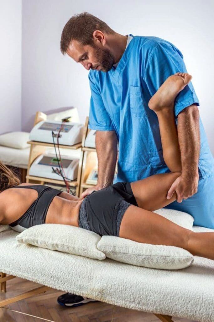
No injection or surgical tool can outrun poor biomechanics. My approach always begins with restoring movement capacity and load tolerance across the hip-pelvis-lumbar complex. This is where integrative chiropractic care shines alongside physical therapy.
What we aim to correct:
- Pelvic alignment and SI joint stability: misalignment or instability amplifies shear forces into the hip.
- Gluteal strength and timing: the gluteus medius/minimus stabilize frontal-plane load; deficits increase compressive stress on the joint and greater trochanter.
- Core integration: a well-coordinated deep core (diaphragm, pelvic floor, transversus abdominis, multifidus) reduces axial load on the hip and improves lumbopelvic rhythm.
- Hip mobility: safe restoration of internal rotation and extension enables proper gait mechanics and reduces anterior joint stress.
How integrative chiropractic care fits:
- High-velocity, low-amplitude (HVLA) adjustments for the SI joint and lumbar segments can restore segmental motion and reduce neurogenic guarding.
- Low-force mobilizations and instrument-assisted soft tissue techniques for the gluteal fascia, tensor fasciae latae, and iliopsoas reduce myofascial loading and pain.
- Neuromuscular re-education and movement retraining align joint mechanics with muscular effort—this is where chiropractic clinical reasoning complements PT exercise progression.
- Kinetic chain assessments identify upstream/downstream contributors (foot mechanics, thoracolumbar stiffness, asymmetrical gait cycles).
Why this works physiologically:
- Improved alignment and neuromuscular timing reduce aberrant shear, compressive hotspots, and inflammatory signaling within the joint capsule.
- Effective core-gluteal integration redistributes load across tissues adapted to force absorption, reducing stress on compromised cartilage.
- Restored motion reduces synovial stagnation, enhancing nutrient diffusion and clearance of inflammatory byproducts.
My clinical observations:
- Patients who commit to combined PT plus integrative chiropractic protocols progress more consistently, with fewer flares and better long-term function. In complex or athletic cases, this joint strategy is often the difference between symptom reduction and meaningful restoration of performance (Jimenez, n.d.-a; Jimenez, n.d.-b).
Corticosteroid Injections: Short-Term Relief, Diagnostic Utility
Corticosteroid injections into the hip joint are a longstanding tool for pain modulation and diagnostic clarity.
What the evidence shows:
- Randomized trials indicate significant pain relief at approximately 3 months compared with saline or placebo, but benefits often diminish by 6 months.
- Major societies, including the American Academy of Orthopaedic Surgeons, offer moderate-strength recommendations for use focused on short-term pain reduction and for diagnostic purposes.
Physiological rationale:
- Corticosteroids suppress synovial inflammation by inhibiting phospholipase A2 and downstream eicosanoid pathways, thereby reducing prostaglandin and leukotriene production.
- Reduced synovitis decreases joint effusion and intra-articular pressure, relieving nociceptive signaling.
Clinical reasoning:
- I use a targeted diagnostic injection when the pain generator is unclear (hip vs. SI vs. lumbar vs. trochanteric tendinopathy). If pain transiently resolves after an intra-articular injection, it helps confirm that the symptoms originate from the hip joint.
- In patients needing rapid symptom control to engage in rehabilitation, a single injection can jumpstart movement restoration, but it must be paired with biomechanics-focused care. Without strengthening and mobility retraining, benefits fade and may not alter the disease trajectory.
Platelet-Rich Plasma (PRP): Longer-Lasting Relief and Biological Repair Signals
PRP has emerged as a biologic option for hip OA with growing support from randomized controlled trials. While protocols vary, several consistent findings guide clinical practice.
Key insights from pooled studies:
- Across multiple randomized controlled trials, PRP reduces pain at several time points, with low- to moderate-quality evidence supporting clinically meaningful improvements over 3–6 months.
- Single-injection protocols often perform as well or better than series protocols, possibly due to reduced post-injection flares and more precise dosing.
- Lower volumes are associated with better outcomes and fewer adverse events. In practice, volumes of 3–6 mL are well tolerated; large-volume injections (≥15 mL) increase discomfort without clear benefit.
Why PRP works physiologically:
- PRP concentrates platelets and growth factors (e.g., PDGF, TGF-β, VEGF, IGF-1), which can modulate inflammation, enhance matrix synthesis, and support chondral homeostasis.
- Leukocyte-poor PRP in joints may reduce levels of catabolic cytokines (IL-1β, TNF-α) more effectively than leukocyte-rich formulations, thereby decreasing synovial irritation.
- Rebalancing the joint environment helps calm nociceptive signaling and may slow degenerative cascades.
Comparative outcomes:
- Systematic reviews comparing corticosteroids, hyaluronic acid, and PRP frequently show PRP achieving the lowest pain scores at 6 months, while steroids offer earlier relief with shorter durability.
- The conclusion: use steroids for diagnostic and short-term flare control, and consider PRP for medium-term pain reduction and functional gains—always coupled with integrative rehabilitation strategies.
Protocol design:
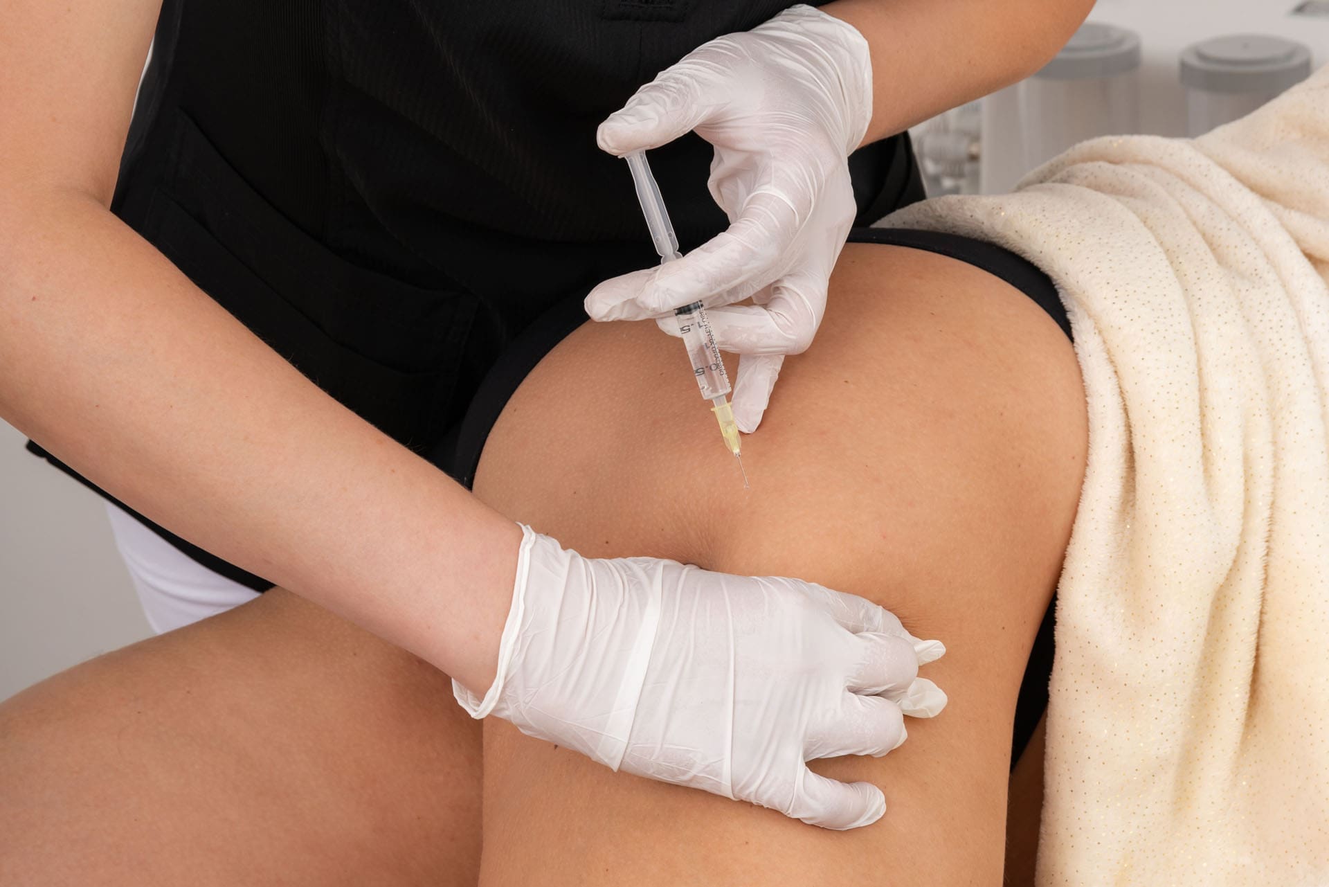
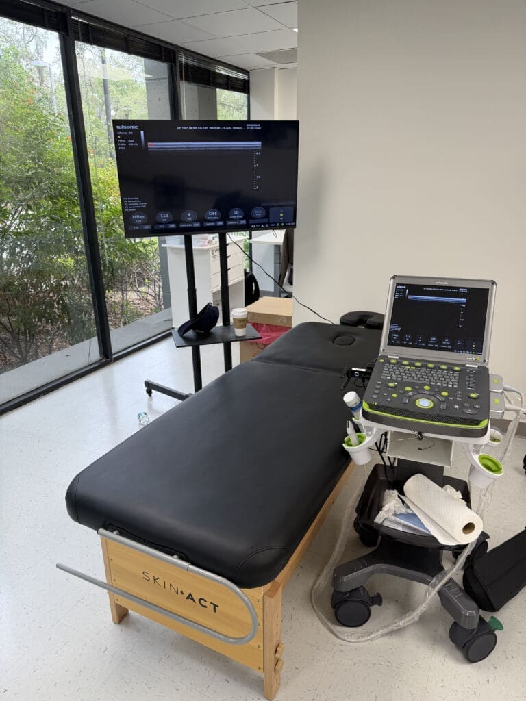
- I favor single, ultrasound-guided intra-articular injections of leukocyte-poor PRP in the 3–6 mL range.
- Post-procedure, I avoid NSAIDs for 5–7 days to preserve platelet signaling, focusing on a gentle range of motion, progressive strength work, and neuromotor retraining as pain allows.
- Based on patient response and goals, repeat injections can be considered at 4–6 months, though many patients do well with a single carefully designed protocol.
Case Study: When Hip OA Masquerades as Spine Pain
I want to share a case that exemplifies how careful diagnosis and integrative care change outcomes.
- A 22-year-old college linebacker transferred into our program with a 6-month history of “low back pain.” He had undergone multiple epidural steroid injections, medial branch blocks, and sciatic injections with no lasting benefit.
- On exam, his hip internal rotation was limited to about 15 degrees, and FABER reproduced deep anterior pain. Lumbar spine imaging showed an L5-S1 disc herniation—consistent with prior studies—but it did not explain his persistent pain patterns.
- Focused hip imaging revealed cortical irregularity near the femoral head-neck junction, suggestive of intra-articular pathology.
Our plan:
- Begin PT focused on core and gluteal strengthening with controlled restoration of hip mobility.
- Perform a diagnostic intra-articular injection—his pain resolved, confirming hip joint origin.
- Follow with a PRP injection during the off-season (about three and a half months later), coupled with progressive biomechanics work.
Outcome:
- He completed the next three years without lost time due to hip or lumbar complaints. The take-home point: accurate identification of the pain generator and integration of chiro-PT protocols enable biologics such as PRP to deliver meaningful, durable relief.
Building a Comprehensive Hip OA Treatment Plan: Step-by-Step
Here is how I structure care for hip OA patients, grounded in physiology and research:
- Assessment and Differential:
- Clarify pain location: anterior (intra-articular), lateral (gluteal/trochanteric), posterior (SI/lumbar—but keep hip in mind).
- Perform targeted tests: FADIR, FABER, log roll, gait analysis.
- Use imaging judiciously: weight-bearing X-rays, targeted MRI for labrum/osteophytes.
- Foundational Integrative Care:
- Begin PT plus chiropractic early:
- Pelvic/SI alignment strategies (HVLA when indicated, gentle mobilization).
- Gluteal and deep core strengthening to reduce shear and improve load sharing.
- Hip mobility restoration—internal rotation, extension—without provoking flares.
- Myofascial release for TFL, iliopsoas, piriformis, and adductors.
- Begin PT plus chiropractic early:
- Pain Modulation:
- Use corticosteroid injections for short-term control and diagnostic clarity.
- Consider PRP for medium-term relief and potential improvement in biological terrain.
- Progression:
- Layer neuromotor retraining (single-leg stance drills, step-down control, hip-hinge mechanics).
- Address kinetic chain issues: foot mechanics, thoracolumbar mobility, contralateral hip stability.
- Long-Term Health:
- Encourage consistent moderate physical activity to counteract the inactivity–mortality link.
- Support metabolic health with anti-inflammatory nutrition and sleep strategies; metabolic syndrome worsens OA outcomes.
Why this works:
- The combination of reduced inflammation (steroids or PRP) and restored biomechanics (PT and chiropractic) interrupts the pain–inactivity cycle.
- Over time, patients build tissue tolerance, restore joint motion, and regain confidence in movement—crucial for maintaining function and preventing relapse.
Biologics in Hip OA: Dosing, Volume, and Future Directions
Key open questions—and how we navigate them now:
- Optimal platelet dose: Current evidence suggests benefit from leukocyte-poor formulations; higher platelet counts might aid repair signaling, but must be balanced against synovial reactivity.
- Volume considerations: Clinical observations and pooled data indicate that lower volumes (3–6 mL) improve comfort and outcomes compared with higher volumes. Larger volumes can elevate intra-articular pressure and irritate the joint.
- Frequency: Several analyses favor single-injection strategies, possibly due to reduced cumulative synovial irritation.
- Concentrated plasma: Novel systems aim to enrich anti-inflammatory and anti-degenerative proteins while minimizing leukocytes—promising in theory, but we need standardized reporting and head-to-head trials to establish superiority.
From bench to bedside:
- Biologic therapies target the catabolic milieu of OA—reducing inflammatory cytokines, encouraging matrix maintenance, and supporting chondrocyte survival. The best outcomes occur when biologics are a bridge to better mechanics, not a stand-alone fix.
Practical Protocol: My Clinic Approach to PRP for Hip OA
Here is a simplified overview of how I implement PRP:
- Patient Selection:
- Symptomatic hip OA with functional limitations despite conservative care.
- No active infection, coagulopathy, or uncontrolled systemic inflammation.
- Preparation:
- Use a benchtop processing system to obtain leukocyte-poor PRP.
- Target 3–6 mL intra-articular injection volume.
- Procedure:
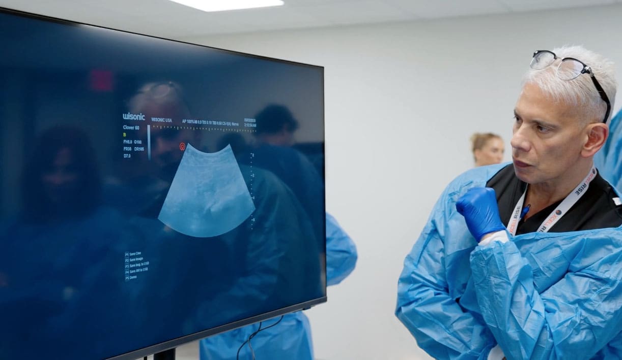
- Ultrasound-guided injection for precision and safety.
- Post-procedure, avoid NSAIDs for 5–7 days; consider acetaminophen for discomfort.
- Rehabilitation:
- Initiate gentle ROM within 24–72 hours, as tolerated.
- Progress gluteal/core strengthening, balance, and hip control drills.
- Incorporate chiropractic mobilization for SI/lumbar segments as needed to normalize load transfer.
- Follow-Up:
- Reassess function and pain at 6–8 weeks and 3–6 months.
- Consider repeat PRP at 4–6 months if pain/function plateaus and patient goals demand.
Physiological reasoning:
- This cadence respects platelet signaling timelines, reduces synovial irritation, and uses the post-injection window to re-pattern movement for lasting benefit.
Clinical Pearls and Red Flags
- If posterior hip pain persists after targeted SI/piriformis/hamstring care, suspect intra-articular hip pathology—retest with FADIR and guided imaging.
- Loss of internal rotation is a small but potent predictor of hip joint involvement.
- Avoid chasing pain with serial injections without improving mechanics—the hip demands a systems approach.
- Encourage consistent activity—the mortality data are a reminder that movement is medicine.
Conclusion: A Modern Framework for Hip OA That Puts Biomechanics First
Hip OA is more than cartilage loss—it is a dynamic interplay between inflammation, mechanics, and behavior. The latest research consistently shows:
- Corticosteroids provide short-term relief and diagnostic help.
- PRP offers more durable pain reduction at 6 months for many patients.
- The best outcomes come when we pair biologics with integrative chiropractic care and targeted physical therapy to restore motion, stability, and confidence.
In my practice, this combined approach delivers meaningful improvements in pain and function, supports systemic health through increased activity, and respects the hip’s complexity. When we treat the hip as both a joint and a node in a kinetic chain, we change lives—not just symptoms.
References
- American Academy of Orthopaedic Surgeons Clinical Practice Guidelines on Osteoarthritis of the Hip (American Academy of Orthopaedic Surgeons, n.d.)
- Global Burden of Disease Study Findings on Musculoskeletal Disorders (GBD Collaborators, 2019)
- PRP in Osteoarthritis: Mechanisms and Clinical Outcomes (Filardo, S., & Kon, E., 2020)
- Platelet-Rich Plasma vs Corticosteroid Injections for Hip OA: Systematic Review and Meta-Analysis (Dai, W., et al., 2021)
- Physical Activity, Osteoarthritis, and Mortality Risk (Booth, F. W., Roberts, C. K., & Laye, M. J., 2019)
- Hip Examination and Special Tests in Clinical Practice (American Academy of Family Physicians, 2017)
- Integrative Chiropractic and Multidisciplinary Care in Musculoskeletal Conditions (Goertz, C. M., et al., 2019)
- Chondroprotection and Biologic Modulation in OA (Mobasheri, A., et al., 2020)
- Dr. Alexander Jimenez Clinical Observations and Practice Insights (Jimenez, A., n.d.-a)
- Professional Profile and Publications of Dr. Alexander Jimenez (Jimenez, A., n.d.-b)