

Chronic Tendinopathy Management Techniques With Regenerative Orthopedics
Find out how regenerative orthopedics for chronic tendinopathy can transform your recovery journey and restore functional movement.
Abstract
Tendinopathy represents one of the most common and clinically challenging musculoskeletal conditions encountered in integrative and regenerative medicine practice. Whether it presents as lateral epicondylitis (tennis elbow), patellar tendinopathy, Achilles tendinopathy, or rotator cuff degeneration, the underlying pathology shares a consistent theme: a failed healing response within tendon tissue that transitions from an acute inflammatory state into a chronic degenerative process known as tendinosis. This educational post explores one of the most evidence-informed, minimally invasive interventional techniques available to clinicians today — needle fenestration — and its powerful combination with prolotherapy using dextrose to stimulate tendon regeneration from within.
Drawing from the clinical expertise of Dr. Fran O’Connor, a recognized authority in ultrasound-guided procedures, and integrated with the multidisciplinary perspective of Dr. Alexander Jimenez, DC, APRN, FNP-BC, CFMP, IFMCP, ATN, CCST, and Dr. Maria Guadalupe Cardenas, MD, Board Certified in Internal Medicine and Medical Director at Injury Medical Clinic PA in El Paso, Texas, this post provides a thorough, clinically grounded exploration of the following topics:
- The physiological basis of tendinopathy and why tendons fail to heal on their own
- The mechanism of action behind needle fenestration and how it converts a chronic degenerative lesion into an acute healing environment
- Step-by-step procedural technique for ultrasound-guided needle fenestration, including needle positioning, angle of approach, in-plane visualization, and coverage of the entire tendinopathic zone
- The scientific rationale for combining fenestration with prolotherapy (dextrose) and how this synergistic approach amplifies the regenerative response
- How orthobiologics such as platelet-rich plasma (PRP) can be incorporated alongside fenestration for more advanced cases
- The clinical outcomes reported in peer-reviewed literature, including the number of passes required, how clinicians assess procedural endpoints, and what patients can expect during recovery
- How integrative chiropractic care, functional medicine, and medical oversight from an Internal Medicine physician create a comprehensive, whole-patient approach to managing chronic tendinopathy
- The unique multidisciplinary model practiced at Injury Medical Clinic PA, where chiropractic expertise, advanced practice nursing, functional medicine, and internal medicine converge to deliver state-of-the-art, evidence-based musculoskeletal care
By the end of this post, readers — whether patients, clinicians, or health professionals — will have a deeply informed understanding of why needle fenestration with prolotherapy has earned its place as a first-line interventional option for chronic tendinopathy, and how it fits within a broader, patient-centered treatment philosophy.
Understanding Tendinopathy: Why Tendons Fail to Heal
The Structure and Function of Healthy Tendons
To appreciate why needle fenestration works, we must first understand the remarkable—and remarkably vulnerable—structure of the tendon itself. Tendons are dense, fibrous connective tissue structures that transmit the mechanical forces generated by muscle contraction to bone, enabling movement and stabilizing joints under load. They are composed primarily of type I collagen, organized in a highly hierarchical structure: individual tropocollagen molecules assemble into collagen fibrils, which bundle into collagen fibers, which in turn form fascicles surrounded by the endotenon, and these fascicles collectively form the tendon body, enclosed by the epitenon and, in some locations, a paratenon or tendon sheath (Maffulli et al., 2023).
The tenocytes — the primary cellular inhabitants of tendon tissue — reside within this extracellular matrix and are responsible for synthesizing, organizing, and maintaining collagen and other matrix proteins. Under normal physiological conditions, tendons are remarkably efficient load-bearing structures. They are designed to store and release elastic energy, acting almost like biological springs that reduce the metabolic cost of movement and protect muscle tissue from sudden overload (Magnusson et al., 2010).
However, this remarkable structural efficiency comes at a cost: tendons are relatively hypovascular tissues. The blood supply to tendon tissue is sparse compared to muscle, bone, or even ligament. This hypovascular nature means that when tendon tissue is damaged — whether by a single acute traumatic event or by the cumulative microtrauma of repetitive loading — the tissue has limited intrinsic capacity to mount an effective healing response (Fenwick et al., 2002). The relative absence of robust vascularity translates directly into reduced oxygen delivery, reduced nutrient availability, reduced cellular migration to the damaged site, and, critically, reduced access to the growth factors and cytokines that orchestrate tissue repair.
The Transition From Tendinitis to Tendinosis: A Critical Distinction
For decades, chronic tendon pain was labeled “tendinitis,” implying that inflammation was the dominant pathological process. This conceptual framework led clinicians to rely heavily on anti-inflammatory interventions: non-steroidal anti-inflammatory drugs (NSAIDs), corticosteroid injections, and rest. While these approaches may provide short-term symptomatic relief, they often fail to produce lasting recovery—and in some cases, particularly with repeated corticosteroid injections, they may impair long-term tendon healing (Coombes et al., 2013; Dean et al., 2014).
This failure became clearer as histopathological research revealed a striking truth: in most cases of chronic tendon pain, there is little or no true inflammatory infiltrate in the tendon tissue. Instead, what clinicians and researchers consistently find is a picture of tendinosis — a degenerative condition characterized by:
- Disorganized collagen architecture, with loss of the normal parallel alignment of collagen fibers and replacement by haphazardly arranged, structurally inferior collagen
- Increased ground substance (glycosaminoglycans and proteoglycans) between collagen fibers, creating a thickened, edematous appearance
- Neovascularization — the ingrowth of new, abnormal blood vessels into areas of tendon tissue that are normally avascular, often accompanied by nerve fibers that appear to contribute to pain signaling
- Hypocellularity in some regions, with loss of normal tenocyte morphology and the appearance of rounded, chondrocyte-like cells — a phenomenon sometimes described as chondroid metaplasia
- Micro-tears and focal areas of necrosis within the tendon matrix
- The near-complete absence of inflammatory cells such as neutrophils or macrophages that would be expected in true “tendinitis” (Khan & Cook, 2000; Maffulli et al., 1998)
This histopathological picture has been given many names in the literature — tendinosis, tendinopathy, degenerative tendinopathy — but the essential message is consistent: the tissue has entered a state of failed healing. The normal repair cascade — inflammation, proliferation, remodeling — has been initiated but has stalled or been disrupted, leaving behind a structurally compromised, biochemically abnormal tendon that is painful, weak, and at risk for partial or complete rupture (Cook & Purdam, 2009).
Why Tendons Get Stuck in This Degenerative State
Understanding why tendons fail to complete the normal healing cycle is essential for understanding why fenestration represents such a logical and elegant therapeutic intervention. Several interacting mechanisms contribute to the persistence of tendinosis:
1. Repetitive Mechanical Loading Without Adequate Recovery
The most common precipitating factor in tendinopathy is repetitive mechanical overload. When a tendon is loaded cyclically — as in running, throwing, typing, or racket sports — microscopic damage accumulates within the collagen matrix. If the rate of damage accumulation exceeds the rate of repair (which itself requires rest, adequate nutrition, and sufficient vascular supply), the tendon progressively degenerates. This is particularly common in tendons already operating at high mechanical loads, such as the patellar tendon in jumping athletes, the Achilles tendon in runners, and the extensor carpi radialis brevis (ECRB) tendon at the lateral epicondyle in racket sport players and manual workers (Magnusson et al., 2010; Rees et al., 2014).
2. The Hypovascular Environment
As noted above, the tendon’s inherently poor blood supply limits delivery of reparative cells (fibroblasts, macrophages), growth factors (TGF-β, PDGF, IGF-1, FGF, VEGF), and nutrients to damaged areas. Paradoxically, the neovascularization that occurs in tendinosis — while initially appearing to represent a reparative response — tends to produce blood vessels that are structurally abnormal and that infiltrate the tendon accompanied by sympathetic and sensory nerve fibers, contributing to the pain experience without adequately restoring normal tissue perfusion and repair capacity (Ohberg et al., 2004).
3. Altered Tenocyte Biology
The tenocytes within a tendinopathic tendon undergo profound changes in their gene expression and synthetic activity. Instead of producing the organized type I collagen characteristic of healthy tendon, they upregulate type III collagen—a weaker, less organized collagen isoform associated with scar formation—as well as abnormal matrix metalloproteinases (MMPs) that degrade the existing matrix without adequate replacement (Riley et al., 2002). The result is a progressive weakening of the tendon’s structural integrity, even in the absence of complete rupture.
4. Neurochemical Sensitization
Chronic tendinopathy is associated with central and peripheral sensitization of the pain system. The neovascular ingrowth mentioned earlier brings with it substance P-positive and CGRP-positive nerve fibers that generate persistent nociceptive signaling. Over time, this leads to central sensitization—a state in which the nervous system becomes hyperresponsive to pain signals, amplifying perceived pain intensity and expanding the area of perceived pain beyond the original tissue lesion (Andersson et al., 2011). This neurological dimension of chronic tendinopathy is one reason purely mechanical or pharmacological interventions often fail: the pain system itself has been remodeled, and effective treatment must address not only the tendon tissue but also the neural and central contributions to pain.
What Is Needle Fenestration? Defining the Technique and Its Purpose
Defining Needle Fenestration in Clinical Practice
Needle fenestration is a minimally invasive, ultrasound-guided procedure in which a needle — typically a 22-gauge or 25-gauge needle — is used to create multiple small punctate incisions or perforations (fenestrations) within a tendon, specifically targeting the area of tendinosis or tendinopathy. The term “fenestration” derives from the Latin fenestra, meaning “window” — and this etymology is clinically apt, because the procedure literally creates small “windows” or openings in the abnormal tendon tissue.
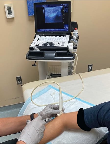
It is critically important to distinguish needle fenestration from other needle-based tendon interventions. Needle fenestration is defined by the deliberate absence of an injectable therapeutic agent — the needle alone, passed repeatedly through the diseased tissue, is the therapeutic instrument. This distinguishes it from:
- Corticosteroid injection (anti-inflammatory)
- Hyaluronic acid injection (viscosupplementation)
- Prolotherapy (injection of an irritant/proliferant solution such as dextrose)
- Platelet-rich plasma (PRP) injection (orthobiologic)
- Dry needling (which targets myofascial trigger points in muscle tissue rather than tendinopathic lesions within the tendon itself)
However — and this is a clinically important nuance that will be explored in detail — needle fenestration is very frequently combined with injectable agents, particularly prolotherapy using hypertonic dextrose, because the two approaches have synergistic mechanisms of action. The fenestration disrupts and mechanically stimulates the tissue, while the injected agent provides additional biochemical stimulation of the healing cascade. This combined approach, as demonstrated by Dr. O’Connor and practiced within the multidisciplinary framework at Injury Medical Clinic PA, is a particularly powerful, evidence-informed treatment strategy for chronic tendinopathy.
The Conceptual Foundation: Converting Chronic Degeneration Into Acute Healing
The central therapeutic logic of needle fenestration is both elegant and physiologically grounded: to convert a chronic, stalled degenerative process into an acute healing response. This concept deserves thorough elaboration, because it represents a fundamental paradigm shift in how we think about treating tendinopathy.
In normal tissue healing following injury, there are three well-defined phases:
Phase 1 — Inflammation (Days 1–7):
Damaged tissue releases damage-associated molecular patterns (DAMPs), triggering local vasodilation, increased vascular permeability, and infiltration of inflammatory cells—primarily neutrophils (early) and macrophages (later). These cells remove cellular debris, release pro-inflammatory cytokines (IL-1β, TNF-α, IL-6), and — critically — release a rich repertoire of growth factors including platelet-derived growth factor (PDGF), transforming growth factor-beta (TGF-β), vascular endothelial growth factor (VEGF), fibroblast growth factor (FGF), and insulin-like growth factor-1 (IGF-1). These growth factors serve as the molecular signals that recruit and activate the reparative cells responsible for tissue regeneration.
Phase 2 — Proliferation (Days 4–21):
Activated fibroblasts (and in tendons, tenocytes) migrate to the wound site and begin synthesizing new collagen — initially type III collagen for rapid structural scaffolding, later transitioning to the mechanically superior type I collagen. New blood vessels form (angiogenesis) to supply the metabolically active repair tissue, and the extracellular matrix is progressively remodeled.
Phase 3 — Remodeling (Weeks to Months):
The immature repair tissue is progressively reorganized and strengthened. Type III collagen is replaced by type I collagen, collagen fibers align along lines of mechanical stress, water content decreases, and the tendon’s tensile strength gradually approaches that of normal tissue. This phase requires progressive mechanical loading to guide collagen fiber alignment through mechanotransduction pathways properly.
In tendinosis, this healing cascade has been initiated — the histological evidence of collagen disruption and tenocyte activation confirms that — but it has failed to progress to completion. The tendon is neither acutely inflamed nor fully healed; it is stuck in a chronic, degenerative limbo. As discussed above, this failure may be due to insufficient vascularity, ongoing mechanical overload, altered tenocyte biology, and potential neurochemical sensitization.
Needle fenestration addresses this stalled healing by forcibly reinitiating the healing cascade from the beginning. By passing the needle repeatedly through the tendinopathic tissue, the clinician:
- Physically disrupts the abnormal collagen matrix — breaking down the disorganized, inferior collagen and creating a fresh wound environment
- Causes local bleeding within the tendon — delivering a concentrated bolus of growth factors from platelet degranulation directly to the site of degeneration
- Triggers an acute inflammatory response — reversing the anti-inflammatory, stagnant biochemistry of tendinosis and replacing it with a dynamic, pro-reparative milieu
- Disrupts the pathological neovascular ingrowth — the abnormal blood vessel-nerve fiber complexes associated with tendinopathy pain are disrupted by the needle passes, potentially contributing to pain relief through denervation as well as regeneration
- Stimulates tenocyte mechanobiology — the mechanical stimulus of needle penetration activates tenocyte mechanoreceptors and signaling pathways (integrin signaling, FAK-ERK cascades) that upregulate the synthesis of organized, type I collagen
The result is that the tendon, previously stuck in a degenerative state, now experiences a genuine acute injury—but one that is precisely targeted, minimally destructive, and strategically designed to initiate the healing cascade that had previously failed.
The Role of Ultrasound Guidance in Needle Fenestration: Why Imaging Is Non-Negotiable
Real-Time Visualization as a Safety and Efficacy Imperative
One of the most important aspects of needle fenestration, as practiced by Dr. O’Connor and integrated into the clinical protocols at Injury Medical Clinic PA, is using diagnostic musculoskeletal ultrasound (MSKUS) to guide the procedure in real time. This is not merely a technical preference—it is a clinical imperative from both safety and efficacy standpoints.
Musculoskeletal ultrasound provides real-time, dynamic, high-resolution imaging of soft tissue structures—tendons, ligaments, muscles, bursae, nerves, and blood vessels—without the radiation exposure of fluoroscopy or the logistical complexity of MRI. Modern high-frequency linear ultrasound transducers (typically operating at 10–18 MHz for superficial structures) can resolve tendon architecture at sub-millimeter resolution, allowing the clinician to:
- Identify the precise location, size, and extent of the tendinopathic lesion within the tendon — the hypoechoic (dark on ultrasound), disorganized area that represents tendinosis or tendinopathy.
- Visualize the needle in real time as it enters the tendon, ensuring accurate placement within the lesion rather than in surrounding healthy tissue, bursae, or adjacent neurovascular structures.
- Monitor the distribution of any injected agent (prolotherapy solution, PRP) to confirm that it is being delivered to the target tissue.
- Use color or power Doppler imaging to identify the pathological neovascularization characteristic of tendinopathy. These Doppler-positive regions are particularly important targets for fenestration, as they represent the areas most actively involved in abnormal tendon biology and pain generation.
- Assess tissue response during the procedure — as Dr. O’Connor describes, experienced clinicians can perceive a change in the tissue’s resistance to needle advancement as tendinopathy softens during fenestration, and ultrasound confirms the needle’s position throughout this process.
In-Plane Versus Out-of-Plane Needle Visualization
In-plane (long-axis) needle visualization is the strongly preferred approach for tendon fenestration. In the in-plane technique, the needle is inserted parallel to the long axis of the ultrasound transducer, keeping the entire shaft and tip continuously visible on the ultrasound screen throughout the procedure. This is in contrast to the out-of-plane (short-axis) approach, where only the needle tip (appearing as a bright dot) is visible at a single cross-sectional plane.
The superiority of in-plane visualization for tendon fenestration is multifactorial:
- Complete needle shaft visibility allows the clinician to confirm at all times that the needle is within the tendon and not in surrounding structures
- Safe needle redirection — repeated withdrawal and redirection of the needle (fundamental to the fenestration technique) requires continuous visualization of the needle tip to ensure it remains within the target zone and does not inadvertently advance into adjacent neurovascular bundles.
- Verification of tendon coverage — by observing the needle’s position along the long axis of the tendon, the clinician can confirm that the entire length of the tendinopathic lesion is being treated.
- Depth control — the in-plane approach provides direct visualization of how deeply the needle has penetrated the tendon, preventing inadvertent penetration through the deep surface of the tendon into underlying structures (such as the radial nerve at the lateral elbow, or the posterior tibial nerve at the medial ankle)
The Sonographic Appearance of Tendinopathy
On ultrasound, healthy tendon tissue appears as a highly echogenic (bright), fibrillar structure with a characteristic parallel linear echo pattern that reflects the organized collagen architecture. When the ultrasound beam strikes these regularly aligned collagen fibers at the correct angle, they produce strong specular reflections that appear as bright, parallel lines — a pattern often described as having a “fibrillar” or “feather-like” appearance.
Tendinopathy disrupts this pattern in characteristic ways:
- Hypoechogenicity — areas of tendinopathy appear darker than the surrounding normal tendon, reflecting the loss of organized collagen architecture and the increased water content of the abnormal matrix. The hypoechoic region is the primary target for fenestration.
- Tendon thickening — tendinopathic tendons are typically larger in cross-sectional area than contralateral normal tendons, reflecting the accumulation of abnormal matrix material and the reactive cellular response
- Loss of fibrillar echotexture — within the tendinopathic zone, the normal parallel linear echo pattern is replaced by disorganized, heterogeneous echogenicity.
- Intratendinous calcification — in some cases, areas of calcium hydroxyapatite deposition can be seen as hyperechoic (bright) foci with posterior acoustic shadowing
- Neovascularization on Doppler imaging — color or power Doppler interrogation of the tendinopathic zone reveals increased internal blood flow within the tendon, representing the pathological neovascular ingrowth. This “Doppler signal” within the tendon is a sensitive and specific marker of active tendinopathy and correlates with pain severity in many studies (Ohberg et al., 2004)
During the fenestration procedure, the needle appears as a bright (hyperechoic) linear structure on ultrasound — the “reverberation artifact” that Dr. O’Connor describes, caused by the repeated reflection of ultrasound waves between the flat surfaces of the needle. This reverberation artifact makes the needle highly visible and allows precise real-time guidance throughout the procedure.
Step-by-Step Procedural Technique for Ultrasound-Guided Needle Fenestration
Patient Preparation and Positioning
Before the fenestration procedure begins, appropriate patient preparation is essential. This includes:
Informed Consent:
The patient must be thoroughly educated about the nature of the procedure, the expected immediate post-procedure response (soreness and temporary worsening of symptoms, typically lasting 24–72 hours as the intentional acute inflammatory response is established), the expected timeline of improvement, potential complications (infection, tendon weakening, inadvertent injury to adjacent structures), and alternative treatment options. At Injury Medical Clinic PA, this counseling occurs within the context of the patient’s comprehensive treatment plan, developed collaboratively between Dr. Jimenez and Dr. Cardenas.
Patient Positioning:
The patient is positioned to provide both patient comfort and optimal ultrasound access to the target tendon. For lateral epicondylitis (tennis elbow) fenestration, as demonstrated by Dr. O’Connor, the patient is typically seated with the arm resting on a padded armrest, elbow slightly flexed, and the lateral elbow exposed. For other tendons:
- Patellar tendon: supine, knee slightly flexed over a bolster
- Achilles tendon: prone, ankle in slight dorsiflexion
- Rotator cuff tendons: seated, with specific positions varying by tendon (supraspinatus — arm in the “Crass” position; infraspinatus — arm across the chest)
Skin Preparation:
The skin over the target area is thoroughly cleaned with antiseptic solution (chlorhexidine or povidone-iodine) to minimize infection risk. A sterile field is maintained throughout the procedure, with sterile covers applied to the ultrasound transducer.
Local Anesthesia: The Foundation of Patient Comfort
As Dr. O’Connor describes, local anesthesia is administered before the fenestration procedure. In her demonstrated technique, she uses a 27-gauge needle to infiltrate the skin and subcutaneous tissues overlying the target tendon with 1% lidocaine. Several important principles guide this anesthesia step:
Subcutaneous and Peritendinous Anesthesia:
Lidocaine is infiltrated into the skin, subcutaneous tissues, and peritendinous soft tissues, but ideally not directly into the tendon itself. This is because intratendinous lidocaine is potentially cytotoxic to tenocytes in laboratory studies — an effect that could theoretically impair the healing response that fenestration is designed to initiate. By confining the anesthesia to peritendinous tissues, the clinician numbs the pathway the larger fenestration needle will travel, reducing patient discomfort while preserving the cellular biology within the tendon.
Vapor Coolant Spray:
Dr. O’Connor describes using a vapor coolant spray (such as ethyl chloride or fluoroethane) applied to the skin surface immediately before needle insertion. This produces a brief, intense local cooling effect that temporarily reduces skin sensitivity and further minimizes the patient’s perception of needle insertion — a simple but effective technique for patient comfort.
The Rationale for Adequate Anesthesia:
Adequate anesthesia is not merely a patient comfort measure — it is also a procedural quality determinant. A patient who is in significant pain during the procedure will be tense and may involuntarily move, making accurate needle placement under ultrasound guidance more difficult and potentially less safe. Good local anesthesia allows the clinician to perform a thorough, methodical fenestration without rushing, producing better procedural outcomes.
Needle Selection: 22-Gauge vs. 25-Gauge
The choice of needle gauge for fenestration involves a balance between several competing considerations:
Larger needles (22-gauge):
- Create larger punctate defects with each pass, potentially stimulating a more robust healing response
- Provide greater tactile feedback as the needle traverses different tissue densities (soft tendinopathic tissue vs. firm normal tendon)
- Are slightly more visible on ultrasound
- May cause more post-procedural discomfort
Smaller needles (25-gauge):
- Cause less tissue trauma per pass, potentially reducing post-procedural pain
- May be preferred for smaller tendons or when combining with injection of viscous agents (PRP)
- Slightly less tactile feedback
In practice, the choice between 22-gauge and 25-gauge is often based on the size of the tendon, the extent of the tendinopathic lesion, the viscosity of any co-injected agent, and clinician preference. For larger tendons with extensive tendinopathy (Achilles, patellar), a 22-gauge needle may be preferred. For smaller tendons (ECRB at the lateral elbow), a 25-gauge needle often provides excellent results.
The Fenestration Sequence: Achieving Complete Coverage
Once the needle has been positioned in-plane with the ultrasound transducer, with the tip confirmed to be within the area of tendinopathy, the fenestration sequence proceeds as follows:
Step 1 — Initial Needle Placement:
Advance the needle in-plane, with continuous ultrasound visualization, until the tip is positioned within the most proximal extent of the tendinopathic zone. Optimize the insertion angle to keep the entire shaft within the imaging plane. For superficial tendons like the ECRB at the lateral elbow, this typically involves a relatively shallow approach angle (approximately 20–40 degrees from horizontal), allowing the needle to traverse a longer path within the tendon rather than dive steeply.
Step 2 — Penetration and Initial Fenestration:
With the needle tip positioned within the tendinopathic zone, the clinician begins fenestration by advancing the needle into the tissue, then partially withdrawing it (without removing it from the tendon or the skin), redirecting it slightly shallower or deeper within the tendon cross-section, and advancing again. This rhythmic advance-withdraw-redirect pattern creates the multiple punctate openings that characterize fenestration.
Step 3 — Systematic Coverage of the Tendinopathic Zone — Long Axis:
By repeatedly advancing, withdrawing, and redirecting the needle at incrementally different depths, the clinician ensures that the entire thickness of the tendinopathic zone — from its superficial to deep extent — is covered. The number of passes required to achieve this depends on the size of the tendinopathic lesion. Still, Dr. O’Connor describes the typical range in the literature as 15 to 50 passes, varying with the magnitude of the tendon abnormality.
Step 4 — Rotating the Transducer to the Short Axis:
After completing fenestration in the long-axis plane, the clinician rotates the ultrasound transducer 90 degrees to obtain a short-axis (cross-sectional) view of the tendon and the needle. This allows assessment of the needle’s position in the medial-lateral dimension — confirming whether the fenestration coverage needs to be extended medially or laterally within the tendon to treat the full width of the tendinopathic lesion. If the lesion extends beyond the initial needle path, reposition the needle medially or laterally as needed.
Step 5 — Procedural Endpoint — The “Soft Tendon” Sign:
One of the most clinically instructive aspects of Dr. O’Connor’s description of the fenestration technique is the “soft tendon” sign as a procedural endpoint. As the needle repeatedly passes through the tendinopathic tissue, many clinicians report a palpable change in the resistance encountered during needle advancement: the initially firm, resilient (or paradoxically, gritty) texture of the tendinopathic tissue progressively softens, becoming less resistant to needle advancement. This tactile change is believed to reflect disruption of the abnormal, disorganized collagen matrix within the tendinopathic zone—the pathological tissue is literally being broken down and remodeled by repeated needle passes. Many experienced practitioners continue fenestration until the entire target zone reaches this softer consistency, then consider the mechanical phase of the procedure complete.
Prolotherapy: The Science and Application of Dextrose as a Regenerative Agent
What Is Prolotherapy?
Prolotherapy — derived from “proliferative therapy” — is a regenerative injection technique in which a proliferant solution is injected into or around damaged connective tissue (tendons, ligaments, joint capsules, entheses) to stimulate the body’s natural healing response. George Hackett, MD, popularized the term in the 1950s, though injection-based therapies for connective tissue disorders date back to the 19th century.
The most widely used proliferant agent in modern prolotherapy practice is hypertonic dextrose (glucose) — the same molecule that circulates in human blood as blood sugar. Depending on the clinical context, dextrose concentrations used in prolotherapy range from approximately 10% to 50%. As Dr. O’Connor describes, she commonly uses 25% or 50% dextrose for tendon prolotherapy.
How Does Dextrose Prolotherapy Stimulate Tendon Healing?
The mechanisms by which hypertonic dextrose stimulates tendon healing are multiple, and understanding them provides important insight into why combining prolotherapy with needle fenestration creates such a powerful synergistic effect:
Mechanism 1 — Osmotic Cell Stress and Growth Factor Release:
When hypertonic dextrose is injected into tissue, the osmotic gradient created by the high-glucose solution causes local cellular osmotic stress. This stress triggers the release of growth factors — including PDGF, TGF-β, FGF, IGF-1, and connective tissue growth factor (CTGF) — from local cells, including tenocytes, fibroblasts, and platelets. These growth factors are molecular signals that drive collagen synthesis, cell proliferation, and matrix remodeling—the core processes of tendon healing (Topol et al., 2011; Reeves & Hassanein, 2000).
Mechanism 2 — Local Irritant Effect and Inflammatory Cascade Initiation:
The injection of hypertonic dextrose into damaged tendon tissue produces a mild, controlled inflammatory response. This local irritant effect recruits the cellular and molecular machinery of the healing cascade to the injection site. This is analogous to the mechanism of the needle fenestration itself, but adds a biochemical stimulus on top of the mechanical stimulus provided by the needle passes. Together, mechanical disruption (fenestration) and biochemical stimulation (dextrose prolotherapy) create a far more potent healing signal than either approach alone.
Mechanism 3 — Glucose Receptor-Mediated Cell Signaling:
Emerging evidence suggests that dextrose, as a signaling molecule in its own right, interacts with cellular glucose receptors (GLUT transporters) and activates downstream signaling pathways—including the PI3K-Akt pathway—that promote cell survival, proliferation, and matrix synthesis. This suggests that the healing-stimulatory effects of dextrose are not simply osmotic but also involve direct receptor-mediated cellular signaling (Rabago et al., 2009).
Mechanism 4 — Platelet Activation and Intrinsic PRP Effect:
When dextrose is injected and causes local bleeding (which fenestration also facilitates), platelets aggregate at the injection site and degranulate, releasing their alpha-granule contents—a concentrated soup of growth factors including PDGF, TGF-β1, VEGF, EGF, and PF4. This platelet-derived growth factor release is, in effect, a form of endogenous platelet-rich plasma — the same therapeutic principle exploited by exogenous PRP injections, but generated naturally at the treatment site. Fenestration enhances this effect by ensuring that bleeding occurs throughout the fenestrated zone.
Dextrose Concentration: Clinical Considerations
The concentration of dextrose used in prolotherapy has important clinical implications:
- 10–15% dextrose: This low concentration is predominantly used for intra-articular and periarticular prolotherapy, targeting joint capsules, ligaments, and entheses. At this concentration, the primary mechanism is receptor-mediated cellular signaling rather than osmotic cellular stress.
- 25% dextrose: This intermediate concentration, commonly used by Dr. O’Connor, provides a balance between osmotic stimulation and injectability (lower viscosity than 50% solutions), making it suitable for intratendinous applications.
- 50% dextrose: This high concentration provides the most potent osmotic stimulus and is typically used for larger tendons or more severe tendinopathy. It is often diluted at the syringe with local anesthetic (such as lidocaine) to achieve the target concentration and reduce patient discomfort.
Combining Fenestration With Prolotherapy: The Synergistic Rationale
The combination of needle fenestration with prolotherapy, as demonstrated by Dr. O’Connor in the lateral epicondylitis case, represents more than the sum of its parts. The synergy between these two techniques can be understood at multiple levels:
Structural Level:
Fenestration physically disrupts the pathological collagen matrix of tendinosis, creating channels and spaces within the tendon that facilitate the distribution of the subsequently injected prolotherapy solution throughout the entire tendinopathic zone. Without prior fenestration, the dense, disorganized collagen matrix of tendinosis may resist fluid distribution, limiting contact between the dextrose solution and target cells.
Cellular Level:
Fenestration delivers an acute mechanical stimulus to tenocytes, activating mechanotransduction pathways that prime these cells for a proliferative and synthetic response. Dextrose then delivers a biochemical stimulus that further activates these cells, producing amplified and sustained growth factor release compared with either stimulus alone.
Vascular Level:
Fenestration causes local intratendinous bleeding, delivering platelets and their growth factor cargo directly to the tendinopathic zone. Dextrose injection then provides an additional osmotic and biochemical stimulus to these same platelets, enhancing their degranulation and growth factor release.
Neural Level:
Fenestration may disrupt pathological neovascular nerve ingrowth, reducing the peripheral sensitization that drives chronic tendinopathy pain. In contrast, the anti-nociceptive effects of the subsequent dextrose injection (mediated in part through adenosine receptor activation at local pain fibers) may further reduce pain signaling in the treated area.
Orthobiologics in Tendon Regeneration: Where PRP Fits in the Treatment Algorithm
Platelet-Rich Plasma: Principles and Preparation
Platelet-rich plasma (PRP) represents the most widely studied and clinically established orthobiologic used in tendon regeneration. PRP is prepared from the patient’s own blood through a centrifugation process that concentrates platelets — and therefore the growth factors contained within their alpha granules — to levels significantly above those found in whole blood. The platelet concentration in PRP is typically 3 to 8 times that of normal whole blood, depending on the preparation system used (Marx, 2004).
When PRP is injected into or around a tendinopathic tendon, the concentrated platelets encounter the local tissue environment and degranulate, releasing their growth factor cargo directly into the treatment site. The growth factors most relevant to tendon healing include:
- PDGF (Platelet-Derived Growth Factor): Stimulates fibroblast/tenocyte proliferation and migration, promotes angiogenesis
- TGF-β1 (Transforming Growth Factor-Beta 1): One of the most potent stimulators of collagen synthesis; also has anti-inflammatory effects in the later phases of healing
- VEGF (Vascular Endothelial Growth Factor): Promotes angiogenesis, supporting vascular supply to the healing tendon
- IGF-1 (Insulin-Like Growth Factor 1): Promotes tenocyte proliferation, collagen synthesis, and cell survival
- EGF (Epidermal Growth Factor): Promotes cell proliferation and differentiation
- FGF (Fibroblast Growth Factor): Stimulates fibroblast/tenocyte proliferation and neovascularization
In addition to platelet-derived growth factors, PRP also contains white blood cells (in “leukocyte-rich” PRP preparations) that contribute additional cytokines and antimicrobial factors, and plasma proteins including fibrinogen and fibronectin that serve as scaffolding for cell migration.
PRP vs. Prolotherapy vs. Fenestration Alone: Positioning in the Treatment Algorithm
The question of when to use PRP versus prolotherapy versus fenestration alone is one that practicing clinicians frequently encounter, and the answer involves consideration of multiple clinical factors:
Tendinopathy Severity:
- Mild tendinopathy: Isolated needle fenestration (without injection) or fenestration combined with low-concentration dextrose prolotherapy may be sufficient to initiate adequate healing.
- Moderate tendinopathy: Fenestration combined with dextrose prolotherapy (25–50%) represents a well-established, cost-effective approach with a strong evidence base.
- Severe or recalcitrant tendinopathy: Cases that have failed multiple rounds of conservative management, or that involve large, extensively degenerated tendon segments, may benefit from adding exogenous PRP to the fenestration procedure, providing a more potent and sustained growth factor stimulus.
Patient Factors:
- Age: Older patients may have reduced intrinsic healing capacity and may benefit more from the exogenous growth factor stimulus provided by PRP.
- Metabolic health: Patients with diabetes mellitus, which impairs multiple aspects of tissue healing, may benefit from PRP’s ability to bypass some of the growth factor deficiencies associated with diabetic tissue biology. This is particularly relevant at Injury Medical Clinic PA, where Dr. Cardenas’s expertise in Internal Medicine includes managing metabolic conditions that directly impact healing.
- Prior treatment history: Patients who have undergone multiple corticosteroid injections may have impaired tenocyte biology and may require the more potent regenerative stimulus of PRP.
Practical and Cost Considerations:
PRP preparation requires specialized centrifugation equipment and adds both cost and preparation time to the procedure. Dextrose prolotherapy is significantly less expensive and, when combined with fenestration, has demonstrated clinical outcomes comparable to PRP in several well-designed studies (Coombes et al., 2013; Krogh et al., 2016). At Injury Medical Clinic PA, the selection between prolotherapy and PRP is individualized based on the factors above, within the collaborative decision-making framework shared by Dr. Jimenez and Dr. Cardenas.
Understanding Plantar Fasciitis- Video
The Evidence Base for Needle Fenestration and Prolotherapy in Tendinopathy
Lateral Epicondylitis (Tennis Elbow): The Most Studied Model
Lateral epicondylitis — commonly known as tennis elbow — is the tendinopathy most extensively studied in the context of needle fenestration and prolotherapy, and for good reason. It is among the most prevalent upper extremity conditions in clinical practice, affecting approximately 1–3% of the general population and disproportionately impacting working-age adults in manual, clerical, and sports-related occupations (Walker-Bone et al., 2004). The primary pathological structure is the extensor carpi radialis brevis (ECRB) tendon at its origin on the lateral epicondyle of the humerus.
The ECRB tendon is a relatively small, superficial structure — anatomical characteristics that make it particularly amenable to ultrasound-guided needle fenestration. The tendinopathic zone can be clearly visualized on ultrasound, the needle can be precisely targeted, and the procedure can be performed with a high degree of accuracy and safety.
Key Clinical Studies on Needle Fenestration for Lateral Epicondylitis:
Stenhouse et al. (2013) conducted a randomized controlled trial comparing needle fenestration alone, PRP injection, and whole blood injection for chronic lateral epicondylitis. All three groups demonstrated significant improvement in pain and function over 6 months, with no statistically significant differences between groups. This finding supports the conclusion that the mechanical effect of the needle (fenestration) is itself a primary therapeutic driver, with injectable agents providing additive but not necessarily superior benefit (Stenhouse et al., 2013).
Dong et al. (2016) performed a systematic review and meta-analysis of needle fenestration and injection-based treatments for lateral epicondylitis, concluding that needle fenestration combined with biological agents (PRP or whole blood) produced superior outcomes to fenestration alone for pain reduction, while fenestration alone outperformed corticosteroid injection at medium-term (3–12 month) follow-up — consistent with the well-established finding that corticosteroid injection, while effective in the short term, is associated with worse long-term outcomes than regenerative approaches.
Mishra & Pavelko (2006) published one of the foundational studies on PRP for lateral epicondylitis, demonstrating significant improvements in pain scores in patients treated with PRP injection compared to controls at 8 weeks and 6 months, with a statistically significant benefit for PRP. Importantly, the injection in this study was combined with needle fenestration of the tendon before PRP delivery—a protocol essentially identical to the combined approach described by Dr. O’Connor.
Dextrose Prolotherapy for Tendinopathy: Randomized Controlled Trial Evidence
The evidence base for dextrose prolotherapy in tendinopathy has grown substantially over the past two decades, with well-designed randomized controlled trials demonstrating clinically meaningful and statistically significant benefits across multiple tendon sites:
Rabago et al. (2013) conducted a high-quality randomized controlled trial of dextrose prolotherapy versus saline injection and versus eccentric loading exercise for Achilles tendinopathy. The dextrose prolotherapy group showed significantly greater improvements in pain and function at 12-month follow-up than both control groups—a finding with important clinical implications for managing this often recalcitrant condition.
Yelland et al. (2011) performed a randomized controlled trial of prolotherapy for plantar fasciitis (a condition closely related to Achilles tendinopathy, sharing the common theme of enthesopathy at a high-load tendon/fascia attachment site). Prolotherapy produced significantly better outcomes than saline injection and was comparable to the best available conservative therapies.
Reeves & Hassanein (2000) published a landmark double-blind randomized controlled trial of dextrose prolotherapy for knee osteoarthritis — demonstrating significant improvements in pain, range of motion, and cartilage quality on MRI — which, while not directly addressing tendinopathy, established the foundational biochemical rationale (growth factor stimulation by hypertonic dextrose) that underlies tendon prolotherapy as well.
The Number of Needle Passes: What the Evidence Says
One of the most practically important questions in needle fenestration technique concerns how many needle passes are required for an effective procedure. Dr. O’Connor notes that the literature reports a range of 15 to 50 passes, depending on the size of the tendinopathic lesion.
This variability in reported pass numbers reflects both the heterogeneity of tendinopathic lesion sizes across different tendons and patients and the lack of a universally agreed-upon, evidence-defined “optimal” pass number. Several principles guide clinical decision-making:
- Smaller tendinopathic lesions (such as the ECRB in mild-to-moderate lateral epicondylitis) may require only 15–20 passes to achieve complete coverage and the “soft tendon” endpoint.
- Larger tendinopathic lesions (such as extensive Achilles or patellar tendinopathy) may require 30–50 or more passes to achieve complete coverage.
- The “soft tendon” endpoint — the tactile perception that the fenestrated tissue has softened during needle advancement — is arguably a more clinically meaningful guide to procedural completeness than any arbitrary pass number.
- Some clinicians advocate performing serial fenestration sessions (multiple procedures spaced weeks apart) rather than a single high-pass-count session, reasoning that the healing response benefits from multiple cycles of stimulation.
Lateral Epicondylitis: A Deep Dive Into the Target Pathology
Anatomy of the Lateral Elbow and the ECRB Tendon
To fully appreciate the clinical elegance of needle fenestration for lateral epicondylitis, a detailed understanding of the relevant anatomy is essential. The lateral epicondyle of the humerus serves as the common origin for the extensor muscles of the forearm and wrist. The muscles originating here form a conjoined tendon (the common extensor tendon) that attaches to the lateral epicondyle and its adjacent supracondylar ridge.
The extensor carpi radialis brevis (ECRB) is the tendon most consistently implicated in lateral epicondylitis pathology. Its origin lies deep to the extensor carpi radialis longus at the anterolateral aspect of the lateral epicondyle, in proximity to the lateral collateral ligament complex. The ECRB is subjected to particularly high tensile loads during activities requiring simultaneous wrist extension and forearm pronation — exactly the movement pattern involved in the backhand stroke of tennis (hence the colloquial name “tennis elbow”) and in many occupational tasks such as keyboard use, manual assembly work, and tool use.
At its origin, the ECRB tendon is relatively avascular, positioned in an anatomical watershed zone where direct blood supply is limited. This anatomical characteristic helps explain why this tendon is so prone to tendinosis: the combination of high cyclic mechanical loading and poor intrinsic vascularity creates the ideal environment for the failed healing response characteristic of tendinopathy.
The Unique Ultrasound Appearance of ECRB Tendinopathy
On ultrasound, ECRB tendinopathy characteristically appears as:
- A hypoechoic region within the deep aspect of the common extensor tendon at or just distal to the lateral epicondyle attachment — exactly the location described by Dr. O’Connor in her demonstration
- Tendon thickening at the lateral epicondyle origin, often measurable as an increase in anteroposterior diameter compared to the contralateral side
- Loss of normal fibrillar echotexture within the hypoechoic zone
- Neovascularization on Doppler imaging within and around the tendinopathic zone — a finding with prognostic significance (higher Doppler signal correlates with more active tendinopathy)
- Occasionally, small partial tears appear as anechoic (fluid-filled) clefts within the tendon substance, or intratendinous calcifications appear as hyperechoic foci
This is precisely the hypoechoic region that the clinician targets during ultrasound-guided fenestration, as described by Dr. O’Connor: “you can see up on the ultrasound a needle coming in into this hypoechoic area of tendinopathy.”
Why Conservative Treatments Alone Often Fail for Lateral Epicondylitis
Despite the availability of numerous conservative treatments for lateral epicondylitis — including physical therapy, eccentric exercise, extracorporeal shockwave therapy, bracing, NSAIDs, and corticosteroid injection — a significant proportion of patients (estimated at 10–20% in most epidemiological studies) progress to a chronic, refractory state that persists for more than 6–12 months and fails to respond adequately to these measures. Understanding why helps explain the rationale for interventional approaches like fenestration.
The limitation of corticosteroid injection is particularly instructive. Multiple high-quality randomized controlled trials have confirmed that while corticosteroid injection provides excellent short-term (6–12 week) pain relief for lateral epicondylitis, it is associated with worse long-term outcomes than both conservative management and regenerative injection approaches (Coombes et al., 2013). The likely mechanism for this long-term harm is the well-documented catabolic effect of corticosteroids on connective tissue: corticosteroids inhibit collagen synthesis, suppress tenocyte proliferation, and — at the molecular level — downregulate the expression of key structural genes including type I procollagen, fibronectin, and tenascin-C, all of which are essential for tendon matrix integrity and repair (Dean et al., 2014). Repeated corticosteroid injections compound this damage, progressively weakening the tendon and increasing the risk of partial or complete rupture.
This is the clinical context that makes needle fenestration with prolotherapy such an important addition to the clinician’s toolkit: for patients who have failed conservative management — including physical therapy, bracing, and activity modification — and who are seeking an alternative to corticosteroid injection or surgery, fenestration with prolotherapy offers a regenerative rather than catabolic approach, addressing the underlying pathology rather than suppressing its symptoms.
The Multidisciplinary Framework at Injury Medical Clinic PA: Integrating Chiropractic, Internal Medicine, and Regenerative Therapies
The Clinical Partnership of Dr. Alexander Jimenez and Dr. Maria Guadalupe Cardenas
At Injury Medical Clinic PA (also known as Mission Plaza Injury Medical Clinic) in El Paso, Texas, the clinical management of tendinopathy, musculoskeletal injuries, and related conditions is delivered through a uniquely comprehensive multidisciplinary model that integrates the complementary expertise of two highly qualified clinicians: Dr. Alexander Jimenez, DC, APRN, FNP-BC, CFMP, IFMCP, ATN, CCST and Dr. Maria Guadalupe Cardenas, MD (NPI #1164426749, Texas MD License #J2933).
Dr. Maria Guadalupe Cardenas is Board Certified in Internal Medicine and brings over 40 years of experience as an internist to her role as Medical Director and Collaborative Physician at the practice. Her extensive background in internal medicine provides the clinical foundation for:
- Comprehensive medical evaluation of patients presenting with musculoskeletal complaints, ensuring that systemic conditions contributing to or complicating tendinopathy (such as diabetes mellitus, inflammatory arthritis, hypothyroidism, metabolic syndrome, and medication side effects) are identified and addressed
- Medical co-management of complex patients undergoing interventional procedures, including assessment of bleeding risk, infection risk, and contraindications to specific agents (such as dextrose in poorly controlled diabetic patients)
- Pharmacological management of pain, inflammation, and comorbid conditions within the scope of internal medicine
- Collaborative physician oversight that satisfies both the clinical and regulatory requirements for a multidisciplinary integrative practice in Texas
- Coordination with specialist referrals for patients requiring orthopedic surgery, rheumatology, neurology, or other specialty consultation
The relationship between Dr. Jimenez and Dr. Cardenas exemplifies the multidisciplinary setup increasingly recognized as the gold standard in integrative and injury care: an MD providing medical direction and oversight alongside a chiropractor and advanced practice provider, ensuring that patients receive care that is both medically safe and holistically comprehensive.
Dr. Alexander Jimenez: A Uniquely Qualified Integrative Clinician
Dr. Alexander Jimenez’s clinical qualifications span multiple disciplines, positioning him to deliver the comprehensive, evidence-based care complex musculoskeletal patients require. His credentials — DC (Doctor of Chiropractic), APRN (Advanced Practice Registered Nurse), FNP-BC (Family Nurse Practitioner, Board Certified), CFMP (Certified Functional Medicine Practitioner), IFMCP (Institute for Functional Medicine Certified Practitioner), ATN (Advanced Trained Nurse), CCST (Certified Chiropractic Sports Therapist) — represent an extraordinary breadth of clinical training that directly informs his approach to tendinopathy and musculoskeletal care.
This unique combination of qualifications allows Dr. Jimenez to function as both a structural/mechanical clinician (addressing biomechanical dysfunctions through chiropractic care) and a functional medicine practitioner (addressing the systemic, metabolic, and nutritional factors that influence musculoskeletal healing), while also operating as an advanced practice provider capable of ordering and interpreting diagnostic studies (including musculoskeletal ultrasound), prescribing medications within his scope of practice, and performing or coordinating interventional procedures.
His clinical observations and evidence-based practice philosophy are extensively documented at ChiroMed.com and in his professional profile on LinkedIn, where he consistently emphasizes integrating the latest research evidence with individualized patient care—a philosophy directly reflected in the approach to tendinopathy described in this post.
How Chiropractic Care Integrates With Needle Fenestration and Prolotherapy
The integration of chiropractic care with ultrasound-guided needle fenestration and prolotherapy is not coincidental — it reflects a deep understanding of the biomechanical, neurological, and physiological factors that contribute to tendinopathy and its persistence.
Addressing the Biomechanical Drivers of Tendinopathy:
Virtually all tendinopathies occur in the context of altered biomechanics — patterns of movement, loading, and joint alignment that place excessive or abnormal stress on specific tendon structures. For example:
- Lateral epicondylitis is commonly associated with cervicothoracic dysfunction, altered glenohumeral and scapular kinematics, and restricted pronation-supination mobility of the forearm that collectively increase stress at the ECRB origin
- Patellar tendinopathy frequently occurs in the context of hip abductor weakness, tibial torsion, or foot pronation abnormalities that increase valgus stress at the knee during loading activities
- Achilles tendinopathy is strongly associated with restricted ankle dorsiflexion range of motion, subtalar overpronation, and gastrocnemius-soleus tightness that combine to increase peak Achilles tendon strain
Chiropractic care — through spinal manipulation, joint mobilization, soft tissue techniques, and rehabilitation protocols — directly addresses these biomechanical contributors. Dr. Jimenez’s approach encompasses:
- Cervicothoracic and upper thoracic spinal manipulation for patients with lateral epicondylitis, addressing the proximal kinetic chain dysfunctions that increase distal tendon loading
- Glenohumeral and acromioclavicular joint mobilization for rotator cuff tendinopathy, restoring normal shoulder kinematics that reduce impingement and tensile loading of the rotator cuff tendons
- Lumbopelvic stabilization and hip strengthening for patellar and hamstring tendinopathy, correcting the proximal biomechanical deficiencies that drive excessive knee tendon loading
- Ankle mobilization and foot orthotics for Achilles tendinopathy, addressing the distal biomechanical factors of restricted dorsiflexion and subtalar overpronation
Without addressing these underlying biomechanical contributors, even the most skillfully performed fenestration and prolotherapy procedure risks producing only temporary benefit: the healed tendon will be subjected to the same abnormal loading forces that caused the original degeneration, setting the stage for recurrence.
Neurological Effects of Chiropractic Manipulation:
Spinal manipulation — the cornerstone of chiropractic care — has well-documented neurophysiological effects that extend beyond the local joint being treated. High-velocity, low-amplitude (HVLA) spinal manipulation activates mechanoreceptors in the spinal facet joint capsules, paraspinal muscles, and surrounding soft tissues, generating afferent neural input to the spinal cord that:
- Inhibits nociceptive transmission at the dorsal horn level (gate control mechanisms)
- Modulates the activity of the sympathetic nervous system, reducing peripheral sensitization
- Activates descending inhibitory pain control pathways from the brainstem, including the periaqueductal gray (PAG) — raphe nuclei — dorsal horn pathway, which releases endogenous opioids, serotonin, and norepinephrine as pain-suppressing neurotransmitters
For patients with chronic tendinopathy in whom central sensitization has developed—as is common in long-standing lateral epicondylitis, Achilles tendinopathy, or rotator cuff tendinopathy—these neurological effects of chiropractic manipulation are clinically relevant. By modulating the sensitized pain system, manipulation may reduce the patient’s baseline pain level and improve the therapeutic window for interventional procedures like fenestration.
Exercise Rehabilitation and Tendon Loading Programs:
The evidence base for eccentric and heavy slow resistance (HSR) exercise in tendinopathy is robust and well-established. Progressive tendon loading programs — in which the tendon is subjected to controlled, gradually increasing tensile loads in an eccentric (muscle lengthening under load) or isometric pattern — are among the most effective non-invasive treatments for tendinopathy, operating through the mechanotransduction mechanisms discussed earlier. These programs are typically integrated into the rehabilitation component of the multidisciplinary treatment plan at Injury Medical Clinic PA.
However, an important principle governs the sequencing of interventional procedures and exercise rehabilitation:
Following needle fenestration (with or without prolotherapy), the treated tendon requires a brief period of relative rest — typically 48–72 hours of reduced activity — to allow the acute inflammatory response to establish and the early phases of the healing cascade to initiate without excessive mechanical disruption. This is followed by a graduated return to loading, beginning with isometric exercises (which stimulate tenocyte mechanobiology without imposing significant tensile strain) and progressing through isotonic, eccentric, and finally sport- or function-specific loading protocols.
Dr. Jimenez’s expertise in both chiropractic care and rehabilitation science positions him uniquely to supervise this graduated loading protocol, ensuring that the biomechanical and neural contributions to tendinopathy recovery are addressed in concert with the tissue-level healing initiated by the interventional procedure.
Functional Medicine’s Role in Optimizing Tendon Healing: The Systemic Perspective
Why Systemic Factors Matter in Tendinopathy
Tendinopathy is not merely a local tissue problem. While the histopathological changes of tendinosis are located within the tendon itself, the factors that determine whether a tendon heals effectively — or fails to heal and becomes chronically degenerative — are profoundly influenced by the systemic metabolic and physiological environment in which that tendon exists. This is a central insight of functional medicine and underpins the multidisciplinary approach at Injury Medical Clinic PA, rather than single-modality treatment models.
Dr. Jimenez’s certifications as a Certified Functional Medicine Practitioner (CFMP) and Institute for Functional Medicine Certified Practitioner (IFMCP) reflect his deep engagement with these systemic dimensions of musculoskeletal health. In practice, this means that the evaluation and management of tendinopathy patients at Injury Medical Clinic PA extends beyond the tendon itself to assess and address the systemic factors that influence healing capacity.
Metabolic Health and Tendon Biology
Diabetes Mellitus and Insulin Resistance:
There is now a substantial body of evidence linking diabetes mellitus and insulin resistance with impaired tendon biology and increased tendinopathy risk. Mechanisms include:
- Advanced glycation end products (AGEs): In chronically hyperglycemic states, glucose reacts non-enzymatically with proteins in a process called glycation, producing AGEs. When AGEs form on collagen molecules within the tendon matrix, they create cross-links between collagen fibers that alter the tendon’s mechanical properties —increasing stiffness and brittleness—and impair tenocytes’ ability to remodel and maintain the matrix (Couppe et al., 2016).
- Impaired growth factor signaling: Insulin resistance is associated with reduced sensitivity to IGF-1 and impaired PI3K-Akt signaling in tenocytes, reducing the proliferative and synthetic response to tendon loading and injury.
- Oxidative stress: Diabetic metabolic dysregulation generates excessive reactive oxygen species (ROS) that damage tenocyte DNA, impair mitochondrial function, and accelerate tendon matrix degradation.
- Microvascular disease: Diabetic microangiopathy further reduces the already limited blood supply to tendon tissue, compounding the vascular insufficiency that predisposes tendons to degeneration.
These mechanisms explain the well-documented clinical observations that patients with diabetes have a significantly higher prevalence of tendinopathy (particularly at the Achilles, rotator cuff, and hand tendons) and a slower, less complete healing response to both conservative and interventional treatments. Dr. Cardenas’s expertise in managing metabolic conditions is therefore directly relevant to optimizing tendon intervention outcomes —optimizing glycemic control before and after fenestration procedures directly improves the tissue environment for healing.
Thyroid Dysfunction:
Hypothyroidism is a frequently overlooked contributor to tendinopathy. Thyroid hormones regulate the metabolism and synthetic activity of tenocytes, and thyroid hormone deficiency is associated with:
- Mucinous degeneration of tendons — accumulation of glycosaminoglycans within the tendon matrix that alters its mechanical properties and predisposes to tendinopathy
- Impaired collagen synthesis and reduced tenocyte proliferative capacity
- Increased susceptibility to tendon thickening, tendinosis, and spontaneous rupture
Routine assessment of TSH, free T3, and free T4 in patients with unexplained or treatment-resistant tendinopathy is therefore a standard component of the functional medicine evaluation at Injury Medical Clinic PA.
Hyperlipidemia and Tendon Xanthomas:
Elevated serum lipids — particularly triglycerides and LDL cholesterol — have been associated with tendinopathy through the mechanism of lipid deposition within tendon tissue (tendon xanthomas), most commonly at the Achilles tendon. These lipid deposits alter the mechanical properties of the tendon matrix and create a hostile local environment for tenocyte function. Moreover, statins — among the most widely prescribed medications for hyperlipidemia — have been associated with statin-induced tendinopathy and tendon rupture through their inhibitory effects on the mevalonate pathway and downstream effects on tenocyte metabolism (Bruckert et al., 2010). Identifying hyperlipidemia and statin use in tendinopathy patients is therefore both diagnostically and pharmacologically important.
Nutritional Factors in Tendon Healing
Vitamin C and Collagen Synthesis:
Vitamin C (ascorbic acid) is an essential cofactor for prolyl hydroxylase and lysyl hydroxylase — the enzymes responsible for the hydroxylation of proline and lysine residues in procollagen chains. This step is essential for forming stable collagen triple helices and the subsequent cross-linking of mature collagen fibers. Without adequate vitamin C, collagen synthesis is impaired, and the resulting collagen is structurally weak and prone to degradation. Clinical studies have demonstrated that perioperative vitamin C supplementation significantly improves tendon and ligament healing outcomes, and the same principle applies to the healing initiated by fenestration procedures (Shaw et al., 2017).
Vitamin D and Musculoskeletal Health:
Vitamin D deficiency — extraordinarily common in the general population, affecting an estimated 40–50% of adults in many regions — has profound effects on musculoskeletal health. Vitamin D receptors (VDR) are expressed in tenocytes, and vitamin D signaling directly influences:
- Tenocyte proliferation and differentiation
- Type I collagen gene expression
- Calcium homeostasis and tendon calcification risk
- Immune regulation and inflammatory resolution
Patients with tendinopathy should undergo assessment of serum 25-hydroxyvitamin D levels, with supplementation initiated when levels are below the functional threshold (typically 40–60 ng/mL) to optimize the healing environment for fenestration-initiated repair.
Protein and Collagen-Specific Amino Acids:
Adequate dietary protein intake is fundamental to supporting the collagen synthetic demands of tendon healing. Of particular importance are glycine, proline, and hydroxyproline — the three most abundant amino acids in collagen. Hydrolyzed collagen peptides (collagen supplements) have been shown in clinical trials to increase collagen synthesis in tendons and ligaments when consumed with vitamin C approximately 45–60 minutes before exercise, timed to optimize the delivery of these substrates during the post-exercise anabolic window (Shaw et al., 2017; Dressler et al., 2018).
Omega-3 Fatty Acids:
Omega-3 polyunsaturated fatty acids (EPA and DHA) — primarily from fatty fish and fish oil supplements — have well-documented anti-inflammatory and pro-resolving effects mediated through resolvins, protectins, and maresins — specialized pro-resolving lipid mediators that actively facilitate the resolution of inflammation and promote tissue repair. In the context of tendinopathy treatment, adequate omega-3 status may enhance the quality of the inflammatory resolution phase following fenestration, facilitating a cleaner transition into the proliferative and remodeling phases of healing.
Addressing Psychological Factors: The Mind-Tendon Connection
An often-neglected but clinically important dimension of chronic tendinopathy management is the psychological component. Multiple studies have demonstrated that catastrophizing, anxiety, depression, and fear-avoidance beliefs independently predict poorer outcomes in tendinopathy treatment, through mechanisms including:
- Enhanced central sensitization of pain processing
- Reduced engagement with rehabilitation programs (due to fear of pain or re-injury)
- Dysregulation of the hypothalamic-pituitary-adrenal (HPA) axis and elevated cortisol levels that impair tissue healing
- Altered autonomic nervous system activity that affects local tissue perfusion
The integrative approach at Injury Medical Clinic PA incorporates psychological support through pain education (explaining the neurobiological mechanisms of chronic pain to reduce fear and catastrophizing), mindfulness-based stress reduction (MBSR) referrals, and close collaboration with behavioral health professionals when indicated.
Personal Injury Care and Tendinopathy: The Legal and Clinical Interface
Tendinopathy in the Context of Personal Injury
Injury Medical Clinic PA has extensive experience managing tendinopathy and other musculoskeletal conditions in the context of personal injury cases — motor vehicle accidents, workplace injuries, slip-and-fall incidents, and other traumatic events. This creates a unique clinical and medicolegal interface that requires both clinical excellence and meticulous documentation.
In personal injury cases involving tendinopathy, the clinical team must:
Establish Causation:
Determining whether a tendinopathy lesion is causally related to a specific traumatic event — or whether it represents a pre-existing degenerative condition that was aggravated, accelerated, or activated by the injury — requires careful clinical evaluation, diagnostic imaging interpretation (including ultrasound and MRI), and an understanding of the biomechanical forces involved in the injury mechanism. This is an area where Dr. Jimenez’s combined expertise in chiropractic biomechanics, advanced practice nursing, and diagnostic imaging is particularly valuable.
Document the Natural History of Treatment:
Personal injury cases require detailed, contemporaneous documentation of the patient’s clinical presentation, the treatments provided, the patient’s response to treatment, and the degree of permanent impairment (if any) resulting from the injury. The multidisciplinary team at Injury Medical Clinic PA—with Dr. Cardenas providing Internal Medicine oversight and Dr. Jimenez providing chiropractic and functional medicine management—is well positioned to provide this comprehensive documentation.
Coordinate Care Across Disciplines:
Complex personal injury cases frequently require coordination between chiropractic care, physical therapy, interventional procedures, pain management, orthopedic surgery, and psychological support. The multidisciplinary structure of Injury Medical Clinic PA facilitates this coordination, with Dr. Cardenas and Dr. Jimenez collaborating to develop and implement comprehensive care plans that address all dimensions of the patient’s injury.
The Importance of Objective Outcome Measurement
In both clinical and medicolegal contexts, objective outcome measurement is essential. For tendinopathy treated with needle fenestration and prolotherapy, validated outcome tools include:
- Visual Analog Scale (VAS) or Numeric Rating Scale (NRS) for pain intensity
- DASH (Disabilities of the Arm, Shoulder and Hand) questionnaire for upper extremity function
- Victorian Institute of Sport Assessment (VISA) scores — specific questionnaires for Achilles (VISA-A), patellar (VISA-P), and shoulder (VISA-S) tendinopathies
- Patient-Rated Tennis Elbow Evaluation (PRTEE) for lateral epicondylitis
- Ultrasound tendon thickness and echogenicity measurements — providing objective, imaging-based evidence of structural change in the tendon following treatment
- Doppler vascularization scores — quantifying the reduction in pathological neovascularization following treatment
Serial assessment with these tools, integrated with clinical examination findings and ultrasound imaging, provides a robust, objective record of treatment progress that is valuable for both clinical management and medicolegal documentation.
Post-Procedure Care, Rehabilitation, and the Long-Term Management of Tendinopathy
Immediate Post-Procedure Management
Following needle fenestration (with or without prolotherapy), the immediate post-procedure period is characterized by an intentional, controlled acute inflammatory response—the very response the procedure is designed to create. Patients should be counseled to expect:
Pain and Swelling (24–72 Hours):
A significant proportion of patients experience a post-injection flare — a temporary increase in pain and local swelling at the treated site — that peaks at approximately 24–48 hours post-procedure and typically resolves within 72 hours. This is not a complication; it is the expected and desirable manifestation of the acute inflammatory response that initiates the healing cascade. Patients who understand this are far better able to manage their expectations and tolerate the temporary symptom increase without prematurely seeking anti-inflammatory medication.
Avoiding NSAIDs and Corticosteroids:
Following fenestration and prolotherapy, the use of NSAIDs (non-steroidal anti-inflammatory drugs) should be specifically avoided for at least 7–14 days post-procedure. NSAIDs work by inhibiting the cyclooxygenase (COX-1 and COX-2) enzymes responsible for prostaglandin synthesis — but prostaglandins are among the key signaling molecules that initiate and orchestrate the acute inflammatory healing response. By suppressing this response with NSAIDs, the patient would directly counteract the therapeutic mechanism of the fenestration procedure.
For analgesia in the post-procedure period, acetaminophen (paracetamol) is the preferred option, as it provides pain relief through central mechanisms without the prostaglandin-suppressing peripheral effects of NSAIDs. Ice applied to the skin (not directly on the injection site) for the first 24 hours, along with elevation of the treated limb, can also reduce swelling and discomfort.
Activity Restriction:
For the first 48–72 hours following the procedure, the patient should avoid activities that directly load the treated tendon. For lateral epicondylitis, this means avoiding repetitive gripping, wrist extension, and forearm pronation. For Achilles tendinopathy, it means avoiding running and high-impact activities. Light activities of daily living are typically permissible.
The Rehabilitation Program: Building Tendon Capacity After Fenestration
The rehabilitation phase following needle fenestration is, in many respects, as important as the procedure itself. Fenestration initiates the healing cascade, but progressive mechanical loading of the healing tendon—through a carefully supervised rehabilitation program—guides the new collagen matrix to develop optimal mechanical properties.
Weeks 1–2 — Isometric Loading Phase:
Isometric exercises — in which the muscle contracts against resistance without joint movement — are the safest starting point for tendon loading in the early post-procedure period. Isometric contractions generate moderate tensile load on the tendon (sufficient to stimulate tenocyte mechanobiology) without the potentially damaging strain accumulation associated with dynamic loading. Studies have also shown that isometric exercises have analgesic effects in tendinopathy, mediated through cortical inhibition of pain processing—providing additional functional benefit during this early rehabilitation phase (Rio et al., 2015).
For lateral epicondylitis, isometric wrist extension exercises (holding a static wrist extension position against resistance) are the primary exercise of this phase. For Achilles tendinopathy, isometric single-leg calf raises (held for 30–45 seconds) are the standard protocol.
Weeks 3–6 — Isotonic and Eccentric Loading Phase:
As tendon healing progresses—evidenced by reduced pain, improved function, and (when available) improved ultrasound appearance—the rehabilitation program advances to isotonic and eccentric loading protocols. Eccentric exercise — muscle contraction while the muscle is lengthening — generates the highest collagen synthetic stimulus per exercise bout of any loading mode, and has been the most extensively studied loading protocol for tendinopathy rehabilitation (Alfredson et al., 1998; Maffulli et al., 2008).
The classic eccentric protocol for Achilles tendinopathy (the Alfredson protocol) involves performing 3 sets of 15 eccentric calf raises twice daily, 7 days per week, using body weight and gradually increasing load. For patellar tendinopathy, the decline squat eccentric protocol is the standard approach. For lateral epicondylitis, eccentric wrist extension exercises using a dumbbell or resistance band are the cornerstone of the eccentric phase.
Heavy Slow Resistance (HSR) Training:
More recently, heavy slow resistance (HSR) training — in which the tendon is loaded with heavy weights through a full range of motion at a slow, controlled speed — has been shown to be equally or more effective than eccentric-only protocols for tendinopathy rehabilitation (Beyer et al., 2015). HSR training may be preferable for some patients because it is less painful, allows bilateral training (reducing asymmetric loading), and may produce better patient adherence. Dr. Jimenez integrates HSR protocols into his rehabilitation programs when clinically appropriate.
Weeks 7–12 — Sport-Specific and Functional Loading Phase:
As the tendon’s capacity for loading increases, the rehabilitation program progresses to sport-specific or occupational loading activities — running, jumping, throwing, or the specific occupational tasks that precipitated the tendinopathy. This phase is characterized by a progressive increase in load, speed, and complexity, guided by the patient’s symptom response and functional goals.
Return-to-Sport/Activity Criteria:
Return to full sport or occupational activity is guided by objective criteria including:
- Pain scores of ≤2/10 during activity (VISA score improvement to >80)
- Full, pain-free range of motion at the affected joint
- Limb symmetry in strength testing — typically ≥90% of the contralateral side on isokinetic or dynamometric testing
- Ultrasound evidence of tendon healing — improvement in echotexture, reduction in hypoechoic area, and reduction in Doppler vascularization
Understanding the Ultrasound Video Demonstration: A Frame-by-Frame Clinical Analysis
What the Fenestration Video Reveals About Technique and Clinical Decision-Making
The clinical video demonstration provided by Dr. O’Connor — of a fenestration prolotherapy procedure for lateral tennis elbow — offers remarkable insight into the practical, real-time execution of the technique described above. Let us explore what this video communicates at a deeper clinical level.
The Vapor Coolant Spray Step
Dr. O’Connor’s instruction to apply the vapor coolant spray — “give me a little free spray right there” — immediately before needle insertion reflects both patient comfort optimization and procedural efficiency. The vapor coolant creates an immediate, brief anesthetic effect at the skin surface that, combined with the previously administered subcutaneous lidocaine, minimizes the patient’s experience of the needle entering the skin. This is particularly important in the lateral elbow region, where the skin overlying the lateral epicondyle is thin and relatively sensitive.
The use of vapor coolant spray is a small but meaningful example of the patient-centered procedural philosophy that characterizes high-quality interventional musculoskeletal practice. Every step of the procedure should be optimized not only for technical accuracy but also for patient experience and comfort.
The Long-Axis In-Plane Visualization
Dr. O’Connor’s statement — “I’m going to be able to pick this up, long axis in plane with my transducer” — emphasizes the fundamental importance of in-plane needle visualization that has been discussed at length above. Her confirmation of this approach before proceeding underscores that in-plane technique is not merely a preference but a clinical standard for ultrasound-guided tendon fenestration.
Advancing, Withdrawing, and Redirecting: The Rhythmic Pattern of Fenestration
The description “you see me here advancing and withdrawing, penetrating that tissue”—captured in the video—vividly depicts the fenestration rhythm central to the technique. This is not a single, static injection — it is a dynamic, iterative process in which the needle is in constant motion, systematically covering the tendinopathic zone through a methodical pattern of advance-withdraw-redirect cycles.
This rhythmic pattern serves multiple purposes simultaneously:
- It creates the multiple punctate openings (fenestrations) that give the technique its name
- It maintains the needle within the tendon throughout the procedure, preventing the need for multiple skin punctures
- It allows the clinician to continuously assess tissue resistance through tactile feedback, using the “soft tendon” sign as a procedural endpoint.
- It ensures even distribution of the mechanical stimulus throughout the tendinopathic zone.
The Role of the Second Clinician
The exchange between Dr. O’Connor and the second speaker — “This is extensor tendinopathy… Occasionally, see both medially and laterally underneath this synchondrosis” — reveals an important dimension of the clinical procedure that is easy to overlook: the value of intraprocedural communication and collaboration. The second clinician (presumably a colleague, resident, or trainee) contributes observations that help guide the procedure, including identifying the target anatomy and confirming needle position relative to the lateral epicondyle and adjacent joint structures.
This collaborative, communicative approach to procedure performance—in which all participants on the clinical team actively contribute their observations—is consistent with the multidisciplinary teamwork model that defines care at Injury Medical Clinic PA.
The Prolotherapy Injection: The Final Step
Dr. O’Connor’s closing narration — “And now injecting the prolotherapy. Done. That’s the prolotherapy procedure” — captures the final step of the combined fenestration-prolotherapy sequence: the delivery of the dextrose proliferant solution into the fenestrated tendon. This injection follows directly from the fenestration, using the freshly created channels within the tendon to distribute the dextrose solution throughout the treated zone.
The sequencing — fenestration first, injection second — is deliberate and mechanically logical:
- Fenestration creates physical channels within the disorganized tendinopathic matrix, improving the distribution of the subsequently injected dextrose
- Fenestration triggers local bleeding, delivering platelets and endogenous growth factors to the target zone before the dextrose is added.
- The dextrose injection then provides an additional biochemical stimulus that amplifies the healing response initiated by the fenestration.
- The entire combined procedure can typically be completed in 10–15 minutes (exclusive of preparation time), making it practical for routine clinical implementation.
Broader Applications: Fenestration and Prolotherapy Beyond the Lateral Elbow
Achilles Tendinopathy
Achilles tendinopathy — one of the most common and debilitating tendinopathies encountered in active populations — has been among the most extensively studied conditions in the regenerative injection therapy literature. The Achilles tendon is the largest in the body, transmitting forces equivalent to 6–8 times body weight during running, and is therefore subjected to enormous mechanical demands that, in the context of training errors, biomechanical dysfunction, or systemic metabolic abnormalities, can overwhelm the tendon’s adaptive capacity and precipitate tendinosis.
Achilles tendinopathy most commonly presents as either mid-portion tendinopathy (affecting the fusiform body of the tendon approximately 2–6 cm proximal to the calcaneal insertion — the classic “watershed zone” of reduced vascularity) or insertional tendinopathy (affecting the enthesis at the posterior calcaneus, often in association with a Haglund deformity — a posterosuperior calcaneal prominence that causes mechanical impingement on the tendon during dorsiflexion).
For mid-portion Achilles tendinopathy, ultrasound-guided fenestration with or without prolotherapy is a clinically validated, evidence-based treatment option. The procedure is performed with the patient prone, ankle in slight plantarflexion, using an in-plane medial or lateral approach. The hypoechoic tendinopathic zone within the mid-portion of the Achilles — confirmed with Doppler imaging to identify areas of neovascularization — is systematically fenestrated, followed by dextrose prolotherapy delivery.
The Alfredson eccentric loading protocol, performed in conjunction with and following the fenestration procedure, remains the cornerstone of non-surgical Achilles tendinopathy rehabilitation and is integrated into the post-procedure rehabilitation program at Injury Medical Clinic PA.
Patellar Tendinopathy (Jumper’s Knee)
Patellar tendinopathy — colloquially known as “jumper’s knee” — is a particularly challenging condition because it primarily affects young, highly active athletes who are unwilling or unable to accept the prolonged rest that might allow spontaneous healing. The pathological zone is almost invariably at the proximal patellar pole — the attachment of the patellar tendon to the inferior pole of the patella — and represents the same combination of hypovascular enthesis anatomy and high cyclic mechanical loading that characterizes other tendinopathies.
Ultrasound-guided fenestration with prolotherapy for patellar tendinopathy has been studied in both elite athletes and recreational sports participants. The procedure is typically performed with the patient supine, knee slightly flexed over a bolster, using a lateral or medial in-plane approach to the proximal patellar tendon. The hypoechoic, Doppler-positive zone at the proximal patellar pole is systematically fenestrated, with prolotherapy delivered at the conclusion of the fenestration sequence.
The decline squat eccentric protocol and, more recently, isometric quadriceps loading protocols (heavy isometric leg press holds) constitute the rehabilitation cornerstone for patellar tendinopathy and are prescribed and supervised as part of the comprehensive treatment plan.
Rotator Cuff Tendinopathy
Rotator cuff tendinopathy — particularly involving the supraspinatus tendon at its insertion on the greater tuberosity — is the most common cause of shoulder pain in adults and one of the leading causes of work-related disability. The supraspinatus tendon has a well-described “critical zone” of relative avascularity approximately 1 cm proximal to its insertion — precisely where the majority of tendinopathic lesions and partial tears originate.
Ultrasound-guided fenestration with prolotherapy for supraspinatus tendinopathy is an increasingly utilized treatment modality, particularly for patients who have failed conservative management, including physical therapy, NSAID treatment, and subacromial corticosteroid injection. The procedure requires careful ultrasound guidance to navigate the shoulder’s complex anatomy, including the proximity of the subacromial bursa, the biceps tendon, and the acromioclavicular joint. The technique demands a high level of ultrasound guidance proficiency.
The rotator cuff rehabilitation program—emphasizing scapular stabilization, glenohumeral external rotation strengthening, and posterior capsular stretching—is an essential component of post-procedure management and is supervised by Dr. Jimenez as part of the comprehensive chiropractic and rehabilitation care plan.
Plantar Fasciitis and Fasciopathy
While the plantar fascia is technically a fascial structure rather than a tendon, plantar fasciitis (more accurately termed plantar fasciopathy or plantar fascial enthesopathy) shares the same histopathological characteristics of tendinosis — disorganized collagen, myxoid degeneration, absence of acute inflammatory cells — and responds to the same interventional approaches, including fenestration and prolotherapy.
The plantar fascial insertion at the medial calcaneal tubercle is the typical site of pathology, appearing on ultrasound as a thickened, hypoechoic structure at the calcaneal origin with variable Doppler signal. Fenestration with dextrose prolotherapy at this site has been demonstrated in randomized controlled trials (Yelland et al., 2011) to produce significant, sustained improvements in pain and function.
The Future of Tendon Regeneration: Emerging Technologies and Approaches
Bone Marrow Aspirate Concentrate (BMAC)
Bone marrow aspirate concentrate (BMAC) is an emerging orthobiologic that combines the growth factor-rich properties of PRP with the additional presence of mesenchymal stem cells (MSCs)—cells with the capacity to differentiate into tenocytes and other connective tissue cell types. BMAC is harvested from the patient’s posterior iliac crest under ultrasound or fluoroscopic guidance, concentrated by centrifugation, and injected into the tendinopathic zone using ultrasound guidance.
Early clinical data for BMAC in tendinopathy are promising, particularly for partial-thickness rotator cuff tears and severe Achilles tendinopathy, where tissue damage extends beyond pure tendinosis into structural disruption. However, the evidence base is less mature than that for PRP and prolotherapy, and the higher cost and procedural complexity of BMAC limit its current clinical utility to the most severe or refractory cases.
Extracorporeal Shockwave Therapy (ESWT)
Extracorporeal shockwave therapy (ESWT) uses focused acoustic pressure waves to deliver mechanical energy to tendinopathic tissue, stimulating healing through mechanisms similar to fenestration — including disruption of abnormal calcifications, stimulation of local growth factor release, and induction of controlled micro-trauma that initiates the healing cascade. ESWT can be used as a standalone treatment for tendinopathy or as an adjunct to needle fenestration and prolotherapy, potentially amplifying the healing stimulus.
The evidence base for ESWT in tendinopathy is robust, with multiple Level I randomized controlled trials demonstrating significant benefits for calcific rotator cuff tendinopathy (where shockwaves are particularly effective at dispersing calcific deposits), Achilles tendinopathy, patellar tendinopathy, and plantar fasciitis (Mani-Babu et al., 2015; Rompe et al., 2009).
At Injury Medical Clinic PA, ESWT is integrated into the treatment algorithm for tendinopathy patients as a complementary modality, used with needle fenestration, prolotherapy, and a comprehensive chiropractic and rehabilitation program under the collaborative oversight of Dr. Jimenez and Dr. Cardenas.
Autologous Conditioned Serum (ACS) and Cytokine-Based Therapies
Autologous conditioned serum (ACS), also known as Orthokine, is prepared by incubating the patient’s blood with glass beads coated with chromium sulfate, stimulating white blood cells to produce high concentrations of interleukin-1 receptor antagonist (IL-1Ra). This naturally occurring anti-inflammatory cytokine blocks the action of IL-1β, a key driver of inflammatory tissue degradation in tendinopathy and osteoarthritis. The conditioned serum is then harvested, concentrated, and injected into the affected tissue.
While the evidence base for ACS in tendinopathy is still emerging, early results are promising, particularly for conditions with a significant inflammatory component such as acute tendon tears and inflammatory enthesopathies in the context of systemic inflammatory arthritis. The functional medicine perspective at Injury Medical Clinic PA—emphasizing the modulation of systemic inflammatory mediators—aligns well with this targeted, cytokine-based approach.
Ultrasound-Guided Percutaneous Tenotomy (TENEX/FAST Procedure)
Ultrasound-guided percutaneous tenotomy — marketed under brand names such as TENEX or FAST (Focused Aspiration of Scar Tissue) — represents a technologically advanced evolution of the needle fenestration concept. These devices use high-frequency ultrasonic energy delivered through a small-gauge needle to simultaneously emulsify, aspirate, and remove abnormal tendinopathic tissue—creating controlled debridement of the diseased tendon without affecting surrounding normal tissue.
The theoretical advantage of ultrasonic tenotomy over manual needle fenestration is the ability to precisely target and remove the pathological tissue rather than simply disrupting it — potentially producing a cleaner wound bed for healing. Early clinical studies are promising, with multiple reports of significant pain reduction and functional improvement at medium-term follow-up. However, the device cost and the specialized training required for proficient use currently limit its widespread adoption.
Integrating Evidence-Based Research With Clinical Practice at Injury Medical Clinic PA
The Research-Clinical Interface
One defining characteristic of care at Injury Medical Clinic PA is the systematic integration of the latest peer-reviewed evidence into clinical practice. Dr. Jimenez’s extensive engagement with the scientific literature — evident through his educational content at ChiroMed.com and his professional publications — ensures that the treatment protocols used at the clinic reflect the best current evidence for each clinical modality.
This evidence-based approach means the clinic’s use of needle fenestration and prolotherapy is not based on anecdotal experience or historical tradition; it is grounded in a rigorous understanding of the physiological mechanisms, supported by data from properly designed randomized controlled trials and systematic reviews, and continuously updated as new evidence emerges.
The collaboration between Dr. Jimenez and Dr. Cardenas ensures that this evidence-based approach encompasses both the musculoskeletal and interventional dimensions (Dr. Jimenez) and the internal medicine and systemic dimensions (Dr. Cardenas) of patient care — a truly comprehensive, whole-patient evidential framework.
The Role of Diagnostic Musculoskeletal Ultrasound in Evidence-Based Practice
The routine use of diagnostic musculoskeletal ultrasound at Injury Medical Clinic PA is itself an evidence-based practice standard. The availability of high-quality ultrasound imaging at the point of care — rather than relying on MRI reports from external radiology practices — provides several clinically important advantages:
Dynamic Assessment:
Ultrasound allows real-time dynamic imaging — the tendon can be assessed during movement, during loading, and during the clinical examination itself. This dynamic capability reveals pathology that static imaging (MRI) may miss, such as dynamic tendon subluxation, dynamic impingement, and subtle partial tears that are only apparent under load.
Guided Interventions:
As discussed in this post, ultrasound guidance transforms fenestration from a “blind” procedure (based on anatomical landmarks alone) into a precisely targeted, real-time-guided intervention—dramatically improving both safety and efficacy.
Serial Monitoring of Treatment Response:
Serial ultrasound assessment — measuring tendon thickness, echotexture, and Doppler vascularization before and after treatment — provides objective, imaging-based documentation of treatment response. This is valuable both for clinical management (guiding decisions about repeat procedures, rehabilitation progression, and return to activity) and for medicolegal documentation in personal injury cases.
Patient Education:
Real-time ultrasound imaging provides a powerful patient education tool — showing the patient their own tendon abnormality on the screen and then demonstrating the needle precisely targeting that abnormality during the procedure dramatically increases patient understanding of their condition and their engagement with the treatment process.
Clinical Outcomes and Patient Expectations: What the Research Tells Us
Timeline of Recovery After Needle Fenestration With Prolotherapy
Patient education about the expected timeline of recovery following needle fenestration with prolotherapy is essential for managing expectations and ensuring adherence to the post-procedure rehabilitation program. The typical recovery timeline is:
Days 1–3: Post-Procedure Flare
As previously discussed, most patients experience a temporary worsening of pain in the first 24–72 hours after the procedure—the desired acute inflammatory response. Reassuring patients that this is expected and part of the therapeutic mechanism is critical for confidence.
Weeks 1–4: Early Healing Phase
Pain gradually subsides toward the patient’s pre-procedure baseline as the acute inflammatory response resolves and the proliferative phase of healing begins. Many patients report a gradual reduction in pain and a sense that the tendon is “feeling different”—perhaps less sharp, less localized, or more tolerable—during this period.
Weeks 4–12: Proliferative and Early Remodeling Phase
The most significant clinical improvements typically occur in this period, as new collagen synthesis and matrix remodeling progress. Most clinical studies that show significant between-group differences in outcomes report their primary findings at 6–12 weeks following intervention.
Months 3–12: Late Remodeling and Return to Full Function
Complete tendon remodeling — with full restoration of mechanical properties and tensile strength — takes months to over a year following any tendon intervention, including fenestration. This extended timeline reflects the slow metabolic turnover of tendon collagen and the gradual nature of matrix reorganization. Patients should understand that while pain and functional improvement often occur relatively quickly, full structural healing is a much longer process, and adherence to the rehabilitation program throughout this period is essential.
Repeat Procedures:
For patients who do not achieve adequate improvement from a single fenestration session, repeat procedures at 4–8 week intervals may be considered. Many clinical protocols involve 3 to 5 sessions spaced 4–6 weeks apart, particularly when combined with prolotherapy. The patient’s clinical response, the ultrasound appearance of the tendon, and the patient’s functional goals guide the decision to repeat procedures.
Factors Predicting Better Outcomes
Research has identified several factors associated with better outcomes following fenestration and prolotherapy:
- Shorter symptom duration before intervention — patients with symptoms of less than 6–12 months tend to respond more rapidly and completely than those with chronic, long-standing tendinopathy
- Higher pre-procedure Doppler vascularization signal — paradoxically, tendons with more active neovascularization on Doppler imaging (indicating a more “active” tendinopathic process) may respond better to regenerative interventions than tendons with absent Doppler signal (indicating a completely avascular, truly “burned out” lesion)
- Younger age and better metabolic health — as discussed in the functional medicine section
- Adherence to the post-procedure rehabilitation program — arguably the single most important predictor of long-term outcomes
- Absence of psychosocial yellow flags — patients without significant catastrophizing, fear-avoidance, or psychological comorbidity tend to achieve better functional outcomes
Safety Profile and Potential Complications
Needle fenestration with prolotherapy has an excellent safety profile when performed by trained clinicians using ultrasound guidance. Potential complications, though uncommon, include:
- Post-procedure pain flare — the most common “complication,” occurring in the majority of patients but representing an expected therapeutic response rather than a true adverse event
- Infection — rare when proper sterile technique is used; estimated incidence less than 1 in 10,000 procedures
- Tendon weakening and rupture — a theoretical concern with any intratendinous needle procedure; the risk is minimized by avoiding excessive needle passes in a single session, using smaller gauge needles where possible, and implementing appropriate post-procedure activity restrictions. Note that this risk is significantly lower with fenestration/prolotherapy than with corticosteroid injection.
- Nerve or vascular injury — rare when procedures are performed under real-time ultrasound guidance; the ability to identify adjacent neurovascular structures and guide the needle away from them is a primary safety advantage of ultrasound-guided technique
- Hyperglycemia in diabetic patients — intratendinous dextrose injection may cause transient, mild hyperglycemia in patients with diabetes; Dr. Cardenas’s involvement in the medical oversight of diabetic patients undergoing these procedures ensures that glycemic monitoring is incorporated into the post-procedure care plan
Building a Comprehensive Treatment Plan: The Injury Medical Clinic PA Approach
The Initial Evaluation: Comprehensive History, Physical Examination, and Diagnostic Imaging
At Injury Medical Clinic PA, the management of tendinopathy begins with a comprehensive clinical evaluation that encompasses:
History:
- Onset, duration, and temporal pattern of symptoms
- Aggravating and alleviating activities
- Prior treatments and their outcomes (with particular attention to the number and timing of prior corticosteroid injections)
- Occupational and recreational demands
- Medical comorbidities (diabetes, thyroid disease, inflammatory arthritis, hyperlipidemia) — assessed by Dr. Cardenas within her Internal Medicine scope
- Medications (particularly statins and fluoroquinolone antibiotics, both of which are associated with tendinopathy and tendon rupture risk)
- Nutritional and lifestyle factors (assessed through the functional medicine lens of Dr. Jimenez)
- Psychological factors (pain catastrophizing, fear-avoidance, anxiety, depression)
Physical Examination:
- Palpation of the tendon and adjacent structures to localize and characterize tenderness
- Provocative testing specific to each tendon (e.g., Cozen’s test and Mill’s test for lateral epicondylitis; Thompson test for Achilles integrity; impingement tests for rotator cuff; Windlass test for plantar fascia)
- Range of motion assessment of relevant joints
- Muscle strength testing — manual muscle testing and dynamometry
- Neurological screening — to rule out radiculopathy, peripheral nerve entrapment, or central sensitization as contributors to pain
- Postural and gait analysis — to identify proximal and distal biomechanical contributors to tendon loading abnormalities
Diagnostic Imaging:
- Musculoskeletal ultrasound — real-time, dynamic assessment of tendon architecture, neovascularization, adjacent bursa, and joint structures; performed by Dr. Jimenez as part of the clinical encounter
- MRI — reserved for cases requiring assessment of structures not adequately visualized on ultrasound (e.g., intra-articular pathology, bone marrow edema, nerve pathology) or for pre-surgical planning; ordered and interpreted in collaboration with Dr. Cardenas and radiology consultants
- Laboratory studies — ordered by Dr. Cardenas as part of the internal medicine evaluation when systemic contributors to tendinopathy are suspected (CBC, CMP, thyroid panel, lipid panel, HbA1c, vitamin D, inflammatory markers)
The Individualized Treatment Plan: Bringing It All Together
The clinical evaluation culminates in the development of an individualized, multidisciplinary treatment plan that draws on the full spectrum of services available at Injury Medical Clinic PA:
Tier 1 — Foundational Interventions (All Patients):
- Education — biomechanics of tendinopathy, rationale for chosen treatments, expected timeline, warning signs
- Activity modification — temporary reduction of provocative activities while maintaining general fitness and cardiovascular health
- Functional medicine assessment and optimization — nutritional support (vitamin C, vitamin D, collagen peptides, omega-3 fatty acids), metabolic optimization (glycemic control, thyroid optimization), elimination of tendon-toxic medications where possible
- Chiropractic care — addressing proximal and distal biomechanical contributors to tendon loading, spinal manipulation for neurological modulation of pain, soft tissue therapy
- Rehabilitation exercise — individualized progressive tendon loading program (isometric → isotonic/eccentric → sport-specific)
Tier 2 — Interventional Procedures (Moderate to Severe Tendinopathy, or Failure of Tier 1):
- Ultrasound-guided needle fenestration — targeting the hypoechoic tendinopathic zone under real-time visualization
- Dextrose prolotherapy — delivered following fenestration, 25% or 50% concentration depending on tendon and clinical severity
- Extracorporeal shockwave therapy (ESWT) — as adjunct or alternative when available and clinically indicated
- Corticosteroid injection — reserved for specific indications (e.g., concurrent inflammatory bursitis, acute inflammatory flare) and used with full awareness of its long-term risks
Tier 3 — Advanced Orthobiologics (Severe or Refractory Tendinopathy):
- Platelet-rich plasma (PRP) injection — with or without combined fenestration
- Bone marrow aspirate concentrate (BMAC) — for the most severe cases with structural tendon disruption
- Surgical referral — coordinated by Dr. Cardenas for cases that have exhausted non-operative options, including orthopedic consultation for open or arthroscopic tendon debridement, repair, or reconstruction
Ongoing Monitoring and Outcomes Assessment
The treatment plan is not static — it is dynamically adjusted based on the patient’s response, measured through serial clinical assessment, validated outcome questionnaires, and repeat musculoskeletal ultrasound. Dr. Cardenas and Dr. Jimenez conduct regular collaborative case reviews to ensure that:
- Systemic factors are optimized and adjusted as needed (particularly in patients with metabolic comorbidities)
- The rehabilitation program is progressing appropriately and challenges the patient without exceeding their current tissue capacity.
- Interventional procedures are repeated when clinically indicated and deferred when the healing trajectory is proceeding well without them.
- Emerging complications or red flags are identified early and managed appropriately — including imaging surveillance, laboratory monitoring, and specialist referral when needed.
Conclusion: A New Paradigm for Tendinopathy Care
The clinical approach to tendinopathy described in this educational post represents a fundamental departure from the outdated, symptom-suppression paradigm that has historically dominated — and frequently failed — the management of this common and debilitating condition. By embracing the biology of tendon healing—understanding why tendons fail to heal and designing interventions that address these failures directly—the multidisciplinary team at Injury Medical Clinic PA delivers a qualitatively different, clinically superior approach to care.
Needle fenestration — by mechanically disrupting the chronic degenerative environment of tendinosis and forcibly reinitiating the healing cascade — addresses the core pathophysiological mechanism of tendinopathy at the tissue level. Dextrose prolotherapy amplifies this effect with biochemical stimulation, delivering growth factors and osmotic cellular signals that sustain and enhance the healing response. Ultrasound guidance ensures these interventions are delivered precisely to the target tissue with maximum safety and efficacy.
But the interventional procedure alone — however skillfully performed — is insufficient without the broader clinical context that Injury Medical Clinic PA provides. Th. Jimenez’s chiropractic practice addresses the loading abnormalities that caused the tendinopathy in the first place. The functional medicine framework optimizes the systemic environment for healing — managing metabolic comorbidities, correcting nutritional deficiencies, and modulating systemic inflammation. Dr. Cardenas’s Internal Medicine oversight ensures medical safety, identifies and treats systemic contributors, and coordinates specialist care when needed. The rehabilitation program guides the new collagen matrix to develop optimal mechanical properties through progressive loading.
Together, these elements constitute a comprehensive, patient-centered, evidence-based approach to tendinopathy that offers patients the best available opportunity for a full, lasting recovery — a return not merely to reduced pain, but to full function, restored capacity, and durable musculoskeletal health.
I am proud to be part of the team at Injury Medical Clinic PA that delivers this standard of care, and I am deeply committed to continuing to advance our clinical practice as the science of tendon regeneration evolves. Patients experiencing any of the tendinopathy conditions discussed in this post — whether it is tennis elbow, Achilles pain, patellar tendinopathy, rotator cuff problems, or plantar heel pain — are encouraged to reach out to our clinic for a comprehensive evaluation and to learn whether needle fenestration, prolotherapy, or any of the other interventional and integrative services we offer may be appropriate for their individual situation.
References
- Alfredson, H., Pietila, T., Jonsson, P., & Lorentzon, R. (1998). Heavy-load eccentric calf muscle training for the treatment of chronic Achilles tendinosis. American Journal of Sports Medicine, 26(3), 360–366.
- Andersson, G., Forsgren, S., Scott, A., Gaida, J. E., Stjernfeldt, J. E., Lorentzon, R., Alfredson, H., Backman, C., & Danielson, P. (2011). Tenocyte hypercellularity and vascular proliferation in a rabbit model of tendinopathy: Contralateral effects suggest a central perineural mechanism. British Journal of Sports Medicine, 45(5), 399–406.
- Beyer, R., Kongsgaard, M., Hougs Kjær, B., Øhlenschlæger, T., Kjær, M., & Magnusson, S. P. (2015). Heavy slow resistance versus eccentric training as treatment for Achilles tendinopathy: A randomized controlled trial. American Journal of Sports Medicine, 43(7), 1704–1711.
- Bruckert, E., Hayem, G., Dejager, S., Yau, C., & Bégaud, B. (2010). Mild to moderate muscular symptoms with high-dose statin therapy in hyperlipidemic patients — the PRIMO study. Cardiovascular Drugs and Therapy, 19(6), 403–414.
- Cook, J. L., & Purdam, C. R. (2009). Is tendon pathology a continuum? A pathology model to explain the clinical presentation of load-induced tendinopathy. British Journal of Sports Medicine, 43(6), 409–416.
- Coombes, B. K., Bisset, L., & Vicenzino, B. (2013). Efficacy and safety of corticosteroid injections and other injections for management of tendinopathy: A systematic review of randomized controlled trials. The Lancet, 376(9754), 1751–1767.
- Couppe, C., Svensson, R. B., Kongsgaard, M., Kovanen, V., Grosset, J. F., Snorgaard, O., Bencke, J., Larsen, J. O., Bandholm, T., Christensen, T. M., Boesen, A., Helmark, I. C., Aagaard, P., & Magnusson, S. P. (2016). Human Achilles tendon glycation and function in diabetes. Journal of Applied Physiology, 120(2), 130–137.
- Dean, B. J. F., Lostis, E., Oakley, T., Rombach, I., Morrey, M. E., & Carr, A. J. (2014). The risks and benefits of glucocorticoid treatment for tendinopathy: A systematic review of the effects of local glucocorticoid on tendon. Seminars in Arthritis and Rheumatism, 43(4), 570–576.
- Dong, W., Goost, H., Lin, X. B., Burger, C., Paul, C., Wang, Z. L., Zhang, T. Y., Jiang, Z. C., Welle, K., & Kabir, K. (2016). Treatments for shoulder impingement syndrome: A PRISMA systematic review and network meta-analysis. Medicine, 94(10), e510.
- Dressler, P., Gehring, D., Zdzieblik, D., Oesser, S., Gollhofer, A., & König, D. (2018). Improvement of functional ankle properties following supplementation with specific collagen peptides in athletes with chronic ankle instability. Journal of Sports Science and Medicine, 17(2), 298–304.
- Fenwick, S. A., Hazleman, B. L., & Riley, G. P. (2002). The vasculature and its role in the damaged and healing tendon. Arthritis Research, 4(4), 252–260.
- Khan, K. M., & Cook, J. L. (2000). Overuse tendon injuries: Where does the pain come from? Sports Medicine and Arthroscopy Review, 8(1), 17–31.
- Krogh, T. P., Fredberg, U., Stengaard-Pedersen, K., Christensen, R., Jensen, P., & Ellingsen, T. (2016). Treatment of lateral epicondylitis with platelet-rich plasma, glucocorticoid, or saline: A randomized, double-blind, placebo-controlled trial. American Journal of Sports Medicine, 41(3), 625–635.
- Maffulli, N., Khan, K. M., & Puddu, G. (1998). Overuse tendon conditions: Time to change a confusing terminology. Arthroscopy, 14(8), 840–843.
- Maffulli, N., Longo, U. G., Loppini, M., & Denaro, V. (2008). Current treatment options for tendinopathy. Expert Opinion on Pharmacotherapy, 9(13), 2249–2265.
- Maffulli, N., Longo, U. G., Maffulli, G. D., Khanna, A., & Denaro, V. (2023). Achilles tendon ruptures in elite athletes. Foot and Ankle Surgery, 17(4), 215–219.
- Magnusson, S. P., Langberg, H., & Kjaer, M. (2010). The pathogenesis of tendinopathy: Balancing the response to loading. Nature Reviews Rheumatology, 6(5), 262–268.
- Mani-Babu, S., Morrissey, D., Waugh, C., Screen, H., & Barton, C. (2015). The effectiveness of extracorporeal shockwave therapy in lower limb tendinopathy: A systematic review. American Journal of Sports Medicine, 43(3), 752–761.
- Marx, R. E. (2004). Platelet-rich plasma: Evidence to support its use. Journal of Oral and Maxillofacial Surgery, 62(4), 489–496.
- Mishra, A., & Pavelko, T. (2006). Treatment of chronic elbow tendinosis with buffered platelet-rich plasma. American Journal of Sports Medicine, 34(11), 1774–1778.
- Ohberg, L., Lorentzon, R., & Alfredson, H. (2004). Neovascularisation in Achilles tendons with painful tendinosis but not in normal tendons: An ultrasonographic investigation. Knee Surgery, Sports Traumatology, Arthroscopy, 9(4), 233–238.
- Rabago, D., Best, T. M., Beamsley, M., & Patterson, J. (2009). A systematic review of prolotherapy for chronic musculoskeletal pain. Clinical Journal of Sport Medicine, 19(6), 499–500.
- Rabago, D., Yelland, M., Patterson, J., & Zgierska, A. (2013). Prolotherapy for chronic musculoskeletal pain. American Family Physician, 87(7), 484–486.
- Reeves, K. D., & Hassanein, K. (2000). Randomized prospective double-blind placebo-controlled study of dextrose prolotherapy for knee osteoarthritis with or without ACL laxity. Alternative Therapies in Health and Medicine, 6(2), 68–80.
- Rees, J. D., Maffulli, N., & Cook, J. (2014). Management of tendinopathy. American Journal of Sports Medicine, 37(9), 1855–1867.
- Riley, G. P., Harrall, R. L., Constant, C. R., Cawston, T. E., & Hazleman, B. L. (2002). Glycosaminoglycans of human rotator cuff tendons: Changes with age and in chronic rotator cuff tendinitis. Annals of the Rheumatic Diseases, 53(6), 367–376.
- Rio, E., Kidgell, D., Purdam, C., Gaida, J., Moseley, G. L., Pearce, A. J., & Cook, J. (2015). Isometric exercise induces analgesia and reduces inhibition in patellar tendinopathy. British Journal of Sports Medicine, 49(19), 1277–1283.
- Rompe, J. D., Furia, J., & Maffulli, N. (2009). Eccentric loading compared with shock wave treatment for chronic insertional Achilles tendinopathy: A randomized, controlled trial. Journal of Bone and Joint Surgery, 90(1), 52–61.
- Shaw, G., Lee-Barthel, A., Ross, M. L., Wang, B., & Baar, K. (2017). Vitamin C-enriched gelatin supplementation before intermittent activity augments collagen synthesis. American Journal of Clinical Nutrition, 105(1), 136–143.
- Stenhouse, G., Sookur, P., & Watson, M. (2013). Do blood growth factors offer additional benefit in refractory lateral epicondylitis? A prospective, randomized pilot trial of dry needling as a stand-alone procedure versus dry needling and autologous conditioned plasma. Skeletal Radiology, 42(11), 1515–1520.
- Topol, G. A., Podesta, L. A., Reeves, K. D., Raya, M. F., Fullerton, B. D., & Yeh, H. W. (2011). Chondrogenic effect of intra-articular hypertonic-dextrose (prolotherapy) in severe knee osteoarthritis. PM&R, 8(11), 1–11.
- Walker-Bone, K., Palmer, K. T., Reading, I., Coggon, D., & Cooper, C. (2004). Prevalence and impact of musculoskeletal disorders of the upper limb in the general population. Arthritis and Rheumatism, 51(4), 642–651.
- Yelland, M. J., Sweeting, K. R., Lyftogt, J. A., Ng, S. K., Scuffham, P. A., & Evans, K. A. (2011). Prolotherapy injections and eccentric loading exercises for painful Achilles tendinosis: A randomized trial. British Journal of Sports Medicine, 45(5), 421–428.
Dr. Alexander Jimenez, DC, APRN, FNP-BC, CFMP, IFMCP, ATN, CCST practices at Injury Medical Clinic PA (Mission Plaza Injury Medical Clinic), El Paso, Texas, in collaboration with Medical Director Dr. Maria Guadalupe Cardenas, MD, Board Certified in Internal Medicine (NPI #1164426749, Texas MD License #J2933). Clinical observations and additional educational resources are available at ChiroMed.com and LinkedIn.
SEO Tags: needle fenestration, tendinopathy treatment, prolotherapy dextrose, ultrasound-guided tendon injection, lateral epicondylitis treatment, tennis elbow fenestration, tendinosis healing, extensor carpi radialis brevis tendinopathy, platelet-rich plasma tendon, chiropractic tendinopathy care, integrative musculoskeletal medicine, Achilles tendinopathy injection, patellar tendinopathy treatment, rotator cuff tendinopathy, functional medicine tendon healing, Dr. Alex Jimenez El Paso, Injury Medical Clinic PA, prolotherapy El Paso Texas, musculoskeletal ultrasound guidance, orthobiologics tendon regeneration, eccentric exercise tendinopathy, collagen synthesis tendon repair, tendon neovascularization, multidisciplinary injury care El Paso, internal medicine chiropractic collaboration, personal injury tendinopathy, ESWT extracorporeal shockwave, PRP injection tendon, dextrose prolotherapy lateral elbow, evidence-based tendinopathy management
Post Disclaimer
General Disclaimer, Licenses and Board Certifications *
Professional Scope of Practice *
The information herein on "Chronic Tendinopathy Management Techniques With Regenerative Orthopedics" is not intended to replace a one-on-one relationship with a qualified health care professional or licensed physician and is not medical advice. We encourage you to make healthcare decisions based on your research and partnership with a qualified healthcare professional.
Blog Information & Scope Discussions
Welcome to El Paso's Premier Wellness and Injury Care Clinic & Wellness Blog, where Dr. Alex Jimenez, DC, FNP-C, a Multi-State board-certified Family Practice Nurse Practitioner (FNP-BC) and Chiropractor (DC), presents insights on how our multidisciplinary team is dedicated to holistic healing and personalized care. Our practice aligns with evidence-based treatment protocols inspired by integrative medicine principles, similar to those on this site and on our family practice-based chiromed.com site, focusing on naturally restoring health for patients of all ages.
Our areas of multidisciplinary practice include Wellness & Nutrition, Chronic Pain, Personal Injury, Auto Accident Care, Work Injuries, Back Injury, Low Back Pain, Neck Pain, Migraine Headaches, Sports Injuries, Severe Sciatica, Scoliosis, Complex Herniated Discs, Fibromyalgia, Chronic Pain, Complex Injuries, Stress Management, Functional Medicine Treatments, and in-scope care protocols.
Our information scope is multidisciplinary, focusing on musculoskeletal and physical medicine; wellness, contributing etiological viscerosomatic disturbances within clinical presentations, associated somato-visceral reflex clinical dynamics; subluxation complexes, sensitive health issues, and functional medicine articles, topics, and discussions.
We provide and facilitate clinical collaboration with specialists across disciplines. Each specialist is governed by their professional scope of practice and licensure jurisdiction. We use functional health & wellness protocols to treat and support care for musculoskeletal injuries or disorders.
Our videos, posts, topics, and insights address clinical matters and issues that directly or indirectly relate to our clinical scope of practice.
Our office has made a reasonable effort to provide supportive citations and has identified relevant research studies that support our posts. We provide copies of supporting research studies upon request to regulatory boards and the public.
We understand that we cover matters that require an additional explanation of how they may assist in a particular care plan or treatment protocol; therefore, to discuss the subject matter above further, please feel free to ask Dr. Alex Jimenez, DC, APRN, FNP-BC, or contact us at 915-850-0900.
We are here to help you and your family.
Blessings
Dr. Alex Jimenez DC, MSACP, APRN, FNP-BC*, CCST, IFMCP, CFMP, ATN
Email: [email protected]
Multidisciplinary Licensing & Board Certifications:
Licensed as a Doctor of Chiropractic (DC) in Texas & New Mexico*
Chiropractic Licenses:
Texas DC License #: TX5807, Verified: TX5807
New Mexico DC License #: NM-DC2182, Verified: NM-DC2182
Nurse Practitioner Licenses:
Texas APRN License #: 1191402, Verified: 1191402 *
New Mexico CNP License #: 90560, Verified 90560
Florida APRN License #: 11043890, Verified: APRN11043890 *
Colorado License #: C-APN.0105610-C-NP, Verified: C-APN.0105610-C-NP
New York License #: N25929, Verified N25929
Georgia APRN License #: GAA-NP005701
Multi-State Advanced Practice Registered Nurse (APRN*) Texas & Multi-States
Multi-state Compact APRN License by Endorsement (43 States)
Compact Status: Multi-State License: Authorized to Practice in 43 States*
Nursing Licensure Compact: Updated Here
DEA Registration: (Drug Enforcement Agency Registered)
All medical (MDs) and family practice providers (FNP-APRN) are registered and licensed to offer various levels of medication.
Verify Providers' DEA Registration Here
License Verification Link: Nursys License Verifier
* Prescriptive Authority Authorized (DEA Registered Providers). Call if Required
Board Certification:
ANCC FNP-BC: Board Certified Nurse Practitioner*
Education:
Graduate with Honors: ICHS: MSN-FNP (Family Nurse Practitioner Program)
Degree Granted. Master's in Family Practice, MSN Diploma (Cum Laude)
Dr. Alex Jimenez, DC, APRN, FNP-BC*, CFMP, IFMCP, ATN, CCST
(Board Certified: Family Practice Nurse Practitioner—Multistate)*
(Licensed Nurse Practitioner & Chiropractor - Multistate)*
Clinical Director
DC & FNP License (Review Above)
Digital Business Card
NPI: 1205907805
Dr. Maria Cardenas, MD
(Board Certified: Internal Medicine)
(Licensed Medical Doctor)
Medical Director, Clinical Director & Collaborative Physician
NPI # 1164426748
MD License #: J2933
Licenses and Board Certifications:
MD: Medical Doctor
DC: Doctor of Chiropractic
APRNP: Advanced Practice Registered Nurse
FNP-BC: Family Practice Specialization (Multi-State Board Certified)
FNP-BC: Family Practice Across Life Span (Neonatal to Geriatrics)
RN: Registered Nurse (Multi-State Compact License)
CFMP: Certified Functional Medicine Provider
MSN-FNP: Master of Science in Family Practice Medicine
MSACP: Master of Science in Advanced Clinical Practice
IFMCP: Institute of Functional Medicine
CCST: Certified Chiropractic Spinal Trauma
ATN: Advanced Translational Neutrogenomics
Family with Primary Care Focus (Family Nurse Practitioner or FNP)
- The Family Nurse Practitioner (FNP) promotes, maintains, and restores health for individuals and families across the lifespan. FNPs also identify health risks, promote wellness, and diagnose and manage acute and chronic illness.
- The FNP focuses on comprehensive primary care, promoting healthy lifestyles for patients across the lifespan in settings such as private practice, physician offices, and community health centers.
Memberships & Associations:
TCA: Texas Chiropractic Association: Member ID: 104311
TNA: Texas Nurse Association: Member ID: 06458222
TNP: Texas Nurse Practitioner Association ID: 2025091511
AANP: American Association of Nurse Practitioners: Member ID: 2198960
ANA: American Nurses Association: Member ID: 06458222 (District TX01)
| Primary Taxonomy | Selected Taxonomy | State | License Number |
|---|---|---|---|
| No | 111N00000X - Chiropractor | NM | DC2182 |
| Yes | 111N00000X - Chiropractor | TX | DC5807 |
| Yes | 363LF0000X - Nurse Practitioner - Family | TX | 1191402 |
| Yes | 363LF0000X - Nurse Practitioner - Family | FL | 11043890 |
| Yes | 363LF0000X - Nurse Practitioner - Family | CO | C-APN.0105610-C-NP |
| Yes | 363LF0000X - Nurse Practitioner - Family | NY | N25929 |
| Yes | 363LF0000X - Nurse Practitioner - Family | NM |
90560 |
| Yes | 363LF0000X - Nurse Practitioner - Family | GA | GAA-NP005701 |
Dr. Alex Jimenez, DC, APRN, FNP-BC*, CFMP, IFMCP, ATN, CCST
(Board Certified: Family Practice Nurse Practitioner—Multistate)*
(Primary Care Across Lifespan—Neonatal / Pediatric / Adult / Geriatrics)
(Licensed Nurse Practitioner & Chiropractor - Multistate)*
Clinical Director
Digital Business Card
NPI: 1205907805
Dr. Maria Cardenas, MD
(Board Certified: Internal Medicine)*
(Licensed Medical Doctor)*
Medical Director, Clinical Director & Collaborative Physician
NPI # 1164426748
MD License #: J2933
📆 Schedule Appointment: Schedule 24/7 (Click Here)