Gut health made simple: A step-by-step gut reset guide

How Dysbiosis Starts, How to Rebalance, and How Integrative Care Supports Recovery
Your gut holds trillions of microbes that help break down food, protect your gut lining, train your immune system, and even influence mood and energy. When helpful and harmful microbes fall out of balance—too many “unhelpful” species and not enough “helpful” ones—you get dysbiosis. Dysbiosis can look like gas, bloating, irregular stools, food sensitivities, skin changes, fatigue, or brain fog. The important part: your daily choices and your care plan can push the gut back toward balance. (Penn State Health, 2018; Cleveland Clinic, 2022). (Penn State Health News)
This article keeps things simple and actionable. You’ll learn how and why dysbiosis starts, how specific habits can fix it, and how an integrative chiro-medical team can connect gut health with musculoskeletal recovery, stress care, and, when needed, imaging and documentation.
Dysbiosis in Plain Language
Dysbiosis means the gut ecosystem is out of balance. That can be too many of certain microbes, not enough of others, or lower overall diversity. Diets high in sugar and ultra-processed foods, repeated antibiotics, alcohol and toxins, stress, and short sleep can all nudge the gut in the wrong direction. (Cleveland Clinic, 2024; Better Health Channel, 2023; USDA ARS, 2025). (Cleveland Clinic)
Think of the gut like a garden. Fiber-rich plants feed “good” bacteria, helping them grow and produce protective compounds. Ultra-processed foods are like empty soil—little to no fiber—and may include additives that disturb the gut barrier. Antibiotics (essential when needed) can clear infections but also sweep away helpful species, opening space for invasive strains until balance is restored. Stress and sleep loss tilt the brain–gut axis toward poor motility and inflammation. (Healthline, n.d.; Cleveland Clinic, 2023; Cleveland Clinic, 2024). (Healthline)
SIBO: A Special Case of Dysbiosis
Small Intestinal Bacterial Overgrowth (SIBO) happens when bacteria overgrow in the small intestine—a place that normally carries far fewer microbes. SIBO can cause bloating, fullness after meals, diarrhea, weight loss, and nutrient problems. The usual care includes treating the root cause (like slow motility, adhesions, or structural loops), correcting nutrition gaps, and using targeted antibiotics when appropriate. (Mayo Clinic, 2024a; Mayo Clinic, 2024b). (Mayo Clinic)
SIBO often recurs if the underlying driver isn’t addressed. That’s why an organized plan (nutrition + motility support + follow-ups) matters. Breath testing can help, but it has limits; clinicians weigh test results with symptoms and history. (Mayo Clinic Professionals, 2024). (Mayo Clinic)
How “Bad” Bacteria Gain Ground
Unhealthy bacteria flourish when the environment favors them. Three common patterns:
- Fiber-poor, ultra-processed diets
Helpful microbes eat plant fibers and resistant starches from beans, whole grains, fruits, and vegetables. When meals lack fiber and rely on refined flours, added sugars, and certain additives, beneficial species starve while opportunistic ones thrive. (Cleveland Clinic, 2023; Nova, 2022). (Cleveland Clinic) - Antibiotics and antimicrobial exposure
Antibiotics can be lifesaving. They also reduce helpful species. During recovery, “unhelpful” species can take over unless you rebuild the ecosystem with food-based fiber and, in some cases, probiotics. (Cleveland Clinic, 2024). (Cleveland Clinic) - Stress and sleep loss
Chronic stress and short sleep change motility, increase gut permeability, and alter immune signals, pushing the biome toward imbalance. (Cleveland Clinic, 2022; Better Health Channel, 2023). (Cleveland Clinic)
What the Science Says (Quick Tour)
- Diet is powerful. Changes in what you eat can shift the microbiome’s makeup and activity—sometimes within days. Diverse plants and resistant starches support short-chain fatty acids (SCFAs) like butyrate, which help protect your gut lining. (Singh et al., 2017; Nova, 2022; Washington Post, 2025). (PMC)
- Fermented foods help many people. Yogurt with live cultures, kefir, kimchi, and sauerkraut can increase microbial diversity. Not all fermented foods contain live microbes (e.g., some breads and beers), so check labels for “live and active cultures.” (Cleveland Clinic Magazine, 2023; Health.com, 2025). (magazine.clevelandclinic.org)
- Small steps add up. Simple upgrades—more plants, fewer ultra-processed foods, steady sleep—can move digestion and comfort in the right direction. (Penn State Health, 2018). (Penn State Health News)
A Chiromed-Style Gut-Reset You Can Start This Week
Goal: build a friendlier environment for helpful microbes and a calmer gut-brain axis. Keep it simple and repeatable.
1) Plant-Forward, Not Perfect
- Aim for 4–6 cups of colorful vegetables and fruit most days.
- Include beans or lentils at least 4 days/week.
- Choose whole grains like oats, barley, quinoa, and brown rice.
These foods feed microbes that make SCFAs, which help calm inflammation and seal the gut lining. (Nova, 2022; Washington Post, 2025). (PMC)
2) Fermented Food “Starter Pack”
- Daily yogurt or kefir with live cultures.
- Kimchi or sauerkraut as a spoonful on bowls, tacos, or salads.
- Optional kombucha (watch added sugar).
Look for “live and active cultures.” (Cleveland Clinic Magazine, 2023; Health.com, 2025). (magazine.clevelandclinic.org)
3) Swap the Usual Suspects
- Replace sugary drinks with water or unsweetened tea.
- Swap white bread/treats for whole-grain options.
- Keep ultra-processed snacks for rare treats, not daily habits.
These swaps support diversity and reduce the additives and refined sugars that disadvantage helpful microbes. (Cleveland Clinic, 2023). (Cleveland Clinic)
4) Stress & Sleep—The Hidden Drivers
- Walk 20–30 minutes most days; add 2 short strength sessions weekly.
- Breathe: 4–6 slow breaths/min for 5 minutes, especially before bed.
- Sleep: target 7–9 hours with a consistent wind-down.
Stress and sleep shape motility and the gut barrier, which are key to lasting results. (Cleveland Clinic, 2022; Better Health Channel, 2023). (Cleveland Clinic)
5) Medications—Partner With Your Clinician
If you need antibiotics or other meds that affect the gut, do not stop them on your own. Ask about food-first strategies (fiber, fermented foods) and whether a probiotic is reasonable in your case. (Cleveland Clinic, 2024). (Cleveland Clinic)
6) Hygiene Basics Still Matter
Wash hands, rinse produce, and avoid cross-contamination in the kitchen to lower exposure to harmful bacteria. (Better Health Channel, 2023). (Better Health Channel)
What If You Suspect SIBO?
Talk with your clinician if you have persistent bloating, abdominal pain, diarrhea, unintended weight loss, or symptoms that wake you from sleep. Testing and treatment are individualized. If SIBO is confirmed, nutrition is often phased: address overgrowth and root causes first, then gradually re-expand fiber and fermented foods under guidance to support a resilient microbiome. (Mayo Clinic, 2024a; 2024b). (Mayo Clinic)
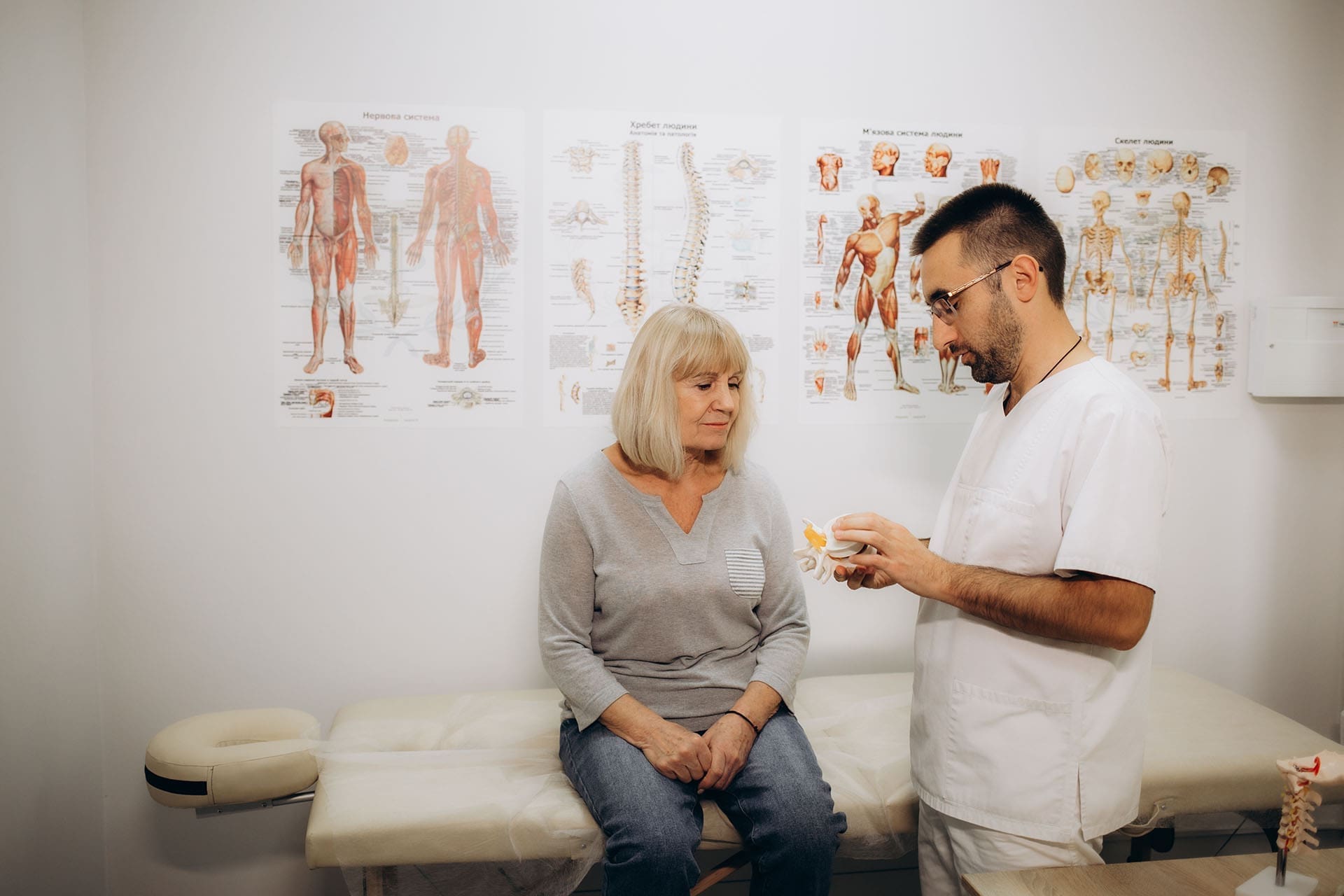
Where Chiropractic and Medical Care Fit (The Chiro-Med Advantage)
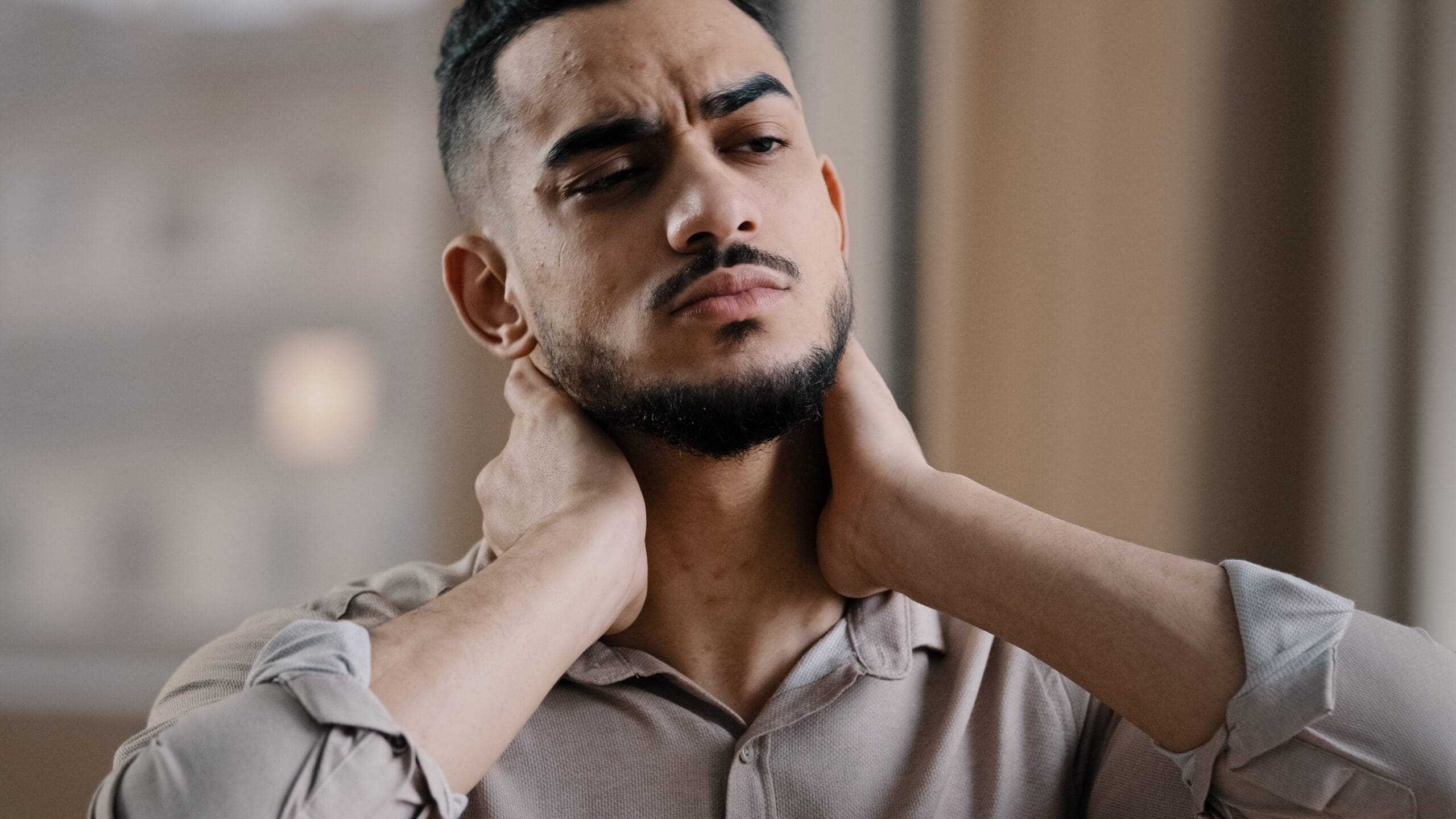
Many Chiromed readers also deal with neck or back pain, sports strains, work injuries, or motor-vehicle accidents (MVAs). Pain, poor sleep, and high stress can worsen gut symptoms through the brain–gut axis. A coordinated chiro-medical model can address both fronts at the same time.
1) Dual-Scope Assessment and Imaging (When Indicated)
A combined clinical exam can separate joint, nerve, and soft-tissue drivers of pain. When needed, X-ray or MRI helps confirm the picture so your plan is safe and specific. (Jimenez Clinic Site; A4M profile). (El Paso, TX Doctor Of Chiropractic)
2) Conservative Therapies That Calm the System
- Spinal adjustments to improve joint motion and ease nerve irritation.
- Targeted exercise therapy to restore mobility and strength.
- Massage therapy for soft-tissue pain, circulation, and relaxation.
- Acupuncture (when available) for pain relief and stress reduction.
These approaches can reduce pain and nervous-system “overdrive,” which often helps gut comfort too. (Sciatica.clinic articles, 2025). (sciatica.clinic)
3) Nutrition & Lifestyle Coaching Built Into Care
An integrated team can translate gut-friendly science into your reality—food swaps, stress skills, and sleep routines that fit busy schedules. The focus is on small wins that add up. (Penn State Health, 2018; Cleveland Clinic, 2022). (Penn State Health News)
4) Injury Documentation and Care Coordination
For work injuries or MVAs, you may need clear medical records, imaging reports, and functional assessments. An integrated clinic can coordinate your care and provide the documentation insurers and legal teams request, while keeping your recovery plan unified. (Jimenez Clinic Site; Scheduler). (El Paso, TX Doctor Of Chiropractic)
Clinical observation (Jimenez): Patients with spine pain and poor sleep often report IBS-like flares. When we combine adjustments or mobilization with gradual activity, breath work, and a simple plant-forward plan (plus one fermented food daily), reports of bloating and meal-related discomfort tend to drop—especially as sleep improves. (Jimenez Clinic Site). (El Paso, TX Doctor Of Chiropractic)
Sample 2-Week “Ease-In” Plan
Week 1: Foundations
- Breakfast: Oats with yogurt or kefir, berries, and nuts.
- Lunch: Grain bowl (quinoa or barley) + beans + mixed veggies; add a spoon of sauerkraut/kimchi.
- Dinner: Chili or lentil curry + salad with olive oil.
- Daily: 20–30 min walk, 5-minute breathing before bed, lights-out window set.
- Limit: one ultra-processed snack per day, max.
Week 2: Build
- Add beans/lentils 5 days/week.
- Add a second fermented food for two days.
- Replace one sweet drink with water or tea each day.
- Add two short strength sessions (15–20 minutes).
- Keep a simple symptom log (bloating, energy, stools, sleep).
Small steps, big difference over time. (Penn State Health, 2018). (Penn State Health News)
When to Seek Care Promptly
- Unintended weight loss, blood in stool, fever, severe or night-time symptoms, or a history of GI surgery.
- Persistent pain and gut complaints despite steady changes.
Talk with your clinician; ask about testing, SIBO evaluation, and tailored treatment. (Mayo Clinic, 2024a). (Mayo Clinic)
Key Takeaways for Chiromed Readers
- Dysbiosis is common and usually fixable with realistic habit changes.
- A plant-forward pattern, along with live-culture foods, stress management skills, and better sleep, can steady the gut and the nervous system.
- When injuries, pain, or SIBO are part of the picture, a coordinated chiro-medical team can blend diagnostics, hands-on care, lifestyle coaching, and documentation—so your gut and your musculoskeletal system improve together. (Cleveland Clinic, 2022; Jimenez Clinic Site). (Cleveland Clinic)
References
- Small intestinal bacterial overgrowth (SIBO) – Symptoms & causes. (2024a, Nov 11). Mayo Clinic. https://www.mayoclinic.org/diseases-conditions/small-intestinal-bacterial-overgrowth/symptoms-causes/syc-20370168 (Mayo Clinic)
- Small intestinal bacterial overgrowth (SIBO) – Diagnosis & treatment. (2024b, Nov 11). Mayo Clinic. https://www.mayoclinic.org/diseases-conditions/small-intestinal-bacterial-overgrowth/diagnosis-treatment/drc-20370172 (Mayo Clinic)
- An updated appraisal of the SIBO hypothesis and the limits of breath testing. (2024, Oct 22). Mayo Clinic for Medical Professionals. https://www.mayoclinic.org/medical-professionals/digestive-diseases/news/an-updated-appraisal-of-the-sibo-hypothesis-and-the-limits-of-breath-testing/mac-20574581 (Mayo Clinic)
- Dysbiosis: What it is, symptoms, causes, treatment & diet. (2024, Apr 16). Cleveland Clinic. https://my.clevelandclinic.org/health/diseases/dysbiosis (Cleveland Clinic)
- What is your gut microbiome? (2023, Aug 18). Cleveland Clinic. https://my.clevelandclinic.org/health/body/25201-gut-microbiome (Cleveland Clinic)
- How your gut microbiome impacts your health. (2022, Jun 9). Cleveland Clinic Health Essentials. https://health.clevelandclinic.org/gut-microbiome (Cleveland Clinic)
- Gut check. (2023, Spring). Cleveland Clinic Magazine. https://magazine.clevelandclinic.org/2023-spring/gut-check (magazine.clevelandclinic.org)
- Nova, E. (2022). The influence of dietary factors on the gut microbiota. Nutrients. https://pmc.ncbi.nlm.nih.gov/articles/PMC9318379/ (PMC)
- Singh, R. K., et al. (2017). Influence of diet on the gut microbiome and implications for human health. Journal of Translational Medicine. https://pmc.ncbi.nlm.nih.gov/articles/PMC5385025/ (PMC)
- Gut health. (2023, Mar 23). Better Health Channel. https://www.betterhealth.vic.gov.au/health/healthyliving/gut-health (Better Health Channel)
- The Medical Minute: Small changes make big differences in digestion. (2018, Mar 7). Penn State Health News. https://pennstatehealthnews.org/2018/03/the-medical-minute-small-changes-make-big-differences-in-digestion/ (Penn State Health News)
- What happens to your gut health when you eat fermented foods regularly. (2025, Oct 9). Health.com. https://www.health.com/what-happens-to-gut-health-when-you-eat-fermented-foods-11825391 (health.com)
- Corbin, K. (2025, Oct 8). What a gut microbiome scientist wants you to eat every day. The Washington Post. https://www.washingtonpost.com/wellness/2025/10/08/what-to-eat-healthy-gut-microbiome/ (The Washington Post)
- El Paso, TX Doctor of Chiropractic (Dr. Alex Jimenez). (2025). https://dralexjimenez.com/ (El Paso, TX Doctor Of Chiropractic)
- Appointment scheduler – Dr. Alex Jimenez. (2025). https://dralexjimenez.com/scheduler/ (El Paso, TX Doctor Of Chiropractic)
- Chiropractic care guide for reducing chronic inflammation. (2025). Sciatica.clinic. https://sciatica.clinic/chiropractic-care-guide-for-reducing-chronic-inflammation/ (sciatica.clinic)
- Chiropractic care techniques for five musculoskeletal issues. (2025). Sciatica.clinic. https://sciatica.clinic/chiropractic-care-techniques-for-five-musculoskeletal-issues/ (sciatica.clinic)