Restoring Musculoskeletal Function: Integrative Care
Abstract
In this educational post, I will take you on a journey into the future of musculoskeletal treatment, moving beyond isolated symptom management to a comprehensive, whole-body paradigm. We will explore interventional orthopedics, which uses precise, image-guided techniques to target the root causes of pain. Building on this, I will introduce a concept I call functional orthopedics and the functional unit approach—a philosophy that integrates the principles of osteopathic medicine, physical medicine, and regenerative science. This approach emphasizes understanding the intricate connections between structure and function, the body’s innate healing capacities, and the importance of treating the entire biomechanical chain rather than just the site of pain. We will delve into the latest evidence-based research by leading experts, examining the critical roles of subchondral bone, intraosseous injections, and comprehensive treatment strategies for conditions such as osteoarthritis. By combining these advanced concepts with the foundational principles of integrative chiropractic care, we can create truly personalized and effective treatment plans that offer lasting relief and restore optimal function.
Understanding the “How” and “Why” of Modern Musculoskeletal Treatment
Thank you for joining me on this exploration of a truly transformative approach to musculoskeletal health. What we are about to discuss is an integral part of a new way of thinking in medicine, and I believe it can fundamentally change how we help our patients heal. Today, we’re not just talking about another treatment method; we’re diving into the “how, why, and what” of a more profound, evidence-based strategy.
- The How: The “how” is our interventional orthopedic approach.
- The Why: The “why” is rooted in functional orthopedics and the functional unit approach.
- The What: The “what” is the application of these principles to deliver comprehensive, patient-centered care.
Let’s unpack what this all means for you and your health journey.
What is Interventional Orthopedics?
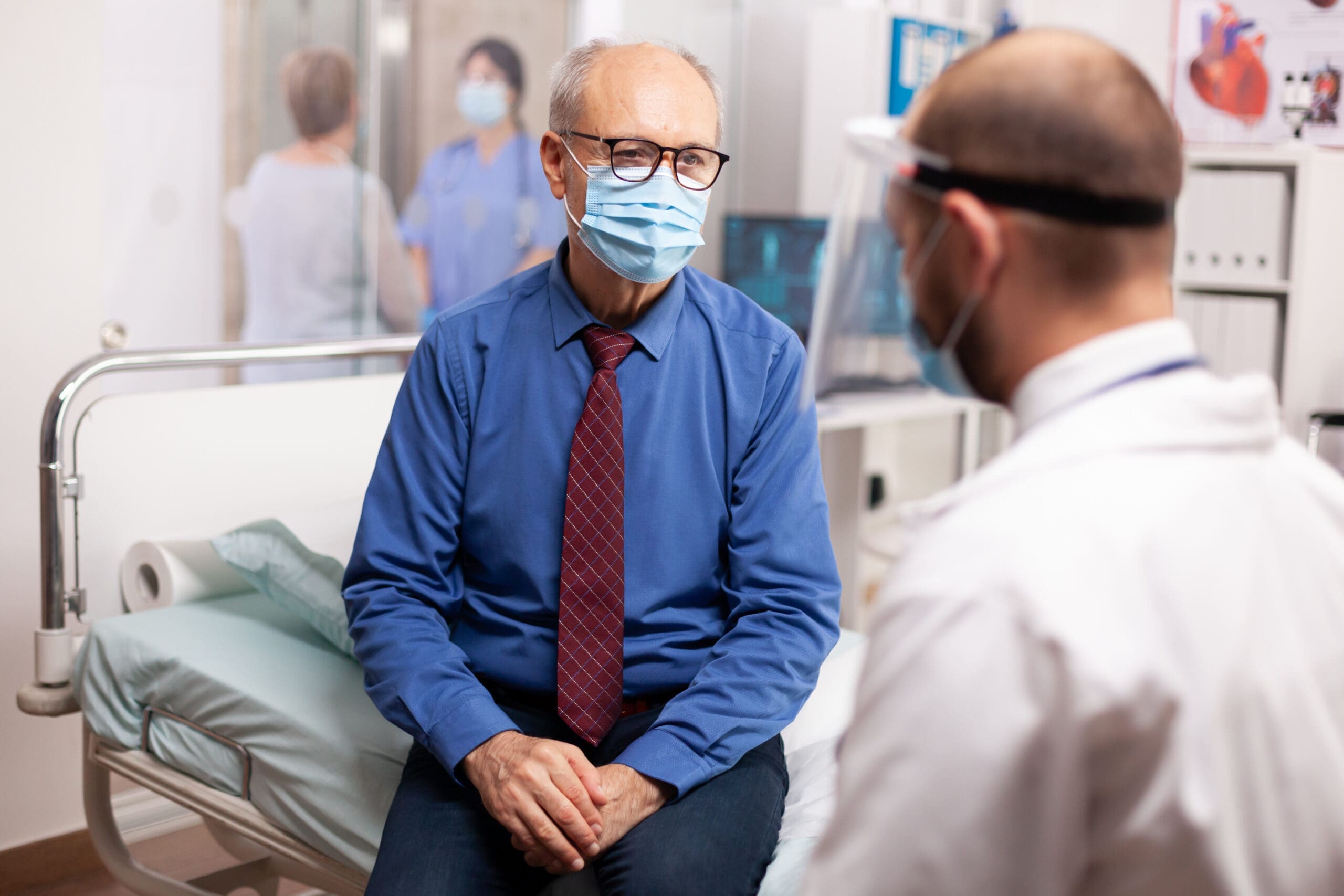
Interventional orthopedics represents a significant evolution from traditional pain management. It’s a specialized field that focuses on using the body’s own healing potential to repair and regenerate damaged tissue. The core principle is precision. Instead of just managing symptoms, we aim to treat the underlying source of the problem.
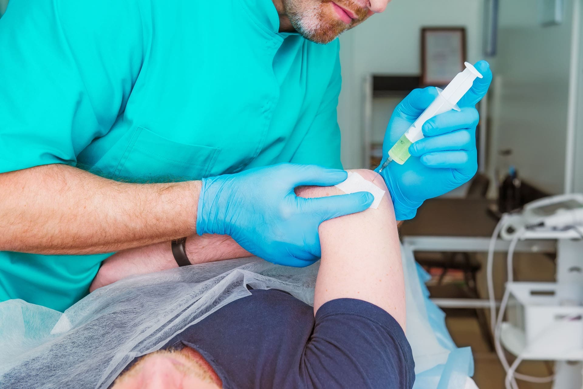
This isn’t about simply injecting a painful joint and hoping for the best. It’s about a meticulous process in which we use advanced imaging, such as ultrasound and fluoroscopy, to visualize and precisely target specific structures. Whether it’s a torn ligament, a damaged tendon, or degenerative changes within a joint, we can deliver orthobiologic treatments—such as Platelet-Rich Plasma (PRP) or Bone Marrow Aspirate Concentrate (BMAC)—directly to the site of injury with pinpoint accuracy.
The goal is to move beyond treating “the thing that is causing the pain” and instead look at the entire picture. But how do we decide what to target? That’s where the “why” comes into play.
Introducing Functional Orthopedics: A Philosophy of Whole-Body Healing
This brings me to a concept that is the cornerstone of my clinical philosophy: functional orthopedics. While you might not find this term in a standard medical textbook (I coined it to describe my integrated approach), its principles are not new. They are deeply rooted in my training as an osteopathic physician, a chiropractor, and a functional medicine practitioner.
Functional orthopedics is guided by several core tenets:
- The body is a unit: No part of the body exists in isolation. A problem in your foot can affect your knee, which can in turn impact your hip and spine. Everything is connected.
- Structure and function are interrelated: The way your body is built (structure) directly influences how it moves and operates (function), and vice versa. An imbalance in one will inevitably affect the other.
- The body has self-healing mechanisms: it possesses an incredible, innate ability to heal and regenerate. The role of a physician is to facilitate and optimize these natural processes.
- Rational treatment is based on these principles: The most effective and lasting treatments are those that honor and work with the body’s integrated design.
This philosophy is a synthesis of my background in Physical Medicine & Rehabilitation (PM&R), which focuses heavily on structure and function, and regenerative medicine, which harnesses the body’s self-healing capabilities. By applying the functional medicine model, we look for the root causes of a condition, considering all the factors—biomechanical, nutritional, and environmental—that contribute to a patient’s health state.
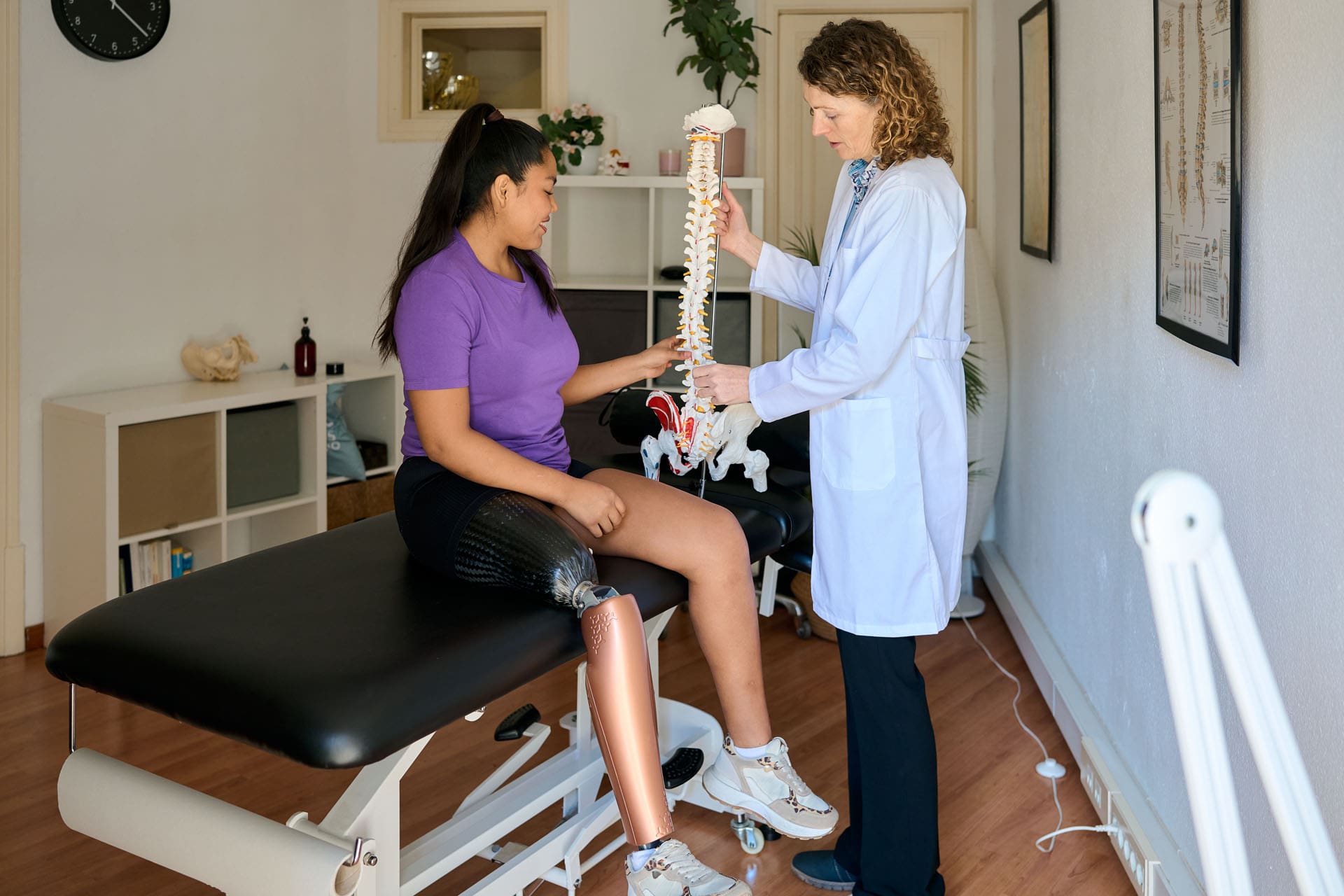
The Functional Unit Approach: Treating the System, Not Just the Symptom
The practical application of functional orthopedics is what I call the functional unit approach. This concept was first described in an old surgical textbook by Dudley and White, who defined the “functional spinal unit” (Dudley & White, n.d.). They recognized that treating a single vertebra or disc was often insufficient because the spine functions as an interconnected system.
We now see this comprehensive approach being validated by modern research in orthobiologics. Several recent studies have demonstrated the superior, long-term benefits of treating the entire functional unit of the spine. For example, researchers have published compelling papers on the use of PRP and BMAC not only in the epidural space but also in the facet joints, ligaments, and paraspinal muscles to treat lumbar and cervical spine issues (Centeno et al., 2017). By addressing all the components that contribute to spinal stability and function, patients experience more profound and lasting results.
This isn’t limited to the spine. A landmark study on knee osteoarthritis compared outcomes between patients who received only an intra-articular (in-joint) injection and those who received both intra-articular and extra-articular (outside the joint) treatments. The results were clear: while both groups improved, the group treated more comprehensively experienced significantly better and more durable outcomes (Centeno et al., 2020).
Think about it from a clinical perspective. How many times have I seen a patient with mild knee osteoarthritis who also has pes anserine bursitis, hamstring tendinopathy, or tenderness along the ligaments? Pain isn’t just coming from the joint space. It’s coming from the entire functional unit that supports and moves that joint. The paradigm shift is from a narrow, intra-articular focus to a comprehensive view encompassing all intra-articular and extra-articular structures.
Beyond the Joint: The Critical Role of Subchondral Bone
But does it stop there? The answer is no. A growing body of research is revealing another crucial layer to this puzzle, especially in osteoarthritis: the subchondral bone. This is the layer of bone directly beneath the cartilage.
For decades, the conventional wisdom propagated to patients was that osteoarthritis is primarily a disease of cartilage loss. We’ve all heard patients say, “My doctor told me I’m bone on bone” or “My cartilage is gone.” However, we also know that the degree of cartilage loss on an X-ray does not always correlate with the level of pain a person experiences.
So what’s the missing link? It’s often the health of the subchondral bone. When cartilage wears away, the underlying bone is exposed to increased stress and inflammation. This bone is not inert; it’s a living, dynamic tissue rich with blood vessels, nerves, and even a population of stem cells (pericytes) that are vital for healing.
Dr. Philippe Hernigou, a pioneering orthopedic surgeon from France, conducted groundbreaking research on this topic. He compared the number of reparative cells in the iliac crest bone marrow (a common site for harvesting bone marrow) with the number of cells in the subchondral bone of an osteoarthritic knee. His findings were astonishing. As osteoarthritis progressed and patients aged, the concentration of these crucial healing cells in the subchondral bone declined dramatically, whereas levels in the iliac crest remained relatively stable (Hernigou et al., 2013). This suggests that the local healing environment within the knee itself becomes depleted.
This discovery has paved the way for a new and powerful treatment strategy: intraosseous injections. By injecting orthobiologics such as PRP or BMAC directly into the subchondral bone, we can replenish the depleted cellular environment and address the “bone” component of osteoarthritis.
- A recent meta-analysis and a consensus statement we published for the American Academy of PM&R have recognized that intraosseous PRP injection has significant merit, particularly for more advanced stages of knee osteoarthritis.
- Perhaps the most compelling evidence comes from a pair of sister studies looking at intraosseous BMAC. In one study, patients had one knee replaced and the other treated with an intraosseous bone marrow injection. With an average follow-up of 15 years, over 80% of patients avoided a knee replacement in their treated knee. Remarkably, they overwhelmingly preferred their “bone marrow knee” to their artificial one (Hernigou et al., 2021).
- The sister study involved patients who wanted to avoid surgery altogether. They received an intra-articular injection in one knee and an intraosseous injection in the other. Both knees improved, but the knee that received the intraosseous injection had a significantly lower rate of conversion to a total knee replacement (Hernigou et al., 2020).
The takeaway is clear: for severe osteoarthritis, we must look beyond the joint space and the surrounding soft tissues. We must also treat the bone. This is the essence of treating the whole functional unit.
The Art of Diagnosis: How We Decide What to Treat
So, how do we put all this together in a clinical setting? How do we decide which structures to treat? It’s not a matter of just guessing; it’s a combination of deep anatomical knowledge, a thorough physical exam, and the art of clinical reasoning.
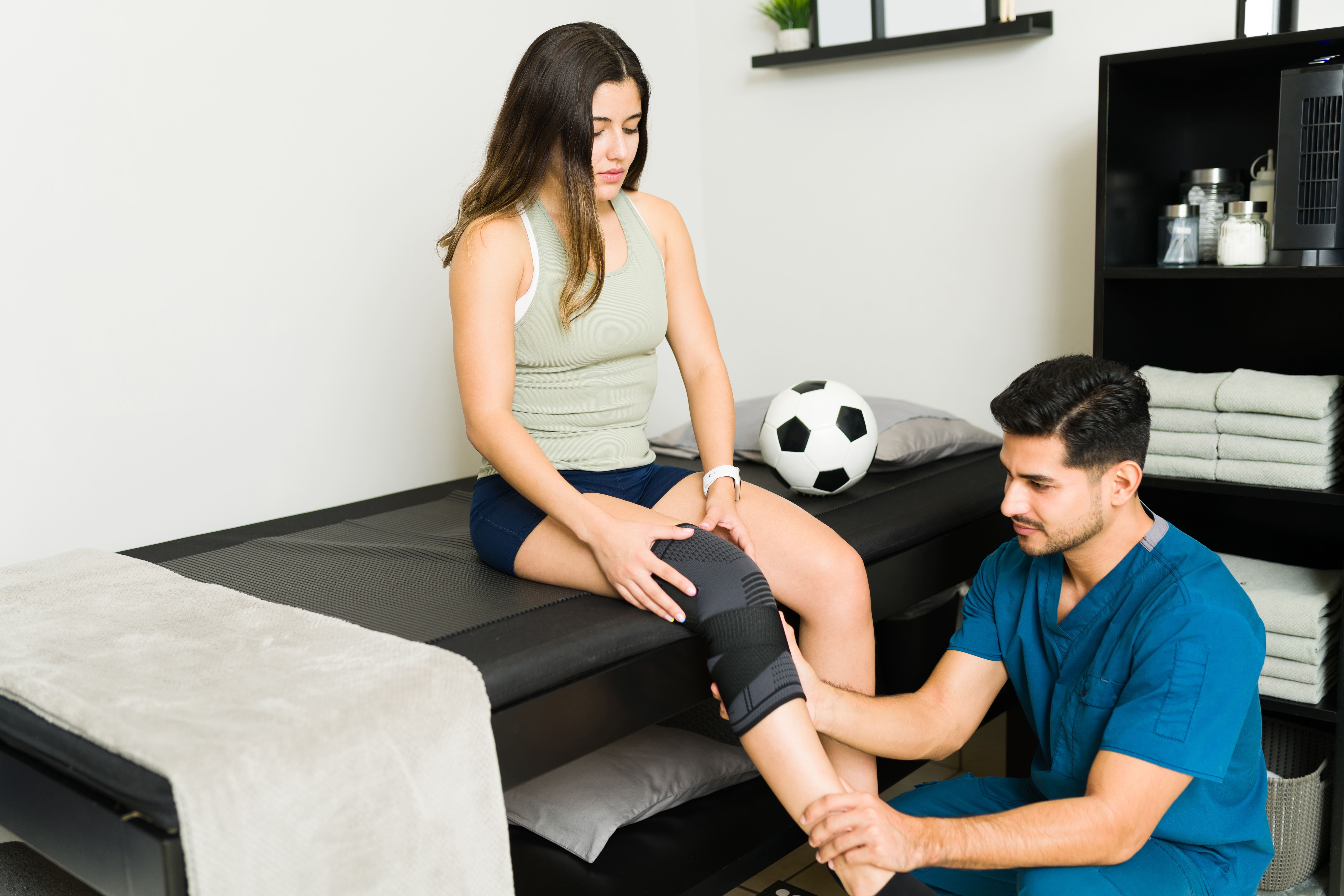
This is where we put on our thinking caps. Let’s consider a patient with medial (inner) knee osteoarthritis.
- The Exam: A physical exam might reveal a varus deformity (bow-legged stance), which places excessive stress on the medial compartment of the knee.
- The Analysis: This varus stress not only compresses the medial meniscus and cartilage but also stretches and weakens structures on the lateral (outer) side of the knee, such as the lateral collateral ligament (LCL).
- The Treatment Plan: A comprehensive treatment plan wouldn’t just address the medial joint space. It would also involve treating the LCL to restore stability and correct the biomechanical imbalance that is driving the degeneration.
Conversely, if a patient has a valgus moment (knock-kneed) and lateral compartment arthritis, we would assess the lateral structures as well as the medial ligaments that are being overstretched.
Or consider a case of patellofemoral pain or maltracking, where the kneecap is being pulled laterally. The solution isn’t just to treat the cartilage behind the kneecap. We must ask why it’s being pulled. Often, the medial patellofemoral ligament (MPFL), which acts as a checkrein, is weak or damaged. Treating and strengthening this ligament is key to restoring proper tracking.
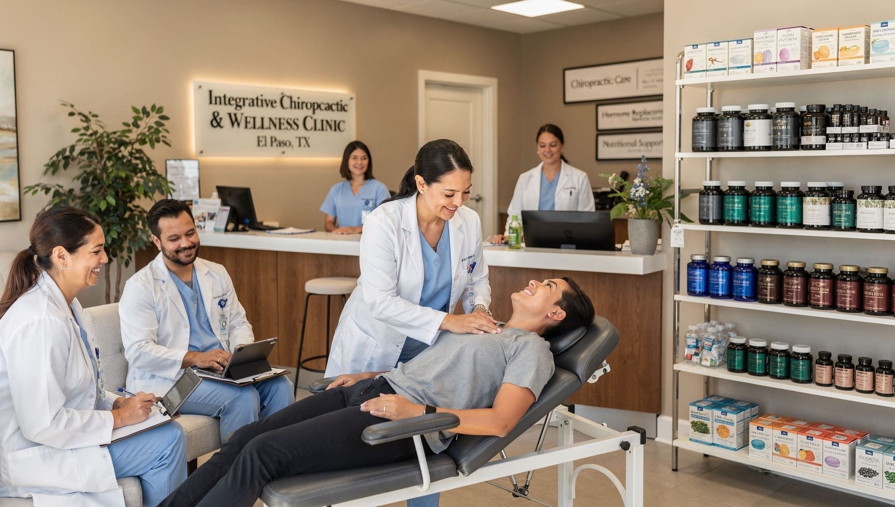
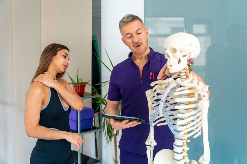
Chiropractic Integration: The Bigger Biomechanical Picture
This is where integrative chiropractic care becomes indispensable. The buck doesn’t stop at the knee. We must ask: why did this atraumatic knee issue develop in the first place?
As a chiropractor, I am trained to look at the entire kinetic chain.
- Look Distally: We must examine the ankle and foot mechanics. Is there excessive foot pronation causing the tibia to internally rotate, creating a valgus stress at the knee?
- Look Proximally: We must evaluate the hip and gluteal muscles. One of the most critical muscles for knee (and hip) stability is the gluteus medius. Weakness in this muscle is a common driver of lower-extremity dysfunction.
- Look to the Spine: Could there be a subclinical radiculopathy? A slight nerve impingement in the lumbar spine can cause weakness in key muscles, such as the EHL (the muscle that lifts the big toe), disrupting the entire gait cycle and placing abnormal stress on the knee.
In my practice, I perform detailed muscle strength testing along the kinetic chain, assess for nerve tension, and use chiropractic adjustments to restore proper alignment and nervous system function. By treating only the knee, will we achieve long-term success if the underlying hip weakness or foot dysfunction remains unaddressed? The answer is a resounding no.
By integrating precise orthobiologic injections with comprehensive chiropractic care, physical therapy, and functional medicine principles, we can address the problem from every angle. This is what I mean when I say we must treat the whole person, not just the pain generator. In doing so, we turn the problem into a “treatment generator”—an opportunity to restore health to the entire system.
This is the future of musculoskeletal medicine. It requires us to go back to our roots in anatomy, physiology, and biomechanics, but to apply that knowledge with the most advanced tools and a holistic, integrated mindset. It’s a truly fulfilling way to practice, and it offers our patients the best possible chance for a long-term, functional recovery.
References
Centeno, C. J., Markle, J., Dodson, E., Stemper, I., Williams, C. J., Kisiday, J. D., … & Steinmetz, N. J. (2017). The use of lumbar epidural injection of platelet lysate for treatment of radicular pain. Journal of Experimental Orthopaedics, 4(1), 38. https://dx.doi.org/10.1186%2Fs40634-017-0113-5
Centeno, C., M.D., Pitts, J., M.D., Al-Sayegh, H., M.D., & Freeman, M., D.C., PhD. (2020). Efficacy of autologous, micro-fragmented adipose tissue with leukocyte poor-platelet rich plasma for the treatment of knee osteoarthritis: a randomized controlled crossover study. Journal of Translational Medicine, 18(131). https://doi.org/10.1186/s12967-020-02285-3
Dudley, H. A. F., & White, J. C. (n.d.). Operative Surgery: Fundamental International Techniques.
Hernigou, P., Poignard, A., Beaujean, F., & Rouard, H. (2013). Percutaneous autologous bone-marrow grafting for nonunions. The Journal of Bone and Joint Surgery. American Volume, 87 Suppl 1(Pt 2), 896-903. https://doi.org/10.1302/0301-620X.87B1.15783
Hernigou, P., Bouthors, C., Bastard, C., Flouzat-Lachaniette, C. H., Rouard, H., & Dubory, A. (2021). Subchondral bone marrow concentrate injection is more effective than intraarticular injection in severe osteoarthritis of the knee: a 15-year-follow-up of a randomized controlled trial. International Orthopaedics, 45(2), 341-349. https://doi.org/10.1007/s00264-020-04871-3
Hernigou, P., Delattre, L., Dubory, A., & Flouzat-Lachaniette, C. H. (2020). Intra-articular injection of bone marrow concentrate is a better choice than intra-osseous injection in less advanced osteoarthritis of the knee. International Orthopaedics, 44(7), 1293-1302. https://doi.org/10.1007/s00264-020-04535-2