Orthobiologic Insights for Patients and Musculoskeletal Health
Delve into the science of musculoskeletal health and orthobiologic methods to boost recovery and maintain joint well-being.
Abstract
Welcome. I’m Dr. Alex Jimenez, and I am excited to share my perspective on a transformative shift happening in musculoskeletal (MSK) medicine. This educational post, from my viewpoint as a Doctor of Chiropractic and a Family Nurse Practitioner, explores the move from volume-driven to precision-based care in orthobiologics and regenerative medicine. For too long, physicians have been constrained by outdated systems, and patients have been offered limited solutions. This post is for my colleagues in the medical field and for patients seeking a deeper understanding of their health. We will journey through the latest evidence-based findings in orthobiologics, exploring why this field, despite its compelling science, has faced challenges in execution. I will outline a comprehensive framework—the Joint Vitality System—that I have developed to ensure consistent, superior outcomes. This system emphasizes precision diagnosis, biologic matching, and a structured, guided recovery plan.
We will delve into the physiological underpinnings of orthobiologics such as Platelet-Rich Plasma (PRP), contrast them with traditional treatments, and highlight the importance of an integrative model that combines chiropractic care, functional medicine, and medical oversight. The discussion will also cover the critical role of data collection, the importance of understanding the physiological drivers of disease—whether inflammatory, degenerative, or structural—and how our integrative approach at Injury Medical Clinic PA serves as a model for this new paradigm of care. Finally, I will explain how our multidisciplinary team, under the medical direction of Dr. Maria Guadalupe Cardenas, MD, provides a comprehensive framework for restoring function and delivering the transformative, whole-person health outcomes our patients deserve. My goal is to empower you with the knowledge to build or seek a practice that is not only sustainable and independent but also delivers the highest standard of patient-centered, regenerative care.
Our Collaborative and Integrative Approach in El Paso, TX
At Injury Medical Clinic PA (also known as Mission Plaza Injury Medical Clinic) in El Paso, Texas, our model is built on a multidisciplinary, patient-centered foundation. I am Dr. Alex Jimenez, and I am honored to work alongside our Medical Director and Collaborative Physician, Dr. Maria Guadalupe Cardenas, MD. Dr. Cardenas is Board Certified in Internal Medicine (NPI #1164426749, Texas MD License #J2933) and brings over 40 years of invaluable experience as our Medical Director and Collaborative Physician. This collaboration between a DC/APRN and an MD is a powerful synergy and is common in modern integrative and injury clinics.

This setup allows us to merge the distinct strengths of different medical disciplines to provide truly holistic patient solutions.
- Dr. Cardenas (MD, Internal Medicine): Dr. Cardenas provides essential medical oversight, manages complex internal medicine conditions that impact musculoskeletal health, and ensures our protocols meet the highest standards of medical safety and efficacy. Her deep knowledge of systemic disease is critical when evaluating a patient’s candidacy for regenerative procedures, managing lab results, and ensuring our treatments are medically sound.
- Dr. Jimenez (DC, APRN, FNP-BC, CFMP, IFMCP, ATN, CCST): As a Doctor of Chiropractic and a Board-Certified Family Nurse Practitioner with extensive certifications in functional medicine, I focus on the biomechanical, musculoskeletal, and functional aspects of health. My role involves using integrative chiropractic care to address spinal alignment, nervous system function, and structural integrity. As a nurse practitioner and functional medicine expert, I investigate the underlying physiological imbalances—in nutrition, hormones, and inflammation—that contribute to injury and disease.
Together, our team seamlessly integrates chiropractic adjustments, functional medicine diagnostics, medical management, rehabilitation, personal injury care, and orthobiologic therapies into a single, cohesive care plan. This allows us to address the patient as a whole person, not just an injured joint. For instance, before a regenerative procedure, we might use chiropractic care to ensure proper joint mechanics, functional medicine to optimize nutrient levels and reduce systemic inflammation, and medical oversight from Dr. Cardenas to manage a patient’s previously undiagnosed pre-diabetes—all of which are crucial for a successful outcome.
Rethinking the Business of Medicine: From Fear to Freedom
Many of us in the medical field come from the “School of Hard Knocks” when it comes to business. We’re trained to believe that if we don’t know every single detail about a subject, we shouldn’t even start. This mindset is rooted in our primary directive: “first, do no harm.” We fear that an error in judgment could have devastating consequences for a patient. However, I want to offer a different perspective: business is not as hard as medicine. The risks are fundamentally different.
- What’s the worst that can happen in a small business venture? You don’t charge as much as you could have. You lose a little money one month, which you can make up the next. You buy ten units of a product instead of twenty to save on upfront costs, even if the per-unit price is slightly higher.
- Were these devastating choices? Did anyone get harmed? Perhaps your bank account was temporarily a few dollars lighter, but that’s just the price of doing business and learning.
Most entrepreneurs “build the airplane while they’re flying it.” They just get started and figure things out along the way. As clinicians, we are incredibly smart and adept at learning. My own journey into private practice started with a copy of Medical Practices for Dummies. It got me surprisingly far! I missed a step about getting a business license right away, but it was easily corrected. No harm, no foul. The point is, it can all be figured out.
The Orthomolecular Micro-Practice: Precision Over Volume
The model I champion is what I call the orthomolecular micro-practice. This is not a volume-driven enterprise; it is a precision practice. In the traditional insurance-based world, the only way to increase revenue is to see more patients because the price per visit is fixed and often low. This leads to burnout, rushed appointments, and mountains of paperwork.
Consider this brutal statistic we’ve observed: the ratio is approximately 15:1. To earn the same revenue from a single orthobiologic cash-based procedure, I would need to see 15 insurance-based patients. If I see 30 patients in a day under the insurance model, I make the same amount as seeing just two or three orthobiologic patients. Think of the administrative burden: would you rather write 30 clinical notes or just two? The answer is obvious.
This is where technology like an AI scribe becomes a game-changer. I personally use a system (DeepScribe) that requires no clicks from me. I record my patient interactions, and by the time I leave the room, the note is fully and directly imported into my EMR. It saves an incredible amount of non-compensated time and allows me to be fully present with my patients.
The Precision Practice is built on a few core principles:
- Precision over Volume: Focusing on a smaller number of patients allows for deeper, more comprehensive care.
- Systems-Driven Approach: Every patient touchpoint, from the initial phone call to the follow-up, is standardized. This ensures a predictable, high-quality experience and makes the practice scalable without sacrificing consistency.
- The Right Patients: We focus on attracting patients who are actively seeking the transformative solutions we offer. We don’t convince or use high-pressure sales tactics. We educate, present the expected outcomes, and empower them to make an informed decision.
Seeing just five to ten of these ideal patients a month can build a thriving, sustainable practice. Ten patients a month at an average of 50,000 in cash revenue. That’s a legitimate business that can cover payroll, rent, and more—all while seeing only ten patients a month.
Patient-Centered Outcomes Over Procedures: Why Value Is About Transformation
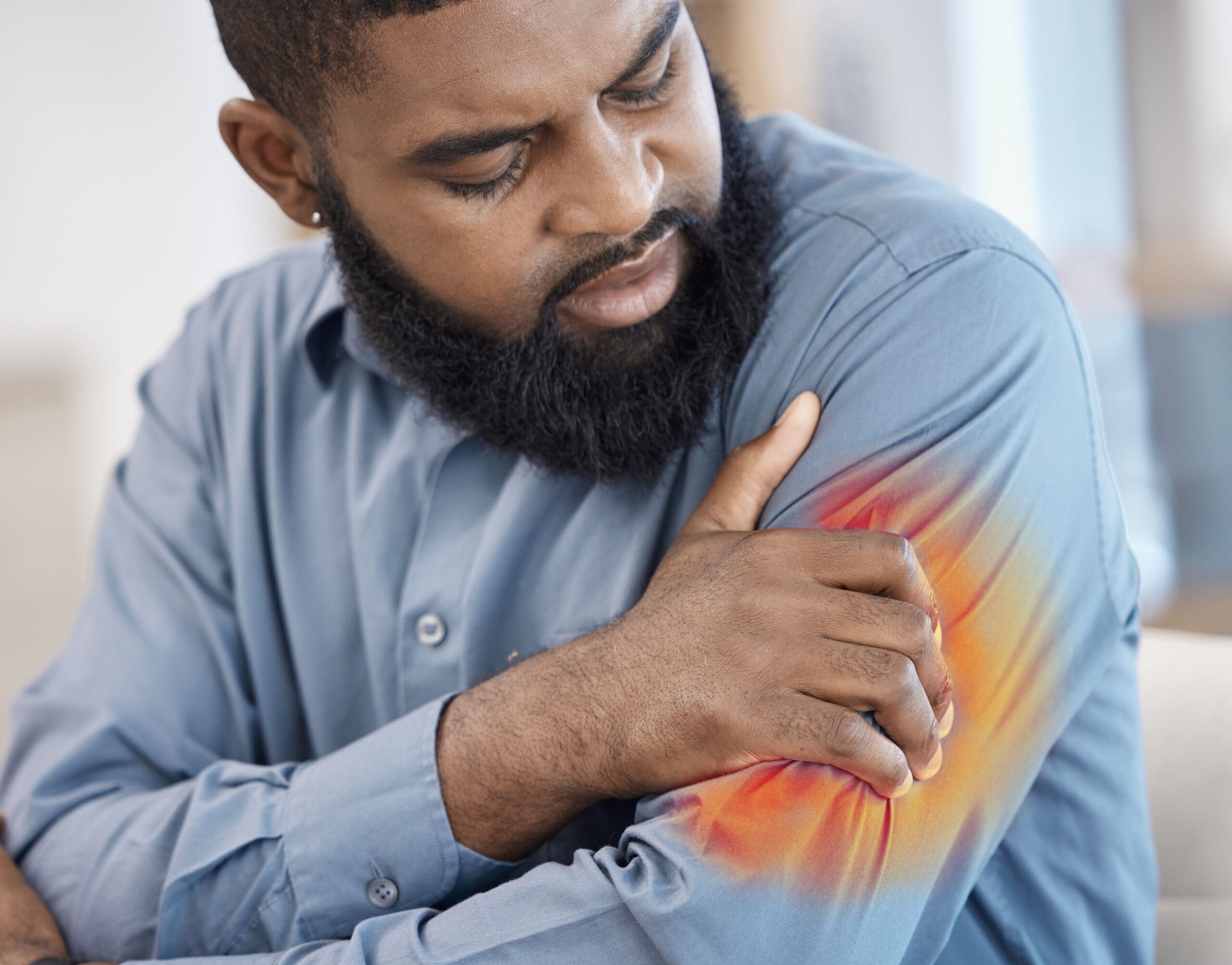
As a clinician, I have learned that patients do not come to us for injections—they come for transformation. They want their lives back: to lift grandchildren, to play pickleball, to work without pain, to sleep through the night, and to feel strong and capable again. This distinction guides everything we do.
- Patients are not purchasing a syringe; they are investing in a meaningful outcome.
- When care results in real, measurable functional restoration, it justifies expert-level compensation because it delivers lasting value.
- The ethical compass remains true when we provide evidence-based methods, conduct meticulous data collection, and set transparent expectations for recovery.
In our clinic’s integrative system, we package what matters: a pathway that blends orthopedic and nervous system restoration, lifestyle changes, and structured rehabilitation. The end goal is simple: unlock the patient’s innate capacity for repair, and then guide it with science-backed steps.
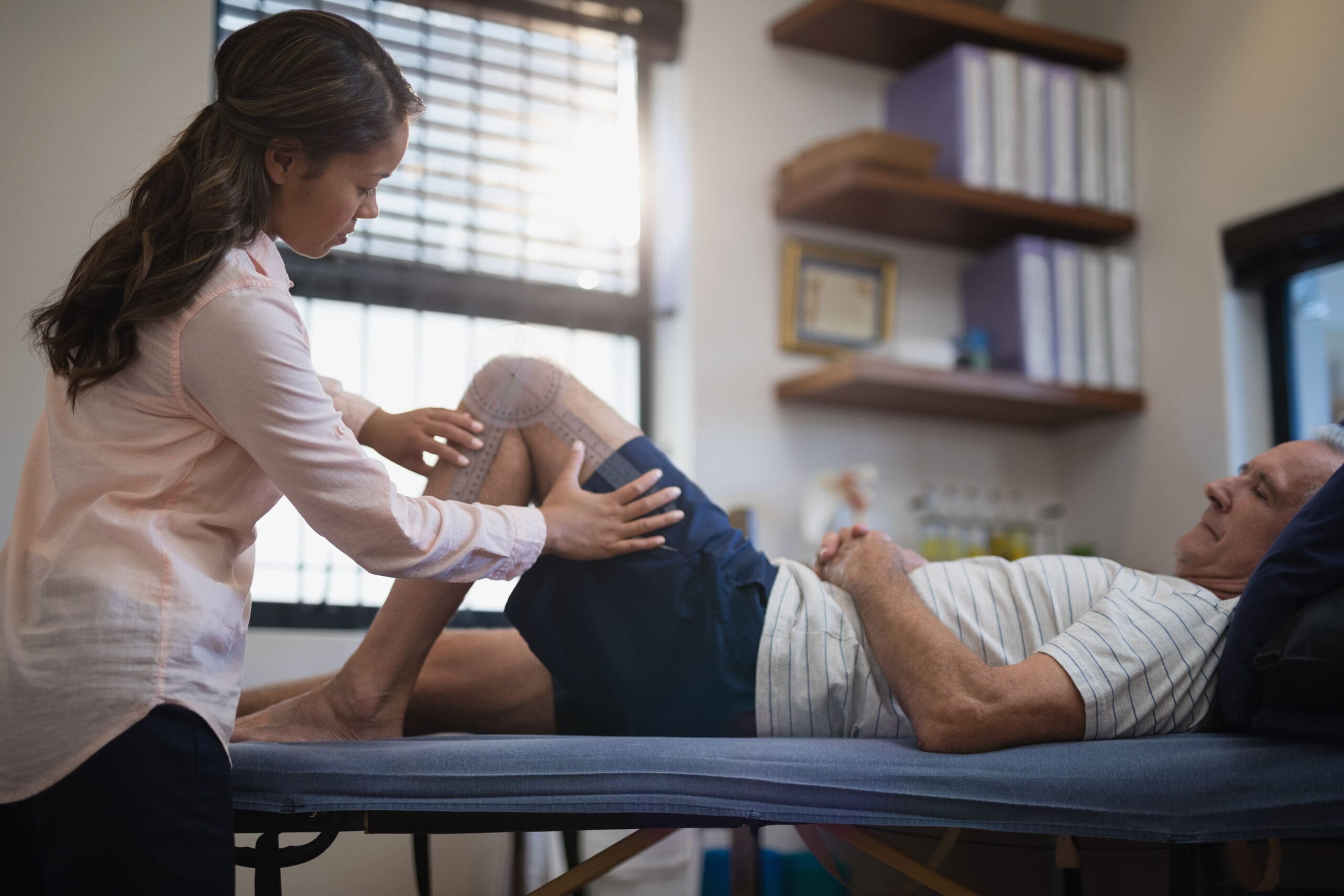
Unlocking Pain Relief: How We Assess Motion to Alleviate Pain- Video
The Challenge of Execution in Orthobiologics
On May 2, 2026, I reflected on the state of orthobiologics, and it became clearer than ever that the science is not the issue. The research supporting the use of biologics such as Platelet-Rich Plasma (PRP) and Bone Marrow Concentrate (BMC) is compelling and continues to grow daily. We have evidence showing we can modify the inflammatory environment of a joint and even stimulate the replication of tenocytes (tendon cells).
So why isn’t this the first-line treatment for every appropriate MSK condition? The answer lies in execution. We’ve seen significant problems that have eroded patient trust and caused physician hesitation:
- Inconsistent Outcomes: Clinic A’s PRP protocol differs markedly from Clinic B’s. This lack of standardization leads to unpredictable results.
- Poor Patient Selection: A common pitfall is offering a single therapy for every condition. PRP is fantastic for many tendon-based issues, but it won’t cure severe, bone-on-bone hip arthritis with significant bone marrow edema. Using the wrong tool for the job is a recipe for failure.
- Overpromising in Marketing: We’ve all seen the “stem cell” clinics that make miraculous claims without proper diagnostics or patient evaluation. This “Wild West” atmosphere, particularly prevalent in places like Florida, erodes public trust. The key is to under-promise and over-deliver.
- Lack of Standardization and Measurement: As a field, we must be rigorous. Leading researchers have shown that a platelet dose above 5.5 billion is associated with more beneficial outcomes (Everhart et al., 2019). Are we measuring the platelet concentration in every PRP sample we prepare? We should be. If you can’t measure, you should at least know your system’s validated output. For instance, in our clinical observations, using a specific 60 cc draw with the Apex kit consistently yields approximately 10.8 billion platelets. This knowledge allows us to ensure we are delivering a therapeutic dose every single time.
These execution failures drive patients away from a field with immense potential and cause good physicians to second-guess their approaches. To ensure consistency and scalability, I developed the Joint Vitality System. This is not just a procedure; it is a comprehensive framework that guides our entire process, from initial consultation to full recovery.
The Joint Vitality System Part 1: Precision Diagnosis Beyond the Obvious
A successful outcome starts with an accurate and precise diagnosis. We cannot afford to guess. This requires a multifaceted approach:
- Thorough History and Physical Exam: We must listen to our patients and touch our patients. You can have two patients with identical MRI reports but completely different sources of pain. One might have true intra-articular knee pain from synovitis, while the other’s “knee pain” is actually referred pain from an L4 radiculopathy or hip arthritis.
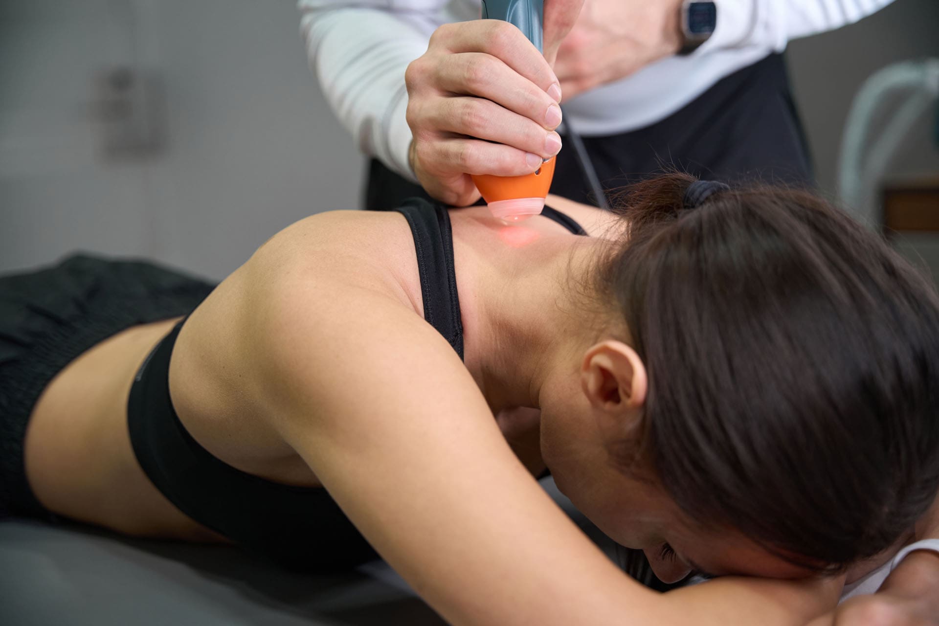
- Diagnostic Musculoskeletal Ultrasound: This is not optional. It is a vital point-of-care tool that allows us to visualize tissues in real time, assess for inflammation, and pinpoint the exact source of pain. I can move the joint and ask, “Does it hurt right here?” while visualizing the underlying anatomy.
- Reviewing Your Own Imaging: While radiologists’ reports are important, MSK-trained clinicians often see subtleties that general radiologists may miss. They might not comment on a high-intensity zone in a disc, a low-grade partial tendon tear, or a meniscocapsular junction sprain—all of which are significant pain generators that we can treat with orthobiologics.
- Diagnostic Injections: I am a firm believer in the “show me” principle. If I am not 100% certain of the pain generator, I use diagnostic injections (e.g., with a local anesthetic) to confirm the source. This is a powerful tool. If numbing a specific structure provides significant temporary relief, we have confirmed our target. It’s a “try it before you buy it” approach for orthobiologics that gives both the patient and me confidence in the treatment plan.
We must differentiate between an inflammatory driver, a degenerative driver, and a structural driver. For example, a hot, swollen knee might be driven by inflammation (synovitis). In contrast, a chronically achy, mechanically unstable knee might be driven by degeneration (arthritis) or a structural problem (e.g., a meniscus tear). Each requires a different approach.
The Joint Vitality System Part 2: Biologic Matching for the Right Job
Once we have a precision diagnosis, we must match it with the appropriate biologic therapy. It is not a one-size-fits-all solution. The key question is: What does this specific tissue need to achieve our therapeutic goal?
- Inflammation Control: If the primary problem is inflammation, our goal is immunomodulation. We need to flip pro-inflammatory M1 macrophages to an anti-inflammatory M2 phenotype. When this occurs in the knee synovium, something remarkable happens: the synovium begins to produce its own endogenous hyaluronic acid (Morigi et al., 2020). We can stimulate the body to heal itself.
- Cellular and Growth Factor Support: For degenerative conditions like tendinopathy or mild-to-moderate arthritis, PRP is an excellent choice. It delivers a high concentration of growth factors that signal tissue repair and reduce inflammation. We tailor the leukocyte profile, using leukocyte-rich PRP for ligament/tendon issues and leukocyte-poor PRP for intra-articular arthritis to better modulate local inflammation (Dohan Ehrenfest et al., 2009).
- Structural Scaffolding and Cellular Regeneration: For more significant issues, like a partial tendon tear with a visible gap or more advanced arthritis, we need more. Adipose tissue provides a structural scaffold (the extracellular matrix) and a rich source of mesenchymal stem cells (MSCs) and other regenerative cells. If there is a gap in a tendon that needs bridging, adipose is a superb option.
- Bone and Cartilage Healing: For severe arthritis with associated bone marrow edema (a sign of stress and inflammation within the bone itself), Bone Marrow Concentrate (BMC) is often the superior choice. BMC contains MSCs and other progenitor cells that are crucial for bone and cartilage health. It is also a flowable product that can be injected intraosseously (directly into the bone) to treat bone marrow lesions, a capability not available with thicker adipose grafts.
We must also consider the delivery method. Putting an adipose graft (which requires an 18-gauge needle) into an intervertebral disc is not a sound application. The biologic must be appropriate for the tissue, the pathology, and the delivery method.
The Joint Vitality System Part 3: Structured Care and Guided Recovery
The procedure is just one part of the journey. A structured care plan is essential for guiding the patient and managing their expectations from start to finish. This includes a comprehensive rehabilitation program that turns improved biology into durable function. We use staged, criterion-based protocols:
- Early phase (days 1–14): Protect the site, restore pain-free range of motion, gentle isometrics, and controlled closed-chain loading to stimulate mechanotransduction without overstrain.
- Mid phase (weeks 3–8): Progressive resistance, eccentric training for tendons, neuromuscular control (balance, perturbation training), and pattern correction (hip hinge, scapular setting).
- Late phase (weeks 9–16): Power development, return-to-sport drills, and task-specific conditioning (e.g., pickleball pivot work, lifting technique for grandparents).
If we perform a procedure on a tendon, the patient must follow a progressive tendon-loading program. This is non-negotiable. The mechanical signals from proper physical therapy are essential for guiding the new tissue as it remodels and strengthens. This is known as mechanotransduction, where cells sense load and trigger gene expression for collagen synthesis and alignment (Wang et al., 2012). Eccentric loading, in particular, promotes tendon remodeling.
Why We Start 30 Days Before the Procedure: Health Optimization and Risk Reduction
True recovery begins before the day of the procedure. Our pre-procedure window—often 30 days—allows us to “stack the deck” for repair. This whole-person approach is critical because healing is metabolically expensive.
Key optimization targets:
- Hematologic readiness: We review complete blood count and iron studies. Adequate oxygen-carrying capacity is vital for cellular respiration and ATP production during healing (Stoltzfus et al., 2019).
- Endocrine balance: We test thyroid function and sex hormones, such as estrogen and testosterone. We now know there are estrogen receptors in the knee, and estrogen has a protective effect on cartilage. Its decline during menopause is linked to an earlier onset of arthritis in women—sometimes 20 years sooner than in men (Sniekers et al., 2008). Optimizing hormones when clinically indicated supports collagen synthesis, bone density, and muscle integrity (Khosla & Monroe, 2018).
- Nutritional status: We test for Vitamin D and other key nutrients. Ensuring adequate intake of vitamin D, omega-3 fatty acids, magnesium, and protein supports immune modulation and connective tissue repair (Calder, 2017; DiNicolantonio et al., 2018).
- Glycemic control: An elevated Hemoglobin A1C indicates poor blood sugar control, which severely impairs healing. Lowering HbA1c improves microvascular function, reduces glycation end products, and enhances wound-healing quality (Singh et al., 2020).
- Sleep and circadian alignment: Consistent sleep boosts growth hormone pulses and tissue repair, while circadian regularity improves insulin sensitivity and inflammatory tone (Luyster et al., 2012).
The Role of Integrative Chiropractic Neuromechanics in Recovery
Integrative chiropractic care is foundational for translating biological repair into functional performance. It is a key component of our guided recovery, helping prevent the recurrence of underlying mechanical stresses that may have caused the problem in the first place.
The physiology behind this approach is powerful:
- Joint alignment and segmental mobility restore optimal arthrokinematics, reducing shear stress on healing tissues.
- Proprioceptive enhancement recalibrates spinal and peripheral reflex loops, improving muscle firing patterns and reducing compensatory overuse. Pain alters motor control via central sensitization. Chiropractic adjustments help normalize afferent input to the nervous system, reducing hypervigilant reflexes.
- Fascial release and myofascial remodeling improve glide planes, reducing nociceptive input and allowing normalized movement arcs.
- Improved joint centration and balanced muscle co-contraction decrease joint microinstability, protecting healing cartilage and tendons from irregular load vectors.
My clinical observations confirm that pairing PRP with chiropractic-guided kinetic chain correction leads to faster time-to-function milestones and fewer relapses, especially in shoulder, knee, and lumbar dysfunctions (Jimenez, n.d.-a; Jimenez, n.d.-b). It ensures the body is optimally aligned to heal.
The Power of Relationships and Your Existing Patient Base
So, how do you find these patients? The growth of a successful orthobiologics practice comes from relationships. The two most powerful and durable sources of growth are:
- Clinician Referrals: Building a referral-based practice is the most sustainable model. We position ourselves as problem-solvers for our colleagues. An orthopedic surgeon sees many patients with non-surgical conditions, such as greater trochanteric bursitis (lateral hip pain). These cases rarely proceed to surgery and can be frustrating for a surgeon to manage. For us, it’s a perfect opportunity to apply orthobiologics.
- Your Existing Patient List: Your most valuable asset is the group of patients who already know, like, and trust you. The cost to reach a patient who is already in your system is zero. They haven’t heard from you about these new treatments because you haven’t offered them yet!
Let me share an example. I used to perform a lot of hyaluronic acid (HA), or “gel,” injections for knee arthritis. When I decided to stop, I contacted all my HA patients and explained that based on the latest evidence, PRP offered a superior outcome. I gave them the choice: transition to PRP with me or receive a referral for HA. The result? Thirty percent of my HA patients transitioned to orthobiologic care. The research supports this move. Studies, such as the one by Meheux et al. (2016), consistently show that PRP outperforms HA at every time point in treating knee osteoarthritis. When we educate patients, many will opt for the better outcome.
Data Collection: The DNA of Continuous Improvement
We collect data because better measurement produces better outcomes. If you are not collecting data on your patients, you are flying blind. I strongly advocate for using a registry like DataBiologics, founded by physicians for physicians. It provides an IRB-approved platform to track outcomes, allowing us to publish our data and, most importantly, tell our patients with confidence what they can expect from our specific treatments in our clinic.
What we track:
- Pain scores (NRS/VAS), function scales (e.g., DASH, LEFS, Oswestry Disability Index), and patient-reported improvements.
- Baseline and follow-up metrics for strength, mobility, and balance.
- Adherence markers for nutrition, sleep, and activity.
A nominal $25 data fee added to the care package can cover system costs and foster engagement. This is how we move from anecdote to evidence. This is how we build trust.
Conclusion: A Modern, Evidence-Based Pathway to Recovery
The train of regenerative medicine is leaving the station. Our integrative framework, guided by medical oversight from Dr. Maria Guadalupe Cardenas, MD, and chiropractic leadership from me, delivers a measured, ethical, and effective route to patient transformation. We start early, combine biologic precision with biomechanical intelligence, optimize metabolism, and move patients through staged rehabilitation. We measure relentlessly, learn constantly, and stay aligned as a team. By embracing a systematic, evidence-based, whole-person approach, we can provide our patients with the exceptional care they deserve while building practices that are professionally and financially rewarding. This is how we practice medicine on our own terms, driven by science and a genuine desire to help our patients heal.
References
- Calder, P. C. (2017). Omega-3 fatty acids and inflammatory processes: From molecules to man. Biochemical Society Transactions. https://doi.org/10.1042/BST20160474
- Chahla, J., et al. (2017). Bone marrow aspirate concentrate for the treatment of chondral defects of the knee: A systematic review of outcomes. Orthopedic Journal of Sports Medicine. https://doi.org/10.1177/2325967117702367
- Dohan Ehrenfest, D. M., et al. (2009). Classification of platelet concentrates (PRP, PRF) and injectable platelet-rich fibrin (i-PRF): Rationale and perspectives. Trends in Biotechnology. https://doi.org/10.1016/j.tibtech.2009.06.004
- DiNicolantonio, J. J., et al. (2018). Magnesium for the prevention and treatment of cardiovascular disease. Open Heart. https://doi.org/10.1136/openhrt-2018-000775
- Everhart, J. S., Cavendish, P. A., & Flanigan, D. C. (2019). Platelet-Rich Plasma Preparation and Composition. In Platelet-Rich Plasma in Orthopedics and Sports Medicine (pp. 43-60). Springer, Cham. https://link.springer.com/chapter/10.1007/978-3-030-01957-2_4
- Filardo, G., et al. (2020). PRP intra-articular injections for knee osteoarthritis: A systematic review of clinical outcomes. Arthroscopy. https://doi.org/10.1016/j.arthro.2020.01.044
- Griffin, T. M., & Scanzello, C. R. (2019). Innate inflammation and adiposity in osteoarthritis. Current Opinion in Rheumatology. https://doi.org/10.1097/BOR.0000000000000602
- Jimenez, A. (n.d.-a). Clinical insights and integrative chiropractic care. ChiroMed.
- Jimenez, A. (n.d.-b). Professional profile and clinical posts. LinkedIn.
- Khosla, S., & Monroe, D. G. (2018). Regulation of bone metabolism by sex steroids. Cold Spring Harbor Perspectives in Medicine. https://doi.org/10.1101/cshperspect.a031211
- Laudy, A. B., et al. (2015). Efficacy of PRP injections in the treatment of tendinopathy: A systematic review and meta-analysis. British Journal of Sports Medicine. https://doi.org/10.1136/bjsports-2014-094700
- Luyster, F. S., et al. (2012). Sleep health and positive aging: A systematic review. Journal of Aging and Health. https://doi.org/10.1177/0898264311428167
- Meheux, G. J., McCulloch, P. C., Lintner, D. M., Varner, K. E., & Harris, J. D. (2016). Efficacy of Intra-articular Platelet-Rich Plasma Injections in Knee Osteoarthritis: A Systematic Review. Arthroscopy: The Journal of Arthroscopic & Related Surgery, 32(3), 495–505.
- Messier, S. P., et al. (2013). Weight loss reduces knee-joint loads in overweight and obese adults with knee osteoarthritis. Arthritis & Rheumatism. https://doi.org/10.1002/art.34684
- Morigi, M., Introna, M., Remuzzi, G., & Rota, C. (2020). The multifaceted roles of mesenchymal stromal cells in homeostasis, autoimmunity, and cancer. EMBO Journal, 39(22), e106536. https://www.embopress.org/doi/full/10.15252/embj.2020106536
- Nabavi, S. M., Šamec, D., Tomczyk, M., Milella, L., Ribaudo, L., Said, R. B., … & Devi, K. P. (2017). Flavonoid biosynthetic pathways in plants: a good source of health-promoting molecules. International Journal of Molecular Sciences, 18(9), 2011.
- Singh, V. P., et al. (2020). Advanced glycation end products and diabetic complications. The Korean Journal of Physiology & Pharmacology. https://doi.org/10.4196/kjpp.2020.24.1.1
- Sniekers, Y. H., Weinans, H., Bierma-Zeinstra, S. M. A., van Leeuwen, J. P. T. M., & van Osch, G. J. V. M. (2008). Animal models for osteoarthritis: the effect of ovariectomy and estrogen treatment – a systematic review. Osteoarthritis and Cartilage, 16(5), 533–541. https://www.osteoarthritisandcartilage.com/article/S1063-4584(07)00350-0/fulltext
- Stoltzfus, R. J. (2019). Research and policy implications of the iron-endowed woman. The Journal of Nutrition, 134(4), 867- 871. https://doi.org/10.1093/jn/134.4.867
- Wang, J. H.-C., et al. (2012). Mechanobiology of tendon. Journal of Biomechanics. https://doi.org/10.1016/j.jbiomech.2011.11.002
SEO Tags: orthobiologics, regenerative medicine, chiropractic care, integrative medicine, integrative chiropractic, Dr. Alex Jimenez, Dr. Maria Cardenas, El Paso TX, Platelet-Rich Plasma, PRP, PRP knee osteoarthritis, Bone Marrow Concentrate, BMC, musculoskeletal health, functional medicine, precision diagnosis, Joint Vitality System, stem cell therapy, sustainable medical practice, patient-centered care, evidence-based medicine, ultrasound-guided injections, tendon loading, hormone therapy, menopause and joint pain, guided rehabilitation, El Paso chiropractor, internal medicine medical director, weight loss arthritis, data collection outcomes, mechanotransduction, collagen synthesis, bone marrow aspirate concentrate, multidisciplinary injury clinic, Mission Plaza Injury Medical Clinic, Injury Medical Clinic, Precision Medicine, Knee Pain, Arthritis Treatment, Business of Medicine, Systems-Driven Practice, Whole Person Health