Integrative Platelet-Rich Plasma Care for Scalp, Joint, and Skin
A Practical, Evidence-Based Guide from the Procedure Room
Abstract
In this educational post, I share a first-person, inside-the-room perspective on how our team prepares and delivers platelet-rich plasma (PRP) and peptide-enhanced therapies for scalp health, joints, and skin rejuvenation—integrated with chiropractic, rehabilitation, and functional medicine principles. I explain the science behind PRP fractionation, gel separators, and sterility; why contact with the separator matters; and how we combine precise technique with patient-centered comfort strategies. I also describe how Dr. Maria Guadalupe Cardenas, MD (Board Certified in Internal Medicine; NPI #1164426749; Texas License #J2933) serves as Medical Director and Collaborative Physician at Injury Medical Clinic PA (Mission Plaza Injury Medical Clinic) in El Paso, Texas, working alongside me, Dr. Alexander (Alex) Jimenez, DC, APRN, FNP-BC, CFMP, IFMCP, ATN, CCST, to provide a multidisciplinary care model that integrates chiropractic care, internal medicine oversight, functional medicine, rehabilitation, and personal injury services.
You will learn:
- What PRP is and why separator gel contact matters
- How we handle tubes, air pockets, and plasma layers to maintain quality
- How peptides and topical carriers can complement PRP
- What patients typically feel during scalp treatments (and why)
- How integrative chiropractic care coordinates with medical oversight, functional medicine, and rehabilitation to optimize outcomes
- The physiologic mechanisms behind PRP for musculoskeletal and skin/scalp applications
- Clinical observations from my practice regarding outcomes and best practices
Throughout, I highlight key terms, summarize recent research using modern evidence-based methods, and explain clinical reasoning so the “why” is as clear as the “how.”
Integrative PRP and Peptide Therapy: A First-Person Walkthrough
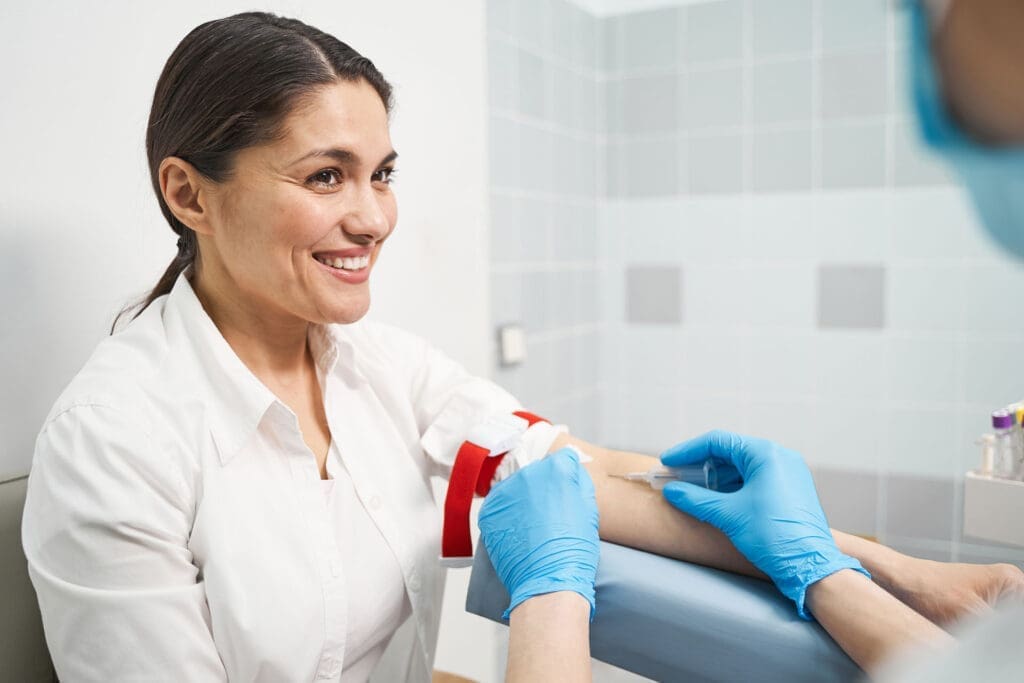
When I step into the procedure room to prepare platelet-rich plasma (PRP), I’m focused on two things: preserving the biologic integrity of the sample and ensuring the patient’s experience is calm, informed, and comfortable. Our team’s back-and-forth—confirming tube labeling, separator position, and plasma depth—reflects the real-world vigilance that high-quality PRP requires.
Key steps I emphasize during PRP prep
- Clear labeling and allocation: Each tube is designated for a specific target (e.g., ankle, knee, hip, scalp, microneedling). This ensures dosing consistency and avoids cross-use.
- Attention to the separator gel: We verify that the separator gel is properly positioned after centrifugation, and we keep the collection needle tip from touching it. Even “just a teeny, tiny” contact can mix layers and reduce PRP quality.
- Air pocket awareness: When aspirating the plasma, I maintain a minor air interface in the syringe to prevent fluid turbulence and keep the tip away from the gel.
- Fraction control: We access the platelet-rich layer just above the buffy coat while minimizing contamination from red cells or separator gel.
- Patient-centered handling: We narrate each step in plain language, respond to sensations (pressure, tension), and integrate gentle scalp massage when appropriate to reduce sympathetically-mediated discomfort.
These details may sound small, but together they protect the bioactive payload—the growth factors and cytokines that make PRP a potent, autologous tool.
Why Separator Gel Contact Matters: The Physiology and Physics
The separator gel is designed to create discrete layers after centrifugation: red cells below, a gel barrier, and plasma above. The platelet-rich fraction sits near the buffy coat. If the collection tip contacts the gel, two things can happen:
- Back-mixing: As my colleague said in the room, plasma components can “leak back” across the boundary. This dilutes the platelet concentration and risks contaminating the PRP with red cells or gel residues.
- Re-spin necessity: We may need to re-centrifuge to re-establish separation, which risks platelet activation at the wrong time, reducing the efficacy of the injection.
Physiologically, the effectiveness of PRP hinges on platelet alpha granules releasing growth factors such as PDGF, TGF-β, VEGF, IGF-1, and EGF at the target tissue. Maintaining an optimal platelet concentration (often 3–5x baseline, depending on protocol) and minimizing pre-activation during handling enhances the burst and sustained release of these factors at the desired site, promoting:
- Angiogenesis and microvascular support (VEGF)
- Chemotaxis and fibroblast proliferation (PDGF)
- Collagen deposition and matrix remodeling (TGF-β)
- Tenocyte and chondrocyte support in musculoskeletal tissue (IGF-1)
- Keratinocyte and dermal regeneration in skin/scalp (EGF)
Modern evidence-based methods—ranging from absorbance-based platelet counts to flow cytometry and standardized classification systems for PRP composition—consistently show that handling variability can affect outcomes (Dohan Ehrenfest et al., 2014; Mautner et al., 2015).
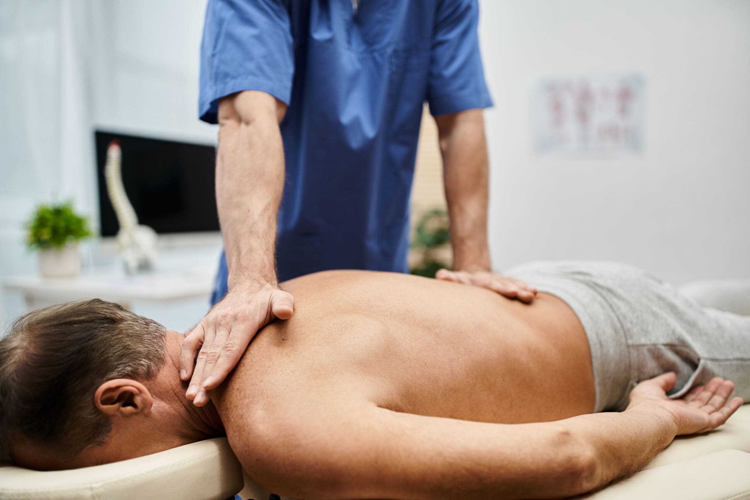
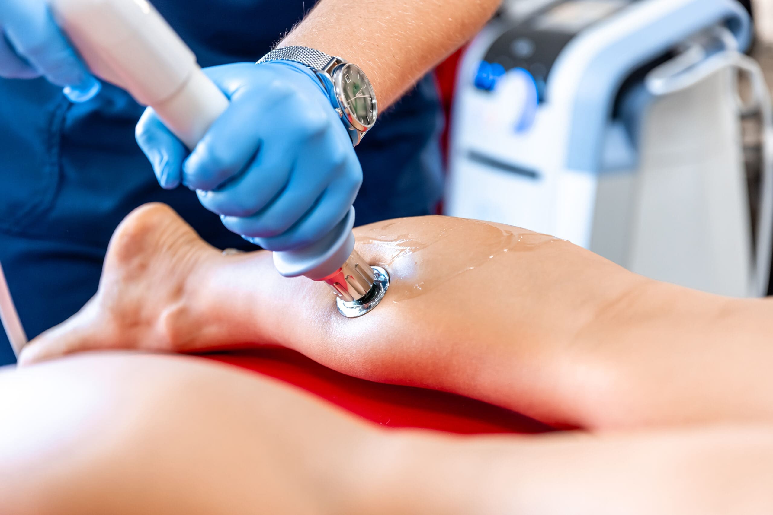
Patient Sensations During Scalp PRP: What They Feel and Why
When I inject PRP into the scalp, patients often describe transient sensations:
- “Head in a vice” or local tension: This is due to the galeal aponeurotica and scalp fascia briefly distending with injected volume. Local nociceptors respond to pressure.
- “Tension headache” quality: Short-lived, typically minutes, as tissue pressure equilibrates and autonomic tone settles.
- Rapid resolution: Gentle scalp massage and parasympathetic engagement reduce sympathetic arousal and help disperse fluid micro-gradients.
We frequently add a topical peptide layer post-PRP. Massaging in the peptides over recently treated tissue taps into mechanotransduction—light pressure can improve microcirculatory spread and may help with nociceptive modulation through gate control mechanisms. Patients often say, “That’s the best part,” because the soothing element reframes the experience and supports adherence to care.
Adding Peptides and Topical Carriers: Synergy with PRP
In the room, we discussed mixing a portion of autologous PRP into a carrier cream formulated to maintain bioactivity—colleagues referenced a “Pro-Cell” moisturizing carrier designed to hold PRP for cosmetic application. The rationale:
- Barrier-compatible vehicle: A well-formulated cream can maintain moisture and protect growth factors at the skin surface long enough to interact with microchannels left by microneedling.
- Microneedling synergy: PRP plus microneedling creates a microenvironment where growth factors enhance keratinocyte proliferation, dermal fibroblast activation, and collagen/elastin remodeling.
- Peptide augmentation: Topical peptides (e.g., copper peptides, signaling peptides) may complement PRP by supporting matrix integrity, angiogenesis, and cell signaling pathways.
We use only carriers known to be compatible with autologous biologics and discuss expectations: topical PRP is an adjunct, not a substitute for injected PRP, where deeper regenerative goals are targeted.
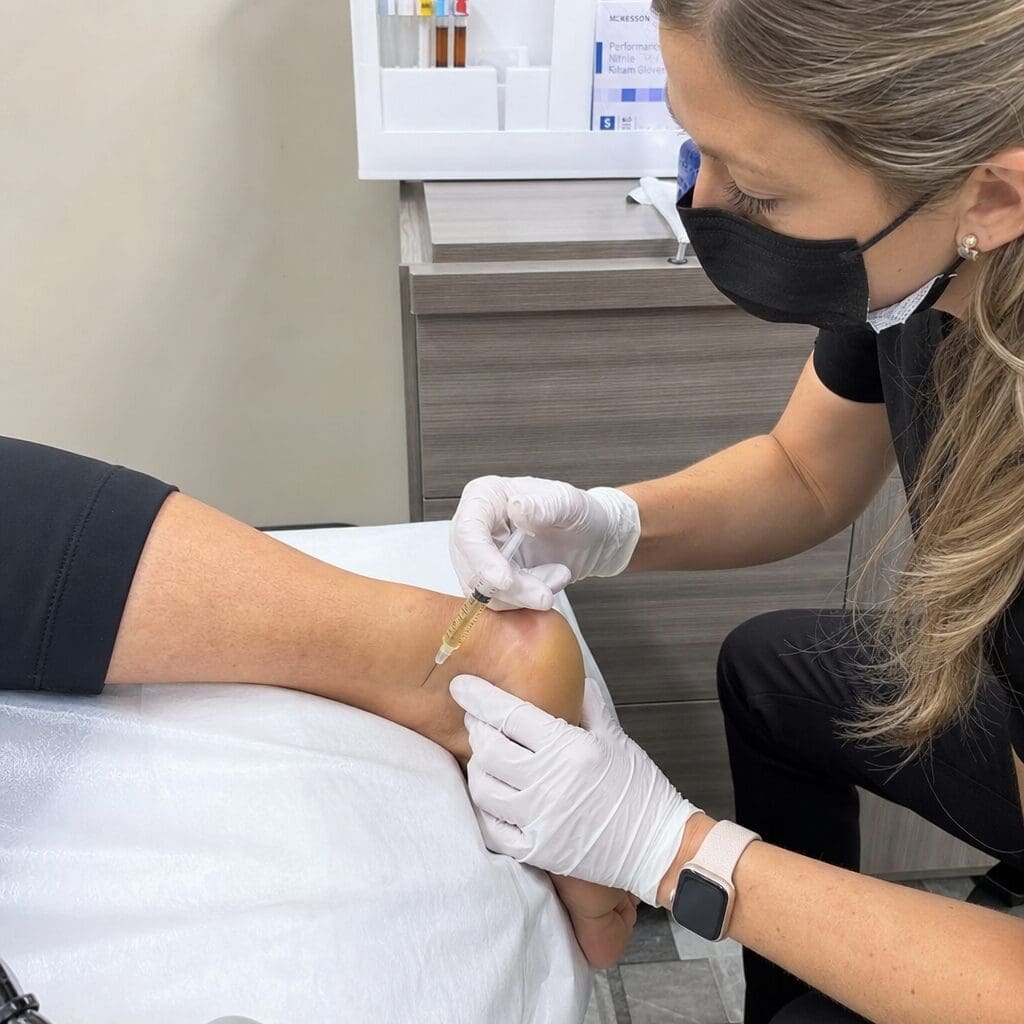
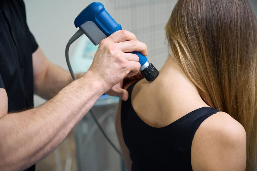
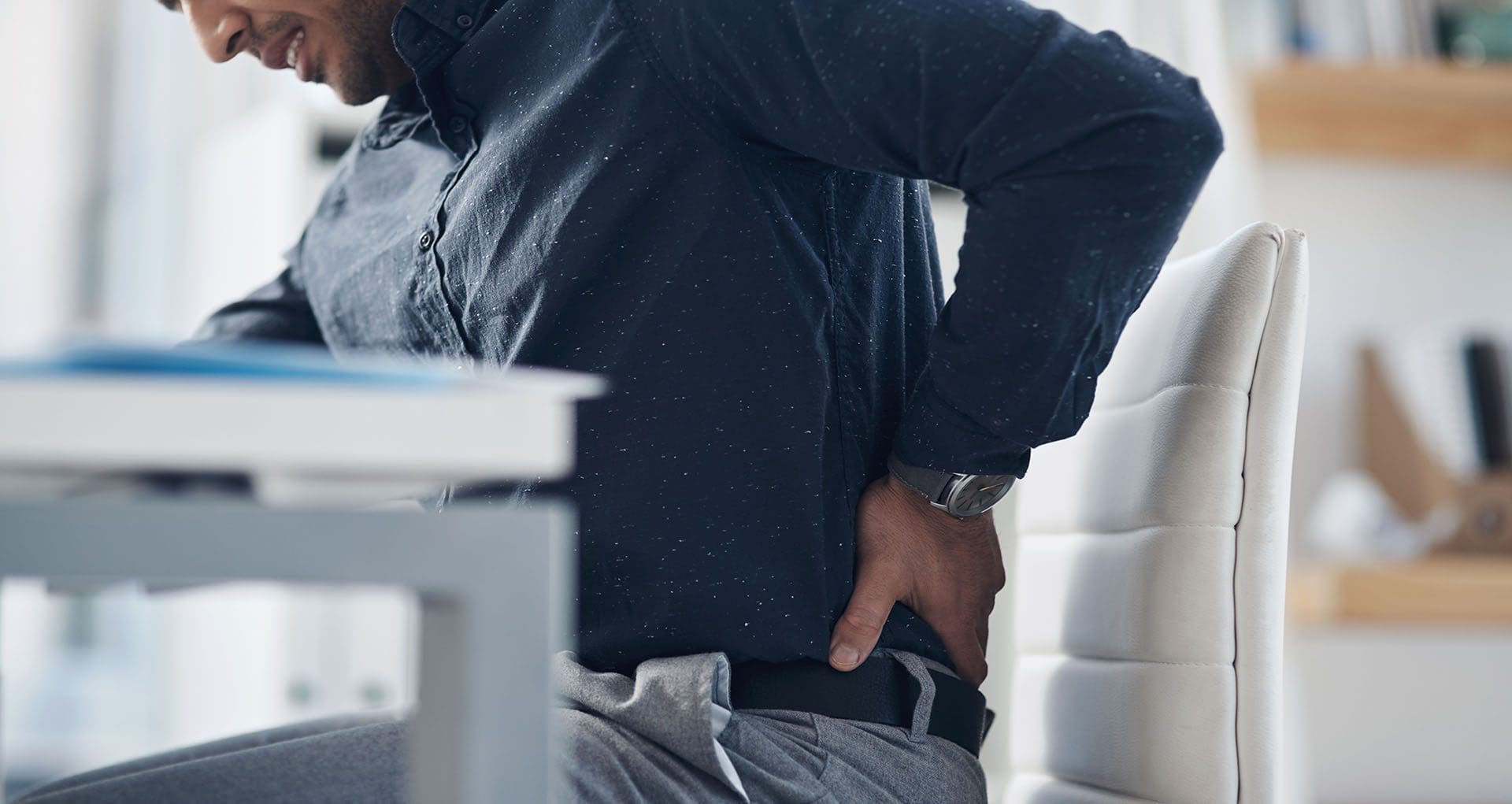
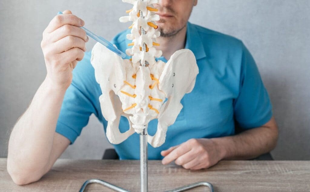
PRP for Joints and Soft Tissue: Mechanisms that Matter
On the musculoskeletal side, we commonly allocate full tubes for joints—ankle, knee, hip—and occasionally for small joints that need targeted attention. The goal is to deliver a platelet concentration appropriate to the tissue type and pathology (e.g., tendinopathy vs. mild osteoarthritis).
Physiologic mechanisms
- Modulated inflammation: PRP can shift the local milieu from catabolic to anabolic by down-regulating NF-κB pathways and promoting anti-inflammatory cytokines (e.g., IL-10) in some formulations.
- Cellular recruitment: PDGF attracts progenitor cells and stimulates fibroblasts and tenocytes; TGF-β drives matrix deposition.
- Cartilage support: While not recreating hyaline cartilage, PRP may improve symptoms and function in early osteoarthritis by encouraging chondrocyte anabolism and modulating synovial inflammation (Laudy et al., 2015; Filardo et al., 2017).
- Tendon and ligament repair: Enhanced collagen synthesis and crosslinking support improved tensile properties over time (Mishra et al., 2014).
Outcome optimization depends on leukocyte content (LP-PRP vs. LR-PRP), activation timing, and injection technique. For intra-articular applications, we typically prefer leukocyte-poor PRP to minimize post-injection flare, while leukocyte-rich PRP may be considered for some tendinopathies where a stronger inflammatory “reset” is useful.
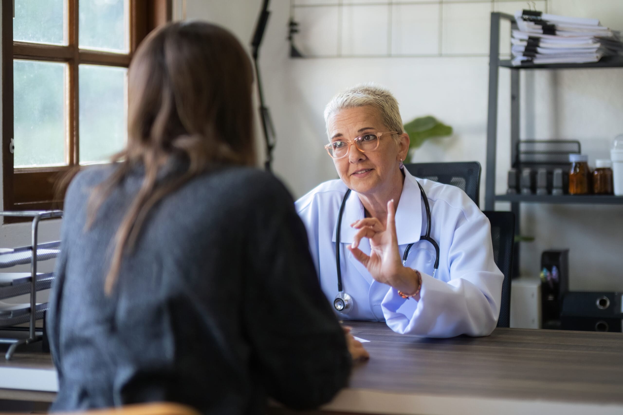
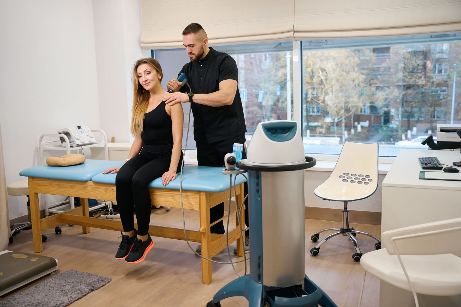
Integrative Chiropractic Care in a Multidisciplinary PRP Framework
I practice as a chiropractor and family nurse practitioner within a comprehensive, medically supervised environment. Dr. Maria Guadalupe Cardenas, MD (NPI #1164426749; Texas License #J2933), serves as Medical Director and Collaborative Physician at Injury Medical Clinic PA (Mission Plaza Injury Medical Clinic) in El Paso, Texas. Our model is multidisciplinary—common in integrative and injury care—blending medical oversight with chiropractic, functional medicine, rehabilitation, and personal injury care.
How we integrate care
- Medical direction and safety: Dr. Cardenas provides oversight on indications, contraindications, and medication management. She ensures adherence to standards for biologics, sterile technique, and emergency preparedness.
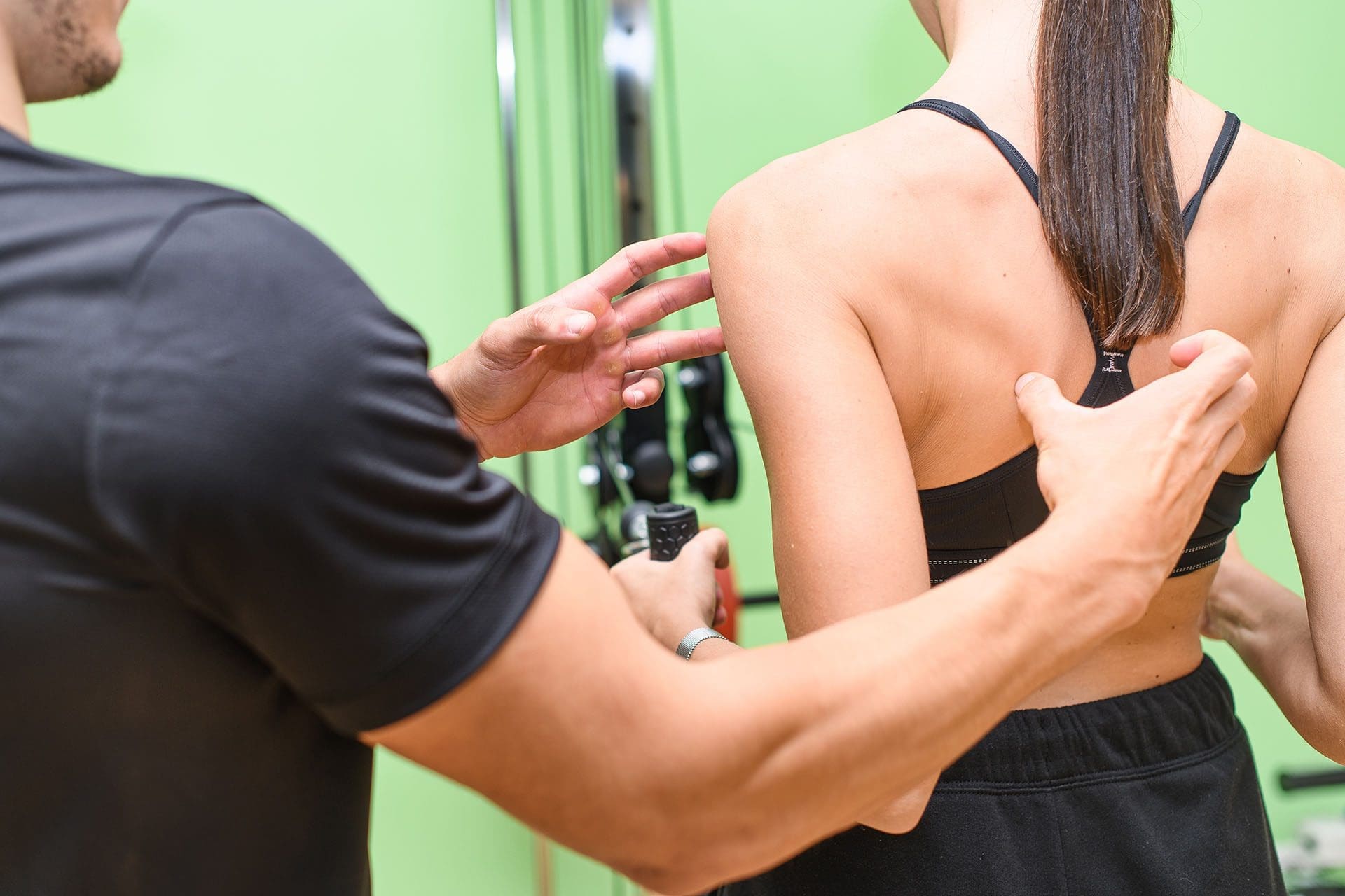
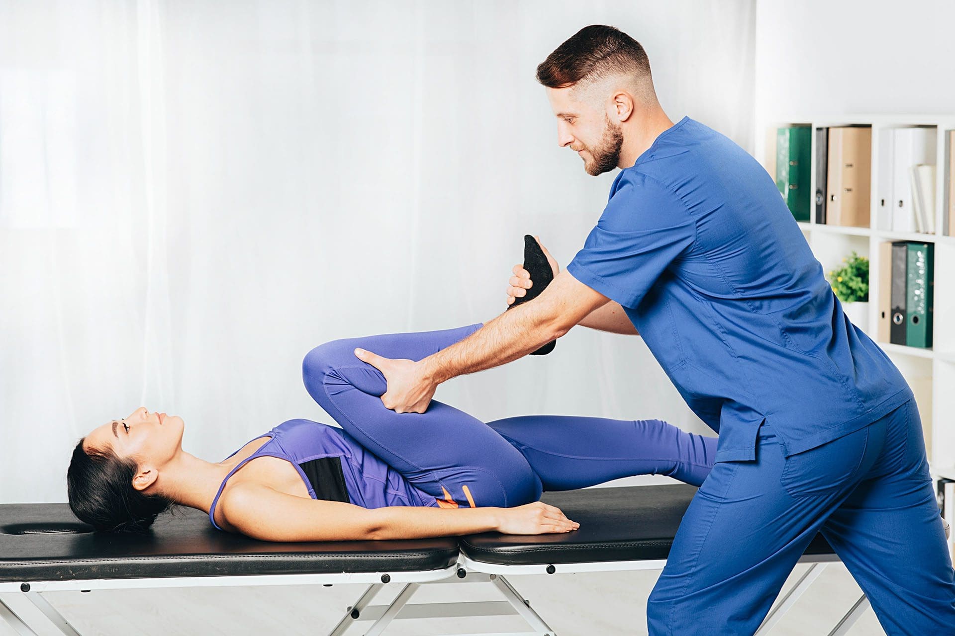
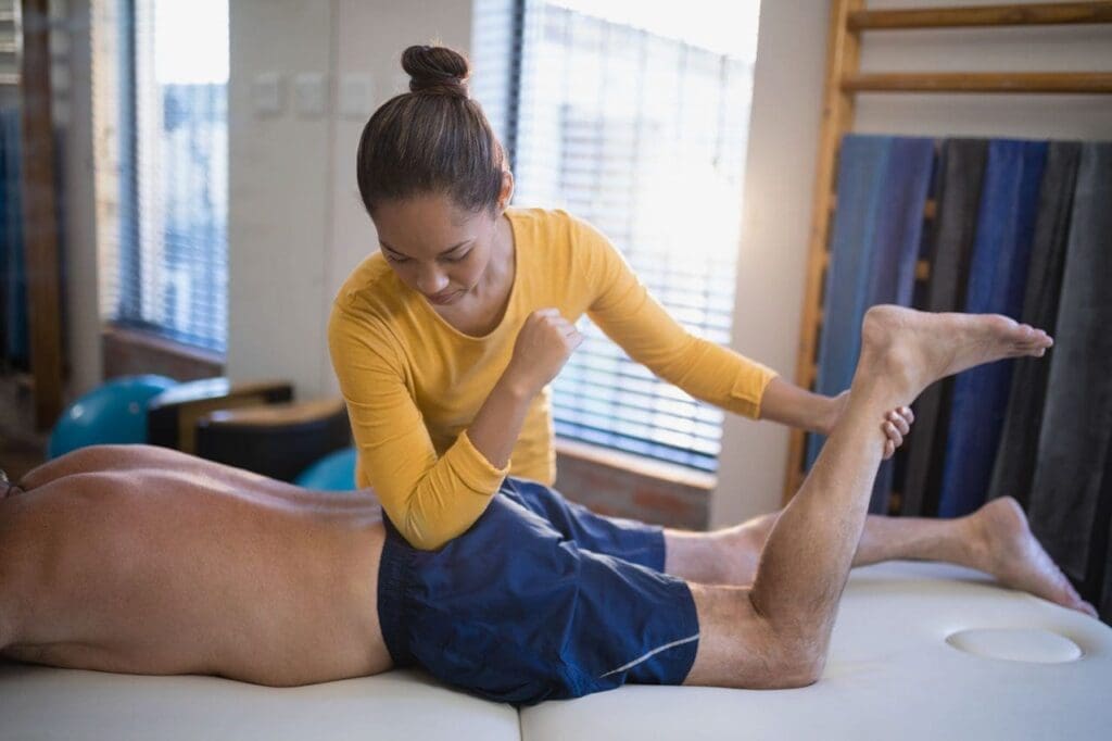
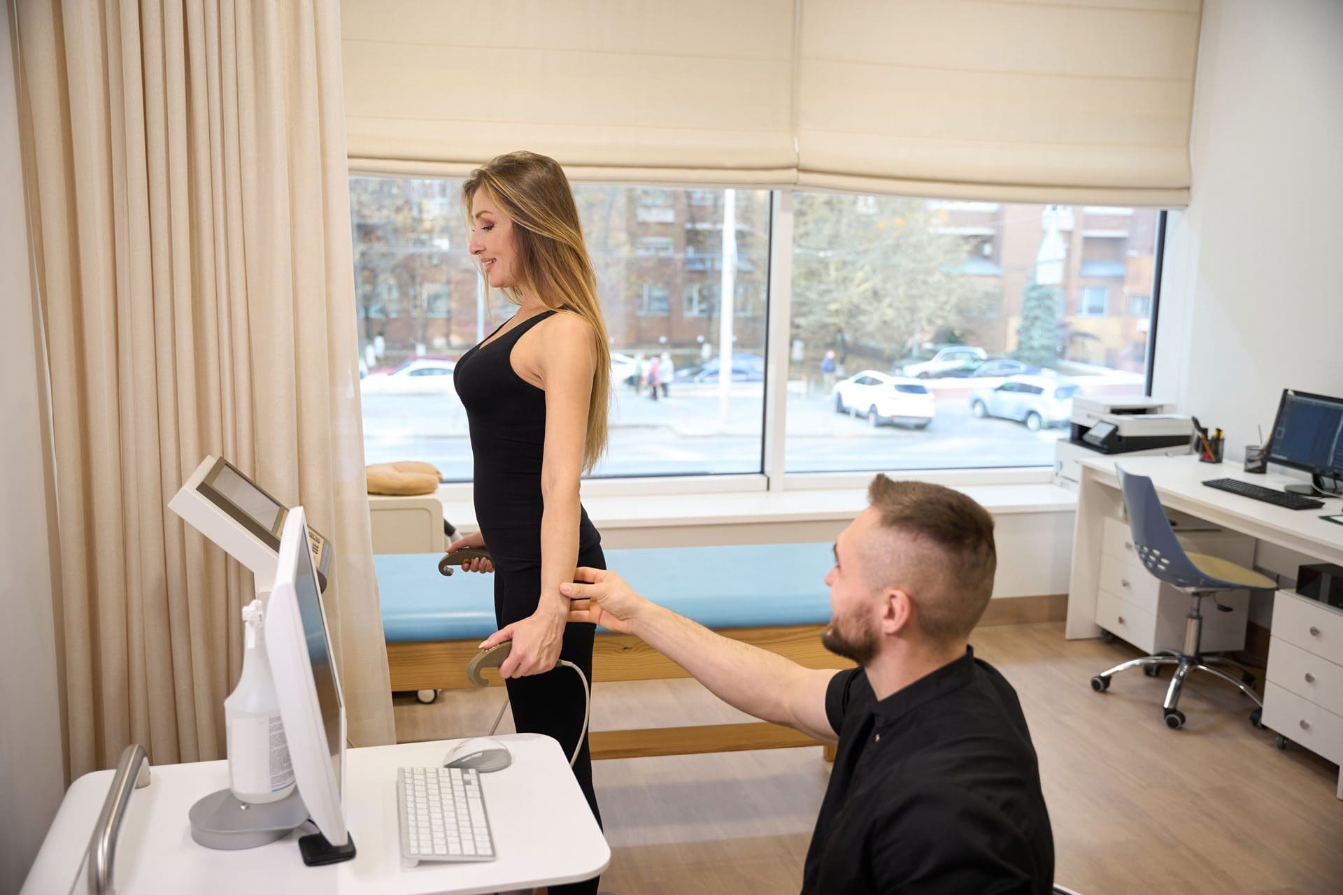
- Chiropractic precision and biomechanics: I evaluate regional interdependence, joint mechanics, and motor control. By restoring segmental mobility and optimizing kinematic chains, we reduce aberrant loading that can undermine PRP’s regenerative intent.
- Functional medicine: We assess metabolic drivers of impaired healing—glycemic control, micronutrient status (e.g., vitamin D, zinc), inflammatory load, sleep, and stress. Good nutrition and lifestyle modulation support collagen synthesis, mitochondrial function, and tissue remodeling.
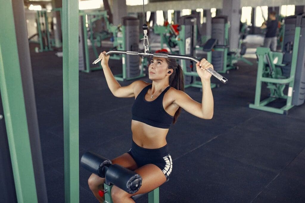
- Rehabilitation: We implement graded loading protocols, isometrics to eccentrics, and neuromuscular re-education to align mechanical signals with biologic repair timelines.
- Personal injury care: For post-collision or workplace injuries, we coordinate imaging, documentation, and return-to-function plans that align medical necessity with ethical, evidence-based practice.
This collaboration means patients receive comprehensive reasoning behind each step—from lab draw to last rep.
Clinical Workflow: Reasoning Behind Each Protocol
- Assessment and stratification
- We differentiate indication (e.g., tendinopathy vs. mild OA vs. hair thinning vs. skin rejuvenation), set goals, and plan volume and frequency.
- We evaluate systemic factors (A1c, lipid profile, inflammatory markers) to optimize the terrain for healing.
- PRP preparation
- Use sterile tubes with separator gel appropriate to the protocol.
- Centrifuge to achieve the desired platelet concentration. Avoid over-activation.
- No gel contact: Maintain a clean interface. If contact occurs and layers mix, consider re-spin or discard to maintain quality.
- Injection technique
- Ultrasound guidance for joints/tendons minimizes off-target placement and reduces post-procedural flare.
- For scalp, distribute small aliquots across regions of thinning; consider nerve block when appropriate for comfort, though many patients manage well with topical and massage support.
- Adjuncts
- Topical peptide cream with PRP for microneedling sessions, when indicated, to enhance epidermal and dermal regeneration.
- Chiropractic adjustments/mobilization to normalize joint and soft-tissue mechanics, reducing nociceptive drive and improving movement quality.
- Rehabilitation and loading
- Protect the site in the acute inflammatory phase (24–72 hours), then transition to subacute loading targeting collagen alignment and capacity building.
- Incorporate isometrics early for tendinopathy, progressing to eccentric/concentric as pain allows.
- Follow-up and metrics
- Track patient-reported outcomes (pain scales, function indices), objective measures (ROM, strength), and where appropriate, trichoscopy for scalp density and microneedling outcomes.
Patient Experience: Comfort Measures that Matter
During scalp injections, we coach patients through breathing and normalize expected sensations. A patient recently told me she felt “more scalp than hair up here” and initially experienced a fleeting “vice-like” tension. We explained that this is temporary pressure within superficial fascial planes and not a true headache. Within minutes, the discomfort subsided—consistent with our typical experience.
- Why it resolves quickly: The scalp’s rich vascularization redistributes fluid while the autonomic nervous system resets. Gentle massage augments fluid kinetics and reduces peripheral nociception.
- Nerve blocks when needed: For patients with heightened sensitivity, small peripheral nerve blocks can blunt afferent signaling, making the session smoother without affecting PRP efficacy.
In joint cases, patients may feel pressure or mild post-injection soreness. We recommend relative rest for 24–48 hours, ice as needed, and resume graded activity according to the tissue and plan.
Evidence Base: What Modern Research Shows
- PRP for knee OA: Meta-analyses suggest PRP can improve pain and function versus hyaluronic acid or saline in mild to moderate osteoarthritis, especially in patients with lower BMI and earlier-stage disease (Belk et al., 2021).
- Tendinopathy: Evidence supports PRP in selected chronic tendinopathies (e.g., lateral epicondylitis, some Achilles and patellar cases), with protocols tailored to tissue and leukocyte content (Mishra et al., 2014).
- Scalp/hair: PRP has demonstrated increases in hair density and thickness in androgenetic alopecia adjunctive to standard care, with repeated sessions optimizing outcomes (Gupta & Carviel, 2022).
- Skin/microneedling: PRP applied with microneedling enhances collagen remodeling and accelerates recovery, improving texture and scar appearance (Alam et al., 2018).
Across studies, technique fidelity—including proper centrifugation, concentration targeting, and avoidance of contamination—correlates with outcomes. This underscores why we obsess over details like no tip-to-gel contact and air pocket management.
Why Integrative Chiropractic Fits: Biomechanics Meet Biology
As a chiropractor, I see PRP not as a standalone solution but as a biologic accelerator that thrives in the right mechanical environment. Consider:
- Joint centration: If a knee remains mal-tracking due to proximal hip weakness or foot mechanics, repeated microtrauma persists. Chiropractic assessments uncover these drivers; targeted manual therapy and corrective exercise reduce aberrant load.
- Fascial continuity: Pain and strain travel through myofascial lines. Addressing thoracolumbar junction stiffness or pelvic torsion can unload distal tendons, allowing PRP-mediated remodeling to consolidate.
- Neuromuscular control: Adjustments and motor control drills refine proprioception and muscle recruitment, reducing shear forces that would otherwise disrupt early collagen matrix.
The result: PRP’s biologic work gains a stable foundation, improving durability of results.
Safety, Oversight, and Ethical Practice
Dr. Cardenas’ medical leadership ensures that:
- Patients are screened for contraindications (e.g., platelet disorders, active infections, anticoagulation concerns).
- Informed consent covers expected benefits, alternatives, and risks.
- Sterile technique is standardized, and contingency protocols are in place.
- Documentation meets medical and legal standards, especially in personal injury contexts.
This framework protects patients and supports reproducible, evidence-informed care.
Practical Tips We Use Every Day
- Keep the syringe tip superficial within the plasma; never let it touch the gel.
- Maintain a small air gap in the syringe to reduce turbulence and preserve layer integrity.
- If the layers mix, don’t force it. Re-spin or consider re-draw depending on the scenario.
- Match PRP composition to target tissue: LP-PRP for joints; consider LR-PRP for certain tendons.
- Use ultrasound guidance for accuracy and safety.
- After scalp PRP, apply peptide-enriched carrier when indicated and massage lightly.
- Align rehab progressions with tissue healing timelines; avoid premature high-load activities.
Clinical Observations from My Practice
Based on patterns I’ve discussed publicly and in professional forums, as well as what we routinely document in the clinic:
- Patients with optimized metabolic health (e.g., A1c < 5.7–6.0, adequate vitamin D, omega-3 index) demonstrate more consistent PRP responses.
- In knee OA, two to three LP-PRP sessions spaced 4–6 weeks apart often yield clinically meaningful improvements in pain and function for 6–12 months, especially when paired with gait retraining and weight management.
- For lateral epicondylitis, a single LR-PRP or staged injections with structured eccentric loading can reduce pain within weeks, with maturation over 3–6 months.
- Scalp PRP combined with home microneedling and peptide topicals shows visible improvements in hair density in 3–4 months for many, with best outcomes when DHT and inflammatory factors are co-managed medically as needed.
These observations align with published literature while acknowledging individual variability—a core reason we tailor care.
The Team Behind the Care
- Medical Director & Collaborative Physician: Dr. Maria Guadalupe Cardenas, MD (Board Certified in Internal Medicine; NPI #1164426749; Texas MD License #J2933), directing medical safety, oversight, and clinical protocols.
- Chiropractic and Functional Care: Dr. Alexander (Alex) Jimenez, DC, APRN, FNP-BC, CFMP, IFMCP, ATN, CCST, integrating biomechanics, manual therapy, functional medicine, and rehabilitation.
- Clinic: Injury Medical Clinic PA (Mission Plaza Injury Medical Clinic), El Paso, Texas—delivering multidisciplinary, integrative solutions for musculoskeletal health, skin/scalp regeneration, and personal injury recovery.
Takeaways for Patients and Clinicians
- Quality handling equals quality outcomes: Protect the platelet payload by respecting the physics of separation.
- Integrate for impact: Combine PRP with chiropractic, rehab, and metabolic optimization for durable results.
- Individualize care: Choose PRP composition and protocols based on tissue type and patient factors.
- Comfort is part of care: Anticipate sensations; smooth the experience with communication, massage, and selective nerve blocks.
- Measure and iterate: Track outcomes and refine protocols with evidence and clinical feedback.
If you are considering PRP for joints, tendons, scalp, or skin, a multidisciplinary approach with medical oversight and integrative chiropractic care can help you reach the best version of your health—safely, methodically, and compassionately.
References
- Alam, M., et al. (2018). “Platelet-rich plasma for skin rejuvenation and scars: a systematic review and meta-analysis.” JAMA Dermatology.
- Belk, J. W., et al. (2021). “Platelet-rich plasma versus hyaluronic acid for knee osteoarthritis: a systematic review and meta-analysis.” Orthopaedic Journal of Sports Medicine.
- Dohan Ehrenfest, D. M., et al. (2014). “Classification of platelet concentrates (Platelet-Rich Plasma—PRP, Platelet-Rich Fibrin—PRF) for topical and infiltrative use in orthopedic and sports medicine: current consensus, clinical implications and perspectives.” Muscles, Ligaments and Tendons Journal.
- Filardo, G., et al. (2017). “Platelet-rich plasma intra-articular injections for cartilage degeneration and osteoarthritis: single- versus double-spinning approach.” Knee Surgery, Sports Traumatology, Arthroscopy.
- Gupta, A. K., & Carviel, J. L. (2022). “A randomized, double-blind study of PRP for androgenetic alopecia: efficacy and safety.” Dermatologic Surgery.
- Mautner, K., et al. (2015). “A call for standardization in platelet-rich plasma preparation protocols and composition reporting.” PM&R.
- Mishra, A., et al. (2014). “Treatment of tendon and muscle using platelet-rich plasma.” PM&R.