Integrative Accident and Work Injury Care in El Paso

An auto accident or workplace injury can affect the body in several ways at the same time. A person may experience inflammation, muscle spasms, joint stiffness, ligament damage, nerve irritation, weakness, and poor movement.
Rest may help mild soreness. However, stubborn injuries often need a more complete recovery plan.
At ChiroMed – Integrated Medicine in El Paso, care focuses on the whole injury rather than just covering up pain. An integrative wellness plan may combine chiropractic care, medical assessment, functional medicine, rehabilitation, nutrition, and advanced therapies when appropriate.
The main goals are to:
- Calm pain and inflammation
- Identify the injured tissues
- Restore spinal and joint movement
- Support natural tissue repair
- Rebuild strength and stability
- Help the patient return to work and daily activities
- Reduce the risk of long-term pain
This layered approach gives patients a clear path from the early stages of an injury to long-term functional recovery.
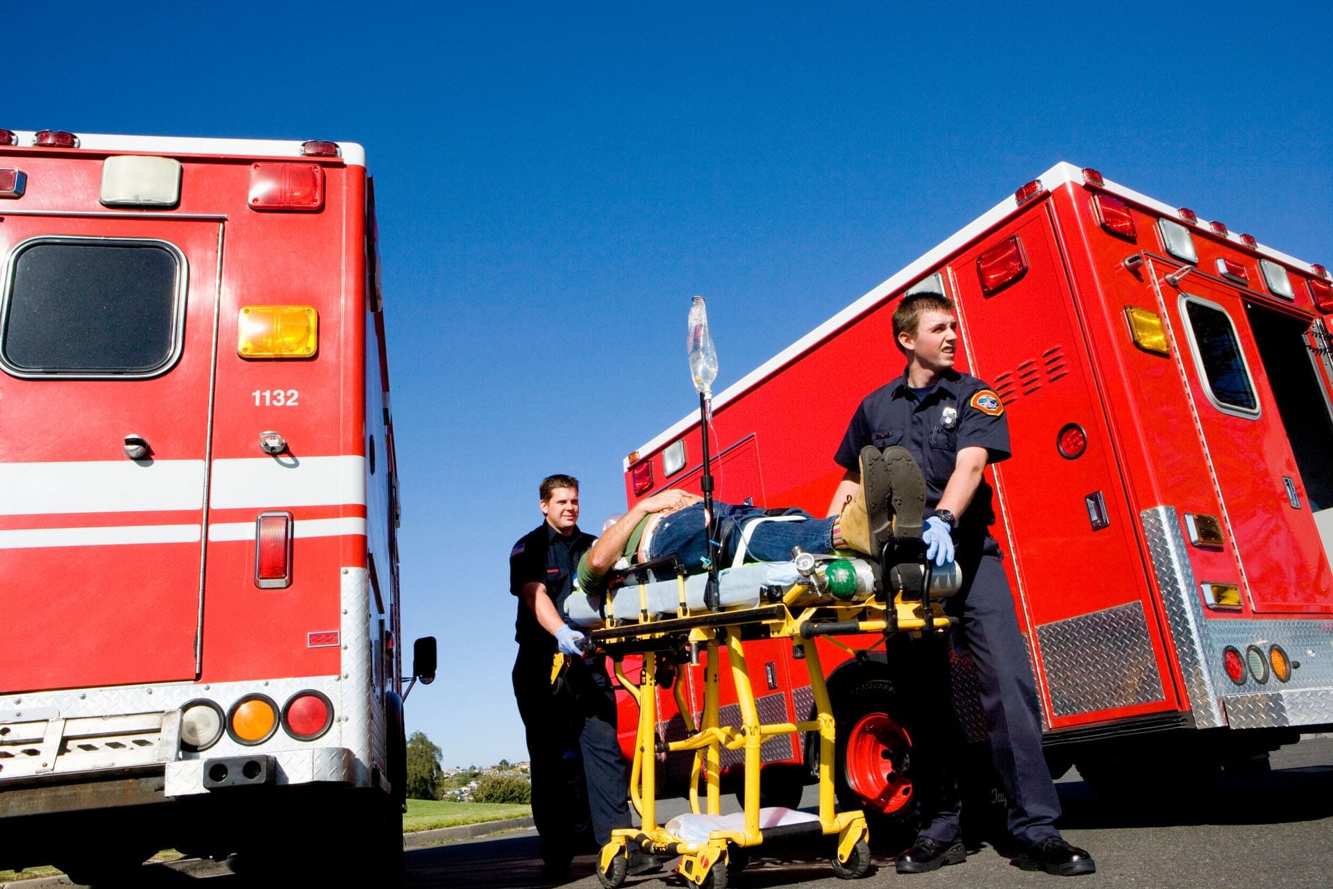
Why Accident and Work Injuries Need a Complete Plan
Accidents can place great force on the spine, joints, muscles, and connective tissues.
During a car crash, the body may move forward, backward, or sideways before a person has time to react. A seat belt can save a life, but it may also place pressure across the shoulder, chest, or hip. Drivers may grip the steering wheel or brace their arms before impact, which can contribute to shoulder, elbow, wrist, or hand injuries.
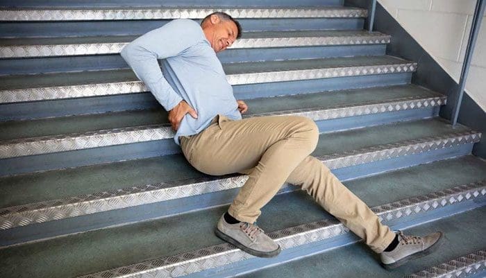
Work injuries can happen during:
- Repeated lifting
- Pushing or pulling
- Slips and falls
- Awkward twisting
- Repetitive arm movements
- Long periods of sitting
- Heavy labor
- Machinery accidents
- Poor workstation setup
Common symptoms include neck pain, back pain, headaches, sciatica, joint stiffness, muscle spasms, numbness, tingling, weakness, and reduced range of motion.
Some symptoms begin right away. Others take several hours or days to become noticeable as swelling and muscle guarding increase. For this reason, early evaluation can be important even when the patient believes the injury is minor (El Paso Chiropractor Blog, 2026a, 2026b).
Phase One: Identify the Injury and Reduce Inflammation
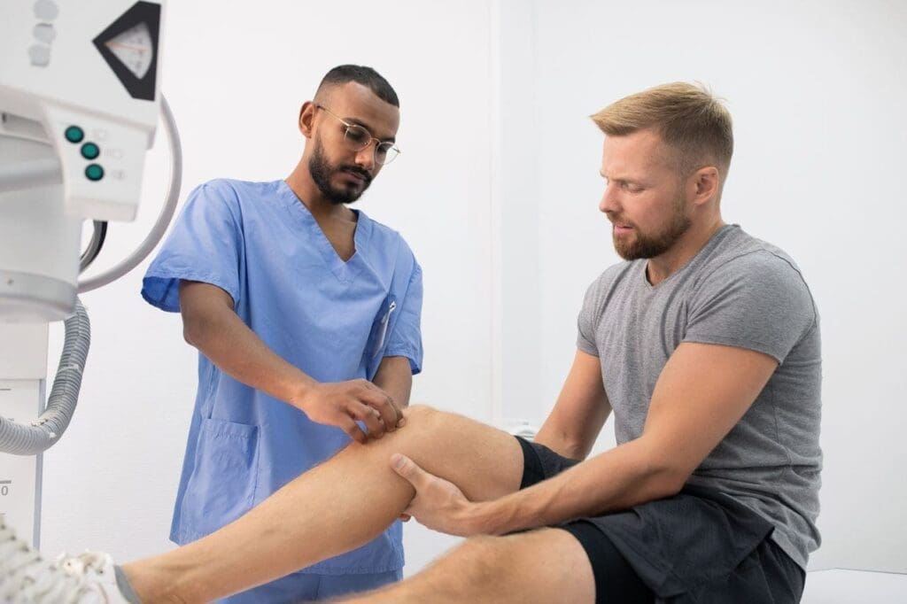
The first stage of care begins with a complete evaluation.
The provider asks how the injury happened, which body parts were affected, when symptoms began, and which activities make the pain better or worse. The examination may include posture testing, range-of-motion measurements, orthopedic tests, neurological screening, muscle strength testing, and movement analysis.
Imaging or referral may be needed when the examination suggests:
- A fracture
- A major ligament tear
- Severe joint instability
- A traumatic brain injury
- Spinal cord involvement
- Progressive muscle weakness
- Loss of bowel or bladder control
- Infection
- A medical emergency
The early treatment plan is usually gentle. The goal is not to force a painful area to move. Instead, care may focus on reducing irritation, protecting injured tissues, controlling muscle spasms, and maintaining safe movement.
Early treatment may include:
- Gentle joint movement
- Soft-tissue therapy
- Cold or heat when appropriate
- Light corrective exercises
- Activity changes
- Supportive taping or bracing
- Nutrition and hydration guidance
A safe plan also considers the patient’s age, medical history, medications, previous injuries, job duties, and overall health.
Phase Two: Restore Spinal and Joint Mechanics
Once the patient can move more safely, treatment may begin addressing restricted joints, poor posture, and abnormal movement patterns.
Chiropractic Care
Chiropractic care focuses on how the spine, joints, muscles, and nervous system work together.
After an accident, muscle guarding may limit normal joint movement. When one area does not move properly, nearby muscles and joints often work harder to compensate for the restriction. This can create a cycle of pain, stiffness, and poor movement.
Carefully selected chiropractic adjustments and joint mobilization may help:
- Restore joint movement
- Reduce mechanical stress
- Improve range of motion
- Decrease muscle guarding
- Support better posture
- Make rehabilitation more comfortable
Chiropractic care does not replace emergency medicine, orthopedic care, or other medically necessary services. It is one part of a coordinated injury-recovery plan.
Research suggests that spinal manipulation may provide modest improvements in pain and function for some patients with neck or back pain. Treatment must be selected according to the patient’s examination, diagnosis, comfort, and risk factors (National Center for Complementary and Integrative Health [NCCIH], n.d.).
Spinal Decompression
Spinal decompression uses controlled traction to gently stretch the spine.
It may be considered for selected patients with:
- Bulging or herniated discs
- Sciatica
- Disc-related neck pain
- Nerve irritation
- Spinal stiffness
The goal is to reduce mechanical pressure and make movement more comfortable. Decompression may also be combined with chiropractic care and corrective exercises.
It is not suitable for every patient. People with fractures, severe osteoporosis, tumors, major spinal instability, or certain medical conditions may need a different form of care.
Spinal decompression should not be presented as a stand-alone cure. Long-term improvement usually also requires stronger muscles, better movement patterns, and changes to activities that continue to place stress on the spine (Sciatica Clinic, 2026a).
Phase Three: Address Stubborn Soft-Tissue Injuries
Muscles, tendons, ligaments, cartilage, and spinal discs do not all heal at the same rate.
Some tissues have a limited blood supply. Others continue to face stress from poor posture, joint instability, repetitive work, or abnormal movement. When an injury does not improve with rest and basic conservative care, additional procedures may be discussed.
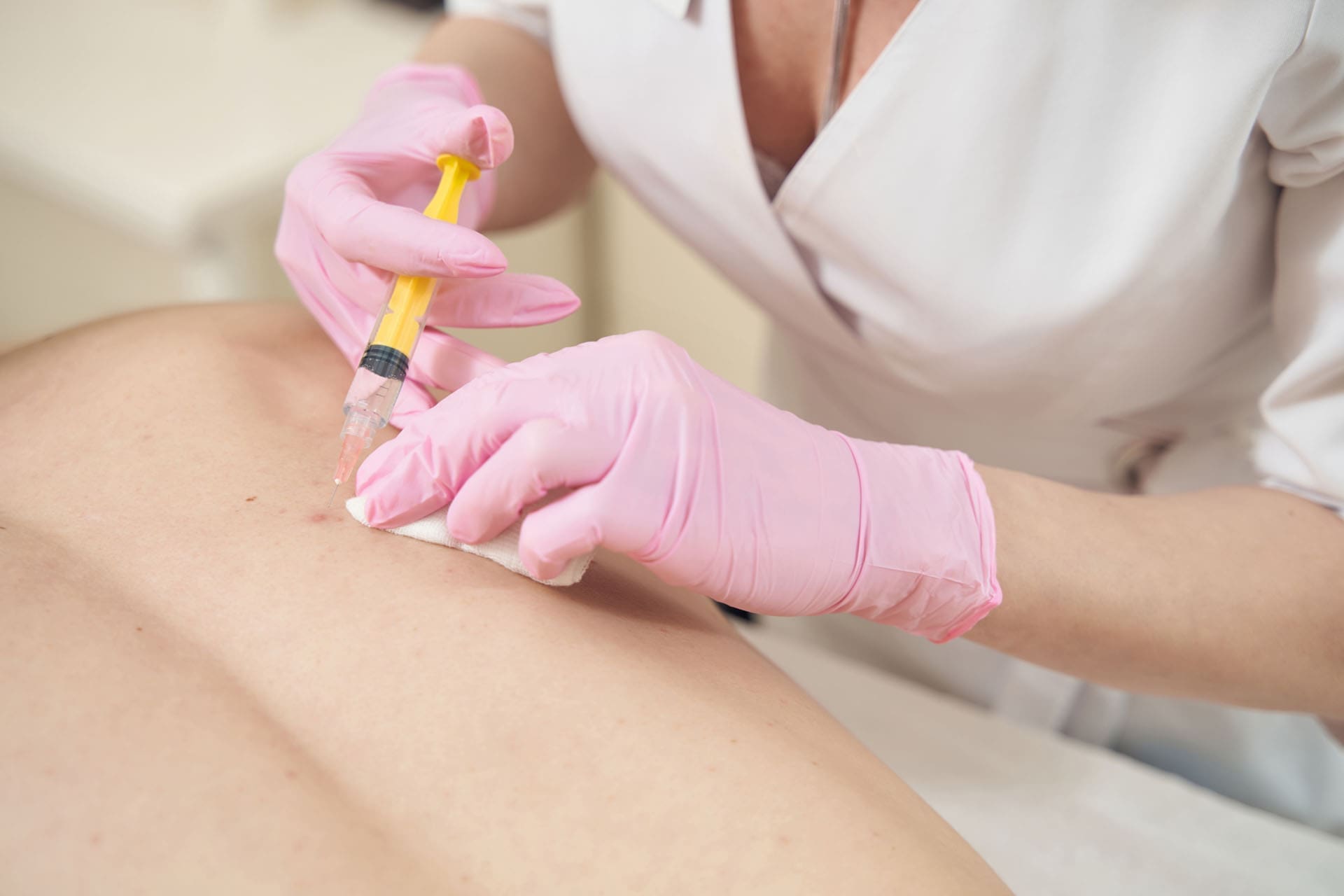
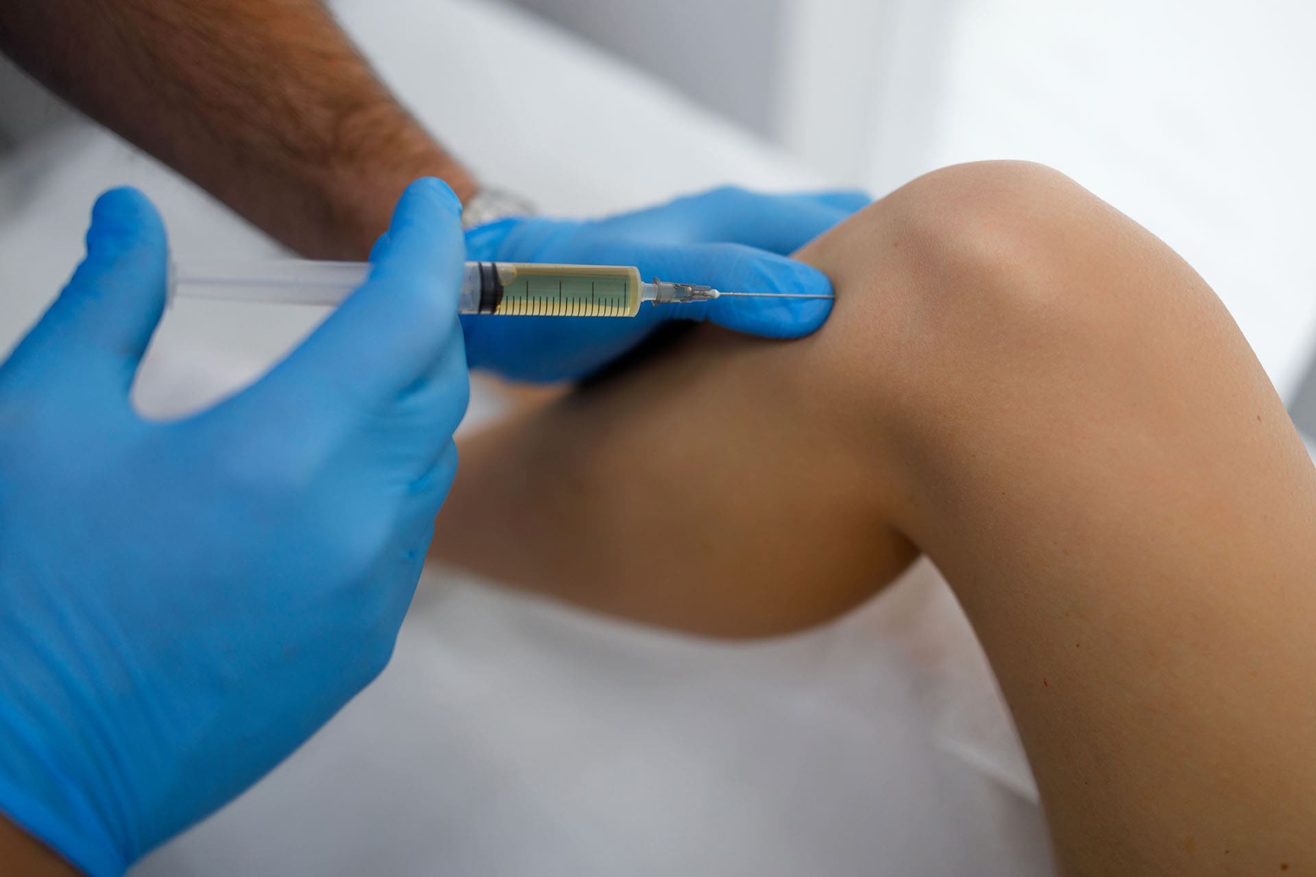
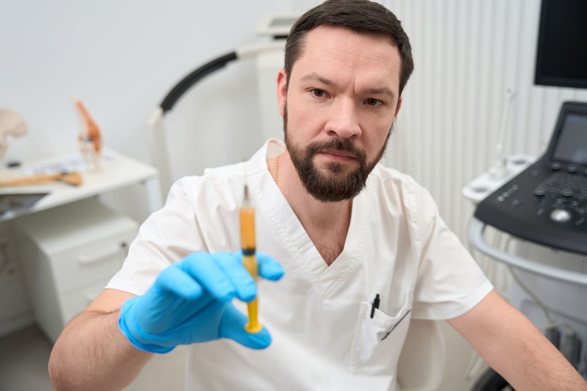
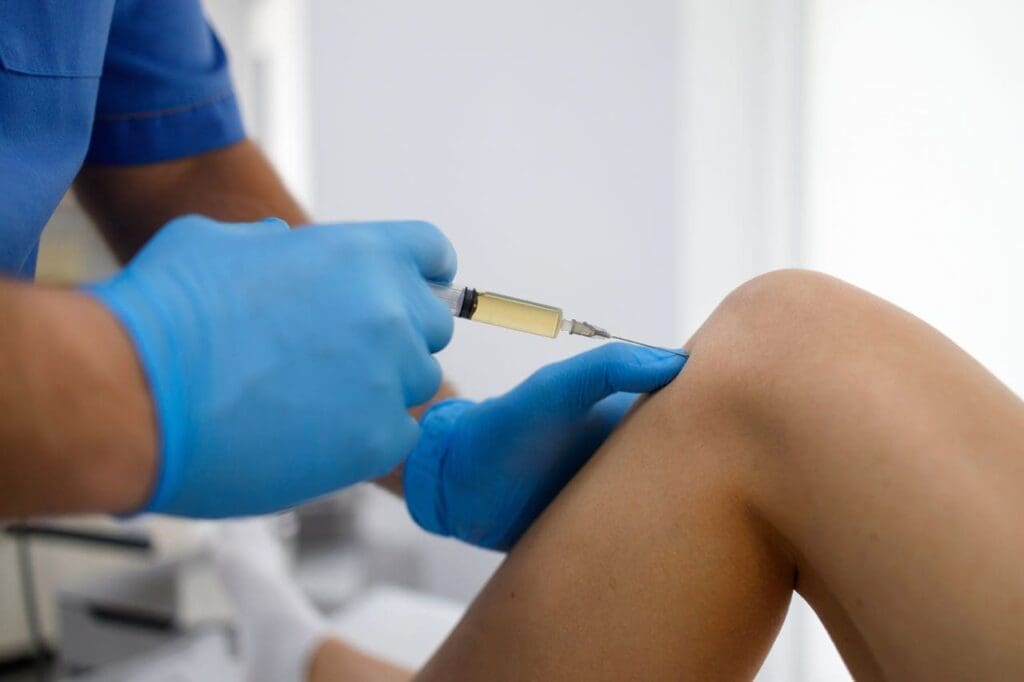
Platelet-Rich Plasma Therapy
Platelet-rich plasma, or PRP, is prepared from the patient’s own blood. A medical professional processes the blood to create a platelet-rich portion.
Platelets contain proteins and growth factors involved in the body’s normal healing response. PRP may be considered for selected tendon, ligament, muscle, or joint injuries.
PRP does not instantly rebuild damaged tissue. It is designed to support the natural repair process. Results may also depend on:
- The type and severity of the injury
- The patient’s health
- The way the PRP is prepared
- How accurately it is placed
- Activity after the procedure
- The rehabilitation plan
- Continued mechanical stress on the area
Dr. Alexander Jimenez’s clinical observations emphasize that regenerative procedures should be combined with improved biomechanics. Treating injured tissue without correcting the movement problem that continues to stress it may limit recovery (Jimenez, 2026).
Microfragmented Adipose Tissue
Microfragmented adipose tissue, commonly called MFAT, is prepared from a small amount of the patient’s fat tissue.
The processed tissue contains structural and signaling components that may support selected orthopedic procedures. It may be considered for some joint, cartilage, or complex soft-tissue conditions.
MFAT requires a fat-harvesting procedure and is more involved than a standard blood draw. PRP and MFAT are not the same treatment.
The choice may depend on:
- The injured structure
- How long symptoms have been present
- Imaging findings
- Previous treatments
- The patient’s overall health
- The expected risks and benefits
No regenerative procedure is best for every patient. A qualified medical provider must first determine whether the person is a suitable candidate (Sports Medicine of the Rockies, 2026).
Patients should also be careful with clinics that promise guaranteed tissue regrowth or market unapproved products as cures. Regenerative procedures should be based on a clear diagnosis, realistic expectations, and proper medical screening.
Laser and Shockwave Therapy
Noninvasive technologies may be used to support pain relief and rehabilitation.
Therapeutic Laser
Therapeutic laser treatment uses selected wavelengths of light over the injured area. This process is often called photobiomodulation.
The therapy may influence cellular activity, local circulation, and inflammatory signals. It may be used as a supportive option for muscle pain, joint irritation, or soft-tissue injuries.
Laser treatment does not physically align the spine or replace exercise. Its role is to help reduce discomfort so the patient can participate more comfortably in movement and rehabilitation.
Shockwave Therapy
Extracorporeal shockwave therapy uses acoustic waves to stimulate targeted tissue.
It is often considered for chronic tendon and soft-tissue problems, including:
- Plantar fasciitis
- Tennis elbow
- Achilles tendon pain
- Calcific shoulder conditions
- Chronic muscle or tendon pain
- Areas with long-standing scar tissue
Shockwave therapy may support circulation, collagen activity, and tissue remodeling. Temporary soreness can occur after treatment.
Patients with certain bleeding risks, infections, tumors, or other medical concerns may not be suitable candidates. Screening should take place before treatment begins (Harrington, n.d.).
Phase Four: Support Healing Through Nutrition
The body needs adequate nutrients to repair injured tissues.
Protein supplies amino acids used to maintain and rebuild muscles, tendons, ligaments, and other tissues. Vitamins and minerals support energy production, nerve function, collagen formation, and immune activity.
A recovery-focused nutrition plan may include:
- Adequate protein
- Vegetables and fruits
- Healthy fats
- Whole-food carbohydrates
- Enough water
- Foods containing vitamin C
- Foods containing magnesium and zinc
- Stable meal timing
- Reduced heavily processed food intake
Sleep also matters. The body performs many repair processes during sleep. Poor sleep can increase pain sensitivity, reduce energy, and make it harder to follow a rehabilitation program.
Functional medicine may help identify other issues that can slow recovery, such as poor blood sugar control, nutrient deficiencies, digestive concerns, chronic inflammation, or unhealthy lifestyle habits.
IV Nutrient Support
IV fluids or nutrients may be considered when there is a clear medical reason, such as dehydration, poor absorption, or a documented deficiency.
IV therapy sends fluids and selected nutrients directly into the bloodstream. It must be provided with proper screening, sterile technique, careful dosing, and medical oversight.
IV therapy should not replace:
- Healthy food
- Water
- Sleep
- Chiropractic care
- Rehabilitation
- Necessary medical treatment
It should also not be promoted as a guaranteed way to heal an injury. Evidence for routine high-dose vitamin infusions in otherwise healthy people remains limited. The treatment must match the patient’s individual medical needs (Alangari et al., 2025).
Phase Five: Rebuild Strength and Function
Pain relief is not the final step.
A patient must regain the ability to walk, bend, lift, reach, work, exercise, and safely complete daily activities. This requires functional rehabilitation.
A rehabilitation program may include:
- Range-of-motion exercises
- Core strengthening
- Hip and leg strengthening
- Shoulder stability exercises
- Balance training
- Posture correction
- Walking or aerobic conditioning
- Work-specific movements
- Gradual lifting practice
- Home exercises
Exercise should progress in stages. Too much activity too soon may irritate healing tissues. Too little movement for too long may lead to stiffness, weakness, and fear of movement.
Progress can be measured through:
- Improved range of motion
- Reduced pain
- Better muscle strength
- Greater walking tolerance
- Improved lifting ability
- Better balance
- Safer work activity
- Increased independence
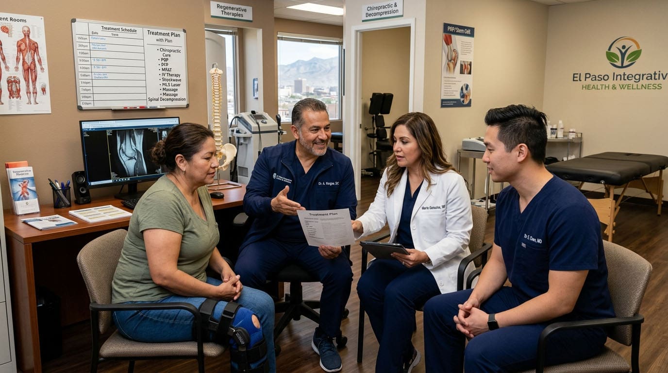
Team-Based Injury Care at ChiroMed
ChiroMed – Integrated Medicine in El Paso uses a multidisciplinary approach to treating accident, work, and sports injuries, as well as chronic musculoskeletal conditions.
The clinic’s services may bring together:
- Chiropractic care
- Nurse practitioner services
- Medical evaluation and oversight
- Functional medicine
- Rehabilitation
- Nutrition counseling
- Soft-tissue treatment
- Spinal decompression
- Therapeutic laser
- Shockwave therapy
- Regenerative medicine consultations
- Personal injury documentation
Dr. Alexander Jimenez, DC, APRN, FNP-BC, CCST, CFMP, IFMCP, ATN, leads chiropractic and integrative clinical care. His combined background in chiropractic, advanced practice nursing, functional medicine, spinal trauma, and rehabilitation allows him to view an injury from several clinical angles.
His clinical observations focus on identifying the cause of ongoing pain rather than treating only the painful area. This includes examining joint mechanics, nerve function, muscle balance, nutrition, inflammation, lifestyle, and the patient’s ability to perform normal activities.
Dr. Maria Guadalupe Cardenas, MD, is board-certified in internal medicine and brings more than 40 years of clinical experience. She serves as Medical Director and Collaborative Physician at Injury Medical Clinic PA.
Her Texas medical license is J2933. Current public provider listings identify her NPI as 1164426748.
Dr. Cardenas provides medical direction alongside Dr. Jimenez’s chiropractic, functional medicine, personal injury, and rehabilitation services. This type of structure is common in multidisciplinary injury clinics.
The chiropractor and rehabilitation team focus on spinal mechanics, joint motion, soft-tissue function, and corrective exercises. The medical physician supports clinical oversight, complex case review, medical safety, and coordination when a patient needs services beyond conservative musculoskeletal care.
A Clearer Path From Injury to Recovery
A complete injury plan is not based on placing every patient into the same treatment program.
The right plan depends on:
- How the injury happened
- Which tissues were damaged
- How severe the symptoms are
- The patient’s overall health
- Work and family responsibilities
- Previous treatments
- Response to care
At ChiroMed, the recovery process may move from inflammation control to structural care, tissue support, and functional rehabilitation.
Chiropractic care and decompression address mechanical stress. PRP, MFAT, laser, and shockwave therapy may support selected injuries. Nutrition and medically appropriate IV therapy support overall health. Rehabilitation helps the patient regain strength and function.
The purpose is not only to manage pain today. It is to help the patient understand the injury, correct contributing problems, support natural healing, and build a stronger foundation for the future.
To learn more about integrative accident or work injury care in El Paso, visit ChiroMed – Integrated Medicine or call 915-850-0900.
References
Alangari, A., et al. (2025). To IV or not to IV: The science behind intravenous vitamin therapy.
ChiroMed. (n.d.-a). About ChiroMed – Integrated Medicine.
ChiroMed. (n.d.-b). Integrated injury care in El Paso, Texas.
ChiroMed. (n.d.-c). Integrated medicine services in El Paso, Texas.
ChiroMed. (2026). Regenerative therapy for auto accident injury recovery.
El Paso Chiropractor Blog. (2026a). Arm and shoulder injuries after auto accidents.
El Paso Chiropractor Blog. (2026b). Speeding and aggressive driving accidents.
Harrington, P. (n.d.). Comparing Class 4 laser therapy, PEMF, and shockwave treatments in chiropractic care.
Jimenez, A. (2026). How PRP composition influences your healing journey.
National Center for Complementary and Integrative Health. (n.d.). Spinal manipulation: What you need to know.
New Regeneration Orthopedics. (2021). Chiropractors: How to integrate regenerative medicine into your practice the right way.
Sciatica Clinic. (2026). Integrated posture care combining multiple therapies.
Sports Medicine of the Rockies. (2026). Comparing PRP, BMAC, and MFAT: Choosing the right regenerative treatment.
The Neck and Back Clinics. (n.d.). What are your chiropractic treatment options after a car accident?.