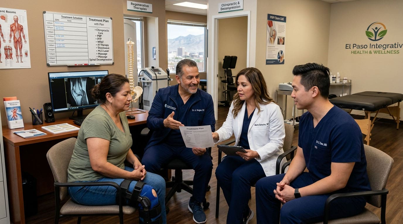
Regenerative Therapies for Wellness, Exercise, and Fitness
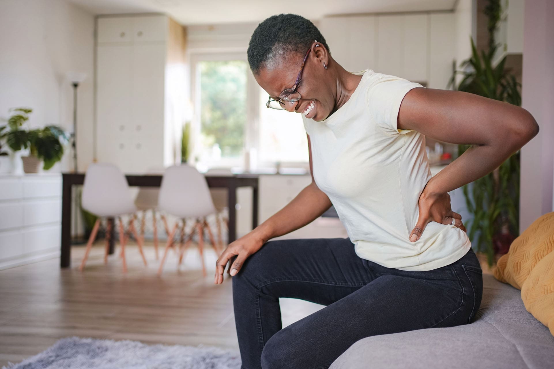
Pain and injuries can make exercise feel difficult or unsafe. A painful knee may change the way you walk. A shoulder injury may prevent you from lifting weights. An irritated spinal nerve may cause pain, numbness, tingling, or weakness that travels into an arm or leg.
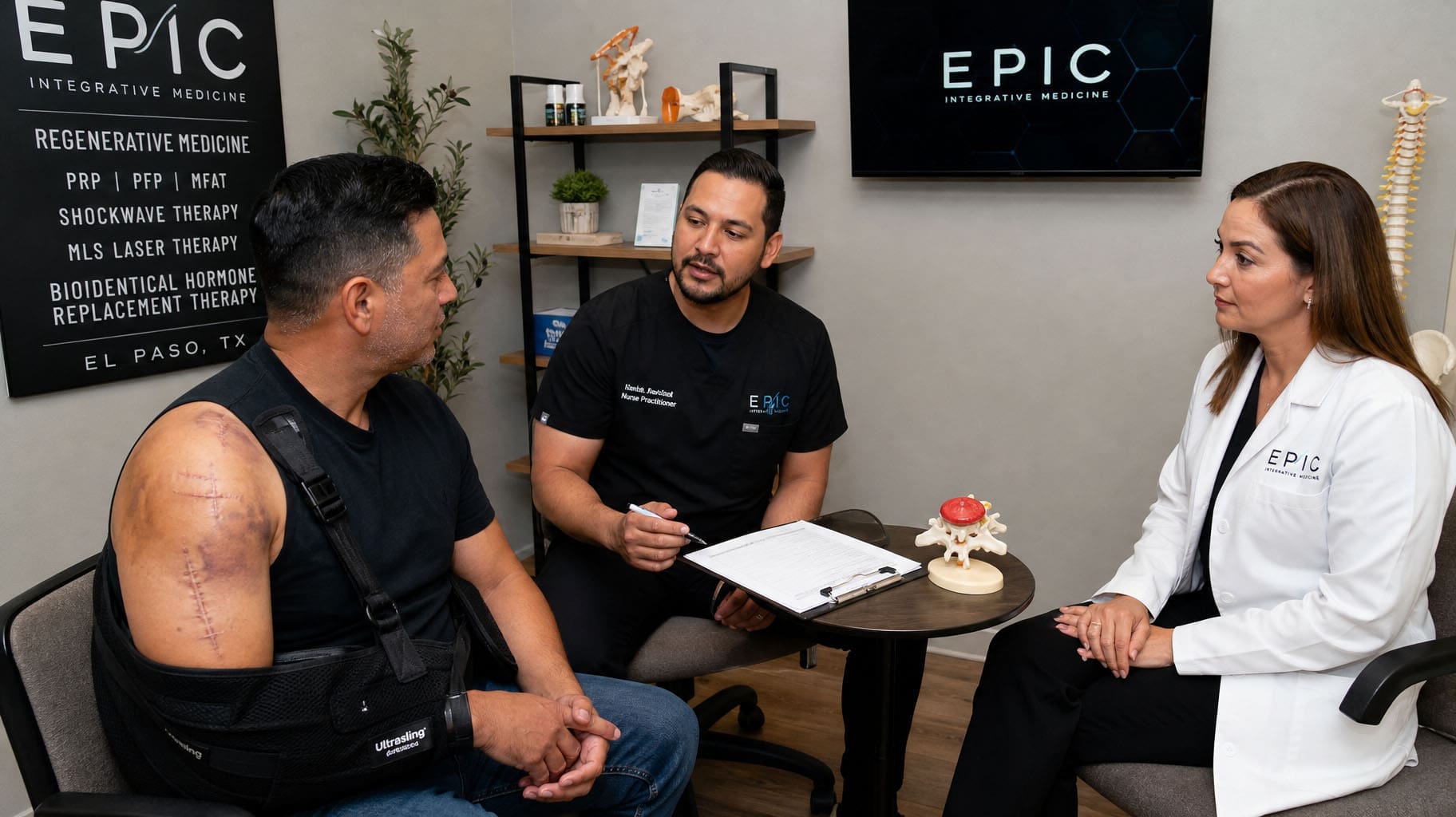
At ChiroMed in El Paso, Texas, recovery is viewed as more than simply lowering pain. The goal is to understand why the pain is happening, support injured tissues, restore healthy movement, and help the patient return to exercise, work, sports, and daily activities safely.
Regenerative therapies such as platelet-rich plasma, platelet-free or platelet-poor plasma, and microfragmented adipose tissue may support the body’s natural repair process in carefully selected patients. Epidural spinal injections may help calm inflammation around irritated spinal nerves. IV infusion nutrient therapy may support hydration and correct certain nutritional deficiencies when medically appropriate.
These treatments may work best when combined with chiropractic care, functional medicine, rehabilitation, and tailored exercise. This team-based approach creates an environment where biological repair, structural alignment, cellular nutrition, and physical conditioning work together.
Building a Better Environment for Recovery
An injury can involve several problems at the same time.
A joint may be inflamed. A ligament may be weak. A spinal nerve may be irritated. Nearby muscles may tighten to protect the area. The patient may then stop moving normally because of pain.
Simply covering the pain may not correct these problems.
An integrative recovery plan may address:
- Tissue irritation and inflammation
- Joint stiffness or poor alignment
- Weak or unbalanced muscles
- Spinal nerve irritation
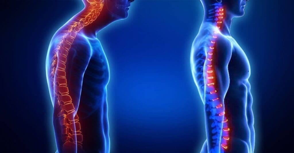
- Poor posture or movement habits
- Dehydration or nutritional deficiencies
- Reduced strength and physical endurance
At ChiroMed, these factors may be evaluated together rather than treated as unrelated conditions.
The “Seed and Soil” Model of Healing
The “seed and soil” model is a simple way to explain integrative recovery.
Regenerative therapies and epidural spinal injections can be viewed as planting the “seed.” They may support the local healing environment or reduce inflammation around an irritated nerve.
Chiropractic care, functional rehabilitation, nutrition, and exercise prepare the “soil.” These treatments help improve joint motion, posture, muscle control, stability, and physical strength.
A seed may not grow well in unhealthy soil. In the same way, a regenerative procedure may have limited value if the patient continues to move poorly, overloads the injured area, or returns to strenuous exercise too quickly.
The ChiroMed recovery model may include:
- Reducing pain and inflammation
- Protecting the injured tissue
- Improving joint and spinal motion
- Supporting tissue repair
- Rebuilding muscle strength
- Correcting harmful movement patterns
- Returning gradually to exercise and fitness
This approach does not promise an instant cure. It creates a more organized path toward recovery.
How Platelet-Rich Plasma May Support Healing
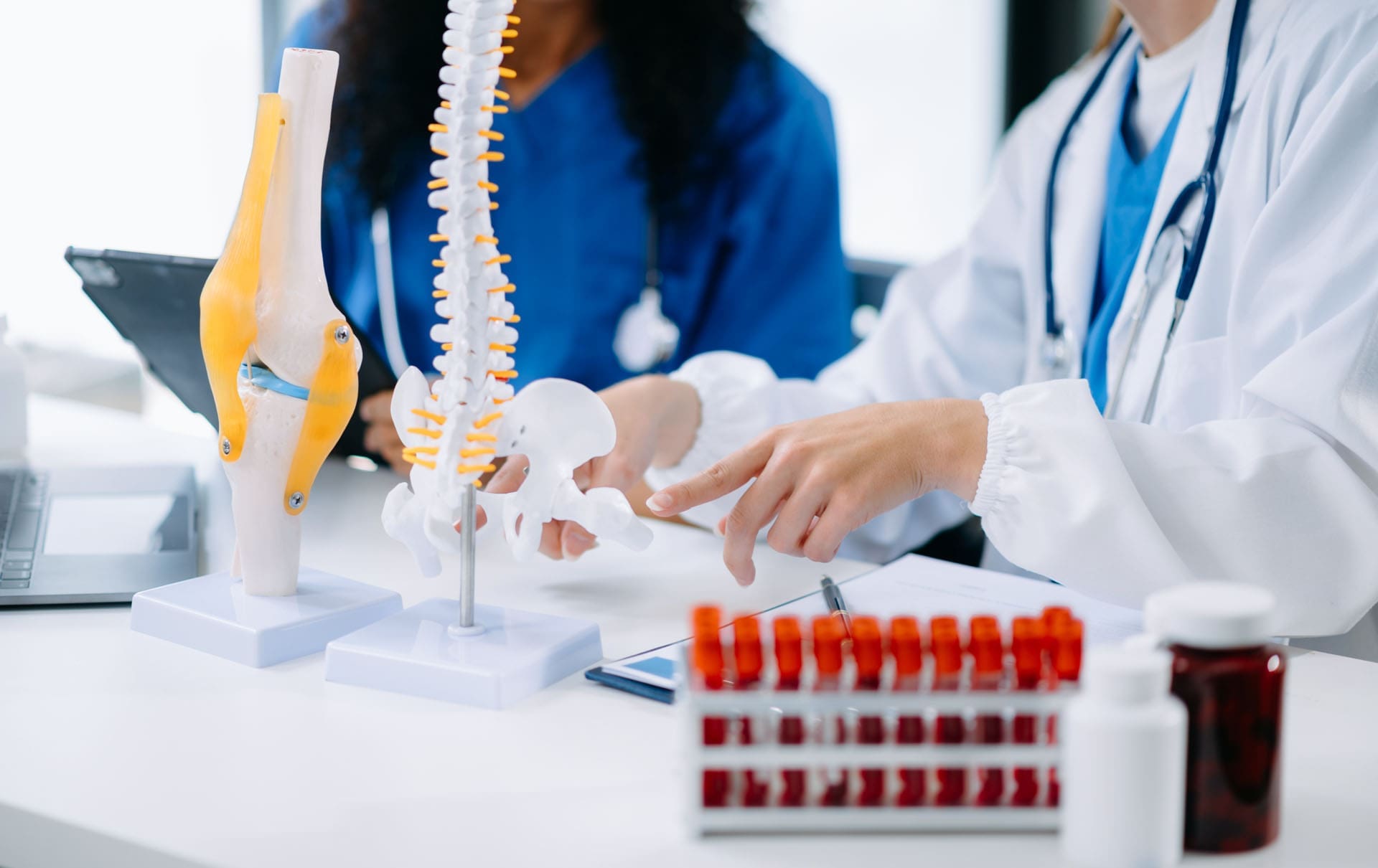
Platelet-rich plasma, commonly called PRP, is prepared from the patient’s own blood.
A small blood sample is collected and placed in a centrifuge. The centrifuge separates the blood into different parts. The platelets and plasma are then concentrated and prepared for injection into a selected joint, tendon, ligament, or muscle.
Platelets contain growth factors and signaling proteins that are involved in the body’s natural healing response. PRP may help create a biological environment that supports tissue recovery.
PRP is sometimes considered for conditions such as:
- Tendon injuries
- Ligament injuries
- Knee osteoarthritis
- Muscle injuries
- Joint irritation
- Certain sports injuries
Research suggests that PRP may reduce pain and improve function in some musculoskeletal conditions. However, results depend on the injury, the patient’s health, the way the PRP is prepared, and the rehabilitation plan used after treatment (Thu et al., 2022).
PRP does not automatically create new tissue or cure every injury. Evidence is stronger for some conditions than for others. Patients should receive a complete evaluation before deciding whether PRP is appropriate (Hospital for Special Surgery, 2024).
After PRP, a rehabilitation plan may progress through:
- Protection and gentle movement
- Light muscle activation
- Controlled resistance exercises
- Balance and stability training
- Heavier strengthening
- Sport- or work-specific activities
Exercise should be increased slowly so the healing area is not overloaded.
Understanding PFP Therapy
PFP may stand for platelet-free plasma or platelet-poor plasma. The exact meaning may vary among clinics, laboratories, and treatment protocols.
These plasma products contain fewer platelets than PRP. They may still contain proteins and other biological materials that support certain treatment goals.
PFP should not be described as the same treatment as PRP. Patients should understand what type of plasma product is being used and why it was selected.
Important questions include:
- Is the product platelet-free or platelet-poor?
- How is it prepared?
- What condition is being treated?
- Is PFP being used alone or with PRP?
- What research supports its use?
- Who will perform the procedure?
- Will ultrasound or other imaging guide the injection?
Research comparing platelet-rich and platelet-poor preparations is still developing. The best option may depend on the tissue, injury, and desired biological response (Raum et al., 2024).
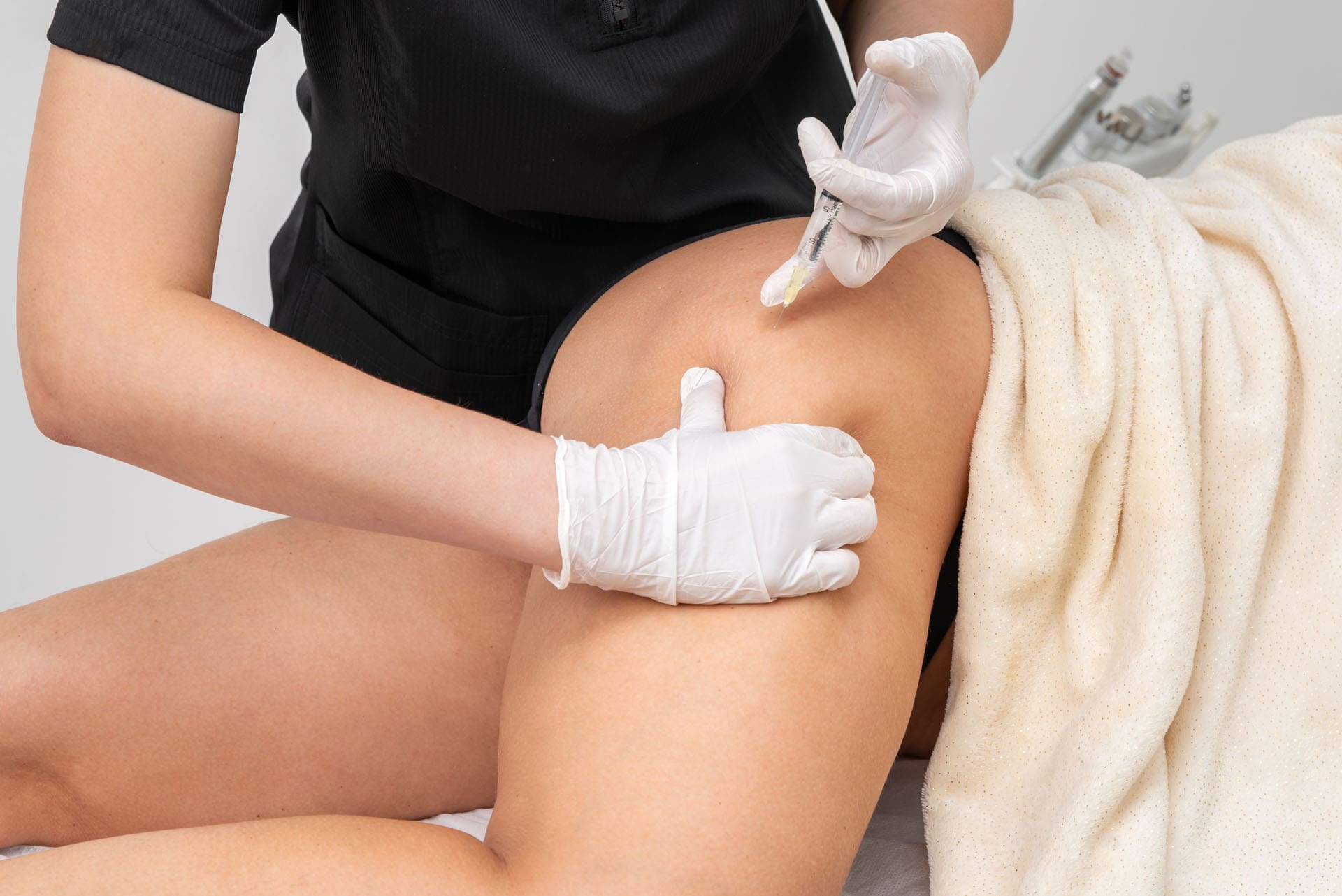
How MFAT May Support Joint Recovery
Microfragmented adipose tissue, also called MFAT, is made from a small amount of the patient’s own fat tissue.
The fat is usually collected through a minor procedure and processed into smaller fragments. The prepared tissue may then be injected into a painful joint or injured soft-tissue area.
MFAT contains structural tissue, blood vessels, signaling cells, and natural biological substances. It may help create a supportive environment around an injured or arthritic joint.
MFAT may be considered for selected patients with:
- Knee osteoarthritis
- Joint degeneration
- Persistent joint pain
- Certain cartilage injuries
- Chronic soft-tissue problems
PRP and MFAT are not identical. PRP uses concentrated platelets from the blood. MFAT uses processed adipose tissue with a more complex structural environment.
Early research suggests MFAT may improve pain and function in some patients with mild to moderate osteoarthritis. However, long-term research is still limited, and results are not guaranteed (Parmar et al., 2026).
The U.S. Food and Drug Administration has warned patients that many regenerative products marketed for orthopedic conditions have not been approved to treat arthritis, disc disease, back pain, or tendon injuries. Patients should be cautious of claims that promise a guaranteed “stem-cell cure” (U.S. Food and Drug Administration, 2021).
Epidural Spinal Injections for Nerve Pain
An epidural spinal injection places anti-inflammatory medicine into the epidural space near an irritated spinal nerve.
It may be considered when a patient has pain that travels from the spine into an arm or leg. This type of pain may be caused by:
- A herniated disc
- A bulging disc
- Spinal stenosis
- Sciatica
- Cervical radiculopathy
- Lumbar radiculopathy
- Degenerative spinal changes
The goal of an epidural injection is to reduce inflammation around the nerve. The treatment does not rebuild a damaged disc or permanently correct spinal alignment.
However, reducing nerve inflammation may create a useful period in which the patient can move, sleep, walk, and participate in rehabilitation with less pain (Cleveland Clinic, 2021).
During this rehabilitation period, the patient may begin:
- Gentle walking
- Core-strengthening exercises
- Hip and leg strengthening
- Posture training
- Nerve mobility exercises
- Safe lifting practice
- Gradual fitness activities
The epidural injection helps calm the irritated nerve. Chiropractic care and rehabilitation address movement, posture, mobility, and strength.
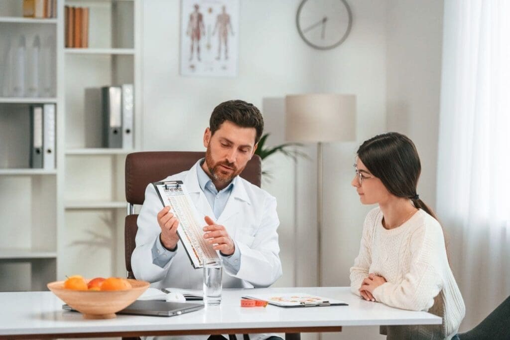
The Role of IV Infusion Nutrient Therapy
IV infusion therapy delivers fluids and selected nutrients directly into the bloodstream.
It may be medically appropriate for patients with dehydration, certain nutrient deficiencies, absorption problems, or other diagnosed needs.
IV therapy may include:
- Saline hydration
- Electrolytes
- Selected vitamins
- Certain minerals
- Amino acids
- Physician-directed medications
In wellness and fitness settings, IV therapy is often promoted for hydration and recovery. However, IV nutrient therapy should not replace drinking water, eating nutritious foods, sleeping well, or resting after physical activity.
Research does not strongly support routine high-dose vitamin infusions for healthy people who are already well hydrated and have no diagnosed deficiency (Alangari et al., 2025).
IV therapy also carries possible risks, including:
- Infection
- Vein irritation
- Fluid overload
- Kidney stress
- Medication interactions
- Abnormal blood pressure
- Electrolyte imbalance
- Heart rhythm changes
A responsible IV program should include a health screening, a clear medical reason for treatment, sterile preparation, accurate dosing, and appropriate monitoring (Mayo Clinic Press, 2024).
At ChiroMed, IV therapy may be considered as one part of a broader wellness plan rather than as a replacement for healthy daily habits.
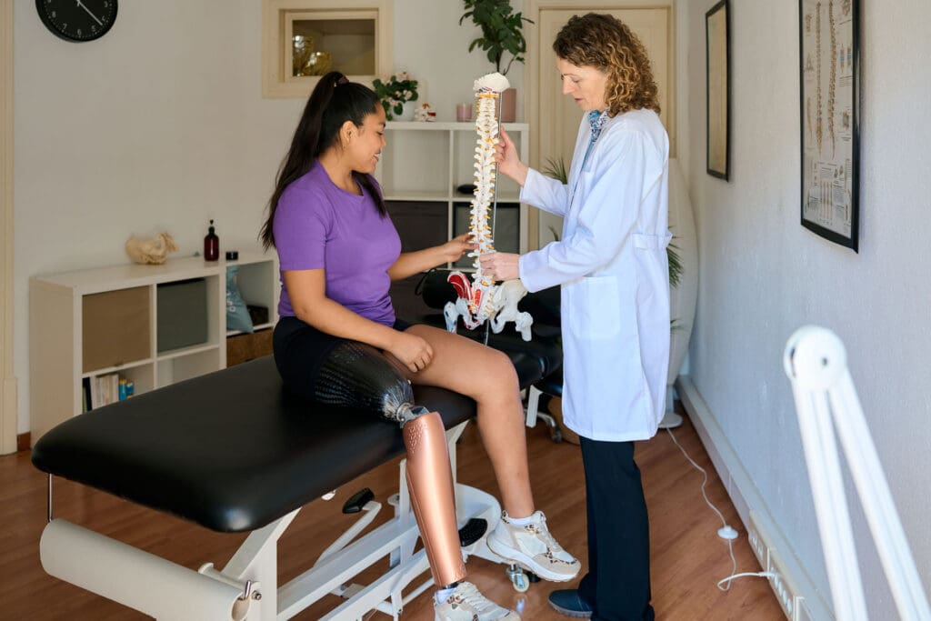
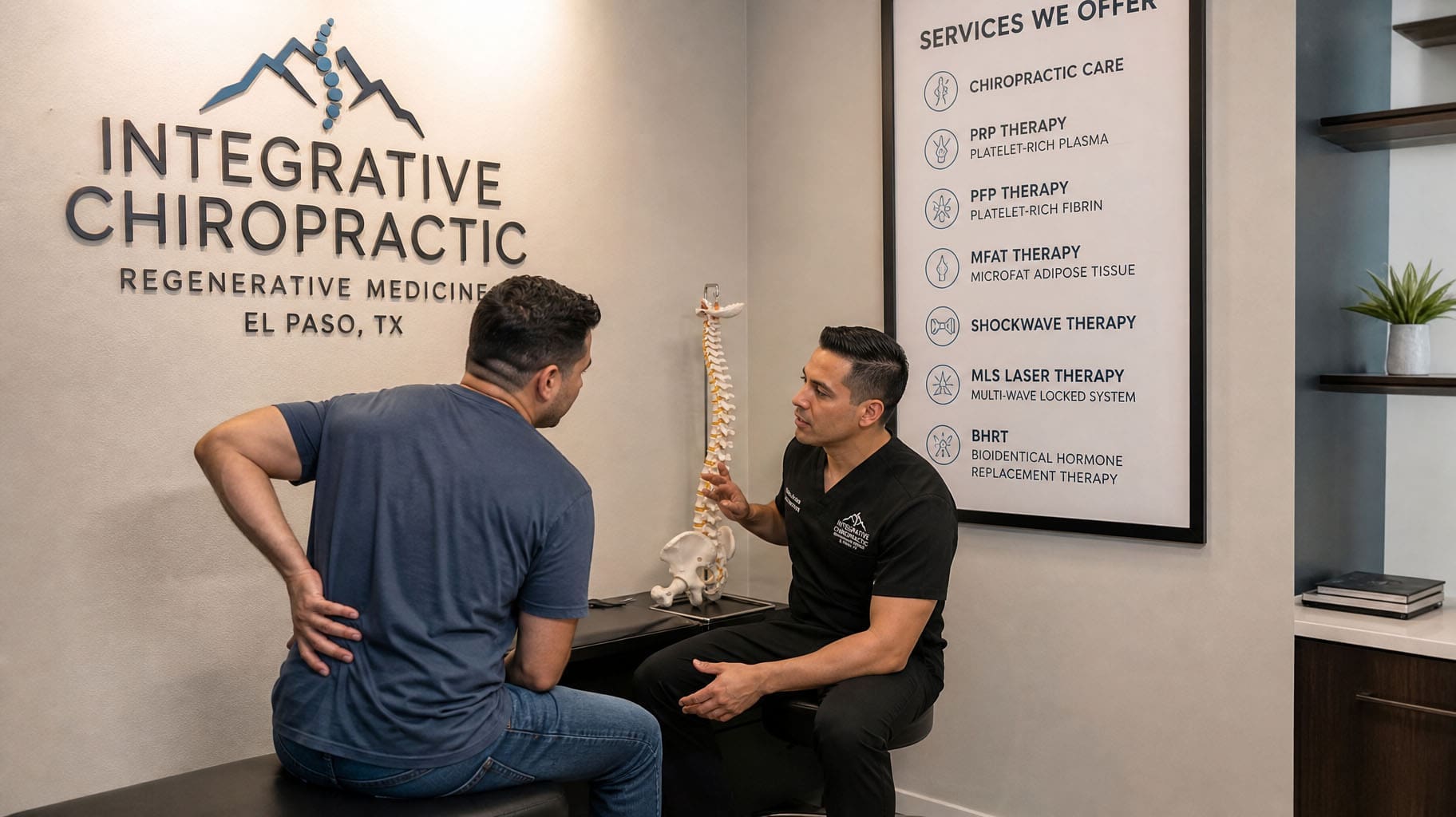
Chiropractic Care Helps Prepare the Body
Regenerative procedures may support tissues, but the body must still move correctly.
If joints remain stiff, muscles stay weak, or poor movement habits persist, the injured area may continue to experience harmful stress.
Chiropractic and rehabilitative care may include:
- Spinal adjustments
- Joint mobilization
- Soft-tissue therapy
- Posture correction
- Mobility exercises
- Core strengthening
- Balance training
- Functional movement exercises
Clinical guidelines support exercise, education, and selected joint mobilization methods as part of conservative low back pain care (George et al., 2021).
At ChiroMed, care is tailored to the patient’s condition. Not every patient receives the same adjustment, injection, therapy, or exercise program.
Returning to Exercise Safely
Feeling less pain does not always mean an injury has fully healed.
Returning to heavy exercise too quickly may cause another injury or increase the original problem. A gradual return-to-fitness plan may move through several stages.
Stage 1: Protect and Calm the Area
The first stage may focus on controlling pain, reducing inflammation, and avoiding movements that worsen symptoms.
Stage 2: Restore Movement
The patient may begin gentle joint motion, stretching, walking, and light muscle activation.
Stage 3: Rebuild Strength
Resistance exercises may be added to improve strength, endurance, balance, and joint stability.
Stage 4: Return to Fitness
The final stage may include heavier lifting, running, sports drills, work tasks, or other goal-specific activities.
Progress should be based on movement quality and function, not only on the amount of time that has passed.
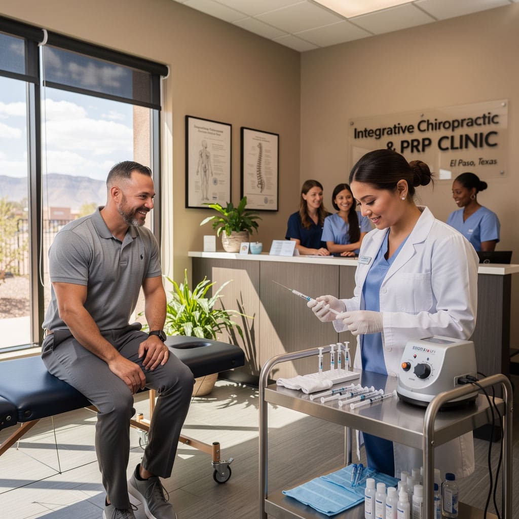
Multidisciplinary Care at ChiroMed
ChiroMed provides a multidisciplinary setting in which chiropractic care, medical oversight, functional medicine, personal injury care, and rehabilitation may work together.
Dr. Alexander Jimenez, DC, APRN, FNP-BC, CCST, CFMP, IFMCP, ATN, combines experience in chiropractic care, advanced practice nursing, functional medicine, and physical rehabilitation.
His clinical observations emphasize that pain and recovery may be influenced by:
- Spinal and joint movement
- Muscle weakness
- Posture
- Nutrition
- Inflammation
- Sleep
- Stress
- Metabolic health
- Previous injuries
Dr. Maria Guadalupe Cardenas, MD, works with Dr. Jimenez as a Medical Director and Collaborative Physician. She is a board-certified internal medicine physician with more than 40 years of clinical experience.
Public provider listings identify Dr. Cardenas under NPI number 1164426748 and Texas medical license J2933.
This professional structure is common in integrative and injury-care clinics. A medical doctor may provide medical direction, health screening, medication review, and oversight while the chiropractor manages structural, musculoskeletal, and rehabilitation concerns.
Dr. Cardenas’s role may include:
- Reviewing chronic medical conditions
- Assessing procedure risks
- Reviewing medications
- Evaluating laboratory findings
- Coordinating medical referrals
- Supporting complex case management
Dr. Jimenez’s role may include:
- Musculoskeletal evaluation
- Chiropractic care
- Functional medicine
- Personal injury care
- Exercise rehabilitation
- Movement correction
- Nutrition and wellness education
This collaboration allows patients to receive care that considers both general health and physical function.
A Complete Approach to Wellness and Fitness
PRP, PFP, MFAT, epidural spinal injections, IV nutrient therapy, chiropractic care, and rehabilitation each have different purposes.
PRP and MFAT may support the healing environment of selected joints and soft tissues. PFP may be used in certain protocols, but its exact preparation should be explained. Epidural injections may calm spinal nerve inflammation. IV therapy may support patients with real hydration or nutrient needs.
Chiropractic care and tailored exercise help prepare the “soil” by restoring motion, improving posture, and rebuilding strength.
At ChiroMed, the goal is not only to make symptoms quieter. The goal is to help patients move better, become stronger, and return to exercise and daily life with greater confidence.
Long-term recovery usually requires more than one treatment. It may involve medical care, chiropractic care, nutrition, movement, rest, and a progressive exercise plan working together.
References
Alangari, A., et al. (2025). To IV or not to IV: The science behind intravenous vitamin therapy.
Carolina Nonsurgical Orthopedics. (2026). PRP vs. MFAT cell therapy: Which regenerative treatment is right for you?.
Cleveland Clinic. (2021). Lumbar epidural steroid injections: What it is, benefits, risks, and side effects.
George, S. Z., et al. (2021). Interventions for the management of acute and chronic low back pain: Revision 2021. Journal of Orthopaedic & Sports Physical Therapy, 51(11), CPG1-CPG60.
Hospital for Special Surgery. (2024). Platelet-rich plasma injection: How it works.
Jimenez, A. (n.d.). Dr. Alexander Jimenez: Chiropractic and integrative medicine.
Jimenez, A. (n.d.). Dr. Alexander Jimenez, DC, APRN, FNP-BC, IFMCP, CFMP.
Jimenez, A. (2026). How PRP composition influences your healing journey.
Mayo Clinic Press. (2024). IV vitamin therapy: Understanding the lack of proven benefit and potential risks.
Open Wellness PDX. (2025). What is regenerative injection therapy? A complete guide to PRP, prolotherapy, and perineural injection.
Parmar, T., et al. (2026). Microfragmented adipose tissue in orthopedic regeneration.
Raum, G., et al. (2024). Platelet-poor versus platelet-rich plasma for the treatment of muscle injury.
Thu, A. C., et al. (2022). The use of platelet-rich plasma in the management of musculoskeletal pain: A narrative review.
U.S. Food and Drug Administration. (2021). Important patient and consumer information about regenerative medicine therapies.