Memorial Day Rear-End Collisions and Chiropractic Care
Why Memorial Day Weekend Can Increase Rear-End Collision Risk
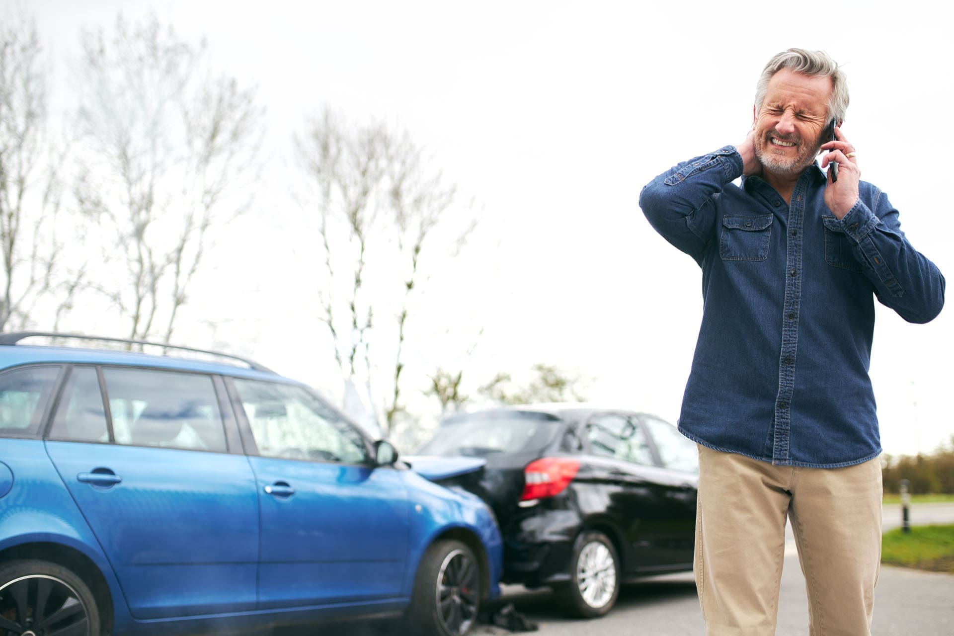
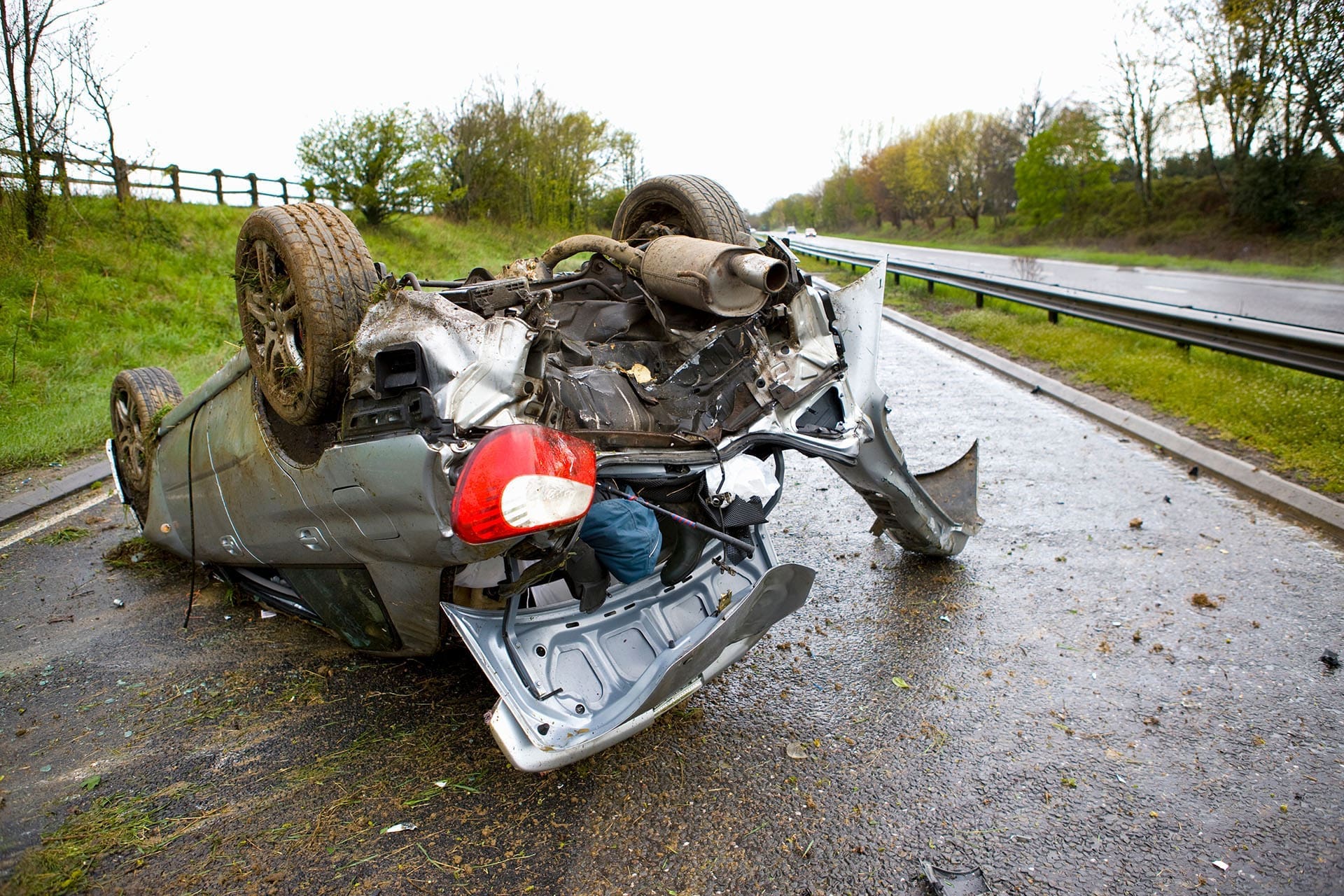
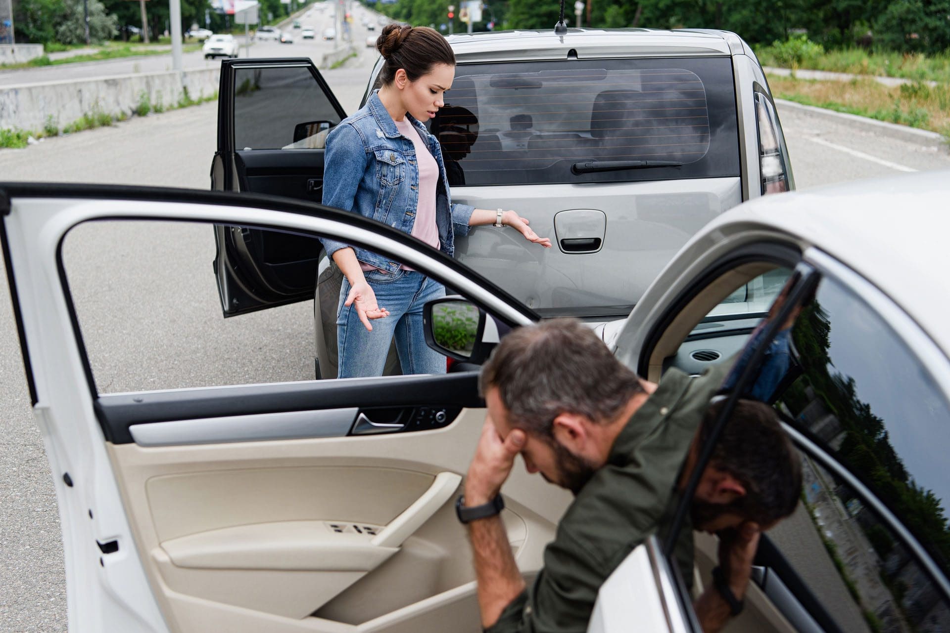
Memorial Day weekend is a busy time for travel. Many people are driving to visit family, attend events, go on vacation, or return home after a long weekend. More cars on the road can mean more traffic, more sudden stops, and more chances for rear-end collisions.
Rear-end collisions happen when one vehicle crashes into the back of another. These crashes are common in:
- Heavy highway traffic
- Stop-and-go traffic
- Construction zones
- Busy intersections
- Parking lot exits
- Sudden slowdowns
- Chain-reaction crashes
During Memorial Day weekend, drivers may also be tired, distracted, or unfamiliar with the roads. A driver may look down at a GPS, check a phone, adjust music, manage passengers, or follow another vehicle too closely. In only a few seconds, traffic can stop, and a rear-end crash can happen.
Rear-end collisions are among the most common types of motor vehicle accidents because they often happen during sudden braking and distracted driving situations (John Price Law Firm, 2024; DeMayo Law Offices, n.d.).
Why Rear-End Collisions Can Injure the Neck and Spine
A rear-end crash can look minor, but the force can still affect the body. When a car is hit from behind, the body may move forward while the head and neck snap back and then forward. This fast motion can create whiplash.
Whiplash can affect the:
- Neck muscles
- Spinal joints
- Ligaments
- Tendons
- Discs
- Nerves
- Upper back
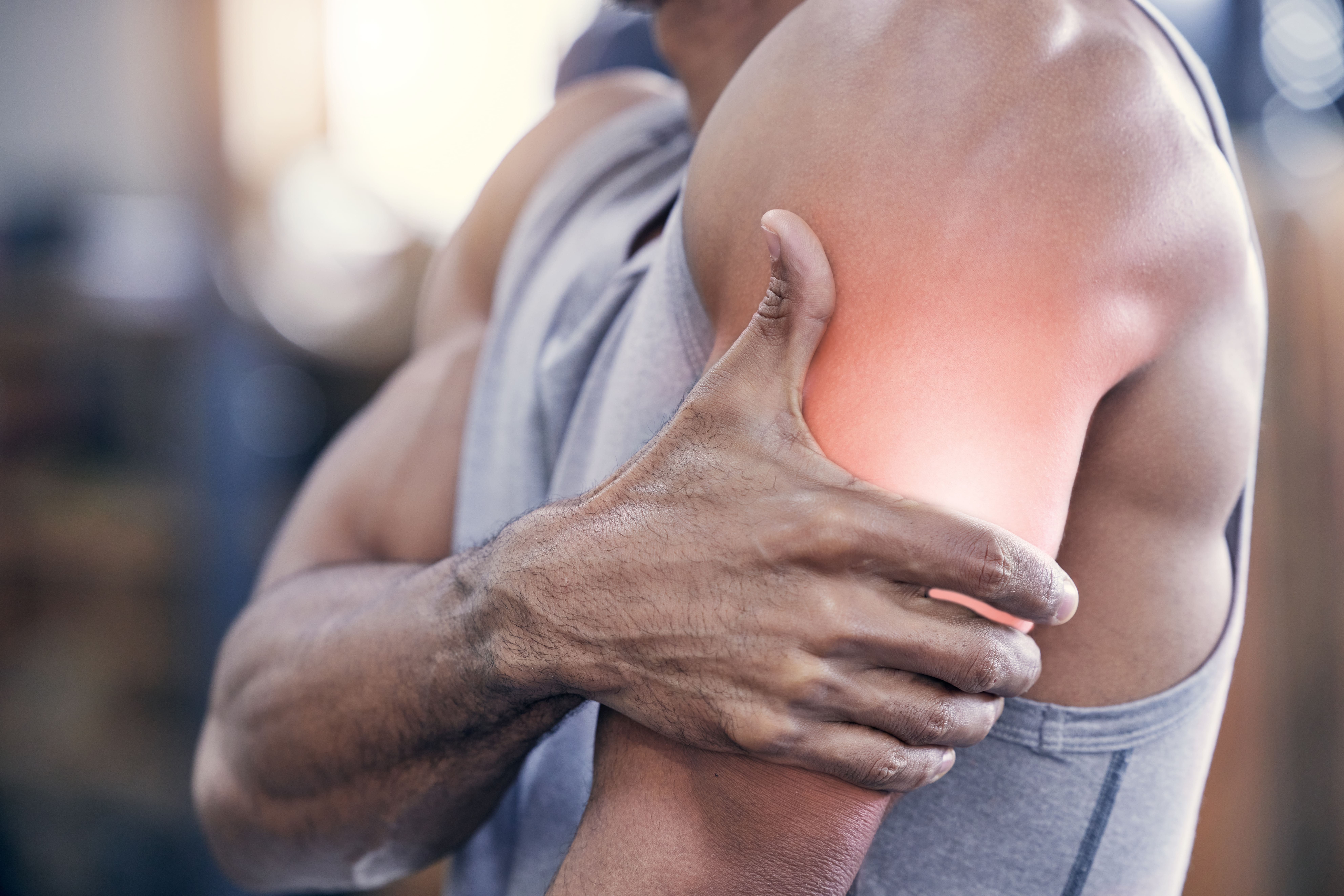
- Shoulders
The neck and spine are not made to absorb sudden crash forces. Even a lower-speed rear-end collision can strain soft tissues and irritate the spinal joints. In more serious crashes, the force may contribute to disc injuries, nerve pain, or long-term stiffness.
Whiplash and neck injuries are often linked to rear-end collisions because the sudden impact can stretch and strain the neck tissues (Accident Clinics, n.d.; Jax Litigation, n.d.).
Common Injuries After a Rear-End Collision
After a rear-end crash, pain may start in one area and then spread over time. Some people feel neck tightness first. Others notice headaches, shoulder pain, low back pain, or numbness later.
Common rear-end collision injuries may include:
- Whiplash
- Neck sprains and strains
- Back sprains and strains
- Muscle spasms
- Herniated discs
- Bulging discs
- Shoulder pain
- Headaches
- Nerve impingement
- Numbness or tingling
- Low back pain
- Sciatica-like symptoms
- Postural changes
Soft tissue injuries can be difficult because they may not always appear clearly on basic imaging. Muscles, ligaments, tendons, fascia, and spinal joints can still sustain injuries even when no bone is broken. Back sprains and strains are common after vehicle accidents and can cause pain, stiffness, swelling, spasms, and limited movement (1-800-NOW-HURT, n.d.).
KNR Legal also notes that car accidents commonly cause whiplash, herniated discs, spinal injuries, and other neck and back problems (Kisling, Nestico & Redick, n.d.).
Why Symptoms Can Show Up Days or Weeks Later
One of the most important things to know is this: pain does not always show up right away.
After a crash, the body releases stress hormones like adrenaline. This can make a person feel alert and less aware of pain. Hours or days later, inflammation may increase, muscles may tighten, and symptoms may become more noticeable.
Delayed symptoms may include:
- Neck stiffness
- Headaches
- Shoulder tightness
- Mid-back pain
- Low back pain
- Dizziness
- Muscle spasms
- Pain when turning the head
- Numbness or tingling
- Trouble sleeping
- Fatigue
- Brain fog
This is why a full evaluation is recommended after a motor vehicle accident, even when the crash seems small. Accident-related symptoms may take time to appear, especially with whiplash, soft-tissue injuries, and nerve irritation (Accident Clinics, n.d.; Zwick Law, 2024).
How a Rear-End Collision Can Affect Posture
A rear-end crash can change how the body holds itself. When the neck or back is injured, muscles may tighten to protect the area. This protective response can change posture and movement.
A person may begin to:
- Hold the head forward
- Raise one shoulder higher than the other
- Limit neck rotation
- Walk differently
- Avoid bending or lifting
- Sit unevenly
- Develop muscle guarding
Poor posture after an accident can place extra stress on the spine. Over time, these factors can make pain worse and slow recovery. De Bruin Chiropractic explains that auto accidents can affect posture and that chiropractic care may help by improving spinal mobility, soft-tissue function, and body alignment (De Bruin Chiropractic, n.d.).
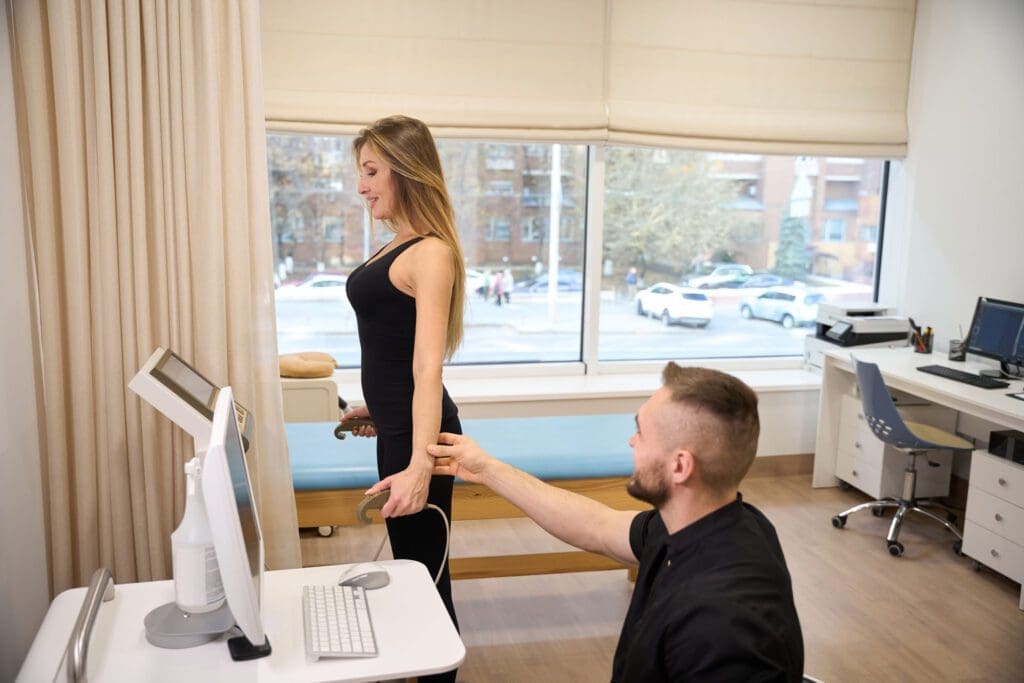
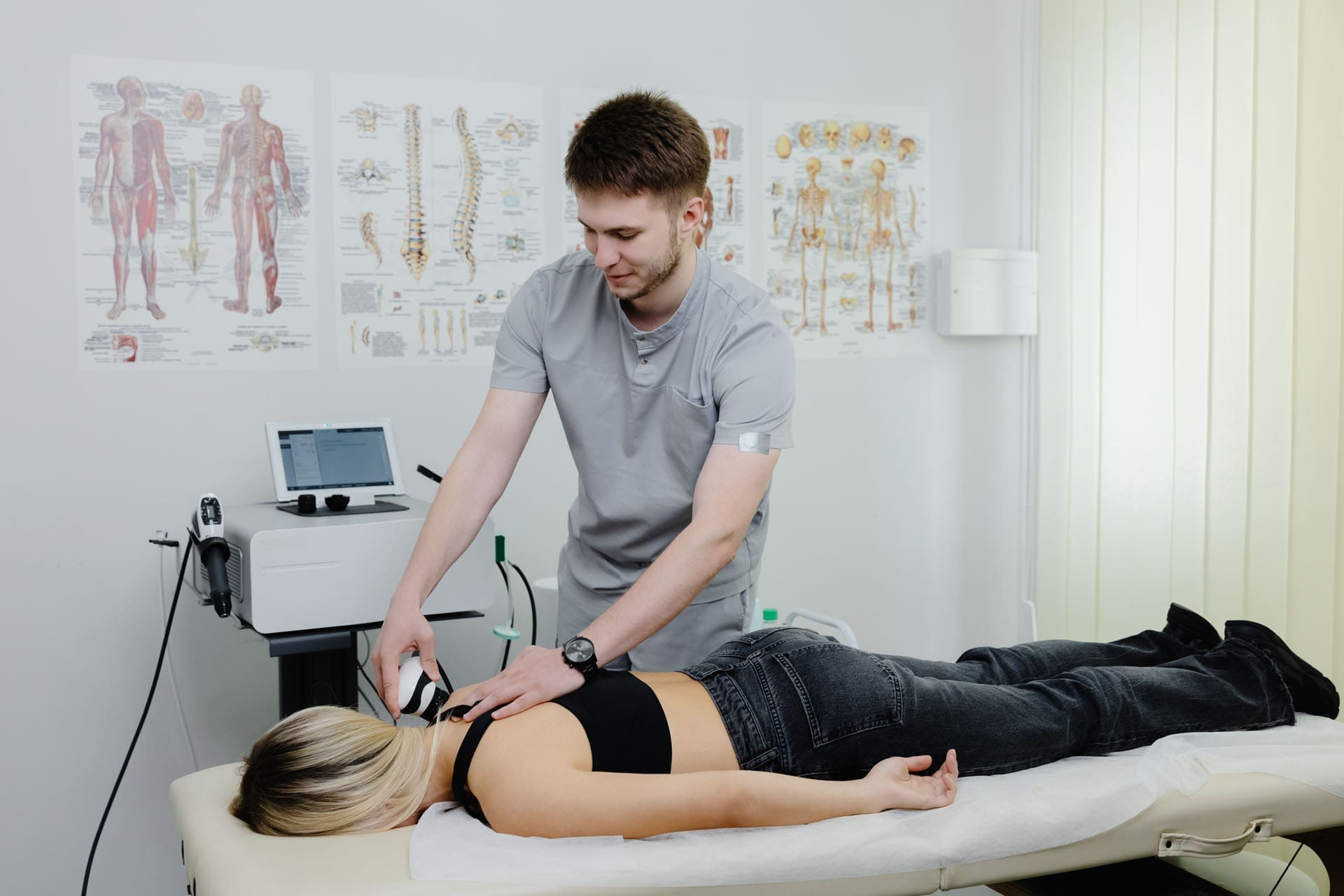
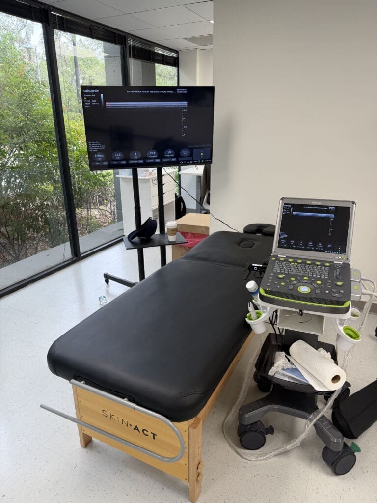
How ChiroMed’s Integrative Approach Fits Into Recovery
For readers of ChiroMed, the key idea is that accident recovery should look at the whole injury pattern. A rear-end collision does not only affect one muscle or one joint. It can affect the spine, nerves, discs, ligaments, soft tissues, posture, inflammation, and movement.
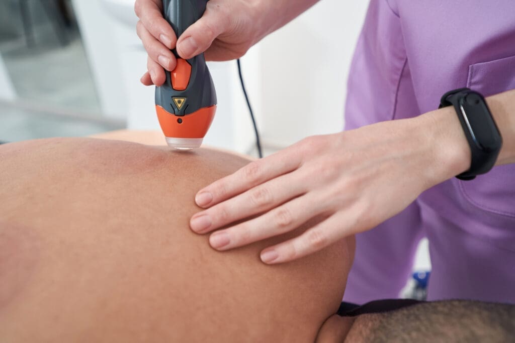
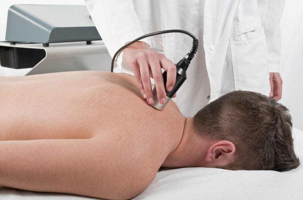
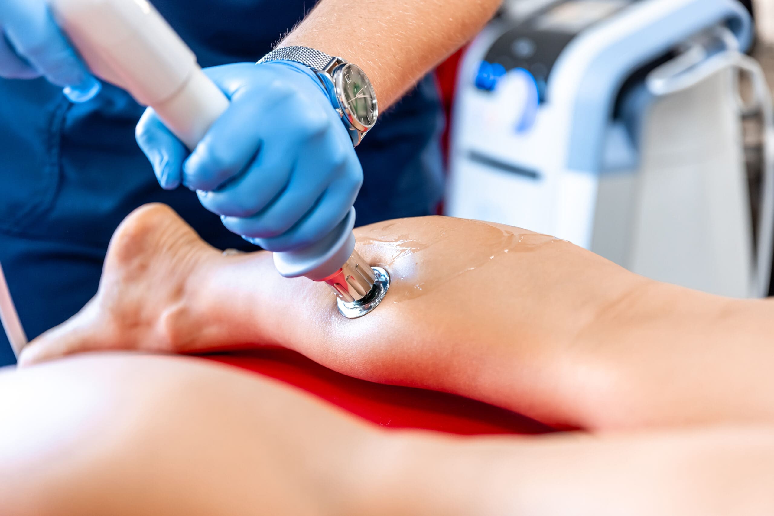
Integrative chiropractic care focuses on helping the body heal naturally by combining different tools and therapies. The goal is not only to reduce pain but also to improve function.
A care plan may include:
- Chiropractic adjustments
- Soft tissue therapy
- Myofascial release
- Corrective exercises
- Stretching and mobility work
- Posture correction
- Spinal decompression when appropriate
- Rehabilitation exercises
- Lifestyle guidance
- Nutrition and inflammation support
- Referrals for imaging or medical care when needed
Doctor Wagner explains that chiropractic care following a car accident may include spinal adjustments, soft-tissue therapy, therapeutic exercise, stretching, postural support, and patient education (Doctor Wagner, n.d.). This type of approach is beneficial because rear-end collision injuries often involve both the spine and the surrounding soft tissues.
Clinical Observations From Dr. Alexander Jimenez, DC, APRN, FNP-BC
Dr. Alexander Jimenez, DC, APRN, FNP-BC, has observed that injuries from motor vehicle accidents often involve more than simple pain. In many cases, the body develops a pattern of joint restriction, muscle guarding, nerve irritation, inflammation, and loss of movement.
Through his integrative clinical approach, Dr. Jimenez emphasizes the importance of identifying the root cause of pain rather than merely treating symptoms. His model combines chiropractic care, functional medicine principles, rehabilitation, diagnostic review, and, when appropriate, personalized recovery planning (Jimenez, n.d.-a).
His clinical observations also highlight that old car accident injuries may continue to cause pain months or years later when the original injury did not heal correctly. These lingering issues may involve muscles, ligaments, spinal joints, discs, nerves, fascia, and chronic inflammation (Jimenez, n.d.-b).
This matters after a Memorial Day rear-end collision because a person may not feel severe pain immediately. But if soft tissue damage, spinal restriction, or nerve irritation is missed, the injury may become harder to treat later.
Why a Full Evaluation Matters After a Memorial Day Crash
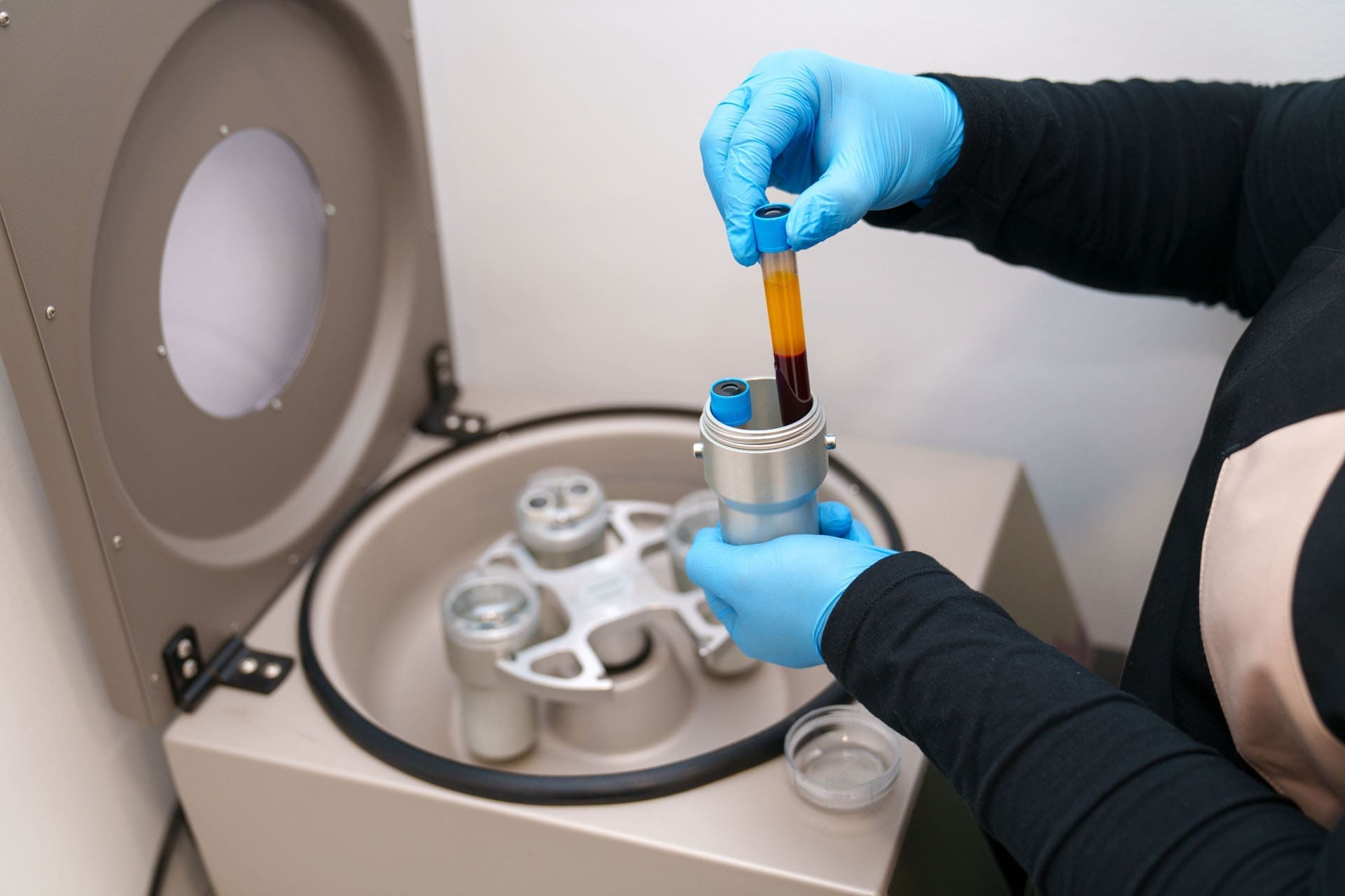
A full evaluation after a rear-end collision can help identify injuries early. This is important for both health and documentation. The evaluation should focus on how the accident affected the body, not just on whether a bone was broken.
A post-accident evaluation may include:
- Review of how the crash happened
- Neck and back pain assessment
- Range-of-motion testing
- Orthopedic testing
- Neurological screening
- Muscle strength checks
- Reflex checks
- Posture analysis
- Functional movement testing
- Imaging referral when needed
This type of exam can help identify whether the person has whiplash, soft tissue injuries, disc irritation, nerve symptoms, or other accident-related problems.
When to Seek Urgent Medical Care
Some symptoms after a crash require immediate medical attention. Chiropractic and integrative care can support recovery, but emergency symptoms should be checked right away.
Seek urgent care if there is:
- Severe headache
- Loss of consciousness
- Confusion
- Vision changes
- Chest pain
- Trouble breathing
- Severe neck or back pain
- Weakness in the arms or legs
- Numbness that spreads
- Loss of balance
- Abdominal pain
- Loss of bowel or bladder control
- Worsening symptoms after the crash
These symptoms may point to a more serious injury and should not be ignored.
Preventing Rear-End Collisions During Holiday Travel
Drivers can reduce risk by planning ahead and staying focused. Memorial Day traffic can be stressful, but safe driving habits can make a big difference.
Helpful safety steps include:
- Leave early to avoid peak traffic
- Keep extra space between vehicles
- Do not tailgate
- Put the phone away
- Let a passenger handle GPS directions
- Avoid eating while driving
- Take breaks on long trips
- Watch for sudden stops
- Slow down in heavy traffic
- Avoid driving tired
- Never drive under the influence
Distracted driving is a major risk because it takes attention away from the road. This includes phone use, GPS adjustments, eating, drinking, and managing passengers.
ChiroMed Takeaway: Do Not Wait for Pain to Become Severe
Memorial Day weekend rear-end collisions are common because of traffic congestion, sudden stops, distracted driving, and long-distance travel. These crashes can cause whiplash, neck pain, back pain, muscle spasms, disc injuries, nerve irritation, and posture problems.
The most important lesson is simple: do not ignore symptoms after a crash.
Even mild stiffness or a small headache may be the first sign of a deeper injury. Since symptoms can take days or weeks to fully appear, a full evaluation is recommended after an accident.
Integrative chiropractic care can support recovery by addressing the spine, soft tissues, posture, movement, and inflammation together. For ChiroMed readers, this approach offers a more complete way to understand and manage accident-related injuries.
References
Accident Clinics. (n.d.). Whiplash and neck pain treatment
De Bruin Chiropractic. (n.d.). How an auto accident impacts your posture and how chiropractic care can help
DeMayo Law Offices. (n.d.). What are the most common types of car accidents?
Doctor Wagner. (n.d.). Chiropractic care after a car accident
Jimenez, A. (n.d.-a). Dr. Alex Jimenez DC, APRN, FNP-BC
Jimenez, A. (n.d.-b). Can old car accident injuries heal with integrative care?
Jimenez, A. (n.d.-c). Dr. Alexander Jimenez LinkedIn profile
John Price Law Firm. (2024). What are the most common car accidents?
Jax Litigation. (n.d.). Car crashes and neck injuries
Kisling, Nestico & Redick. (n.d.). Most common car accident injuries
Sarasota Chiropractor. (n.d.). Auto accident injuries
Tooele Chiropractor. (n.d.). Chronic pain from old car accident injuries
Zwick Law. (2024). Common injuries after a rear-end collision