Recommended Sports Training Gym Workout for Beginners

Simple, Safe, and Athletic
Starting a gym routine can feel confusing because there are so many workouts online. For beginners who want “sports training” (not just bodybuilding), the goal is simple: build a foundation of strength, movement quality, and conditioning—without getting hurt or burning out.
A beginner-friendly sports training plan usually works best as a 3-day-per-week full-body program, built around compound movements (squat, hinge, push, pull, carry) plus core stability and low-impact cardio. This structure appears in many beginner training guides because it provides enough practice to improve while still leaving recovery time for your body to adapt. (Planet Fitness, 2019/2025; Under Armour, n.d.; Mikolo, 2024).
Below is a practical sports training plan you can follow for 4–8 weeks, along with tips on technique, progression, recovery, and how integrative chiropractic care can support your training and help you move better.
What “Sports Training” Means for a Beginner
For beginners, sports training is not about maxing out or doing complicated drills. It’s about learning to produce force safely and efficiently, in patterns that show up in real life and sport:
- Squat (sit, jump, change levels)
- Hinge (bend, pick up, sprint posture)
- Push (push-ups, presses)
- Pull (rows, pulldowns)
- Brace + rotate control (core stability)
- Locomotion + conditioning (walking, rowing, incline treadmill, bike)
A full-body approach is especially helpful early on, because you practice these patterns more often without needing long workouts. Many beginner gym plans also recommend starting with simple machines or stable variations so you can learn form safely (Planet Fitness, 2018/2025; 10 Fitness, 2025).
The “3 Rules” That Make a Beginner Plan Actually Work
1) Keep it simple and repeatable
You want a plan you can do even when you’re tired or busy. If the workout has 25 exercises, it won’t last.
2) Train hard enough, not maximal
Most sets should feel like you could do 2–3 more reps with proper form. That’s how you build strength without turning every day into a recovery problem (Squatwolf, n.d.).
3) Progress slowly on purpose
The beginner’s “secret” is consistency. Small weekly increases add up fast.
Recommended Weekly Schedule (Beginner Sports Training)
A simple week that works for most beginners:
- Monday: Full-Body Workout A
- Wednesday: Full-Body Workout B
- Friday: Full-Body Workout A (next week start with B)
On non-lifting days, add low-impact cardio and mobility (e.g., walking, biking, rowing, or an incline treadmill) (Planet Fitness, 2019; Under Armour, n.d.; Mikolo, 2024).
Warm-Up (8–12 Minutes)
A good warm-up raises your body temperature, wakes up your joints, and teaches your body the positions you need.
Step 1: Easy cardio (3–5 minutes)
- Treadmill walk (flat or slight incline)
- Bike
- Rower
Planet Fitness highlights that beginner cardio doesn’t need to be extreme—simple options work (Planet Fitness, 2019).
Step 2: Dynamic mobility (4–6 minutes)
Pick 4–5 moves, 5–8 reps each:
- Arm circles
- Hip circles
- Leg swings (front/back)
- Bodyweight good mornings
- Deep squat hold (light, comfortable)
Step 3: Movement prep (1–2 minutes)
- 1 set of 8 bodyweight squats
- 1 set of 6 incline push-ups
- 1 set of 8 band rows (or light machine rows)
The Beginner Sports Training Gym Program (3 Days/Week)
Reps, Sets, and Rest (Simple Standards)
- Most strength moves: 3 sets of 8–12 reps
- Core holds: 3 sets of 20–40 seconds
- Rest: 60–90 seconds between sets (longer if needed)
This aligns with the common beginner recommendation to use moderate rep ranges that build skill and strength together (Mikolo, 2024; 10 Fitness, 2025).
Workout A (Full Body Foundation)
1) Squat pattern (choose one)
- Goblet squat (dumbbell) or
- Leg press (machine)
3 sets x 8–12
2) Push pattern (choose one)
- Incline push-up (hands on bench) or
- Chest press machine
3 sets x 8–12
3) Pull pattern (choose one)
- Seated row machine or
- Dumbbell row (bench-supported)
3 sets x 8–12
4) Hinge pattern (choose one)
- Romanian deadlift with dumbbells (light) or
- Hip hinge with cable pull-through
3 sets x 8–12
5) Core stability
- Plank 3 x 20–40 seconds
6) Conditioning finisher (optional)
- Rower: 6 minutes, easy steady pace or
- Incline treadmill walk: 8–12 minutes
Planet Fitness and other beginner guides commonly use incline walking, machines, and simple cardio finishers because they’re easy to scale (Planet Fitness, 2025; 10 Fitness, 2025).
Workout B (Full Body Athletic Balance)
1) Lunge/single-leg pattern
- Reverse lunge (bodyweight or light dumbbells) or
- Step-ups
3 sets x 8 reps each leg
2) Overhead or vertical push (beginner-friendly)
- Dumbbell shoulder press (light) or
- Shoulder press machine
3 sets x 8–12
3) Vertical pull
- Lat pulldown machine 3 sets x 8–12
4) Glute + posterior chain
- Glute bridge or hip thrust (bodyweight or light weight)
3 sets x 10–12
5) Anti-rotation core (beginner sports core)
- Pallof press (cable/band) 3 sets x 10 each side
6) Easy aerobic
- Bike or elliptical 10–15 minutes conversational pace
This “movement-pattern” setup is common in beginner athletic plans because it builds total-body strength and stability without needing complicated programming (Mikolo, 2024; Under Armour, n.d.).
How Heavy Should You Lift?
A beginner-friendly rule that works:
- Pick a weight you can lift for 8–12 reps with correct form
- The last 2–3 reps feel challenging, but you could still do 1–2 more reps if you had to
- If you can easily do 15+ reps, it’s probably time to increase the weight slightly
That “difficult but manageable” guideline is widely recommended for safe progression (Squatwolf, n.d.).
Progression Plan (So You Keep Improving)
Use a simple progression method for 4–8 weeks:
Week-to-week progression
- Option A (reps first):
Keep the same weight and add 1 rep per set until you reach the top of the range (12 reps). Then increase weight slightly and go back to 8 reps. - Option B (small weight jumps):
If the form is stable, add 2.5–5 lb per dumbbell (or the smallest machine increase) when you can complete all sets cleanly.
What to track
- Exercise
- Weight used
- Reps completed
- How it felt (easy/moderate/hard)
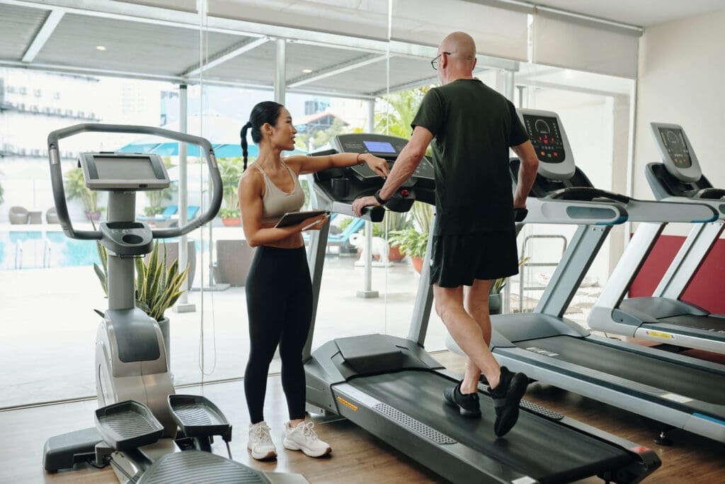
Beginner Cardio That Supports Sports Performance (Without Beating You Up)
A common beginner mistake is going too hard on cardio too soon. Instead, use low-impact cardio that builds your base and helps recovery:
Good beginner options
- Incline treadmill walking
- Rowing machine
- Stationary bike
- Elliptical
- Brisk outdoor walking
Planet Fitness emphasizes beginner-friendly cardio options and the importance of gradually building cardiovascular endurance (Planet Fitness, 2019; Planet Fitness, 2025).
Simple cardio plan
- 2–3 days/week
- 15–25 minutes
- You should be able to talk in short sentences
Recovery Essentials (Where Beginners Actually Get Results)
Training breaks muscle down. Recovery is where your body rebuilds.
Active recovery examples
- Light walking
- Mobility work
- Gentle cycling
- Stretching sessions
Sanford Sports highlights that recovery helps you regenerate and avoid overtraining, and that active recovery can be a smart part of the week (Sanford Sports, 2024).
Basic recovery checklist
- Sleep: aim for consistent, restful sleep
- Protein: include protein at most meals
- Hydration: steady intake throughout the day
- Easy movement on rest days
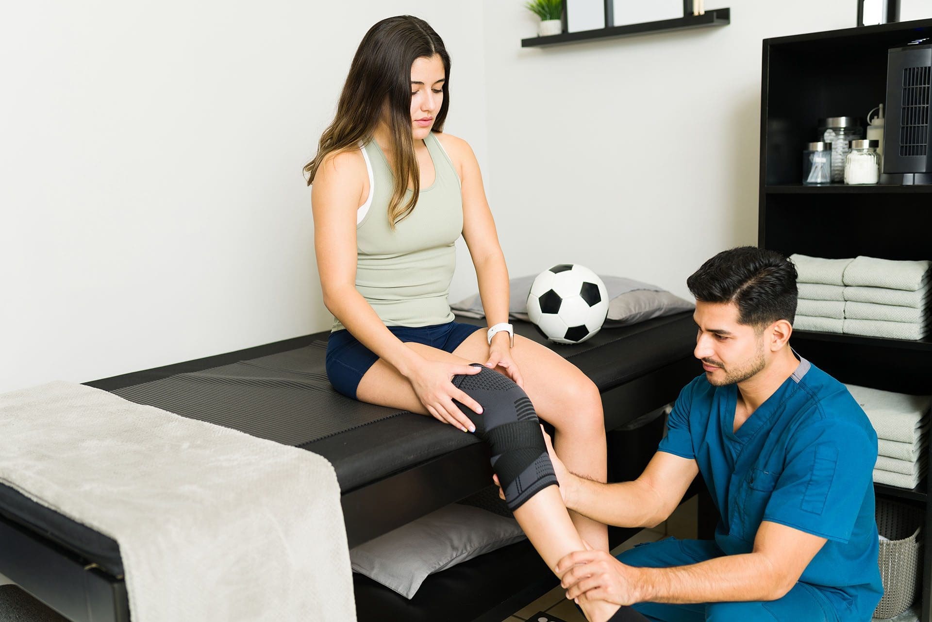
Integrative Chiropractic Care Helps Beginners Train Better
A smart beginner program is not only about exercises—it’s about movement quality. Integrative chiropractic care (when done responsibly and paired with exercise) often focuses on improving joint motion, reducing pain triggers, and correcting movement compensation patterns.
How chiropractic fits into beginner sports training
1) Injury prevention through movement checks
Functional movement evaluations can reveal weak links (hip control, ankle stiffness, and shoulder restriction) before they lead to injury. This is a central theme in Dr. Alexander Jimenez’s integrative sports injury education and movement-focused approach (Jimenez, 2026; PushAsRx, 2026).
2) Mobility and joint mechanics
Better mobility can help you hit safer positions in squats, hinges, and presses. Dr. Jimenez’s clinical content on integrated chiropractic and NP care frequently emphasizes joint mobility, balance, coordination, and reduced risk of re-injury as practical athletic goals (Jimenez, 2026).
3) Recovery support (especially when you’re sore or stiff)
Many chiropractic and sports rehab sources describe combining manual care with exercise to help patients restore function and return to activity (Team Elite Chiropractic, 2022).
Before or after workouts: what’s better?
There isn’t one universal rule, but many clinics describe two common patterns:
- Before training: focus on mobility, joint mechanics, and movement quality
- After training: focus on reducing stiffness and supporting recovery
Some chiropractic guidance suggests that getting adjusted before exercise may help movement feel smoother, while post-workout care may help with soreness and relaxation (Atlas Total Health, 2022).
Practical beginner tip:
If you’re starting out and you tend to get sore easily, schedule chiropractic visits on lighter training days or rest days so you can feel the changes without rushing back into heavy lifting.
Corrective Exercises: The “Bridge” Between Treatment and Training
Corrective exercises are simple drills that restore balance and improve movement patterns. They are often used when someone has tight areas, weak stabilizers, or poor control (Asheville Medical Massage, 2025).
Examples that pair well with beginner lifting
- Glute bridges (glute activation)
- Bird dogs (core + spine control)
- Dead bugs (core bracing)
- Wall angels (posture + shoulder mobility)
- Cat-cow (spinal mobility)
Many chiropractic exercise lists include similar basics because they reinforce better posture and better movement options (Elevate to Life, n.d.; Team Elite Chiropractic, 2022).
Beginner Mistakes to Avoid (So You Don’t Quit)
1) Going too hard in week one
Soreness is normal, but crushing yourself makes consistency harder. Planet Fitness beginner guidance commonly encourages starting with manageable sessions and learning equipment first (Planet Fitness, 2025).
2) Skipping the warm-up
A short warm-up improves performance and helps you move better that day.
3) Changing the plan every workout
Beginners improve faster by repeating key patterns.
4) Ignoring form for heavier weight
The fastest path is controlled reps, full ranges you own, and slow progression.
A Simple 4-Week “Ramp Up” Example
If you want a very clear starting path:
Week 1
- Do Workout A and B with light weights
- Keep cardio easy
- Focus on learning movement
Week 2
- Add 1–2 reps per set or a small weight increase
- Add one extra 10-minute cardio session if energy is good
Week 3
- Increase weight slightly on 1–2 main lifts
- Keep form strict
Week 4
- Keep building reps/weight gradually
- Deload if needed (reduce weights by ~10–15% for a week if you feel beat up)
Under Armour’s beginner schedule also supports the idea of only a few strength days weekly with rest days built in (Under Armour, n.d.).
Safety Notes (Especially for Beginners Who Want Sports Performance)
Stop and get checked if you have:
- Sharp pain, numbness, tingling, or weakness
- Joint swelling that doesn’t settle
- Pain that changes your walking pattern
- Symptoms after a recent injury that are getting worse
If you’re under chiropractic or medical care, your training plan should align with your exam findings and current tolerance.
Bottom Line: The Best Beginner Sports Training Plan Is the One You Repeat
A recommended sports training gym workout for beginners is:
- 3 full-body strength days per week
- Built around squat + hinge + push + pull + core
- Supported by low-impact cardio
- Protected by recovery days
- Improved by movement assessments and corrective exercise
- Enhanced by integrative chiropractic strategies that help restore mobility, reduce compensation, and support training consistency (Jimenez, 2026; PushAsRx, 2026).
If you want the simplest next step: start with the workouts above for 4 weeks, track your progress, and adjust slowly.
References
- A Beginner Workout Plan for Your First Week in the Gym (Planet Fitness, n.d.).
- Strength and Cardio Workouts for Beginners (Planet Fitness, n.d.).
- Building a Beginner Cardio Workout to Increase Cardiovascular Endurance (Planet Fitness, 2019).
- 5 Best Cardio Workouts for Beginners (Planet Fitness, n.d.).
- What to Do at the Gym as a Beginner (Planet Fitness, 2018).
- Beginner Basics: A 30-Minute Gym Machine Workout Routine (Planet Fitness, n.d.).
- Beginner Athlete Workout Guide: Build Your Foundation for Athletic Success (Mikolo, 2024).
- Your Beginner’s 7-Day Gym Plan to Start Strength Training (Under Armour, n.d.).
- Gaining Muscle & Strength: A Complete Guide for Women (Squatwolf, n.d.).
- Beginner Gym Workout Routine: What Should You Do First? (10 Fitness, 2025).
- Why You Should Add Recovery Exercises Into Your Workout Routine (Morando, 2024).
- Corrective Exercises: Restoring Balance and Preventing Injury (Asheville Medical Massage, 2025).
- Top 7 Exercises to Support Your Chiropractic Treatment (Elevate to Life, n.d.).
- At-Home Chiropractic Exercises To Speed Up Recovery (Team Elite Chiropractic, 2022).
- When Should I Get an Adjustment—Before or After I Work Out? (Atlas Total Health, 2022).
- Integrative Chiropractic Prevents Future Injuries for Athletes (PushAsRx, 2026).
- Integrated Chiropractic and NP Care for Sports Injuries (Jimenez, 2026).
- Adaptive Exercises in Integrative Chiropractic Care (Jimenez, n.d.).
- Beginner Gym Routine: Complete Week of Workouts (Hui, 2024).
- The Best Workout Routine for Beginners (YouTube creator, 2023).