Care is the cornerstone of our practice

Give us a Call
+1 (915) 412-6680
Send us a Message
support@chiromed.com
Opening Hours
Mon-Thu: 7 AM - 7 PM
Fri - Sun: Closed

Understanding Nerve Conduction Velocity in Health Care

Should individuals experiencing nerve pain or various sensations get a nerve conduction velocity study to examine nerve health and function?

Nerve Conduction Velocity

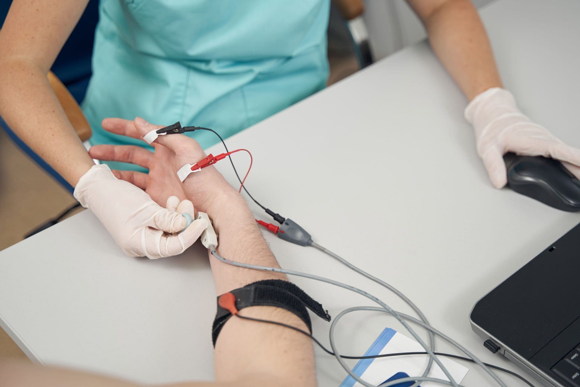
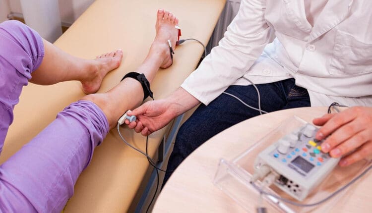
A nerve conduction velocity (NCV) is a noninvasive test that measures the speed and strength of nerve stimulation using electrical probes placed on the skin. It’s used to diagnose nerve damage or disease, often alongside an EMG (electromyogram) to differentiate between nerve and muscle problems. It can also evaluate sensory issues, pain, and weakness of the extremities.

  • This test involves safe electrical shocks that can be slightly uncomfortable but not painful.
  • Nerve conduction velocity (NCV) measures the speed at which electrical impulses travel along a nerve fiber, which measures how quickly electrical signals travel through a nerve.
  • This information indicates nerve health and function.
  • Electromyography (EMG) is a nerve test that involves placing tiny needles into the muscles.
  • A slower NCV can indicate nerve injury or dysfunction.

Test Uses

Generally, the test is ordered to assess peripheral nerve diseases, those that connect from the muscles, organs, and skin to the spinal cord or brain. It can help identify the type and location of nerve damage.

  • Peripheral nerve conditions typically cause pain, sensory loss, tingling, or burning.
  • Mild weakness and diminished reflexes can be detected during a neurological examination.

Conditions

Nerve conduction studies are performed to help diagnose conditions.

  • Herniated disc disease
  • Sciatic nerve problems
  • Carpal tunnel syndrome
  • Guillain-Barré syndrome
  • Nerve damage (neuropathy), such as from diabetes, chemotherapy, or autoimmune disorders
  • Charcot-Marie-Tooth disease

Nerve compression

  • Many different conditions, including trauma, inflammation, and tumors, can compress one or more nerves.
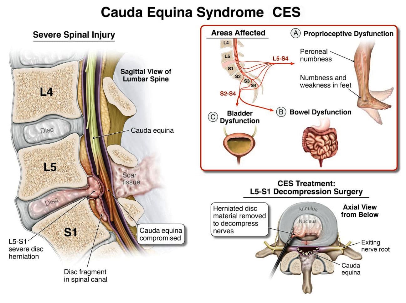
Radiculopathy

  • Often described as a pinched nerve, radiculopathy can affect an arm or a leg, causing pain and weakness.

Peripheral Neuropathy

  • This nerve damage begins in the most distal nerves, those farthest from the center of the body, such as the toes and fingers. It is often due to chronic alcohol misuse, uncontrolled diabetes, nutritional deficits, and inflammatory diseases. (Ferdousi M. et al., 2020)

Carpal Tunnel Syndrome

  • Commonly caused by inflammatory diseases or overuse of the wrists, such as from assembly line work, carpal tunnel syndrome causes numbness, pain, and weakness of the fingers and hands. (Tada K. et al., 2022)

Ulnar neuropathy

  • This common condition causes arm pain and sensory changes, usually due to repetitive movements or a prolonged position that causes pressure on the ulnar nerve.

Guillain-Barré syndrome (GBS)

  • This inflammatory condition causes demyelination, or loss of the insulating covering around nerves, which results in leg weakness.
  • It begins in the motor nerves, which send signals to muscles in the legs. (Shibuya K. et al., 2022)
  • The inflammation travels to nerves of the upper body, often affecting the muscles that control breathing.
  • Respiratory support is necessary until the condition improves.

Chronic Demyelinating Polyneuropathy (CIDP)

  • This condition is a chronic, recurrent form of GBS that usually affects the legs and causes episodes of weakness.

ICU neuropathy

  • Metabolic changes, severe illness, and not moving enough can cause nerves to develop a pattern of weakness and sensory loss.

Myasthenia gravis (MG)

  • This autoimmune condition affects the junction between the nerves and the muscles.
  • Myasthenia gravis causes drooping eyelids and weakness of the arms and shoulders.

Amyotrophic lateral sclerosis (ALS)

  • ALS is a serious, degenerative disease affecting the spinal cord’s motor neurons.
  • Amyotrophic lateral sclerosis progresses rapidly, resulting in substantial weakness of muscles throughout the body.

How it’s Done

  • Surface electrodes are placed on the skin over nerves, and a small electrical current is applied to stimulate the nerve.
  • The time it takes for the electrical signal to travel between the electrodes is measured, and this time is used to calculate the NCV.

Values

Normal NCV values are generally between 50 and 70 meters per second. However, these values can vary depending on the nerve and the individual.

NCV Factors

Various factors can influence NCV.

  • Age
  • Sex
  • Medical conditions like diabetes

Interpretation

  • A slower NCV can indicate nerve damage or demyelination (loss of the myelin sheath, which insulates nerve fibers), while an EMG can help determine if the problem is with the nerve or the muscle.

Results

The results of NCV testing can be used to determine the type, severity, and location of nerve damage. The results will be ready in report form about a week after the test.

  • The test measures velocity (how fast a nerve transmits signals) and amplitude (how many nerve fibers were activated). (Tavee J. 2019)
  • The measurements are transmitted to a computer and shown as waves and numerical values.
  • The values are compared to a standard measurement based on the tested nerve.
  • The distance between the electrodes.
  • The person’s age.

Compared to the standard, the NCV results can identify certain patterns of nerve damage. (Tada K. et al., 2022) Outcomes include: (Tavee J. 2019)

  • If one or more nerves are affected.
  • If motor nerves (control movement), sensory nerves (transmit sensory signals), or both are affected.
  • Whether a nerve is blocked or damaged.
  • The severity of the damage.
  • The type of nerve damage
  • Axonal (damage to the nerve itself)
  • Demyelination (damage to the protective fatty layer around the nerve)

The results can help point to certain diagnoses.

Preparation Before the Test

Individuals will not need to change their diet before having an NCV. However, patients will be asked to avoid lotions or creams on their skin before the test. Individuals who are also having an EMG at the time of their NCV might be asked to stop taking medications or supplements that increase the risk of bleeding and bruising. If a healthcare provider says not to stop taking the medicines for health reasons, the patient might be warned that they could have some bruising after the EMG test.

  • NCV may advise against getting the test for those with electrical device implants.
  • Make sure your healthcare providers are aware of your whole medical history.

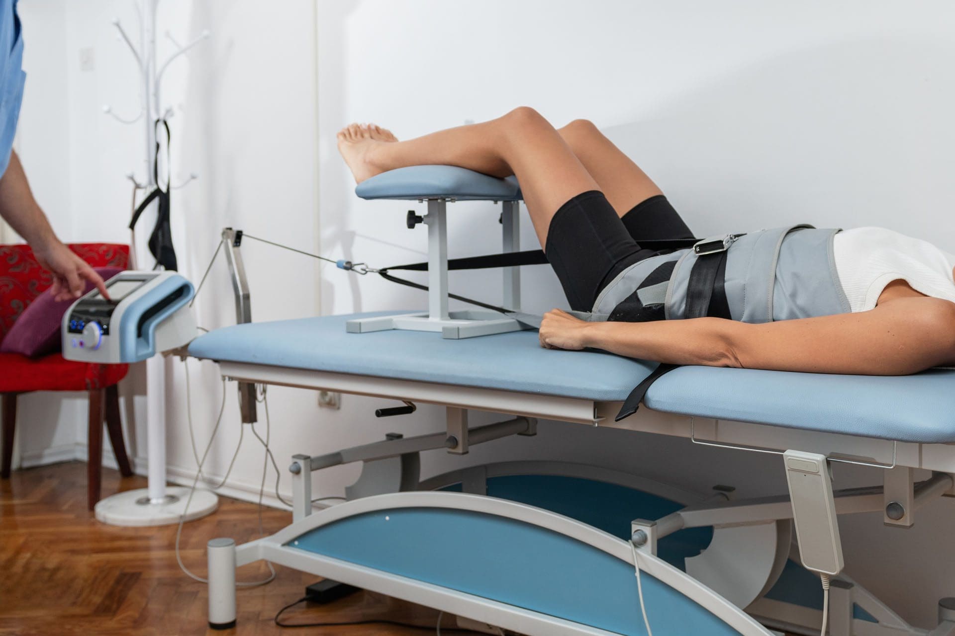
Injury Medical Chiropractic & Functional Medicine Clinic

Injury Medical Chiropractic and Functional Medicine Clinic works with primary healthcare providers and specialists to develop an optimal health and wellness solution. We focus on what works for you to relieve pain, restore function, and prevent injury. Regarding musculoskeletal pain, specialists like chiropractors, acupuncturists, and massage therapists can help mitigate the pain through spinal adjustments that help the body realign itself. They can also work with other medical professionals to integrate a treatment plan to resolve musculoskeletal issues.


Peripheral Neuropathy and Chiropractic Care


References

Ferdousi, M., Kalteniece, A., Azmi, S., Petropoulos, I. N., Worthington, A., D’Onofrio, L., Dhage, S., Ponirakis, G., Alam, U., Marshall, A., Faber, C. G., Lauria, G., Soran, H., & Malik, R. A. (2020). Corneal confocal microscopy compared with quantitative sensory testing and nerve conduction for diagnosing and stratifying the severity of diabetic peripheral neuropathy. BMJ open diabetes research & care, 8(2), e001801. https://doi.org/10.1136/bmjdrc-2020-001801

Tada, K., Murai, A., Nakamura, Y., Nakade, Y., & Tsuchiya, H. (2022). In Carpal Tunnel Syndrome, Sensory Nerve Conduction Velocities Are Worst in the Middle Finger Than in the Index Finger. Frontiers in Neurology, 13, 851108. https://doi.org/10.3389/fneur.2022.851108

Shibuya, K., Tsuneyama, A., Misawa, S., Suzuki, Y. I., Suichi, T., Kojima, Y., Nakamura, K., Kano, H., Ohtani, R., Aotsuka, Y., Morooka, M., Prado, M., & Kuwabara, S. (2022). Different patterns of sensory nerve involvement in chronic inflammatory demyelinating polyneuropathy subtypes. Muscle & Nerve, 66(2), 131–135. https://doi.org/10.1002/mus.27530

Tavee J. (2019). Nerve conduction studies: Basic concepts. Handbook of Clinical Neurology, 160, 217–224. https://doi.org/10.1016/B978-0-444-64032-1.00014-X

Cauda Equina Syndrome: Symptoms and Treatments

Cauda equina syndrome/CES is a rare condition in which the nerves in the lower back are compressed. It can include sciatica as one of its symptoms. Individuals presenting with symptoms that could be CES are advised to see a healthcare provider as soon as possible, as delaying treatment can lead to permanent damage.

Cauda Equina Syndrome

A cluster of nerve roots called the cauda equina, Latin for horse’s tail, sends and receives messages to the legs, bladder, and other body parts. Cauda equina syndrome is a rare condition in which nerve roots in the lower spinal cord are compressed. This compresses the nerves and disrupts motor and sensory function in the bladder and lower extremities. The most common cause is a ruptured or herniated disc in the lumbar area. This usually occurs when a severe disc herniation compresses the nerve bundle at the base of the spinal cord, causing significant neurological dysfunction like bladder/bowel issues and numbness in the saddle area. If found early, it is treated with surgery within 24 to 48 hours of symptom onset.

Initial Signs

Symptoms can occur acutely, in hours, or gradually over weeks or months. One of the first signs and most common symptoms is urinary retention, where the bladder fills, but there is no normal sensation or urge to urinate. (American Association of Neurological Surgeons, 2024) Other symptoms include: (Gardner A., Gardner E., & Morley T. 2011)

  • Severe low back pain
  • Sciatica
  • Pain, numbness, or weakness in one or both legs
  • Saddle anesthesia, which is numbness or other nerve sensations around the anus and the area that sits.
  • A loss of reflexes in the extremities
  • Sexual dysfunction that comes suddenly
  • Loss of bowel control
  • Overflow urination

There are two stages based on urinary symptoms (Fairbank J., & Mallen C. 2014)

CES-Incomplete CES-I

CES-Retention CES-R

  • Bladder control is completely lost.

Causes

The most common cause is a ruptured or herniated disc in the lumbar area. Other possible causes may include: (American Association of Neurological Surgeons, 2024)

Trauma or injury to the spine

  • that leads to fractures or dislocations (gunshots, falls, auto accidents)

Tumors or Infections

  • It can compress the cauda equina nerve bundle.

Spinal Stenosis

  • Narrowing of the spinal column can be either congenital or acquired.

Bone Spurs

Tuberculosis or Pott’s Paralysis

  • Pott’s paralysis is a neurological complication of tuberculosis (TB) of the spine.
  • TB is a bacterial infection that usually affects the lungs but can spread to the spine.

Iatrogenic Side Effects

  • Injuries or illnesses that result from medical or surgical treatment

Spinal Lesions or Malignant Tumors

  • A spinal lesion refers to any abnormal growth or damage within the spine.
  • It can include benign (noncancerous) and malignant (cancerous) tumors.
  • A malignant tumor is a cancerous growth within the spine; essentially, a malignant tumor is a type of spinal lesion with the potential to spread to other parts of the body.

Spinal Infection, Inflammation, Hemorrhage, or Fracture

  • A spinal infection refers to a bacterial, fungal, or viral infection that occurs within the bones of the spine (vertebrae) or the surrounding tissues, potentially causing pain, inflammation, and, in severe cases, neurological complications like weakness or paralysis;
  • Spinal inflammation is a general term for swelling or irritation within the spinal column.
  • Spinal hemorrhage” indicates bleeding within the spinal canal.
  • A spinal fracture refers to a break in one or more of the vertebrae in the spine.

Spinal Arteriovenous Malformations (AVMs)

  • A spinal arteriovenous malformation (AVM) is a rare condition in which the arteries and veins in the spinal cord tangle abnormally.
  • This can damage the spinal cord over time.

Complications from Lumbar Surgery

  • Lumbar surgery can have several complications, including infections, blood clots, nerve damage, and spinal fluid leaks.

Spinal Anesthesia

  • Spinal anesthesia is a regional anesthesia that blocks pain and sensation in the lower body.
  • It involves injecting a local anesthetic medication into the subarachnoid space surrounding the spinal cord.
  • The exact cause is not fully understood, but it can involve direct nerve root injury from the needle, inflammation caused by the anesthetic, or a spinal hematoma compressing the nerve roots.

Spinal Birth Defects

Diagnosis

Diagnosis requires a medical history of symptoms, general health, activity level, and a physical exam to assess strength, reflexes, sensation, stability, alignment, and motion. (​American Association of Neurological Surgeons, 2024) Testing includes: 

Blood Tests

  • These test for infections.

MRI

  • These yield three-dimensional images of the spinal cord, nerve roots, and surrounding areas.

Myelography

  • X-ray or computerized tomography (CT) imaging is enhanced by the injection of contrast material into the cerebrospinal fluid spaces, which can show displacement of the spinal cord or spinal nerves.

Specialized Nerve Testing

  • This could be nerve conduction velocity tests and testing electrical activity in muscles or electromyography.

Treatment

The extent of urinary problems can determine treatment protocols. A CES diagnosis is usually followed by emergency surgery within 24 to 48 hours to relieve compression of the nerves. Moving quickly is essential to prevent permanent complications such as nerve damage, incontinence, or leg paralysis. (American Association of Neurological Surgeons, 2024)

  • Depending on the cause, corticosteroids also may be prescribed to reduce swelling.
  • Antibiotics may be needed if an infection is responsible for CES.
  • For situations in which a tumor is the cause, surgery to remove it may be necessary, followed by chemotherapy and/or radiation.
  • The outcome with CES-I during surgery is generally favorable.
  • Those whose CES has deteriorated to CES-R tend to have a less favorable prognosis.

Post Surgery Therapy

After surgery, CES can be challenging to deal with. If bladder function has been impaired, recovery of control can take time.

  • It may be necessary to use a catheter or to wear disposable underwear. (American Association of Neurological Surgeons, 2024)
  • Frequent urinary infections are also a potential complication.
  • Loss of bladder or bowel control can be psychologically distressing, impacting social life, work, and relationships.
  • Sexual dysfunction can also occur, contributing to relationship difficulties or depression.

Therapy with a mental health professional may be recommended. When damage is permanent, it will be important to include family and friends in the adjustment to living with a chronic condition. Psychological counseling and/or a support group can be helpful. Other specialists who can help include: (American Academy of Orthopaedic Surgeons, 2024)

  • Occupational therapist
  • Physical therapist
  • Physiotherapist
  • Sex therapist
  • Social worker

Injury Medical Chiropractic and Functional Medicine Clinic

Injury Medical Chiropractic and Functional Medicine Clinic works with primary healthcare providers and specialists to build optimal health and wellness solutions. We focus on what works for you to relieve pain, restore function, prevent injury, and mitigate issues through adjustments that help the body realign itself. The clinic can also work with other medical professionals to integrate a treatment plan to resolve musculoskeletal problems.


Disc Herniation


References

American Association of Neurological Surgeons. (2024). Cauda Equina Syndrome. https://www.aans.org/patients/conditions-treatments/cauda-equina-syndrome/

Gardner, A., Gardner, E., & Morley, T. (2011). Cauda equina syndrome: a review of the current clinical and medico-legal position. European Spine Journal: official publication of the European Spine Society, the European Spinal Deformity Society, and the European Section of the Cervical Spine Research Society, 20(5), 690–697. https://doi.org/10.1007/s00586-010-1668-3

Fairbank, J., & Mallen, C. (2014). Cauda equina syndrome: implications for primary care. The British journal of general practice: the journal of the Royal College of General Practitioners, 64(619), 67–68. https://doi.org/10.3399/bjgp14X676988

American Academy of Orthopaedic Surgeons. (2024). Cauda equina syndrome. https://orthoinfo.aaos.org/en/diseases–conditions/cauda-equina-syndrome

The Prevalence of Rotator Cuff Tears and Treatment Approaches

Could older individuals who do not have symptoms of shoulder pain or loss of shoulder and arm function have a rotator cuff tear?

Rotator Cuff Tear Physical Therapy

A rotator cuff tear is a common injury to the four muscles and tendons surrounding and stabilizing the shoulder joint. Studies have shown that (Geary M. B., & Elfar J. C. 2015)

  • 30% of those under the age of 70
  • 70% of those over age 80 have a rotator cuff tear. 

Physical therapy is often recommended as a first-line initial treatment for rotator cuff pain. In most cases, a rotator cuff tear will not need surgical treatment. Determining when surgery is necessary depends on several factors a patient can discuss with their healthcare provider.

Causes

  • Overuse and repetitive motions
  • Trauma (e.g., falls, collisions)
  • Age-related degeneration

Symptoms

  • Shoulder pain, especially with overhead or rotational movements
  • Weakness and difficulty raising the arm
  • Clicking or grinding sounds in the shoulder
  • Limited range of motion

Treatment

The goal of physical therapy for a rotator cuff tear is not necessarily to heal the torn tendon but to relieve pain and improve strength by reducing inflammation and restoring shoulder joint mechanics. This is achieved through physical therapy, ice application, anti-inflammatory treatments like medications, and cortisone injections.

Physical Therapy

The goal of physical therapy is to improve the function of the muscles that surround the shoulder. Physical therapy targets the smaller muscles around the shoulder that are commonly neglected. By strengthening these muscles, the treatment can help compensate for damaged tendons and improve the mechanics of the shoulder joint. A chiropractic physical therapy team will develop a personalized exercise program. Generally, rehabilitation will start with gentle range of motion exercises that can be accomplished using the arms to lift a mobility stick/wand overhead.

Shoulder Pulleys

  • These improve shoulder range of motion and flexibility.

Isometric Exercises

  • These exercises are for the rotator cuff muscles and may then be started.
  • This exercise can improve the contracting of the muscles around the shoulder and offer more support to the shoulder joint.

Scapular Stabilization Exercises

  • These can also be done to improve the muscles surrounding the shoulder blade’s function.
  • This can help improve how the shoulder joint, arm, and scapulae move together when using the arm.

Advanced Strengthening

  • Advanced exercises can be done with a dumbbell or resistance band.

Consult your healthcare provider or physical therapist before starting these or any other exercises for a rotator cuff tear. Doing exercises correctly prevents further pain, injury, or shoulder problems. Specific, focused exercises can help expedite and regain normal shoulder function.

Injury Medical Chiropractic and Functional Medicine Clinic

The prognosis for rotator cuff tears depends on the severity of the tear and the individual’s overall health. With proper treatment, most people can regain the full function of their shoulders. However, some may experience ongoing pain or limitations in severe cases. Injury Medical Chiropractic and Functional Medicine Clinic works with primary healthcare providers and specialists to build optimal health and wellness solutions. We focus on what works for you to relieve pain, restore function, prevent injury, and help mitigate issues through adjustments that help the body realign itself. They can also work with other medical professionals to integrate a treatment plan to resolve musculoskeletal problems.


Shoulder Pain Chiropractic Treatment


References

Geary, M. B., & Elfar, J. C. (2015). Rotator Cuff Tears in the Elderly Patients. Geriatric orthopaedic surgery & rehabilitation, 6(3), 220–224. https://doi.org/10.1177/2151458515583895

Maximizing the Benefits of Napping: Optimal Duration and Effects

Could regular daytime naps help individuals slow the aging process from natural brain shrinkage?

Taking Naps

Research and experts suggest that a short nap between 10 to 40 minutes provides the most benefits, including:

  • Improved mood
  • Cognitive performance
  • Alertness

A study suggests that daytime napping might prevent the brain from shrinking with age. (Paz V., Dashti H. S., & Garfield V. 2023)

Optimal Nap Time

A small study of young adults found that naps lasting 10 to 60 minutes immediately improved mood and alertness. However, most research shows that naps under 30 minutes offer the most benefit because individuals are less likely to enter the deep sleep stage or experience sleep inertia – a period of impaired alertness right after waking.  A meta-analysis also suggested that short naps reduce fatigue, increase productivity, and enhance physical performance. (Dutheil F. et al., 2021) A 10-minute nap is the most effective at improving:

  • Sleep latency or sleep onset latency (SOL)
  • Fatigue
  • Vigor
  • Cognitive performance

Some sleep specialists recommend that patients not take naps longer than 40 minutes, as too much napping could be unhealthy. Studies have shown that excessive daytime sleepiness and napping for over 60 minutes might increase the risk of type 2 diabetes and heart problems. (Yamada T, Nobuhiro S, Takashi K. 2016)

Health

In the study published in Sleep Health, researchers used data from over 30,000 participants aged 40 to 69 from the U.K. Biobank. Researchers examined genetic variants associated with taking naps regularly. (Paz V., Dashti H. S., & Garfield V. 2023) The researchers found a link between regular daytime napping and larger brain volume. The difference in brain volume between individuals who nap regularly and those who don’t was equivalent to 2.6 to 6.5 years of aging. However, no association was found between napping and cognitive performance reaction time or visual memory. The brain naturally shrinks with age, but this process is accelerated in individuals with neurodegenerative diseases and cognitive decline.

How To Avoid Napping for Too Long

Taking naps is good for you. But there’s a difference between healthy daytime sleep and counterproductive excessive sleeping.

  • Sleep specialists recommend setting an alarm for a nap or asking a family member, friend, or coworker to wake you up.
  • Individuals can try placing their phones or alarm clocks far away so they have to move to turn them off.
  • Individuals are recommended to stand up immediately to wake the body through movement and light exposure to avoid feeling groggy after a nap.
  • One study suggests consuming caffeine before a nap may be an effective countermeasure for sleep inertia. (Hilditch C. J., Dorrian J., & Banks S. 2016)
  • Engaging in physical activities before and after naps can also help promote wakefulness.

Sometimes, people feel exhausted for various reasons, such as stress and nutrition, rather than a lack of sleep. In these cases, sleeping more than the body needs will only worsen sleep quality at night. When individuals are experiencing daytime fatigue, rather than lying back down or sitting in bed, sleep specialists suggest walking around. This allows the fatigue to pass, and the patient can sleep better at night.

Injury Medical Chiropractic and Functional Medicine Clinic

Chiropractic’s goals are to help individuals enhance movement with less pain due to a condition after injury or surgery. Injury Medical Chiropractic and Functional Medicine Clinic works with primary healthcare providers and specialists to develop optimal health and wellness solutions. We focus on what works for you to relieve pain, restore function, prevent injury, and help mitigate issues through adjustments that help the body heal itself. They can also work with other medical professionals to integrate a treatment plan to resolve musculoskeletal problems.


Secrets of Optimal Wellness


References

Paz, V., Dashti, H. S., & Garfield, V. (2023). Is there an association between daytime napping, cognitive function, and brain volume? A Mendelian randomization study in the UK Biobank. Sleep health, 9(5), 786–793. https://doi.org/10.1016/j.sleh.2023.05.002

Dutheil, F., Danini, B., Bagheri, R., Fantini, M. L., Pereira, B., Moustafa, F., Trousselard, M., & Navel, V. (2021). Effects of a Short Daytime Nap on the Cognitive Performance: A Systematic Review and Meta-Analysis. International journal of environmental research and public health, 18(19), 10212. https://doi.org/10.3390/ijerph181910212

Yamada T, N. S., Takashi K. (2016). Daytime napping, daytime sleepiness and the risk of metabolic diseases: dose-response meta-analysis using restricted cubic spline model. J Am Coll Cardiol., 67(13), 1951. https://doi.org/https://doi.org/10.1016/S0735-1097(16)31952-0

Hilditch, C. J., Dorrian, J., & Banks, S. (2016). Time to wake up: reactive countermeasures to sleep inertia. Industrial health, 54(6), 528–541. https://doi.org/10.2486/indhealth.2015-0236