Understanding the Impact of Whiplash Before Going Back To Work
Discover the impact of whiplash on your health and learn effective strategies for recovery and management before going back to work.
Returning to Work After Motor Vehicle Accidents: Understanding Whiplash, Treatments, and Clinical Insights
Introduction
Getting back to work after a motor vehicle accident (MVA) can feel like trying to solve a puzzle with missing pieces, especially when whiplash is involved. Whiplash, a frequent injury from car crashes, can turn simple tasks like typing or lifting into a real pain in the neck (pun intended!). But with the right care, many people can return to their jobs and lives with confidence. In this in-depth guide, we’ll explore whiplash, its impact on work, nonsurgical treatments, and the clinical rationale for getting back to work. We’ll also spotlight Dr. Alexander Jimenez, a leading chiropractor and nurse practitioner in El Paso, Texas, whose expertise in personal injury cases helps patients navigate recovery and legal processes. Let’s dive into this journey of healing, with a sprinkle of humor to keep things light—because who said recovery can’t have a few laughs?
What is Whiplash?
Definition and Causes
Whiplash is a neck injury caused by a sudden, forceful back-and-forth movement of the head, much like the crack of a whip. This rapid motion can strain or tear the muscles, ligaments, and tendons in the neck, leading to a range of symptoms. The most common cause is rear-end car collisions, but whiplash can also occur from sports injuries, falls, or even an overly enthusiastic head-banging session at a concert (though we’ll stick to MVAs for now). Research estimates that whiplash affects over one million people annually in the U.S., with societal costs reaching up to $29 billion (Lovell & Galasko, 2002).
The mechanics of whiplash are no joke. When a car is hit from behind, the head accelerates backward and then snaps forward, stretching soft tissues beyond their normal limits. MRI and autopsy studies have shown that whiplash can cause injuries to cervical ligaments, discs, and facet joints, which are critical for neck stability (Kaale et al., 2005). These injuries, often invisible on standard X-rays, can lead to persistent pain if not addressed early.
Symptoms of Whiplash
Whiplash symptoms are as varied as the toppings on a pizza—and just as likely to linger if not handled properly. Common symptoms include:
- Neck Pain and Stiffness: The hallmark of whiplash, often described as a tight, aching sensation.
- Headaches: Frequently starting at the base of the skull.
- Dizziness: Making you feel like you’re on a merry-go-round that won’t stop.
- Fatigue: Because your body is working overtime to heal.
- Shoulder, Back, or Arm Pain: Pain can radiate beyond the neck.
- Numbness or Tingling: Often in the arms, signaling nerve irritation.
- Blurred Vision, Ringing in the Ears: Less common but still disruptive.
- Cognitive Issues: Trouble concentrating or remembering, as if your brain took a vacation.
- Sleep Disturbances, Irritability, Depression: Because chronic pain is a mood-killer.
These symptoms may not show up right away, sometimes taking days or weeks to appear, which is why seeing a doctor ASAP is crucial (Sterner & Gerdle, 2004). Think of it like a sneaky ninja—whiplash can creep up when you least expect it.
| Symptom | Description |
|---|---|
| Neck Pain/Stiffness | Aching or tightness, limiting neck movement. |
| Headaches | Often starting at the skull base, can be persistent. |
| Dizziness | Feeling unsteady or lightheaded. |
| Fatigue | General tiredness due to the body’s healing efforts. |
| Radiating Pain | Pain spreading to shoulders, back, or arms. |
| Numbness/Tingling | Nerve-related sensations in arms or hands. |
| Cognitive/Sleep Issues | Difficulty focusing, remembering, or sleeping; irritability or depression. |
Impact of Whiplash on Work
Whiplash can throw a wrench into your work life faster than a Monday morning traffic jam. The pain and stiffness can make it tough to sit at a desk, lift boxes, or even focus on a computer screen. Research paints a sobering picture:
- A BMC Public Health study found that only 44% of whiplash patients returned to work after two years (Freeman et al., 1999).
- A Danish study of 104 patients reported 56% were still on sick leave two years post-injury (Kasch et al., 2001).
- Between 19% and 60% of patients experience symptoms six months after injury, with up to half unable to return to work within a year (Sterner & Gerdle, 2004).
- A 2001 study noted that 12% of whiplash patients hadn’t resumed normal activities or modified job functions a year later (Spitzer et al., 1995).
These numbers show that whiplash isn’t just a physical hurdle—it’s a career roadblock. Chronic symptoms, like persistent neck pain or cognitive difficulties, can lead to long-term work disability, especially if untreated. Imagine trying to type a report while your neck screams, “Not today!” Early treatment is the key to getting back to your desk, warehouse, or classroom without feeling like you’re auditioning for a role as a human statue.
The Power of Chiropractic Chiropractic Care In Injury Rehabilitation- Video
Clinical Rationale for Returning to Work
Returning to work after an MVA isn’t just about paying the bills (though that’s a big motivator!). It’s also about restoring normalcy, boosting mental health, and preventing the downward spiral of inactivity. From a clinical perspective, early mobilization is a game-changer. Prolonged rest can lead to muscle atrophy and increased stiffness, making recovery harder (Quebec Task Force, 1995). Think of your body like a car—if you leave it in the garage too long, it’s going to get rusty.
Healthcare providers often recommend a gradual return to work as part of rehabilitation. This might mean starting with part-time hours or modified duties, like swapping heavy lifting for lighter tasks. The goal is to keep you moving without overdoing it. Studies suggest that early, active rehabilitation—think chiropractic adjustments or physical therapy—can reduce pain and improve function, paving the way for a smoother return to work (Teasell et al., 2010).
However, the decision to return isn’t one-size-fits-all. It depends on:
- Injury Severity: Mild whiplash might resolve in weeks, while severe cases could take months.
- Job Demands: A desk job is easier to return to than construction work.
- Overall Health: Pre-existing conditions can complicate recovery.
A good doctor, like Dr. Alexander Jimenez, will assess these factors and create a plan that gets you back to work without risking further injury. It’s like planning a road trip—you need a map, not just a destination.
Nonsurgical Treatments for Whiplash
Nobody wants to go under the knife if they can avoid it, and luckily, whiplash responds well to nonsurgical treatments. These approaches aim to reduce pain, restore mobility, and get you back to your daily grind. Here’s a rundown of the top options:
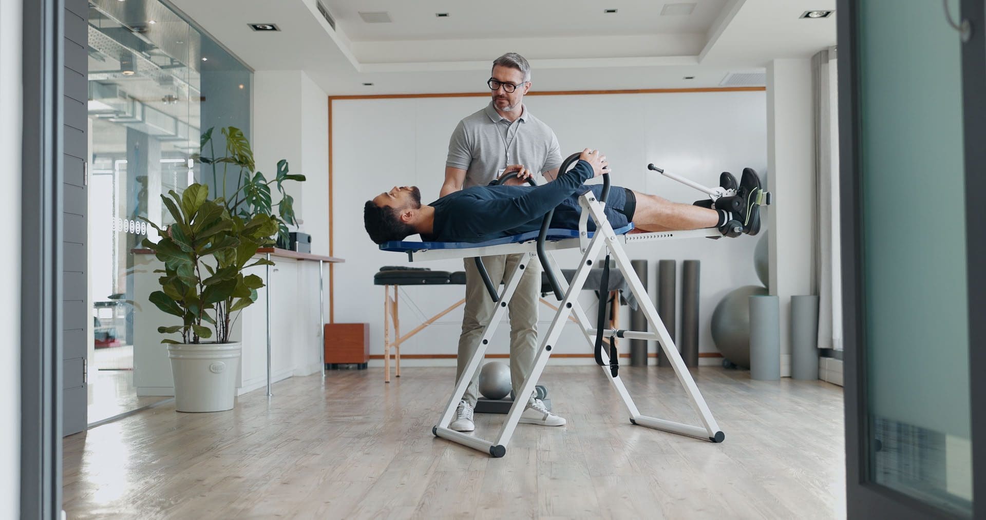
- Chiropractic Care: Chiropractors use spinal manipulation to realign the spine and relieve nerve pressure. It’s like giving your neck a gentle nudge to say, “Get back in line!” Studies show it can significantly reduce pain and improve mobility (Lord et al., 1996).
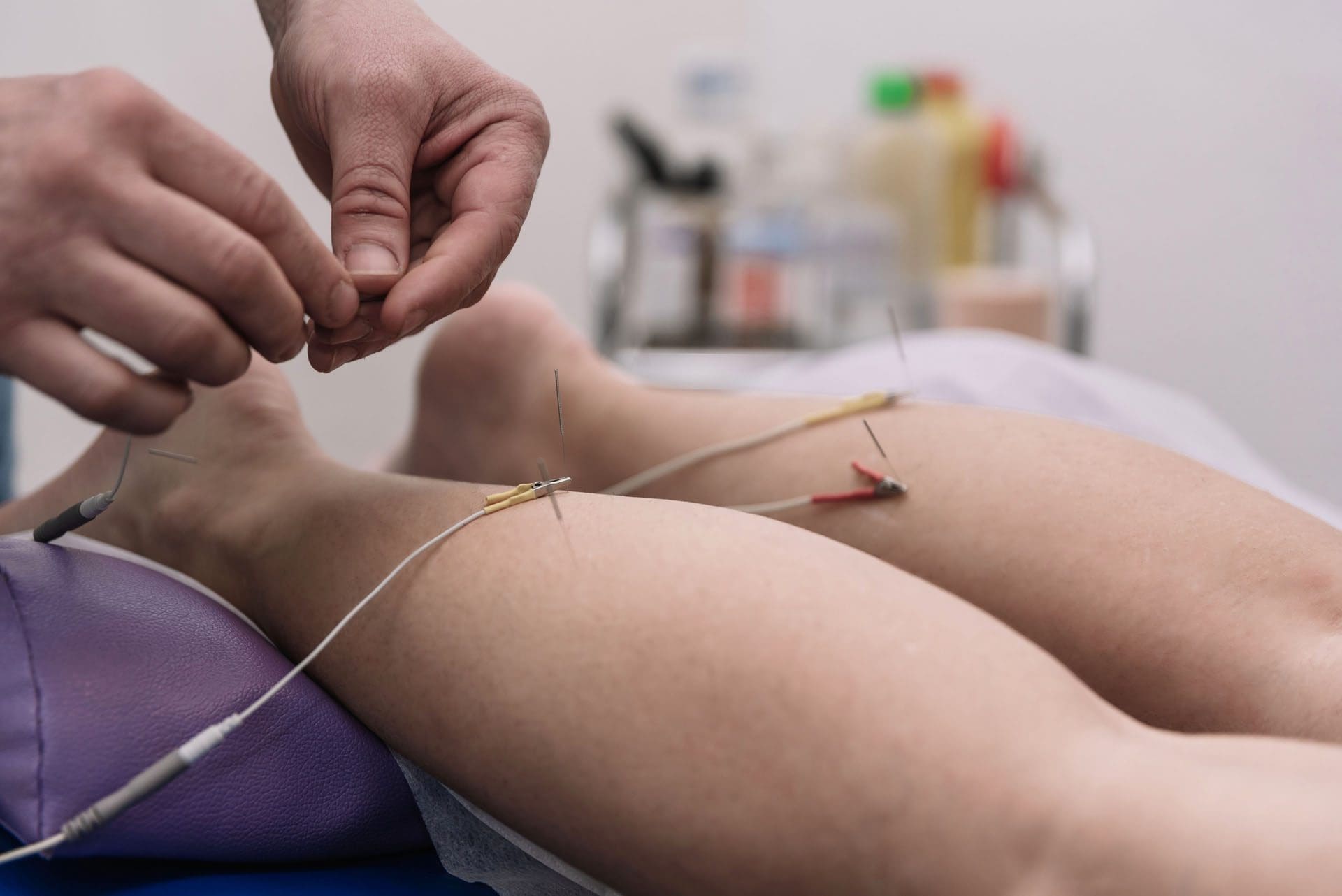
- Physical Therapy: Physical therapists design exercise programs to strengthen neck muscles and improve range of motion. They might also use heat, ice, ultrasound, or electrical stimulation to ease pain. It’s like a gym session tailored for your neck (Teasell et al., 2010).
- Exercise: Gentle exercises, like stretching or swimming, can strengthen the neck and improve posture. Think of it as physical therapy’s fun cousin—less clinical, more doable at home (McLean et al., 2014).
- Medications: Over-the-counter pain relievers (e.g., ibuprofen) or muscle relaxants can help manage pain and inflammation. Prescription meds may be used for severe cases (Childs et al., 2008).
- Injections: Corticosteroid injections can reduce inflammation in stubborn cases, offering relief when other methods fall short (Barnsley et al., 1994).
- Radiofrequency Neurotomy: For chronic pain, this procedure targets specific nerves to block pain signals. It’s like hitting the mute button on your neck’s complaints (Lord et al., 1996).
These treatments work best when started early, preventing symptoms from becoming chronic. Combining them, like pairing chiropractic care with exercise, can supercharge recovery. It’s like assembling a superhero team for your neck—each treatment brings its own powers!
| Treatment | Benefits | Considerations |
|---|---|---|
| Chiropractic Care | Reduces pain, improves spinal alignment and mobility. | Requires skilled practitioner; multiple sessions. |
| Physical Therapy | Strengthens muscles, enhances flexibility, reduces pain. | Time-intensive; needs patient commitment. |
| Exercise | Improves posture, strengthens neck, accessible at home. | Must be done correctly to avoid strain. |
| Medications | Quick pain and inflammation relief. | Temporary; potential side effects. |
| Injections | Targeted relief for severe inflammation. | Invasive; not first-line treatment. |
| Radiofrequency Neurotomy | Long-term pain relief for chronic cases. | Specialized procedure; not widely available. |
Dr. Alexander Jimenez’s Approach to Personal Injury Cases
In El Paso, Texas, Dr. Alexander Jimenez, DC, APRN, FNP-BC, stands out as a beacon of hope for MVA victims. With over 25 years of experience, Dr. Jimenez combines his expertise as a chiropractor and nurse practitioner to offer integrative care that addresses both the physical and medical aspects of whiplash (Injury Medical Clinic). His clinic, Injury Medical & Chiropractic Clinic, specializes in treating severe pain conditions, including neck pain, back pain, sciatica, and whiplash, using advanced therapies focused on mobility, health, and wellness.
Advanced Diagnostics and Treatment
Dr. Jimenez employs advanced imaging, like MRI and CT scans, to pinpoint injuries that might not show up on standard X-rays. These tools help him identify damage to ligaments, discs, or facet joints, ensuring accurate diagnoses (Jimenez, n.d.). He also uses diagnostic evaluations, such as range-of-motion tests and neurological assessments, to create personalized treatment plans. His dual-scope approach—combining chiropractic adjustments with medical management—ensures holistic care that tackles both symptoms and underlying causes.
Bridging Medical and Legal Needs
Personal injury cases often involve legal battles, and Dr. Jimenez excels as a liaison between medical care and legal documentation. He provides detailed reports that link injuries to the MVA, supporting patients’ claims in court. This is crucial in El Paso, where personal injury cases are common due to frequent MVAs. His ability to translate complex medical findings into clear, legally admissible documentation makes him a trusted partner for both patients and attorneys (Personal Injury Doctor).
Patient-Centered Care
Patients rave about Dr. Jimenez’s compassionate approach. One patient noted, “Dr. Jimenez and his staff genuinely care about your recovery,” highlighting his dedication to improving mobility and reducing pain (Yelp Reviews). His clinic also emphasizes education, teaching patients about injury prevention and wellness to prevent future issues. It’s like getting a personal coach for your health, minus the whistle!
Personal Injury Cases in El Paso
El Paso, a bustling border city, sees its fair share of MVAs, leading to numerous personal injury cases. Whiplash and other soft tissue injuries are common, and navigating the medical and legal landscape can be overwhelming. Dr. Jimenez’s expertise is a game-changer here. His ability to provide comprehensive care while supporting legal claims makes him a go-to practitioner for accident victims. His clinic’s focus on conditions like whiplash, sciatica, and neck pain ensures that patients get back on their feet—and back to work—as quickly as possible (Auto Accident Treatment).
In personal injury cases, medical evidence is critical. Dr. Jimenez’s use of advanced imaging and diagnostics provides solid proof of injury, which can make or break a legal case. His integrative approach also means patients don’t have to bounce between specialists—he handles both the chiropractic and medical sides, streamlining recovery and documentation.
A Touch of Humor
Let’s face it—dealing with whiplash is about as fun as a root canal during a power outage. But here’s a silver lining: with the right care, you can go from “ouch” to “I’ve got this!” faster than you can say “chiropractic adjustment.” Picture your neck as a grumpy cat—stiff and uncooperative at first, but with a little TLC from Dr. Jimenez, it’ll be purring (or at least not hissing) in no time. Humor aside, recovery is serious business, and the right treatment can make all the difference.
Conclusion
Returning to work after an MVA, especially with whiplash, requires a strategic approach that blends timely treatment, personalized care, and expert guidance. Dr. Alexander Jimenez’s integrative methods, advanced diagnostics, and role as a medical-legal liaison make him a standout in El Paso’s personal injury landscape. By addressing whiplash with nonsurgical treatments and supporting patients through recovery and legal processes, he helps them reclaim their lives and livelihoods.
Disclaimer: This blog post is for informational purposes only and should not be taken as medical advice. Always consult a qualified healthcare professional for diagnosis and treatment of any medical condition.
Key Citations
- Whiplash Disorders Review
- Neck Disability Index and MRI Findings
- Chronic Whiplash Disorders Review
- Critique of Whiplash Syndrome Literature
- Handicap After Acute Whiplash Injury
- Quebec Task Force on Whiplash
- Radiofrequency Neurotomy for Cervical Pain
- Therapeutic Interventions for Whiplash
- Chronic Symptoms After Whiplash
- Neck Pain Clinical Guidelines
- Pathophysiology of Whiplash
- Dr. Jimenez’s Injury Medical Clinic
- Personal Injury Doctor Services
- Dr. Jimenez LinkedIn Profile
- Dr. Jimenez Healthgrades Profile
- A4M Profile of Dr. Jimenez
- Auto Accident Injury Treatment
- Injury Medical Clinic Yelp Reviews
- Return to Work Rates After Whiplash