Personal TENS Devices: A Modern Approach to Pain Relief
For individuals managing chronic pain conditions, can incorporating a personal TENS device help?
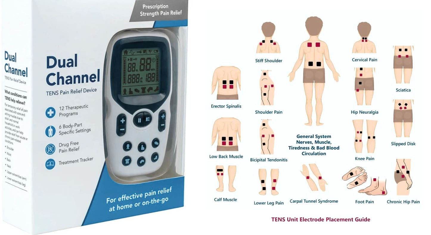
Personal TENS Device
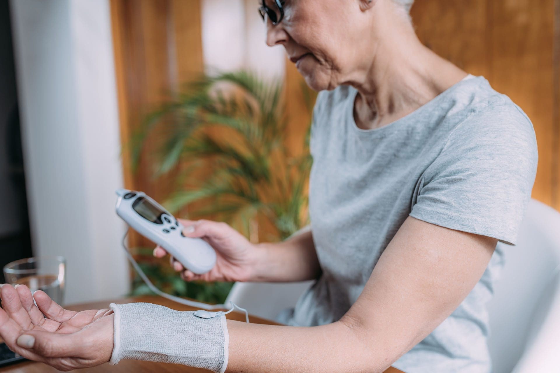
Almost all physical therapy, chiropractic, acupuncture, and massage clinic healthcare providers offer TENS (transcutaneous electrical nerve stimulation) therapy. Individuals can buy a TENS unit for on-the-go and home care use. A personal TENS unit is a small, battery-powered device that uses low-voltage electrical currents delivered through electrodes placed on the skin to help relieve pain.
How It Works
TENS units work by stimulating nerve fibers in pain, which can help block pain signals from reaching the brain or by stimulating the release of endorphins, the body’s natural pain-killing chemicals.
Uses
TENS units are used to treat a variety of conditions, including:
- Arthritis
- Muscle cramps
- Neck pain
- Back pain
- Pelvic pain
- Knee pain
- Osteoarthritis
- Fibromyalgia
- Tendinitis
- Bursitis
- Diabetes-related neuropathy
- Pelvic pain from periods or endometriosis
Features
Portability
- TENS units are small, lightweight, and portable, making them convenient for home or on the go. It can be held in a pocket or clipped to a belt. (National Health Service UK, 2025)
Electrodes
- The unit is connected to a series of electrodes placed on the skin to deliver the electrical charge.
Adjustable Settings
- Many TENS units allow users to adjust the electrical pulses’ intensity, frequency, and duration.
Benefits
Non-Invasive
- TENS therapy is a non-invasive method for relieving pain.
Drug-Free
- It can provide pain relief without the need for medication.
Convenient
- TENS units are small, portable, and relatively discrete.
Precautions
Consult a healthcare provider before using a TENS device to ensure its safety for you and your injury/condition. The treatment should not be used for individuals who are pregnant, have epilepsy, poor sensation, a heart problem, a pacemaker, or another electrical or metal implant in their body. (National Health Service UK, 2025) The electrodes should not be placed on certain areas of the body, including (Teoli D, Dua A, An J. 2025)
- Head
- Eyes
- Mouth
- Front of the Neck
- Chest and upper back at the same time
- Numb areas
- Broken skin
- Tumors
There is a minor risk of skin irritation, particularly if allergic to the adhesive pads.
Effectiveness
Researchers are still determining how effective TENS units are for relieving and reducing pain. A study found that TENS was effective in relieving pain for patients with fibromyalgia. (Dailey D. L. et al., 2013) Another study suggested that TENS may improve bone pain for cancer patients, but the results were inconclusive because of the limited number of randomized trials. (Vance C. G. et al., 2014)
Research suggests that some factors can impact the effectiveness of the intervention. Varying the intensity and frequency may help it be more effective so the body doesn’t develop a tolerance to it. In addition, using the electrodes in areas that are acupuncture points may help to reduce pain. While further study is needed, TENS is considered a safe pain-relief option for many conditions because it’s non-invasive and doesn’t require medication. (Vance C. G. et al., 2014)
Injury Medical Chiropractic & Functional Medicine Clinic
Individuals interested in trying a personal TENS unit should consult their healthcare provider. They may be able to refer them to a physical therapist, who can show them which type and how to use it for their particular condition. Injury Medical Chiropractic and Functional Medicine Clinic works with primary healthcare providers and specialists to develop an optimal health and wellness solution. We focus on what works for you to relieve pain, restore function, and prevent injury. Regarding musculoskeletal pain, specialists like chiropractors, acupuncturists, and massage therapists can help mitigate the pain through spinal adjustments that help the body realign itself. They can also work with other medical professionals to integrate a treatment plan to resolve musculoskeletal issues.
Don’t Ignore Post-Accident Pain
References
National Health Service UK. (2025). TENS (transcutaneous electrical nerve stimulation). https://www.nhs.uk/conditions/transcutaneous-electrical-nerve-stimulation-tens/
Teoli, D., Dua, A., & An, J. (2025). Transcutaneous Electrical Nerve Stimulation. In StatPearls. https://www.ncbi.nlm.nih.gov/pubmed/30725873
Dailey, D. L., Rakel, B. A., Vance, C. G. T., Liebano, R. E., Amrit, A. S., Bush, H. M., Lee, K. S., Lee, J. E., & Sluka, K. A. (2013). Transcutaneous electrical nerve stimulation reduces pain, fatigue, and hyperalgesia while restoring central inhibition in primary fibromyalgia. Pain, 154(11), 2554–2562. https://doi.org/10.1016/j.pain.2013.07.043
Vance, C. G., Dailey, D. L., Rakel, B. A., & Sluka, K. A. (2014). Using TENS for pain control: the state of the evidence. Pain management, 4(3), 197–209. https://doi.org/10.2217/pmt.14.13