Care is the cornerstone of our practice

Give us a Call
+1 (915) 412-6680
Send us a Message
support@chiromed.com
Opening Hours
Mon-Thu: 7 AM - 7 PM
Fri - Sun: Closed

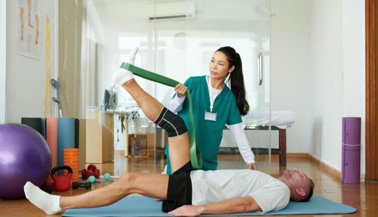
Post Surgery Leg Strengthening Tips for Patients

What are some leg-strengthening exercises that will expedite recovery for athletes and physically active individuals who have undergone leg surgery?

Post Surgery Leg Strengthening

Leg muscles may weaken after hip, knee, ankle, or foot surgery. This happens because those muscles are not used as much during recovery. Gaining back strength and muscle endurance after an injury or surgery is an important step in recovery. Leg exercises can help regain mobility and prevent complications like blood clots and pressure sores after surgery or an injury, which is why engaging in post-surgery leg strengthening is important once the doctor gives the ok.

Benefits

post-surgery leg strengthening exercises have several benefits, including

  • Rebuilds strength and confidence
  • Retraining for optimal mobility and flexibility
  • Prevents pressure sores
  • Prevents blood clots

When the leg muscles are contracted, they move blood like a pump, maintaining proper circulation. Moving in a bed after surgery also helps prevent pressure sores from forming and blood clots. A physical therapy team will determine the right leg exercises for each individual and their injury/condition. This is an important step when moving forward after surgery. (Hoogeboom T. J. et al., 2014)

Starting Out

The first exercises should target all the major muscles of the leg. (Madara K. C. et al., 2019)

Gluteal Sets

This isometric exercise means the muscles contract while no motion occurs at the hip joints. To perform:

  • Lie on your back and tighten your buttock muscles.
  • Hold the muscles tight for 5 seconds, then relax.
  • Repeat 10 or 15 times.
  • Gluteal sets can be done several times per day.

Heel Slide

Heel slides can help regain strength in the major muscles of the leg. To perform:

  • Lie on your back.
  • Bend the knee of the surgical leg and slowly slide the heel toward your butt.
  • Slide as far as possible and hold for 5 seconds.
  • Slowly return to the starting position and repeat.

Short Arc Quad

The short arc quad, or SAQ, is a simple way to get the quadricep muscles working. To perform:

  • Lie on your back with a towel roll, small ball, or something similar under the knee.
  • Slowly straighten the knee.
  • Tighten the quad muscle on the top of the thigh.
  • Hold for 3 seconds, then relax.
  • Repeat 10 to 15 times.

Quad Set

This exercise helps get the quad muscles working. It also helps control the position of the kneecap. To perform:

  • Lie on your back.
  • Place a small towel roll under the knee.
  • Try to press the back of the knee flat against the floor.
  • Hold for 10 seconds and release.
  • Repeat 10 to 15 times.

Individuals can complete quad sets bilaterally or with both knees simultaneously. This makes the stronger leg help strengthen the weaker side.

Straight Leg Raise

To perform:

  • Lie on your back.
  • Lift your leg straight off the floor until it is at the height of the opposite bent knee.
  • Hold for 10 seconds and slowly lower.
  • Repeat 10 to 15 times.

Be sure to keep the knee straight for the entire exercise. Keep the opposite knee bent for comfort. To ensure the knee is straight, individuals can complete a quad set first and then the straight leg raise. The exercise can be more challenging by increasing repetitions or adding a 2- to 3-pound ankle weight on the thigh. For even more challenge, add the ankle weight to the ankle.

Hamstring Strengthening

Working out the hamstrings after injury or surgery is important. The hamstring muscles bend the knee and extend the hip backward. To perform:

  • Lie on your stomach.
  • Bend one knee to raise the lower limb straight in the air.
  • Hold for 5 seconds and lower slowly.
  • Repeat 10 to 15 times.

Once the exercise is easy to do, increase the repetitions to 30. Individuals can also try adding a 2- to 3-pound ankle weight.

Physical therapy can help individuals regain mobility after injury or surgery. A therapist may prescribe exercises as part of an at-home exercise program. Over time progress will go from simple exercises to more challenging ones to improve balance and mobility. (Madara K. C. et al., 2019)

Injury Medical Chiropractic & Functional Medicine Clinic

Before starting this or any other exercise program, consult a doctor and a physical therapist to find the right exercises for your situation. Injury Medical Chiropractic and Functional Medicine Clinic works with primary healthcare providers and specialists to develop an optimal health and wellness solution. We focus on what works for you to relieve pain, restore function, and prevent injury. Regarding musculoskeletal pain, specialists like chiropractors, acupuncturists, and massage therapists can help mitigate the pain through spinal adjustments that help the body realign itself. They can also work with other medical professionals to integrate a treatment plan to resolve musculoskeletal issues.


Are You Recovering From Ankle Sprains?


References

Hoogeboom, T. J., Dronkers, J. J., Hulzebos, E. H., & van Meeteren, N. L. (2014). Merits of exercise therapy before and after major surgery. Current opinion in anaesthesiology, 27(2), 161–166. https://doi.org/10.1097/ACO.0000000000000062

Madara, K. C., Marmon, A., Aljehani, M., Hunter-Giordano, A., Zeni, J., Jr., & Raisis, L. (2019). PROGRESSIVE REHABILITATION AFTER TOTAL HIP ARTHROPLASTY: A PILOT AND FEASIBILITY STUDY. International Journal of Sports Physical Therapy, 14(4), 564–581.

Lidocaine Patch for Pain Relief: An Overview

For individuals experiencing lower back pain and sciatica symptoms, can using a lidocaine patch help?

Lidocaine Patch

Lidocaine patches are in a drug class called local anesthetics (MedlinePlus, 2025) (Food and Drug Administration, 2022). They relieve pain in small body areas by blocking the nerves from sending pain signals. The Food and Drug Administration approved lidocaine 5% or 1.8% patches for relieving postherpetic neuralgia (PHN) pain. These lidocaine patches are prescription-only (Food and Drug Administration, 2022). They are available either by prescription or over the counter and are commonly used for back pain and other areas. Over-the-counter lidocaine, 4% patches, can be used to relieve minor aches and pains, including back pain. However, further research is needed to recommend the use of lidocaine patches as an effective method of pain relief. (Department of Veterans Affairs/Department of Defense, 2022) (North American Spine Society, 2020)

The Patch

They are available in prescription and over-the-counter forms. Over-the-counter forms come as a 4% patch in various brands, such as (MedlinePlus, 2025)

  • Aspercreme
  • Lidocare
  • Salonpas

Prescription-only patches come in 5% or 1.8% patches and are approved to relieve long-term nerve pain from shingles in adults. Zlido is a brand name for a 1.8% lidocaine patch. Five percent lidocaine patches are available generically. (DailyMed, 2018) (Food and Drug Administration, 2021) The safety and effectiveness in children are unknown. (Food and Drug Administration, 2022)

Other Lidocaine Forms

As a local anesthetic, it is available in several other forms, including:

  • Spray
  • Topical cream
  • Lotion
  • Liquid
  • Ear drops
  • Eye gel
  • Injection
  • It is also available as a short-term intravenous infusion for abnormal heart rhythm. (DailyMed, 2025)

Effectiveness

Clinical trials have shown that the patches effectively relieve chronic low back pain. However, these studies were nonrandomized and did not include a control group to compare against the treatment group. (Santana J. A., Klass S., & Felix E. R. 2020) The results may be subject to potential biases. For this reason, further evidence is needed to support lidocaine patches’ effectiveness in reducing low back pain. (North American Spine Society, 2020) High-quality randomized and controlled clinical trials are necessary to study the effectiveness. (North American Spine Society, 2020) (Santana J. A., Klass S., & Felix E. R. 2020)

How to Use Safely

In general, keep the following in mind (MedlinePlus, 2025)

  • Use according to the directions on the box and the healthcare provider’s recommendations.
  • Do not use on broken or inflamed, swollen skin.
  • Do not apply heat like heating pads or electric blankets over patches.
  • Avoid getting water on or around the patch.
  • Avoid letting a patch near your eyes to limit eye irritation.
  • Fold the sticky sides of the used lidocaine patches together and safely throw them away, keeping them away from children and pets.

How to use (MedlinePlus, 2025)

  • Clean and dry the affected area before placing the patch.
  • Apply to the affected body area as directed.
  • Wash your hands after touching the patch.
  • Remove the patch after what is usually recommended after eight hours.

How Quickly Does It Work?

The amount of medication absorbed into the blood depends on how long the patch is placed on the body and how much is covered with the patch. In a study, healthy participants wore three lidocaine patches on their backs for 12 hours on and 12 hours off during 24 hours. Lidocaine levels were highest at 11 hours. At the end of the 24 hours, there was still some lidocaine left in the bloodstream. (Food and Drug Administration, 2022)

Individuals with PHN may notice a difference in pain intensity after four hours of using the patch. (Rowbotham, M. C. et al., 1996) (Food and Drug Administration, 2022) Experts may suggest using patches for four weeks for those who experience pain after their shingles rash disappears. However, if there is still pain after these four weeks, it is recommended to see a pain specialist. (Gross, G. E. et al., 2020)

Side Effects

Common side effects are typically mild skin reactions where the patch is.  These reactions are usually temporary and will disappear within a few minutes or hours. Examples include: (Food and Drug Administration, 2022)

  • Irritation
  • Itchiness
  • Abnormal or burning sensation
  • Redness
  • Swelling
  • Blisters
  • Bruises
  • Skin bumps
  • Skin color changes
  • Skin peeling

Potentially serious side effects include: (Food and Drug Administration, 2022)

Serious Allergic Reaction

  • Severe allergic reactions are rare, but it is possible.
  • Symptoms include breathing problems, itchiness, and rash.

Methemoglobinemia

  • Methemoglobinemia is a condition that makes it hard for red blood cells to carry oxygen.
  • Individuals may experience symptoms of blue-looking skin, headache, lightheadedness, shortness of breath, abnormal heart rhythm, or seizures.

Using too many lidocaine patches to cover large parts of the body or using the patches longer than 12 hours within 24 hours may cause side effects that may include heart-related effects, such as a slow heart rate and low blood pressure.  Individuals may also experience the following side effects (Food and Drug Administration, 2022)

  • Hot or cold sensation
  • Numbness
  • Dizziness
  • Ringing ears
  • Lightheadedness
  • Mood changes
  • Drowsiness to unconsciousness
  • Vision changes
  • Seizures
  • Tremors
  • Vomiting

Injury Medical Chiropractic & Functional Medicine Clinic

Injury Medical Chiropractic and Functional Medicine Clinic works with primary healthcare providers and specialists to develop an optimal health and wellness solution. We focus on what works for you to relieve pain, restore function, and prevent injury. Regarding musculoskeletal pain, specialists like chiropractors, acupuncturists, and massage therapists can help mitigate the pain through spinal adjustments that help the body realign itself. They can also work with other medical professionals to integrate a treatment plan to resolve musculoskeletal issues.


Sciatica Causes and Treatments


References

National Library of Medicine. MedlinePlus. (2025). Lidocaine transdermal patch. Retrieved from https://medlineplus.gov/druginfo/meds/a603026.html

Food and Drug Administration. (2022). Lidoderm label. Retrieved from https://www.accessdata.fda.gov/spl/data/eedfe43b-1019-19a0-e053-2995a90a7696/eedfe43b-1019-19a0-e053-2995a90a7696.xml

Department of Veterans Affairs/Department of Defense. (2022). VA/DoD clinical practice guideline for the diagnosis and treatment of low back pain. Retrieved from https://www.healthquality.va.gov/guidelines/Pain/lbp/VADoDLBPCPGFinal508.pdf

North American Spine Society. (2020). Evidence-based clinical guidelines for multidisciplinary spine care: diagnosis and treatment of low back pain. https://www.spine.org/Portals/0/assets/downloads/ResearchClinicalCare/Guidelines/LowBackPain.pdf

National Library of Medicine. DailyMed. (2018). Label: lidocaine patch. Retrieved from https://dailymed.nlm.nih.gov/dailymed/drugInfo.cfm?setid=5c66f3b9-6e04-47ab-8d94-21e89ceec154

Food and Drug Administration. (2021). Ztlido label. Retrieved from https://www.ztlido.com/wp-content/uploads/2022/12/ZTlido-LABEL.pdf

National Library of Medicine. DailyMed. (2025). Lidocaine-lidocaine hydrochloride injection, solution. Retrieved from https://dailymed.nlm.nih.gov/dailymed/lookup.cfm?setid=f1b26274-a55e-4321-b96c-ce0df830f205

Santana, J. A., Klass, S., & Felix, E. R. (2020). The Efficacy, Effectiveness and Safety of 5% Transdermal Lidocaine Patch for Chronic Low Back Pain: A Narrative Review. PM & R: the journal of injury, function, and rehabilitation, 12(12), 1260–1267. https://doi.org/10.1002/pmrj.12366

Rowbotham, M. C., Davies, P. S., Verkempinck, C., & Galer, B. S. (1996). Lidocaine patch: double-blind controlled study of a new treatment method for post-herpetic neuralgia. Pain, 65(1), 39–44. https://doi.org/10.1016/0304-3959(95)00146-8

Gross, G. E., Eisert, L., Doerr, H. W., Fickenscher, H., Knuf, M., Maier, P., Maschke, M., Müller, R., Pleyer, U., Schäfer, M., Sunderkötter, C., Werner, R. N., Wutzler, P., & Nast, A. (2020). S2k guidelines for the diagnosis and treatment of herpes zoster and postherpetic neuralgia. Journal der Deutschen Dermatologischen Gesellschaft = Journal of the German Society of Dermatology: JDDG, 18(1), 55–78. https://doi.org/10.1111/ddg.14013

Cobb Angle: From Measurement to Treatment Decisions

The Cobb angle is a mathematical measurement tool for assessing the curvature of the spine. Along with physical exams and other tests, how is it used to evaluate scoliosis and kyphosis of the spine?

Cobb Angle

The Cobb angle is used to quantify the curvature of the spine, particularly in conditions like scoliosis. It measures the degree of side-to-side spinal curvature, a deformity called scoliosis. The angle’s size helps determine what kind of treatment is needed. Monitoring may be all that’s necessary for mild curvature. With severe scoliosis, treatment may require spinal fusion surgery. Named for orthopedic surgery pioneer John Robert Cobb, it describes the distance a scoliotic curve may deviate from being straight. (Botterbush K. S. et al., 2023) Generally, it takes at least 10 degrees of deviation from straight before scoliosis is confirmed.

X-Ray and Interpretation

An X-ray is taken to measure the Cobb angle. Side and back views are taken. The healthcare provider or examiner then views the X-rays and locates the most affected vertebra in the curve, called the apical vertebra. In a scoliotic curve, the apical vertebra is the spinal bone with the greatest degree of rotation that takes the biggest curve away from the center of a normal spine column.

Visualizing the Angle

The apical vertebra is where two lines drawn from the X-rays meet. Two lines are drawn along the edge of the top and bottom bones of the curve. The lines extend out as follows:

  • On the top bone, the line starts on the high side, continues along the top edge, and then slopes down according to the angle of the vertebra. (Jin, C. et al., 2022)
  • On the bottom vertebra, the line starts on the low side, continues along the bottom edge, and slopes upward.
  • The Cobb angle is found by measuring the angle of the two intersecting lines where they meet.

Then, the top and bottom vertebrae of the side-to-side curve are identified to create a number for the Cobb angle. These bones have the most tilt but the least rotation and displacement and are located above and below the apical vertebra. Computer software is commonly used to calculate the Cobb angle. (Jin, C. et al., 2022) Treatment is based on the:

10 Degrees Cobb Angle 

Scoliosis is diagnosed when the Cobb angle reaches 10 degrees or more. However, this is not generally considered a significant curvature (American Association of Neurological Surgeons, 2024). In around 80% of cases, the scoliosis is considered idiopathic or without congenital or other underlying causes.

Less Than 25 Degrees Cobb Angle

If a scoliotic curve is less than 25 degrees, individuals may only need to visit their healthcare provider periodically so long as the scoliosis is monitored. These are mild cases, often without symptoms, but there is a chance that the curvature can progress. This usually means reassessing the Cobb angle every four to six months in a growing child or adolescent. (National Scoliosis Foundation, 2015) A 5-degree or more progression can change the diagnosis and treatment. (Jin, C. et al., 2022)

Between 25 and 40 Degrees Cobb Angle

A Cobb angle of 25 to 40 degrees usually requires wearing a back brace and intensive physical therapy. The goal of these treatments is to help halt the curve’s progression. Braces are generally worn 16 to 23 hours every day. (National Scoliosis Foundation, 2015) The healthcare provider will provide a referral for physical therapy. Many report excellent results with the Schroth or other scoliosis-specific exercise methods. A study found that core stabilization exercise programs can decrease Cobb angles in adolescents with idiopathic scoliosis. (Ko K. J. & Kang S. J. 2017)

Scoliosis in Adults

Scoliosis is diagnosed in adults, usually in those who have had the condition, treated or not, that was identified in their youth. A study that followed various cases for 20 years found disease progression occurred in 40% of adults but was usually less than one degree per year. However, degenerative scoliosis can also occur in individuals aged 65 and older. (American Association of Neurological Surgeons, 2024)

40 Degrees or More Cobb Angle

Surgery may be recommended once the Cobb angle reaches 40 to 50 degrees. A spinal fusion is often used to force the curve to stop developing. In adults, surgery may be needed if the angle reaches 50 degrees and they experience complications, such as nerve damage or bowel/bladder dysfunction. Risk factors in adults include older age, a history of smoking, and a diagnosis of other conditions, including being overweight. (American Association of Neurological Surgeons, 2024)

Variations

Variations occur in measuring scoliosis, and it is important to understand the difference between a change in scoliosis and a change in the tools or measurement. Equipment errors, imaging errors, and the subjective reading of the healthcare provider can change the values. (Jin, C. et al., 2022) Scoliosis measurement software and intelligent medical devices continue to improve how scoliosis is evaluated and treated. Physical exams, symptoms, and careful monitoring of changes in posture or function are still critical to an accurate diagnosis. The healthcare provider will explain the Cobb angle and other test results.

Injury Medical Chiropractic & Functional Medicine Clinic

Injury Medical Chiropractic and Functional Medicine Clinic works with primary healthcare providers and specialists to develop an optimal health and wellness solution. We focus on what works for you to relieve pain, restore function, and prevent injury. Regarding musculoskeletal pain, specialists like chiropractors, acupuncturists, and massage therapists can help mitigate the pain through spinal adjustments that help the body realign itself. They can also work with other medical professionals to integrate a treatment plan to resolve musculoskeletal issues.


Academic Low Back Pain: Impact and Chiropractic Solutions


References

Botterbush, K. S., Zhang, J. K., Chimakurty, P. S., Mercier, P., & Mattei, T. A. (2023). The life and legacy of John Robert Cobb: the man behind the angle. Journal of neurosurgery. Spine, 39(6), 839–846. https://doi.org/10.3171/2023.7.SPINE23146

Jin, C., Wang, S., Yang, G., Li, E., & Liang, Z. (2022). A Review of the Methods on Cobb Angle Measurements for Spinal Curvature. Sensors (Basel, Switzerland), 22(9), 3258. https://doi.org/10.3390/s22093258

National Scoliosis Foundation. (2015). Scoliosis Media & Community Guide. https://www.scoliosis.org/nsf2/wp-content/uploads/2015/06/ScoliMediaGuide_9June3.pdf

American Association of Neurological Surgeons. (2024). Scoliosis. https://www.aans.org/patients/conditions-treatments/scoliosis/

Ko, K. J., & Kang, S. J. (2017). Effects of 12-week core stabilization exercise on the Cobb angle and lumbar muscle strength of adolescents with idiopathic scoliosis. Journal of Exercise Rehabilitation, 13(2), 244–249. https://doi.org/10.12965/jer.1734952.476

Finger Pulley Anatomy: Understanding Your Fingers’ Structure

Finger pulley injuries are unique digital injuries distinct from sprains or dislocations. They occur specifically in rock climbers and occasionally in baseball pitchers. What are the symptoms, diagnoses, and treatments available?

Finger Pulley Injury

A finger pulley injury, common in activities like climbing, involves damage to the fibrous bands (pulleys) that hold tendons against bones. This causes pain, swelling, and potentially bowstringing of the tendons.

  • Finger pulleys are structures that hold tendons against the bones of the fingers.
  • Injury symptoms include pain, swelling, and a popping sound heard at the time of the injury.
  • Finger pulley injuries, or ruptures of the digital pulley, are seen almost exclusively in rock climbers. (Miro P. H. et al., 2021)

This activity stresses the digits when maneuvering along uneven surfaces while supporting the entire body’s weight. The injuries result from the mechanics of the finger tendons and joints and the position the fingers hold while rock climbing. Rock climbing has grown in popularity. The only other sport in which this injury has been described is baseball, in pitchers. The forces acting on the finger are very different in these activities, but both place high stress on the finger pulleys.

Digital Pulleys

Everyone has structures in their fingers called digital pulleys. These pulleys hold the tendons against the bones of the fingers. Each finger has eight pulleys, but only two are considered critical to prevent the finger tendons’ bowstringing (when one pulley gives out or ruptures). This can result in various injury outcomes, from a simple strain of the pulley to ruptures of multiple pulleys in a single digit. Pain, stiffness, and an inability to fully flex the finger can occur. (Carruthers K. H., Skie M., & Jain M. 2016) In severe situations, when the tendons are bowstringing, the tendon may lift away from the finger when making a fist.

Symptoms

Pain and Tenderness

  • Localized pain and tenderness at the finger’s base, particularly when gripping or bending. Pain on the palm side of finger and tenderness with pressure

Swelling

  • Swelling and bruising around the affected finger joint, especially on the palm side.

Popping Sound

Stiffness and Difficulty Bending

  • Stiffness and pain when bending the fingers or difficulty gripping. Difficulty forming a fist

Bowstringing

  • Visible displacement of the tendon from its normal position, causing a bulge at the finger’s base.

Most commonly, the middle or index digit is the injured finger. The two critical pulleys in the finger are designated the A2 and A4. (Carruthers K. H., Skie M., & Jain M. 2016) Individuals may see swelling, redness, and inflammation at the base of the finger (A2) and/or in the space between the two finger joints closest to the tip of the finger (A4). In rock climbers, either or both of those pulleys may be injured. In baseball pitchers, the injury is typically isolated to the A4 pulley.

Causes

  • Overuse and Repetitive Strain: Frequent or intense gripping or crimping, common in rock climbing and other activities, can cause pulley injuries.
  • Dynamic or Sudden Movements: Desperate or dynamic moves or poor technique can lead to injury.
  • Excessive Force: Pulleys can rupture when the force exerted on them is too great.
  • Mechanism of injury: The A2 pulley is the most commonly injured, followed by the A4 pulley.

Diagnosis

Emergency treatment is generally unnecessary. However, it is important to have suspected digital pulley injuries examined by a specialist within several days to a week after the injury. The most important aspect of an evaluation is determining whether the injury has caused the bowstringing of the tendons. Imaging tests may be performed to help with the diagnosis and plan treatment. An ultrasound is recommended as the initial imaging technique. (Miro P. H. et al., 2021)

If an ultrasound is inconclusive, an MRI may be advised. Sometimes, an MRI is performed with the finger held straight and then bent to see if the tendons are bowstringing. An X-ray can also help exclude other causes of finger pain, including sprains and fractures.

Treatment

Conservative Care

  • Immobilization, physical therapy, and pulley-protective measures, such as splints or taped fingers, are often used.

Surgery

  • Surgery may be necessary for severe grade IV injuries where conservative care fails.
  • Only in situations where there are multiple pulley ruptures or if there is delayed treatment should surgery be necessary.

Rehabilitation

  • Focuses on regaining flexibility, strength, and grip function through exercises and physical therapy.

If the tendons do not bowstring, treatment usually protects the injured finger until swelling and pain subside. If there is bowstringing of the tendons, more careful management of the injury is needed. Individuals who suspect a pulley injury rest or splint the finger and use nonsteroidal anti-inflammatory drugs as necessary for pain until they can get a medical evaluation. (Carruthers K. H., Skie M., & Jain M. 2016) Physical therapy is recommended for most injuries, along with immobilization, the H-tape method, and a protective pulley splint. (Miro P. H. et al., 2021) Specialized splints and therapy techniques can allow the pulleys to heal properly.

Returning to activity varies significantly with the severity of the injury. With mild pulley strains, full activity can be resumed as soon as swelling and pain have subsided. Treatment for full ruptures being treated non-surgically is typically between one and three months. For individuals requiring surgical reconstruction of a pulley injury, restrictions may apply up to a year after the surgery.

Injury Medical Chiropractic & Functional Medicine Clinic

To prevent complications, a healthcare provider should evaluate pulley injuries as soon as possible. Treatment most often consists of physical therapy, but surgery may be necessary. Injury Medical Chiropractic and Functional Medicine Clinic works with primary healthcare providers and specialists to develop an optimal health and wellness solution. We focus on what works for you to relieve pain, restore function, and prevent injury. Regarding musculoskeletal pain, specialists like chiropractors, acupuncturists, and massage therapists can help mitigate the pain through spinal adjustments that help the body realign itself. They can also work with other medical professionals to integrate a treatment plan to resolve musculoskeletal issues.


Sports Injury Treatments


References

Miro, P. H., vanSonnenberg, E., Sabb, D. M., & Schöffl, V. (2021). Finger Flexor Pulley Injuries in Rock Climbers. Wilderness & environmental medicine, 32(2), 247–258. https://doi.org/10.1016/j.wem.2021.01.011

Carruthers, K. H., Skie, M., & Jain, M. (2016). Jam Injuries of the Finger: Diagnosis and Management of Injuries to the Interphalangeal Joints Across Multiple Sports and Levels of Experience. Sports Health, 8(5), 469–478. https://doi.org/10.1177/1941738116658643

Complex Regional Pain Syndrome: A Patient’s Guide

Individuals dealing with chronic pain following a minor injury, surgery, or trauma could be experiencing complex regional pain syndrome. What are the symptoms, diagnosis, and treatments available?

Complex Regional Pain Syndrome

Reflex sympathetic dystrophy syndrome (RSDS), more commonly known as complex regional pain syndrome (CRPS), is a chronic pain condition where a person experiences severe, persistent pain in a limb, usually following an injury, that is significantly more profound than from the initial trauma. It is considered a neuroinflammatory disorder in which the body’s response to injury is dysregulated. The condition is characterized by severe burning pain, often in an arm or leg, that can occur following a minor injury, surgery, or trauma. It is associated with abnormal changes in skin temperature, swelling, and sensitivity to touch, usually affecting the affected area’s nerves, skin, muscles, blood vessels, and bones. Other names it is known by include:

  • Causalgia
  • Shoulder-Hand Syndrome
  • Sudeck’s Atrophy

Causes

CRPS is a chronic pain condition believed to result from dysfunction in the central or peripheral nervous systems (National Institute of Neurological Disorders and Stroke, 2017). It involves irritation and abnormal excitation of nervous tissue, leading to abnormal impulses along nerves that affect blood vessels and skin. Animal studies indicate that norepinephrine, a catecholamine released from sympathetic nerves, acquires the ability to activate pain pathways after tissue or nerve injury, resulting in CRPS. Another theory is that CRPS, which follows an injury, is caused by triggering an immune response and continuous inflammation symptoms (swelling, redness, warmth). (Goh E. L., Chidambaram S., & Ma, D. 2017) It is believed to have multiple causes producing similar symptoms.

Triggers

There can be numerous triggers, including:

Symptoms

CRPS usually affects one of the extremities (arm, leg, hand, or foot). The primary symptom is intense, continuous pain. (National Institute of Neurological Disorders and Stroke, 2017) Other symptoms can include

  • Burning pain
  • Swelling
  • Increased skin sensitivity
  • Extreme sensitivity to touch, often causing significant disability in the affected limb.
  • Stiffness and swelling in affected joints
  • Skin color changes – blotchy, purple, pale, red.
  • Skin temperature changes – warmer or cooler than the opposing extremity.
  • Skin texture changes – shiny, thin, sweaty.
  • Changes in nail and hair growth patterns.
  • Pain can spread, for example, from the finger to the entire arm and the opposite extremity or from the left to the right arm.
  • Emotional stress can cause symptoms to worsen.

Some experts suggest three stages during which progressive changes occur in the affected area’s skin, muscles, joints, ligaments, and bones (Harvard Health Publishing, 2023). However, further research is needed.

Stages

Stage One

  • Lasts 1 to 3 months
  • Severe, burning pain
  • Muscle spasm
  • Joint stiffness
  • Rapid hair growth
  • Skin color and temperature changes (Stanford Medicine, 2025)

Stage Two

  • Lasts from 3 to 6 months
  • Pain becomes more intense
  • Swelling
  • Decreased hair growth
  • Nails are cracked, brittle, grooved, spotty
  • Softened bones
  • Stiff joints
  • Weak muscle tone

Stage Three

  • Pain is continuous
  • Muscle atrophy
  • Severely limited mobility
  • Irreversible changes to skin and bone
  • Contractions of muscles and tendons – limbs may be twisted

Diagnosis

  • A patient’s clinical history – signs and symptoms are the major factor in diagnosis.
  • The diagnosis is difficult because many symptoms overlap with other conditions. (Goh E. L., Chidambaram S., & Ma, D. 2017)
  • There is no specific blood test or other diagnostic tests.
  • X-rays may show osteoporosis, and nuclear bone scans may show characteristic uptake patterns that help diagnose.

Treatments

Treatment focuses on relieving painful symptoms and can include: (Goh E. L., Chidambaram S., & Ma, D. 2017)

  • Physical therapy and exercise
  • Psychotherapy to alleviate stress, anxiety, and depression
  • Sympathetic nerve blocks
  • Surgery
  • Spinal cord stimulation
  • Intrathecal drug pumps

Medications

These can include:

  • Topical analgesics
  • Anti-seizure drugs
  • Antidepressants
  • Corticosteroids
  • Opioids

It is estimated that there are 50,000 new cases every year in the United States. (Tajerian M., & Clark J. D. 2016)

Injury Medical Chiropractic & Functional Medicine Clinic

Injury Medical Chiropractic and Functional Medicine Clinic works with primary healthcare providers and specialists to develop an optimal health and wellness solution. We focus on what works for you to relieve pain, restore function, and prevent injury. Regarding musculoskeletal pain, specialists like chiropractors, acupuncturists, and massage therapists can help mitigate the pain through spinal adjustments that help the body realign itself. They can also work with other medical professionals to integrate a treatment plan to resolve musculoskeletal issues.


The Root Causes of Pain


References

National Institute of Neurological Disorders and Stroke. (2017). Complex regional pain syndrome fact sheet. Retrieved from https://www.ninds.nih.gov/sites/default/files/migrate-documents/CRPS_FactSheet-E_508C.pdf

Goh, E. L., Chidambaram, S., & Ma, D. (2017). Complex regional pain syndrome: a recent update. Burns & Trauma, 5, 2. https://doi.org/10.1186/s41038-016-0066-4

Bruehl S. (2015). Complex regional pain syndrome. BMJ (Clinical research ed.), 351, h2730. https://doi.org/10.1136/bmj.h2730

Harvard Health Publishing. (2023). Complex Regional Pain Syndrome (CRPS). https://www.health.harvard.edu/a_to_z/complex-regional-pain-syndrome-crps-a-to-z

Stanford Medicine. (2025). Complex Regional Pain Syndrome (CRPS). https://med.stanford.edu/pain/about/chronic-pain/crps.html

Tajerian, M., & Clark, J. D. (2016). New Concepts in Complex Regional Pain Syndrome. Hand Clinics, 32(1), 41–49. https://doi.org/10.1016/j.hcl.2015.08.003

Greenstick Fracture Explained: A Complete Guide

Broken bones are common injuries. Because children’s bones grow rapidly, they have increased flexibility. When injuries, specifically fractures, occur, they do not always break cleanly across the bone or into pieces. What type of fracture is this, and how are they treated?

Greenstick Fracture

A greenstick fracture is a partial break in a bone that occurs when a bone bends and cracks instead of breaking into separate pieces. (Wolfe J. A. et al., 2019) The term is based on a young green branch that bends and splinters but does not break into pieces when bent. Only one side of the bone is broken, while the other side gets bent. Many children experience at least one fracture during their growing years. This is one of multiple fracture types seen in children. They can happen in adults but are uncommon.

Causes

Greenstick fractures mostly occur in infants or toddlers, sometimes in children during their early adolescent and pre-adolescent years. They are partial-thickness fractures (a break in a bone that doesn’t completely break it) characterized by a break on one side and a bend on the other. Buckle fractures and bow fractures are different types of partial-thickness fractures. Greenstick fractures commonly occur:

  • In children under 10 years old
  • Occurs when a child reaches out to break a fall
  • During motor vehicle collisions
  • Sports
  • Direct impacts
  • Non-accidental trauma

It is more common in long bones, including:

  • Radius
  • Ulna
  • Humerus
  • Fibula
  • Tibia
  • Clavicle

The fracture pattern often indicates a limb’s bending or contortion.

Symptoms

Greenstick fracture symptoms can include:(Atanelov Z, & Bentley T.P. 2025)

  • Localized swelling
  • Localized bruising
  • Pain
  • Deformity, such as the affected body part looking crooked or out of alignment.

Treatment

If the bone is not significantly bent out of alignment, a splint or cast may be all that is necessary to treat the break. If the bone is visibly out of alignment, it must be manually straightened before the limb is put into a cast. If the break is severe, surgery may be required. Fortunately, a growing skeleton can remodel bone, so fractured bones can often realign themselves over time with minimal intervention. Healing depends on various factors, including:

  • Age of the child
  • The severity of the break
  • Location of the break

The younger the child is, the faster the recovery will be. (Pountos I., Clegg J., & Siddiqui A. 2010)

Sometimes, the fracture must be bent back and repositioned in a fracture reduction procedure. An anesthetic may be used as the doctor manually realigns the bone into the correct position. After the reduction, a cast or splint will stabilize the bone and maintain proper alignment. Depending on how quickly the bone heals, a cast may be necessary for a few weeks, months, or longer, depending on the patient and/or underlying conditions.

Healing

  • Healing involves specialized cells that gradually rebuild and fine-tune the new bone.
  • The average time for a greenstick fracture to heal completely may take four weeks.

Injury Medical Chiropractic & Functional Medicine Clinic

Injury Medical Chiropractic and Functional Medicine Clinic works with primary healthcare providers and specialists to develop an optimal health and wellness solution. We focus on what works for you to relieve pain, restore function, and prevent injury. Regarding musculoskeletal pain, specialists like chiropractors, acupuncturists, and massage therapists can help mitigate the pain through spinal adjustments that help the body realign itself. They can also work with other medical professionals to integrate a treatment plan to resolve musculoskeletal issues.


Building a Stronger Body


References

Wolfe, J. A., Wolfe, H., Banaag, A., Tintle, S., & Perez Koehlmoos, T. (2019). Early Pediatric Fractures in a Universally Insured Population within the United States. BMC pediatrics, 19(1), 343. https://doi.org/10.1186/s12887-019-1725-y

Atanelov, Z., & Bentley, T. P. (2025). Greenstick Fracture. In StatPearls. https://www.ncbi.nlm.nih.gov/pubmed/30020651

Pountos, I., Clegg, J., & Siddiqui, A. (2010). Diagnosis and treatment of greenstick and torus fractures of the distal radius in children: a prospective randomised single-blind study. Journal of children’s orthopaedics, 4(4), 321–326. https://doi.org/10.1007/s11832-010-0269-3

Pectoralis Minor: Understanding Its Role in Posture

For individuals dealing with posture problems causing neck, back, and shoulder pain, can pectoralis minor stretches designed to work these areas be a part of physical therapy or as regular exercises at home?

Pectoralis Minor Muscle Stretches

The pectoralis minor is a small, triangular muscle situated deep to the pectoralis major in the anterior chest wall. It originates from the margins of the third to fifth ribs adjacent to the costochondral junction and connects to the coracoid process of the scapula. The pectoralis minor helps with posture, mobility, and shoulder stability and aids breathing. Muscle tightness can cause pain in the chest, shoulder, and neck and a restricted range of motion. Strain and injuries can occur during activities involving overhead movements or forceful pushing. Pectoralis minor stretches are designed to work these muscles that span the ribs and connect to the shoulder to help improve posture and relieve pain and chest weakness. They can help reduce muscle tightness and other conditions like thoracic outlet syndrome. (Kaur U. et al., 2023) (Wagner E. R. et al., 2023) Talk with a healthcare provider Before starting any exercise or stretching program.

Corner Pectoralis Stretch

A corner pec stretch is similar to a wall push-up, except the emphasis is on staying in a position that lengthens the chest muscles. It’s important to move the whole body as a unit and not bend.

  • Stand facing a corner with a relaxed, upright posture.
  • Place your feet so they are parallel, and bend your knees slightly.
  • Stay as relaxed as possible during the movement to protect your joints.
  • Keep your gaze forward.
  • Place your forearms and palms over the walls where two walls connect at a right angle.
  • With your elbows bent to 90 degrees, move forward into the corner of the wall until you feel a comfortable stretch in the pectorals.
  • Keep the hips straight.
  • Hold the position for up to 30 seconds.
  • Return to starting position.
  • If you need a deeper stretch, move the arm position up or down. (University of North Carolina School of Medicine, 2020)

Doorway Stretch

The doorway stretch is similar to the corner stretch. It works the pectoralis major and the minor muscles and helps with mobility. To perform: (Maryland Pain & Wellness Center, 2025)

  • Stand in a doorway with your feet placed together.
  • Place the palms and forearms on either side of the doorway.
  • Your elbows should be even with your shoulders and bend at a 90-degree angle.
  • Keep your back straight.
  • Take a step forward, leaning into the doorway.
  • You should feel the stretch in the muscle.
  • Repeat the stretch with the other foot.

Exercise and ergonomic changes to your chair or desk height can help improve posture and relieve muscle tightness. (Kaur U. et al., 2023)

T Stretch

The T stretch stretches the front of the chest and is done on the floor, typically with a foam roller placed directly under the spine. To perform: (OrthoCarolina, N.D.)

  • Lie down on your back with the foam roller aligned to the spine.
  • Make sure your head and tailbone are supported.
  • Open your arms straight out like a T.
  • Hold the position while stretching.

Y Stretch

The Y stretch is similar to the T stretch; both reduce chest muscle tightness and discomfort. To perform: (OrthoCarolina, N.D.)

  • Use the same foam roll position, lying on your back with the head and tailbone supported and aligned.
  • Stretch the arms out above your head, placing them into the shape of a Y.
  • Allow the chest muscles that connect to the arms to relax.

Studies have examined how quickly a prone scapular retraction can help stretch the back and shoulders. Results suggest the exercises must be performed longer before the pectoralis minor is lengthened to improve symptoms. (Dye J., Allyn M., & Frank C. 2024) However, further research is needed.

Health Conditions

Pectoralis minor stretches may be part of a personalized therapy program to improve mobility, posture, and/or breathing and sleep quality with health conditions that include:

Stretching and strengthening exercises can help improve their flexibility and function. Exercises can improve strength and function by standing or lying down, depending on the stretch.

Injury Medical Chiropractic and Functional Medicine Clinic

The pectoralis minor muscles are often overlooked in clinical examinations but can contribute to musculoskeletal pain and dysfunction. A healthcare provider can teach about stretches, how they can help, and whether they are safe for the individual’s injury and/or condition. Injury Medical Chiropractic and Functional Medicine Clinic works with primary healthcare providers and specialists to build optimal health and wellness solutions. Regarding musculoskeletal pain, specialists like chiropractors, acupuncturists, and massage therapists can help mitigate the pain through spinal adjustments that help the body realign itself. The clinic can also work with other medical professionals to integrate a treatment plan to resolve musculoskeletal problems.


Doorway Stretching Routine


References

Kaur, U., Shrestha, D., Hussain, M. A., Dalal, P., Kalita, M., Sharma, V., & Sharma, S. (2023). Prompt Impact of Muscle Energy Technique on Pectoralis Muscle Tightness in Computer Users: A Quasi-Experimental Study. Journal of Lifestyle Medicine, 13(2), 123–128. https://doi.org/10.15280/jlm.2023.13.2.123

Wagner, E. R., Gottschalk, M. B., Ahmed, A. S., Graf, A. R., & Karzon, A. L. (2023). Novel Diagnostic and Treatment Techniques for Neurogenic Thoracic Outlet Syndrome. Techniques in hand & upper extremity surgery, 27(2), 100–114. https://doi.org/10.1097/BTH.0000000000000419

University of North Carolina School of Medicine. (2020). Upper Body Stretching. https://www.med.unc.edu/htcenter/wp-content/uploads/sites/711/2020/04/Upper-Body-Stretching.pdf

Maryland Pain & Wellness Center. (2025). Stretches to Help with Strained Chest Muscles. Maryland Pain & Wellness Center Restoring Hope, Rebuilding Lives. https://www.marylandpainandwellnesscenter.com/blog/stretches-to-help-with-strained-chest muscles#:~:text=With%20your%20knees%20bent%20and,assist%20in%20deepening%20the%20stretch.

OrthoCarolina. (N.D.). Stretching Guide to Ease Tight Muscles. https://www.orthocarolina.com/storage/wysiwyg/stretching_guide_1.pdf

Dye, J., Allyn, M., & Frank, C. (2024). Is there an immediate effect on pectoralis minor length after performing a prone scapular retraction exercise using typical sets and repetitions in pain-free participants? Journal of bodywork and movement therapies, 40, 1014–1019. https://doi.org/10.1016/j.jbmt.2024.07.026

Chankavee, N., Amatachaya, S., Hunsawong, T., Thaweewannakij, T., & Mato, L. (2023). Effects of modified long stick exercise on hyperkyphosis, muscle imbalance, and balance control in elderly community-dwelling women with hyperkyphosis. Journal of back and musculoskeletal rehabilitation, 36(5), 1151–1162. https://doi.org/10.3233/BMR-220350

Liao, Y. X., Saiken, A., Chang, X., Guo, Y. F., Tan, Z., Deng, F., Meng, Q. L., Zhen, H., Li, Y. M., & Fang, B. M. (2025). Associations of fat, bone, and muscle indices with disease severity in patients with obstructive sleep apnea-hypopnea syndrome. Sleep & breathing = Schlaf & Atmung, 29(1), 82. https://doi.org/10.1007/s11325-024-03241-8

Thongchote, K., Chinwaro, U., & Lapmanee, S. (2024). Effects of scapulothoracic exercises on chest mobility, respiratory muscle strength, and pulmonary function in male COPD patients with forward shoulder posture: A randomized controlled trial. F1000Research, 11, 1284. https://doi.org/10.12688/f1000research.126832.2

Dates: Why You Should Include Them in Your Diet

Can adding dates to one’s diet be nutritious and provide various health benefits for individuals looking for a naturally sweet snack?

Dates

Dates are a type of dried fruit from the date palm tree. Dates are a healthy source of fiber, calcium, magnesium, potassium, niacin, folate, vitamin A, beta-carotene, lutein, and selenium. They can be a healthier alternative to refined sugar because they have a low glycemic index and do not cause a rapid spike in blood sugar levels. They are a rich source of nutrients, including:

  • Fiber: High in fiber, which promotes good digestion and helps keep you feeling full.
  • Potassium: This is essential for heart health and muscle function.
  • Magnesium: supports bone health and energy production.
  • Antioxidants: which protect the body from damage caused by free radicals.
  • Vitamins: a good source of vitamins B6, K, and A.

Consuming them in moderation is recommended, especially for individuals who are trying to lose weight or have diabetes. They are high in sugar, so individuals need to be mindful to avoid or limit additional sources of sugar. Nutritionists and dieticians recommend a modest dose of two large or three small dates, which is around 50 grams and provides half the sugar and calories.

Benefits

Lower Cholesterol

Research has shown that eating dates may also help lower cholesterol and triglyceride levels. However, further research is needed as the current evidence is still inconclusive. (Mirghani H. O. 2024) A recent meta-analysis also showed no significant effect on LDL cholesterol but reduced total cholesterol in individuals with type 2 diabetes.

Help Regulate Blood Sugar

Pitted dates generally don’t contain added sugars but can still contain a significant amount of natural sugars in sucrose, glucose, or fructose. Although their high sugar level may prevent individuals from eating dates as a healthy snack, a key difference between dates and sugary drinks, snacks, or other processed foods is that they contain high levels of fiber. Because of the fiber, which slows glucose absorption in the bloodstream, they also have a low glycemic index, which causes a smaller spike in blood sugar levels. (Mirghani H. O. 2021) Using dates as a sweetener can also be a healthier alternative to refined sugar and can be used in recipes to replace sugar. However, portion control is important.

Support Gut Health

Since dates are high in fiber, they can help the gastrointestinal tract but should be eaten in moderation as part of a balanced, healthy diet. However, they are recommended to be eaten with other fiber-rich foods, specifically those with lower sugar, that includes:

  • Leafy greens
  • Non-starchy vegetables
  • Whole grains
  • Legumes
  • Nuts
  • Seeds

Adding these foods to a diet can also benefit the microbiome. Dates and other fruits can improve the ratio of beneficial gut microbiota. (Eid N. et al., 2015) Eating too many dates could also cause diarrhea, but eating a few daily can help constipation.

Alternatives

  • Other dried fruits, such as prunes, figs, and apricots, are alternatives, but fresh fruits are an even better choice as they are much lower in calories.

Injury Medical Chiropractic and Functional Medicine Clinic

Injury Medical Chiropractic and Functional Medicine Clinic works with primary healthcare providers and specialists to develop highly effective treatment plans through an integrated approach for each patient and restore health and function to the body through nutrition and wellness, functional medicine, acupuncture, Electroacupuncture, and integrated medicine protocols. We focus on what works for you to relieve pain, restore function, prevent injury, and mitigate issues through adjustments that help the body realign itself. The clinic can also work with other medical professionals to integrate a treatment plan to resolve musculoskeletal problems.


Personalized Medicine and Wellness


References

Mirghani H. O. (2024). Effect of dates on blood glucose and lipid profile among patients with type 2 diabetes. World journal of diabetes, 15(6), 1079–1085. https://doi.org/10.4239/wjd.v15.i6.1079

Mirghani H. O. (2021). Dates fruits effects on blood glucose among patients with diabetes mellitus: A review and meta-analysis. Pakistan journal of medical sciences, 37(4), 1230–1236. https://doi.org/10.12669/pjms.37.4.4112

Eid, N., Osmanova, H., Natchez, C., Walton, G., Costabile, A., Gibson, G., Rowland, I., & Spencer, J. P. (2015). Impact of palm date consumption on microbiota growth and large intestinal health: a randomised, controlled, cross-over, human intervention study. The British journal of nutrition, 114(8), 1226–1236. https://doi.org/10.1017/S0007114515002780

Hula Hoop Workouts and Their Health Benefits

Can hula hooping be an option for individuals and athletes needing an effective, light, fun workout?

Hula Hoop Benefits for Core and Full Body Fitness

Hula Hoop Workout

A hula hoop workout is a low-impact exercise that involves spinning a hula hoop around your body. It can help improve balance, core strength, and aerobic fitness. Hula hooping is a great aerobic exercise that raises the heart rate and engages the whole body. Exercises can be done with a weighted or non-weighted hula hoop. A standard hoop will increase heart rate after about three minutes. A weighted hula hoop can target and build important core and lower body muscles, including the hamstrings, calves, quadriceps, and glutes. Lifting the weighted hula hoop also works the upper body muscles, giving the all-over body workout.

Weighted Hula Hoop

A weighted hula hoop can offer several health benefits.

Burns Calories

According to a research study, hula hooping can burn an average of 200 calories during a 30-minute workout. Researchers found that hooping is comparable in calories burned to boot camp-style fitness classes, kickboxing, and step aerobics. The average heart rate of the study participants was 151 beats per minute, equal to 84% of the age-predicted heart rate maximum. This can result in improved cardiovascular health and muscle conditioning. (American Council on Exercise, 2011)

Helps Build Muscle Mass

A study found that hula hooping increased trunk muscle mass and decreased waist circumference more than walking alone. Participants hula hooped an average of 12.8 minutes daily and walked almost 10,000 steps daily. The results showed more benefits to the core with hula hooping. The body fat percentage in the core region decreased significantly with hula hooping compared to walking. (Lahelma M. et al., 2019)

Lowers LDL Cholesterol

  • In the same study, researchers found that hula hooping can reduce LDL cholesterol more than walking. The results demonstrated an LDL-lowering effect similar to what resistance training does for cholesterol levels. Hula hooping for 13 minutes daily could benefit anyone with elevated cholesterol levels. (Lahelma M. et al., 2019)

Fun Workout

  • Weighted hula hooping can help individuals get out of a workout rut if they’re bored with a routine. It is recommended as either a warmup or a full workout.

Allows for Multitasking

  • If time to work out is limited, you can multitask using a hula hoop, easily add it to an exercise routine, and get moving while speaking on the phone, during breaks, or watching TV.

Benefits

  • Core strength: The exercise requires core strength to keep the hoop spinning.
  • Balance: Helps improve balance.
  • Aerobic fitness: Hooping can be used as a primary cardio routine.
  • Weight loss: Hooping can help burn calories and contribute to weight loss.

Hula Hoop Workout

  • Stand with a straight spine and feet shoulder-width apart.
  • Draw your abdomen in to engage your core.
  • Place the hoop around your waist, just above your hips.
  • Hold the hoop with both hands and toss it to one side.
  • Keep your back straight and move forward and back as fast as you can.
  • Pulse your hips and feet in a rocking motion.
  • Keep your arms out to the side or above your head.
  • Be mindful of posture, which will help you hoop better.

Weighted Workout

A full-weighted hula hoop workout. Start with a warmup for 5 minutes with a light jog or running in place. Then, perform three sets of the following:

Minute 1

  • 50 seconds of a hula hoop halo with the right arm – swinging the hoop around your arm
  • 10 seconds of rest

Minute 2

  • 50 seconds of a hula hoop halo with the left arm
  • 10 seconds of rest

Minute 3

  • 50 seconds around the waist, hula hooping
  • 10 seconds of rest

Minute 4

  • 50 seconds of hula hoop squats, keeping the hoop around the legs and not dropping it
  • 10 seconds of rest

Minute 5

  • 50 seconds of hula hoop sit-ups, keeping the hoop around your legs and not dropping it
  • 10 seconds of rest

Minute 6

  • 0 seconds of hula jumping front to back, keeping the hoop around your legs and not dropping it
  • 10 seconds of rest

How long you hula hoop each day is a matter of personal preference. To gain cardiovascular benefits, it is recommended that adults engage in at least 150 minutes per week of moderate-intensity aerobic activity. Spreading out the exercise throughout the week is preferable. (American Heart Association, 2024)

Injury Medical Chiropractic and Functional Medicine Clinic

Injury Medical Chiropractic and Functional Medicine Clinic works with primary healthcare providers and specialists to build optimal health and wellness solutions. Regarding musculoskeletal pain, specialists like chiropractors, acupuncturists, and massage therapists can help mitigate the pain through spinal adjustments that help the body realign itself. The clinic can also work with other medical professionals to integrate a treatment plan to resolve musculoskeletal problems.


Can Core Exercises Help With Back Pain?


References

American Council on Exercise. (2011). ACE-sponsored research: Hooping—Effective workout or child’s play? https://www.acefitness.org/certifiednewsarticle/1094/ace-sponsored-research-hooping-effective-workout-or-child-s-play/

Lahelma, M., Sädevirta, S., Lallukka-Brück, S., Sevastianova, K., Mustelin, L., Gylling, H., Rockette-Wagner, B., Kriska, A. M., & Yki-Järvinen, H. (2019). Effects of Weighted Hula-Hooping Compared to Walking on Abdominal Fat, Trunk Muscularity, and Metabolic Parameters in Overweight Subjects: A Randomized Controlled Study. Obesity facts, 12(4), 385–396. https://doi.org/10.1159/000500572

American Heart Association. (2024). American Heart Association Recommendations for Physical Activity in Adults and Kids. https://www.heart.org/en/healthy-living/fitness/fitness-basics/aha-recs-for-physical-activity-in-adults

Back Cracking: Can It Relieve Your Back Pain?

Are there benefits to back cracking, risks, and how can it be done safely?

Back Cracking

Back cracking is intentionally applying pressure or twisting movements, producing a popping or cracking sound in the spine. Back cracking involves stretching or extending the spine. In most cases, it is considered safe when done gently as it can provide temporary relief from back pain and stiffness by:

  • Stretching the ligaments and muscles around the spine
  • Releasing gas bubbles that may be causing pressure
  • Improving joint mobility

Mechanism of Action

  • Cracking your back creates small gas bubbles in the synovial fluid (the lubricating fluid in the joints).
  • These bubbles form when the pressure in the joints is suddenly released, causing a popping or cracking sound.

It is generally safe, but there are certain conditions under which individuals should avoid cracking their backs.

Popping Sound

Research has used a new type of magnetic resonance imaging (MRI), cine MRI, to study the noise source. Cine MRI produces moving images.

  • This study using this MRI found that the formation of bubbles makes a popping sound.
  • The popping sound does not come from the popping of bubbles in the synovial fluid, as previously believed. (Kawchuk G. N. et al., 2015)
  • When someone cracks their back, the force pulls the bones of the joint apart, causing the pressure within the joint to drop and form a bubble, which eventually dissipates. (Kawchuk G. N. et al., 2015)

Crepitus

  • Crepitus is the medical term for cracking or popping noise from joints.
  • It is not a condition or disease but can be a symptom of one.
  • Other terms include clicking or crunching.

Is It Safe To Perform Daily?

Back cracking once a day is generally considered safe. But if it causes pain or swelling, then stop and contact a healthcare provider. If someone feels the need to crack their back more throughout the day, it could be a sign that they need to see a professional chiropractic healthcare provider. (AICA Orthopedics, 2022) Individuals may crack their backs to address certain conditions or to relieve various discomfort symptoms that can include: (National Center for Complementary and Integrative Health, 2025)

  • Headache
  • Neck pain
  • Lower back pain
  • Sciatica

Individuals may often experience mild side effects like headache, stiffness, or pain. These side effects tend to resolve within a day. Though back cracking can provide temporary relief for some conditions, some serious side effects like neurological problems or strokes have been reported. (National Center for Complementary and Integrative Health, 2025)

Stretches and Movements

When someone needs to crack their back, they can perform a spine stretch. Here are a couple of stretches and movements. (American Academy of Orthopedic Surgeons, 2022)

Sitting Rotation Stretch

  • Sit on the floor with both legs straight.
  • Cross the right foot over the left leg.
  • Rotate the upper body to the right side and press against the right knee with the left elbow.
  • Hold the stretch for 30 seconds and come back to the center.
  • Repeat on the other side.

Knee to Chest

  • Lie flat on the ground.
  • Lift one leg and bring the knee to the chest, pulling the knee in with your hands.
  • Hold for five seconds.
  • Repeat with the other leg.

Several back-cracking assistive devices, such as poles and wheels, are available. Talk to a healthcare provider to determine the right type and ensure it is safe for you and your condition or injury.

Individuals Who Should Avoid Back Cracking

Back cracking can cause additional stress or damage to the joints in those with back injuries or other conditions. Individuals with these conditions should avoid back cracking (AICA Orthopedics, 2022)

  • Numbness or tingling of the arms or legs.
  • Osteoporosis
  • Spinal cancer
  • Spinal abnormalities
  • Individuals who have a high stroke risk.

A Professional Back Adjustment

A chiropractor is a healthcare provider who specializes in spine and spinal adjustments. They adjust the spine and other areas of the body to correct misalignment problems, reduce and relieve pain, and allow the body to recover independently. (National Library of Medicine. MedlinePlus, 2023) The chiropractor will take a health history to learn about previous injuries and conditions. Then, they will evaluate the patient and determine the best course of action. Although a chiropractor performs spinal adjustments, they may also incorporate other treatments, including: (National Library of Medicine. MedlinePlus, 2023)

  • Stretching
  • Non-surgical decompression and traction
  • Acupuncture
  • Muscle Energy Technique (MET)
  • Exercise routines
  • Heat
  • Ice
  • Electrical stimulation
  • Dietary supplements
  • Nutrition and lifestyle counseling

Injury Medical Chiropractic & Functional Medicine Clinic

Injury Medical Chiropractic and Functional Medicine Clinic works with primary healthcare providers and specialists to develop an optimal health and wellness solution. We focus on what works for you to relieve pain, restore function, and prevent injury. Regarding musculoskeletal pain, specialists like chiropractors, acupuncturists, and massage therapists can help mitigate the pain through spinal adjustments that help the body realign itself. They can also work with other medical professionals to integrate a treatment plan to resolve musculoskeletal issues.


Chiropractic Secrets


References

Kawchuk, G. N., Fryer, J., Jaremko, J. L., Zeng, H., Rowe, L., & Thompson, R. (2015). Real-time visualization of joint cavitation. PloS one, 10(4), e0119470. https://doi.org/10.1371/journal.pone.0119470

AICA Orthopedics. (2022). Is cracking your back bad? https://aica.com/is-cracking-your-back-bad/

National Center for Complementary and Integrative Health. (2025). Spinal manipulation: what you need to know. Retrieved from https://www.nccih.nih.gov/health/spinal-manipulation-what-you-need-to-know

American Academy of Orthopedic Surgeons. (2022). Spine conditioning program. https://orthoinfo.aaos.org/en/recovery/spine-conditioning-program/

National Library of Medicine. MedlinePlus.  (2023). Chiropractic. Retrieved from https://medlineplus.gov/chiropractic.html