Complex Regional Pain Syndrome: A Patient’s Guide
Individuals dealing with chronic pain following a minor injury, surgery, or trauma could be experiencing complex regional pain syndrome. What are the symptoms, diagnosis, and treatments available?

Complex Regional Pain Syndrome
Reflex sympathetic dystrophy syndrome (RSDS), more commonly known as complex regional pain syndrome (CRPS), is a chronic pain condition where a person experiences severe, persistent pain in a limb, usually following an injury, that is significantly more profound than from the initial trauma. It is considered a neuroinflammatory disorder in which the body’s response to injury is dysregulated. The condition is characterized by severe burning pain, often in an arm or leg, that can occur following a minor injury, surgery, or trauma. It is associated with abnormal changes in skin temperature, swelling, and sensitivity to touch, usually affecting the affected area’s nerves, skin, muscles, blood vessels, and bones. Other names it is known by include:
- Causalgia
- Shoulder-Hand Syndrome
- Sudeck’s Atrophy
Causes
CRPS is a chronic pain condition believed to result from dysfunction in the central or peripheral nervous systems (National Institute of Neurological Disorders and Stroke, 2017). It involves irritation and abnormal excitation of nervous tissue, leading to abnormal impulses along nerves that affect blood vessels and skin. Animal studies indicate that norepinephrine, a catecholamine released from sympathetic nerves, acquires the ability to activate pain pathways after tissue or nerve injury, resulting in CRPS. Another theory is that CRPS, which follows an injury, is caused by triggering an immune response and continuous inflammation symptoms (swelling, redness, warmth). (Goh E. L., Chidambaram S., & Ma, D. 2017) It is believed to have multiple causes producing similar symptoms.
Triggers
There can be numerous triggers, including:
- Injury or trauma
- Surgery (National Institute of Neurological Disorders and Stroke, 2017)
- Degenerative arthritis of the neck
- Shoulder problems
- Heart disease
- Stroke
- Diabetes
- Cancer
- Infection
- Brain diseases
- Thyroid disorders (Bruehl S. 2015)
- Carpal tunnel
- Shingles
- Certain medications
Symptoms
CRPS usually affects one of the extremities (arm, leg, hand, or foot). The primary symptom is intense, continuous pain. (National Institute of Neurological Disorders and Stroke, 2017) Other symptoms can include
- Burning pain
- Swelling
- Increased skin sensitivity
- Extreme sensitivity to touch, often causing significant disability in the affected limb.
- Stiffness and swelling in affected joints
- Skin color changes – blotchy, purple, pale, red.
- Skin temperature changes – warmer or cooler than the opposing extremity.
- Skin texture changes – shiny, thin, sweaty.
- Changes in nail and hair growth patterns.
- Pain can spread, for example, from the finger to the entire arm and the opposite extremity or from the left to the right arm.
- Emotional stress can cause symptoms to worsen.
Some experts suggest three stages during which progressive changes occur in the affected area’s skin, muscles, joints, ligaments, and bones (Harvard Health Publishing, 2023). However, further research is needed.
Stages
Stage One
- Lasts 1 to 3 months
- Severe, burning pain
- Muscle spasm
- Joint stiffness
- Rapid hair growth
- Skin color and temperature changes (Stanford Medicine, 2025)
Stage Two
- Lasts from 3 to 6 months
- Pain becomes more intense
- Swelling
- Decreased hair growth
- Nails are cracked, brittle, grooved, spotty
- Softened bones
- Stiff joints
- Weak muscle tone
Stage Three
- Pain is continuous
- Muscle atrophy
- Severely limited mobility
- Irreversible changes to skin and bone
- Contractions of muscles and tendons – limbs may be twisted
Diagnosis
- A patient’s clinical history – signs and symptoms are the major factor in diagnosis.
- The diagnosis is difficult because many symptoms overlap with other conditions. (Goh E. L., Chidambaram S., & Ma, D. 2017)
- There is no specific blood test or other diagnostic tests.
- X-rays may show osteoporosis, and nuclear bone scans may show characteristic uptake patterns that help diagnose.
Treatments
Treatment focuses on relieving painful symptoms and can include: (Goh E. L., Chidambaram S., & Ma, D. 2017)
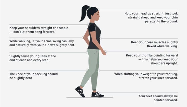
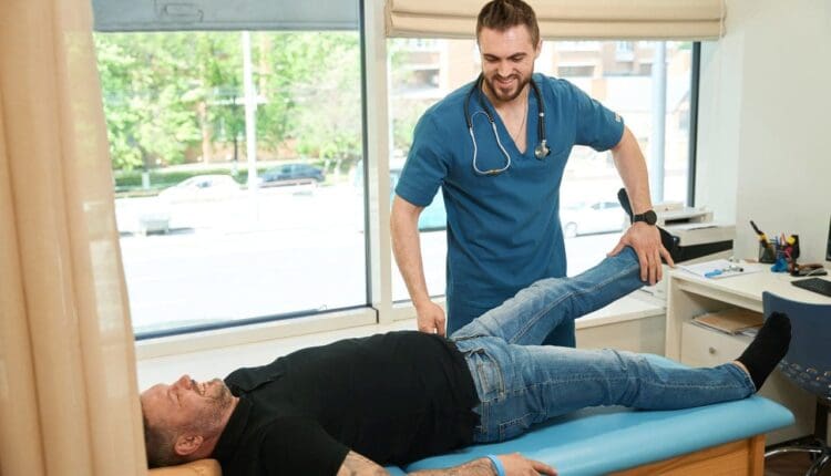
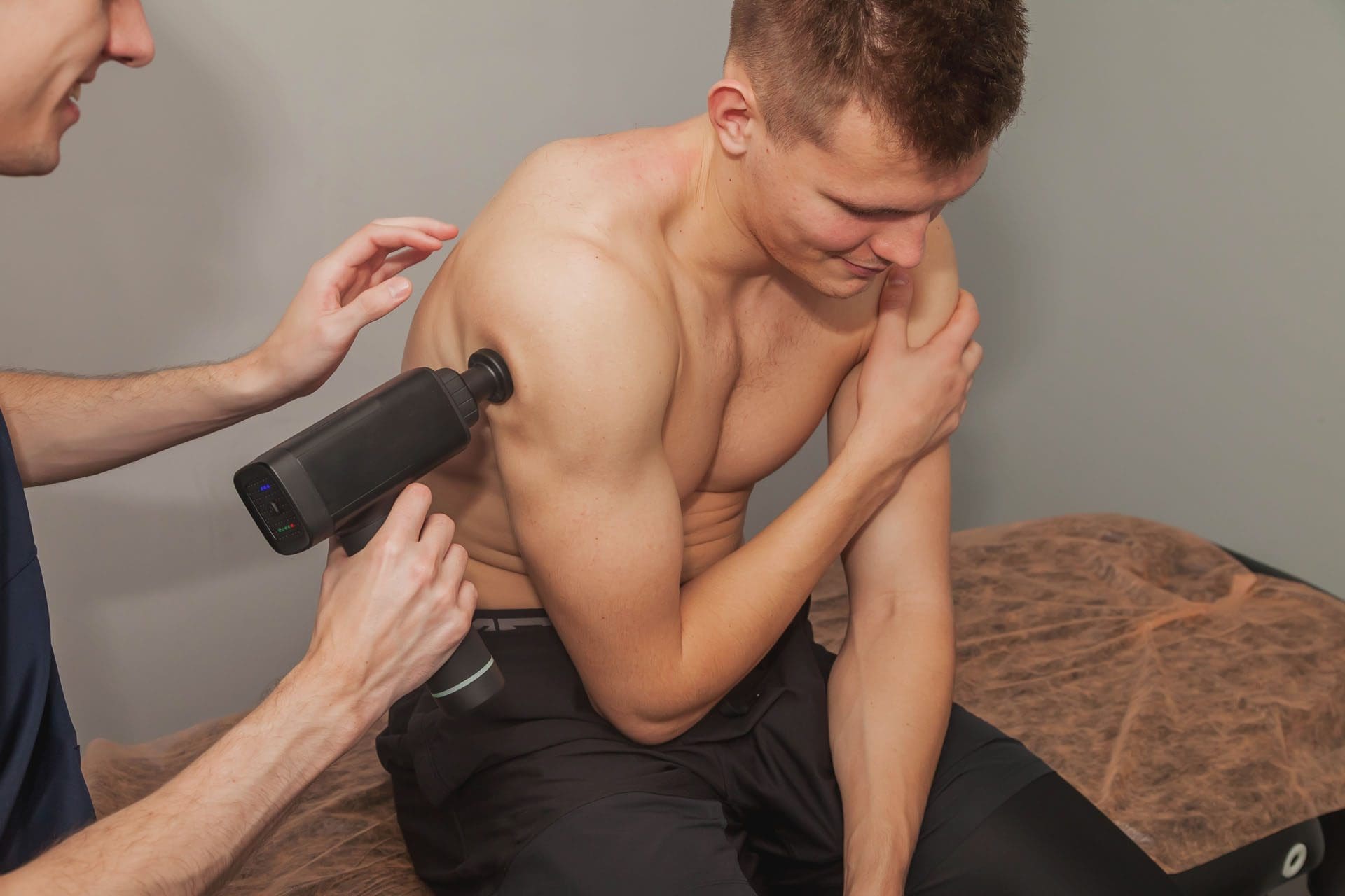
- Physical therapy and exercise
- Psychotherapy to alleviate stress, anxiety, and depression
- Sympathetic nerve blocks
- Surgery
- Spinal cord stimulation
- Intrathecal drug pumps
Medications
These can include:
- Topical analgesics
- Anti-seizure drugs
- Antidepressants
- Corticosteroids
- Opioids
It is estimated that there are 50,000 new cases every year in the United States. (Tajerian M., & Clark J. D. 2016)
Injury Medical Chiropractic & Functional Medicine Clinic
Injury Medical Chiropractic and Functional Medicine Clinic works with primary healthcare providers and specialists to develop an optimal health and wellness solution. We focus on what works for you to relieve pain, restore function, and prevent injury. Regarding musculoskeletal pain, specialists like chiropractors, acupuncturists, and massage therapists can help mitigate the pain through spinal adjustments that help the body realign itself. They can also work with other medical professionals to integrate a treatment plan to resolve musculoskeletal issues.
The Root Causes of Pain
References
National Institute of Neurological Disorders and Stroke. (2017). Complex regional pain syndrome fact sheet. Retrieved from https://www.ninds.nih.gov/sites/default/files/migrate-documents/CRPS_FactSheet-E_508C.pdf
Goh, E. L., Chidambaram, S., & Ma, D. (2017). Complex regional pain syndrome: a recent update. Burns & Trauma, 5, 2. https://doi.org/10.1186/s41038-016-0066-4
Bruehl S. (2015). Complex regional pain syndrome. BMJ (Clinical research ed.), 351, h2730. https://doi.org/10.1136/bmj.h2730
Harvard Health Publishing. (2023). Complex Regional Pain Syndrome (CRPS). https://www.health.harvard.edu/a_to_z/complex-regional-pain-syndrome-crps-a-to-z
Stanford Medicine. (2025). Complex Regional Pain Syndrome (CRPS). https://med.stanford.edu/pain/about/chronic-pain/crps.html
Tajerian, M., & Clark, J. D. (2016). New Concepts in Complex Regional Pain Syndrome. Hand Clinics, 32(1), 41–49. https://doi.org/10.1016/j.hcl.2015.08.003