What Happens If You Don’t Stretch Regularly

How Integrative Chiropractic + NP Care Can Help
Most people don’t skip stretching on purpose. Life gets busy. You sit, drive, work, cook, lift kids or groceries, and move through your day without thinking much about flexibility—until your body starts “talking.”
That “talking” can sound like:
- “My neck feels stuck when I turn.”
- “My lower back is tight every morning.”
- “My hips feel stiff getting out of the car.”
- “My hamstrings feel like guitar strings.”
- “I’m not injured, but everything feels harder.”
While stretching is not magic, regular stretching (and basic mobility work) supports how your muscles, joints, and nervous system work together. When stretching is missing for a long time, muscles can feel tight and stiff, daily movement can feel less smooth, and your risk of strains can go up—especially when you suddenly ask your body to do something harder than usual. (Harvard Health Publishing, 2024; Mayo Clinic Staff, n.d.). Harvard Health+1
This article explains what can happen when you don’t stretch regularly, why stiffness builds over time, and how integrative chiropractic care plus a nurse practitioner (NP) can support better movement, comfort, and function—using a team-based, whole-person approach.
The Simple Truth: Your Body Adapts to What You Repeatedly Do
Your muscles and connective tissues adapt to your habits.
- If you move often, you tend to maintain a usable range of motion.
- If you stay still often, your body gets “good” at being still.
- Some muscles may stay in shortened positions for hours on end if you spend a lot of time sitting or driving.
Stretching helps counter the “always in one position” problem. It’s one reason many medical and fitness education resources describe stretching as supportive for flexibility, joint range of motion, and daily function. (Harvard Health Publishing, 2024; Mayo Clinic Staff, n.d.). Harvard Health+1
What Muscle Stiffness Really Means (In Plain Language)
“Stiffness” is not just one thing. It can come from several common situations:
1) Too little movement for too long
After prolonged periods of minimal movement (such as sitting, bed rest, or low activity), muscles can feel tight and resistant. (Osmosis, n.d.). Osmosis
2) Doing “new” or harder activity than usual
When you do a new exercise or push harder than normal, you can create small amounts of muscle stress, which may lead to soreness and stiffness afterward—especially if you don’t train consistently. (Osmosis, n.d.). Osmosis
3) Hydration and electrolytes can matter
Electrolyte shifts after sweating can affect how muscles contract and how the nervous system communicates with muscles. That’s one reason hydration, nutrition, and recovery routines matter too. (Osmosis, n.d.). Osmosis
If You Don’t Stretch, Do Your Muscles “Shorten”?
You may have heard: “If you don’t stretch, your muscles will shorten.”
A helpful clarification is this:
- For most people living a normal life, the bigger issue is that they become less mobile and less flexible, which can feel like shortening.
- True physical shortening can occur in specific situations (such as prolonged immobilization), but in daily life, it’s more about stiffness, decreased mobility, and reduced tolerance for movement. (adidas, 2025). adidas
So the main risk is practical: movement feels harder, and your body has less “room” to move smoothly.
What Happens Over Time If You Rarely Stretch
When stretching and mobility are missing for weeks or months, several patterns are common.
You may notice a reduced range of motion
Range of motion is how far a joint can move comfortably. Many reputable health resources note that stretching can help joints move through a fuller range of motion and support everyday activity. (Mayo Clinic Staff, n.d.). Mayo Clinic
You may feel “tight,” then weaker in certain positions
Some muscles can become tight and less effective at lengthening when needed. This can alter your ability to squat, reach, rotate, and walk—particularly if you spend a significant amount of time seated. (Harvard Health Publishing, 2024). Harvard Health
Movement efficiency can drop
When your body can’t access normal ranges easily, it often compensates. You might twist through your lower back instead of your hips, shrug your shoulders instead of using your upper back, or flare your ribs instead of using your core. Over time, those compensation patterns can create nagging aches.
Daily tasks can feel harder
This is a big one. Many people don’t care about stretching until it affects real life:
- Looking over your shoulder while driving
- Bending to tie shoes
- Reaching overhead in the kitchen
- Carrying a child or lifting a box
- Standing up from the couch without stiffness
Mayo Clinic notes that stretching can improve the ability to do daily activities and help muscles work more effectively. (Mayo Clinic Staff, n.d.). Mayo Clinic
How Not Stretching Can Increase Injury Risk
“Injury risk” doesn’t mean stretching prevents all injuries. It doesn’t.
But here’s the practical idea: tight, under-prepared tissues can be easier to strain when you suddenly demand more from them.
Harvard Health explains that without regular stretching, muscles can become tight and fail to extend fully during activity, increasing the risk of joint pain, strains, and muscle damage—especially during sudden, strenuous movement. (Harvard Health Publishing, 2024). Harvard Health
Other clinical and rehab-oriented sources also describe that lack of flexibility can contribute to shortened/tight muscles and a higher risk of strains or injury. (OA Orthopaedics, 2024; Aegis Physical Therapy, 2023). OADuluth+1
Common “high-risk moments” when people get hurt
- Weekend yardwork after a week of sitting
- Holiday lifting and carrying (boxes, decorations)
- A rigorous workout after weeks off
- A long drive followed by sudden activity
- Rushing and moving fast with cold muscles
Flexibility vs. Mobility (Why Both Matter)
People mix these terms up:
- Flexibility = how far a muscle can lengthen.
- Mobility = how well you can control movement through a range (often involving joints + muscles + nervous system).
Mobility work typically involves controlled movements through various ranges, whereas stretching can be either held or dynamic. Many fitness education sources describe mobility as supporting a greater range of motion and improved movement quality. (Aaptiv, n.d.). Aaptiv
Real-life takeaway:
If you only stretch but never build control and strength, you may not “own” your range. If you only lift but never work on mobility, your range may slowly shrink.
Stretching Benefits People Commonly Notice
Different people feel different results, but common benefits include:
- Feeling less stiff when waking up
- Smoother movement getting up from a chair
- Better body awareness (posture and alignment)
- Easier walking, squatting, reaching, and rotating
- Better comfort after workouts
Mayo Clinic lists potential benefits like improved range of motion, supporting joints through full motion, increasing muscle blood flow, and supporting daily activity. (Mayo Clinic Staff, n.d.). Mayo Clinic
Some educational resources also describe increased blood flow to tissues with stretching, which supports recovery. (Fitness for Paramedics, n.d.). eCampusOntario Pressbooks
The “Right Way” to Stretch (So You Don’t Make Things Worse)
Stretching is usually safe, but technique matters.
Basic stretching safety rules
Mayo Clinic offers clear, widely used safety tips, including warming up first and avoiding stretching cold muscles. (Mayo Clinic Staff, n.d.). Mayo Clinic
Use these practical guidelines:
- Warm up first: 5–10 minutes of easy walking or light movement.
- Go to mild tension, not pain.
- Breathe: a slow exhale helps your nervous system “downshift.”
- Be consistent: small daily work beats one long session once a week.
- Use dynamic stretching before activity (gentle movement-based stretches).
- Use longer holds after activity (when tissues are warm).
A quick “green light / yellow light / red light” check
Green light (okay):
- mild pulling
- warmth
- gradual easing
Yellow light (slow down):
- sharp pinch
- tingling
- You can’t breathe comfortably through it
Red light (stop and get checked):
- numbness/weakness
- worsening nerve symptoms down an arm/leg
- severe pain, swelling, fever, or unexplained symptoms
A Simple Daily Stretch Routine (10 Minutes)
This is a basic, general routine that many people tolerate well. Modify for comfort.
Lower body (5 minutes)
- Calf stretch (30 seconds each side)
- Hamstring stretch (30 seconds each side)
- Hip flexor stretch (30 seconds each side)
- Glute/hip stretch (30 seconds each side)
Upper body (5 minutes)
- Chest opener (30–45 seconds)
- Upper back reach (30–45 seconds)
- Neck gentle side stretch (20–30 seconds each side)
- Thoracic rotation (open books) (5–8 reps each side)
Harvard Health specifically highlights calves, hamstrings, hip flexors, and quads, as well as shoulders, neck, and low back, as key areas for mobility-focused stretching. (Harvard Health Publishing, 2024). Harvard Health
When Stretching Alone Is Not Enough
If you have persistent stiffness or pain, the problem may not be, “you need to stretch more.” Other factors can drive stiffness, including:
- Joint restriction or irritation
- Overuse patterns
- Poor recovery and sleep
- Past injuries (especially whiplash, falls, sports injuries)
- Underlying conditions (thyroid issues, inflammatory disorders, medication effects)
- Nerve irritation
Osmosis notes that muscle stiffness can come from overuse, immobility, electrolyte issues, and also underlying medical conditions. (Osmosis, n.d.). Osmosis
That’s where integrative care can be useful: you get both a movement-focused approach and a medical lens to rule out deeper causes.
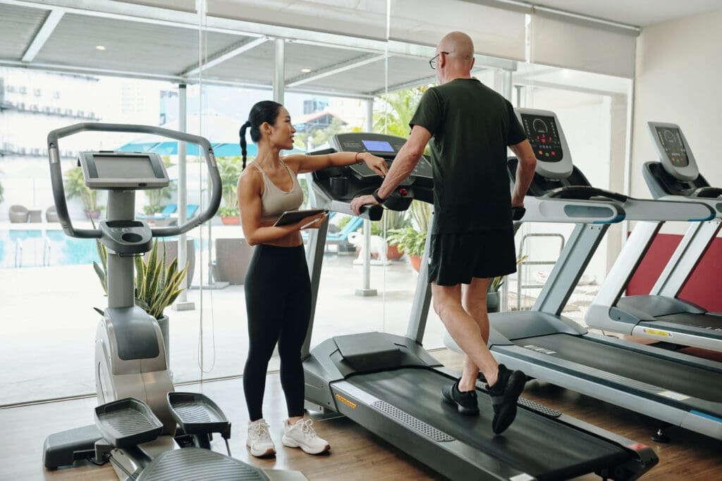
How Integrative Chiropractic Care Can Help (Beyond “Cracking”)
Integrative chiropractic care is not just about one adjustment. A comprehensive approach often includes:
- Examining joint motion and movement patterns
- Addressing areas of restriction and compensation
- Manual care (when appropriate)
- Soft-tissue strategies
- Home mobility and strengthening plans
- Ergonomic guidance (desk, driving, sleep posture)
Dr. Alexander Jimenez, DC, APRN, FNP-BC often emphasizes that people dealing with joint and muscle pain—especially after injury—benefit from keeping the body flexible and using stretching as part of a bigger plan to reduce flare-ups and support function. El Paso, TX Doctor Of Chiropractic
His clinical content also discusses that when muscles are stiff and strained, continuing to force movement can worsen discomfort and further reduce range of motion—and that care may include adjustments and soft-tissue work to support mobility and restore motion. El Paso, TX Doctor Of Chiropractic
Separately, many chiropractic education resources describe adjustments as targeted, controlled techniques used to support mobility and function. (WorkPartners MD, 2024). Work Partners, PLLC
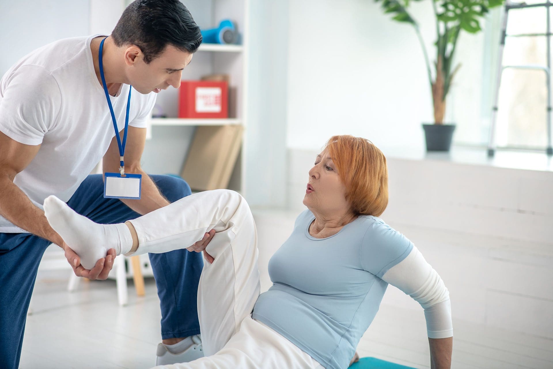
What the Nurse Practitioner Adds (And Why It Matters)
A nurse practitioner (NP) brings medical assessment and management skills to the same movement problem. That matters because stiffness sometimes has medical drivers.
NPs can help by:
- Taking a full health history (sleep, stress, medications, and past injuries)
- Screening for red flags (infection, inflammatory disease, neurological changes)
- Ordering or interpreting appropriate tests (labs or imaging when needed)
- Managing pain safely (when medication is appropriate)
- Coordinating referrals (PT, imaging, specialists)
- Coaching lifestyle factors that affect pain and recovery
Healthgrades summarizes that NPs can evaluate problems, diagnose conditions, interpret diagnostic tests, and provide a wide range of treatments (state rules vary). (Prosser, 2025). Healthgrades Resources
The American Nurses Association describes APRNs as meeting advanced educational/clinical requirements and providing services ranging from primary and preventive care to other specialty services. (ANA, n.d.). ANA
The Power of the Team: Chiropractor + NP Working Together
When chiropractic care and NP care collaborate, it can help patients avoid “one-sided” care (only exercises, only medication, or only manual therapy).
A collaborative plan often looks like this
- Step 1: Clear diagnosis and safety screening
- Rule out serious causes of pain/stiffness
- Identify nerve involvement, red flags, or systemic issues
(Osmosis, n.d.; Prosser, 2025). Osmosis+1
- Step 2: Restore motion safely
- Joint and soft tissue approaches
- Targeted mobility plan
(Mayo Clinic Staff, n.d.; Jimenez, n.d.). Mayo Clinic+1
- Step 3: Build strength to keep the motion
- Strength + control so flexibility “sticks.”
- Simple home program that matches your real life
- Step 4: Reduce flare-ups
- Work, driving, and sleep strategies
- Recovery routines (hydration, stress, sleep)
What patients often like about integrative care
- You don’t have to guess what’s “normal soreness” vs. a real problem.
- You get a plan that fits both your body mechanics and your health history.
- You can track progress with measurable goals (range of motion, function, pain levels).
A Practical Self-Check: Are You Dealing With “Stretching Stiffness” or Something Else?
Ask yourself:
- Does stiffness improve after a warm shower or light movement?
- Does it improve after 5–10 minutes of walking?
- Is it worse after sitting for a long time?
- Do you feel “stuck” more than “injured”?
If yes, you may be dealing with a mobility/flexibility + recovery issue.
But get checked sooner if you have:
- Pain shooting down an arm/leg with numbness or weakness
- New balance problems or frequent falls
- Fever, unexplained weight loss, or severe fatigue with pain
- Symptoms after a significant accident
Because stiffness can sometimes be linked to broader medical conditions, evaluation is important when symptoms are persistent or worsening. (Osmosis, n.d.). Osmosis
Key Takeaways
If you don’t stretch regularly, it’s common to develop:
- Reduced flexibility and usable range of motion
- More stiffness with sitting, driving, or long workdays
- Less efficient movement patterns (more compensation)
- Higher strain risk during sudden activity
Stretching is most helpful when it’s:
- Regular and gentle
- Paired with mobility and strength
- Guided by your symptoms and medical history
Integrative chiropractic care and nurse practitioners can work together to:
- Improve motion and comfort
- Address joint and soft tissue restrictions
- Screen for medical causes of stiffness
- Build a realistic home plan that protects your body long-term
(Mayo Clinic Staff, n.d.; Prosser, 2025; Jimenez, n.d.). Mayo Clinic+2Healthgrades Resources+2
Medical Disclaimer
This article is for educational purposes and is not medical advice. If you have severe pain, numbness, weakness, new neurological symptoms, or symptoms after a serious injury, seek urgent medical evaluation.
References
- Advanced Practice Registered Nurses (APRN) — American Nurses Association. ANA
- Add Stretching to Your Daily Routine to Improve Your Health — Aegis Physical Therapy. Aegis Physical Therapy
- The Three Biggest Myths About Stretching — adidas (April 2025). adidas
- Here’s How Different Methods of Mobility Affect Your Muscle Tone — Aaptiv. Aaptiv
- The importance of stretching — Harvard Health Publishing (April 17, 2024). Harvard Health
- Mobility Flexibility: El Paso, TX — Jimenez, A. (n.d.). El Paso, TX Doctor Of Chiropractic
- Restore Range Of Motion With Chiropractic — Jimenez, A. (n.d.). El Paso, TX Doctor Of Chiropractic
- Benefits of Flexibility and Stretching — Fitness for Paramedics (eCampusOntario Pressbooks). eCampusOntario Pressbooks
- Stretching: Focus on flexibility — Mayo Clinic Staff. Mayo Clinic
- Muscle Stiffness: What Is It, Causes, Treatment, and More — Osmosis. Osmosis
- The Role Of Stretching And Flexibility Exercises — OA Orthopaedics (April 8, 2024). OADuluth
- Treatments a Nurse Practitioner Can Provide — Prosser, A. (Updated July 23, 2025). Healthgrades Resources
- Chiropractic Adjustments for Joint Health: Enhancing Mobility and Function — WorkPartners MD (January 5, 2024). Work Partners, PLLC