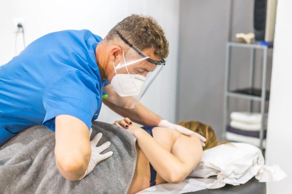
How ChiroMed’s Integrative Care Helps Athletes Recover Faster and Stay Strong
Competitive fastpitch softball pushes the body hard with fast pitches, quick turns, and sudden dives. Pitchers use the underhand windmill motion, which spins the arm in a full circle at high speeds. All players face rapid direction changes on the field. These actions often cause muscle and bone injuries. At ChiroMed – Integrated Medicine in El Paso, TX, athletes find help through a holistic approach. This care combines spinal adjustments, soft-tissue work, and rehabilitation exercises to treat injuries at the root cause. It helps players heal faster, gain more power, and avoid further injury.
The Tough Demands of Fastpitch Softball
The windmill pitch is unique to fastpitch. It creates strong pulls on the shoulder and elbow, unlike overhand throws. Pitchers might throw over 100 pitches in a game, leading to wear and tear. Fielders run, slide, and collide, putting stress on legs and joints. Bases are run in one direction, which twists ankles and knees the same way each time. These repeated movements account for the common injuries in the sport.
Main Overuse Injuries in Fastpitch Softball
Overuse results from performing the same motion too often. It causes most issues for players. Key ones include:
Shoulder problems: Strains in the rotator cuff from constant pitching. This small group of muscles gets inflamed and weak.
Elbow damage: Tears in the ulnar collateral ligament (UCL) from the arm’s twist in the windmill.
Back pain: Twisting during pitches and swings stresses the lower spine.
Hand and wrist strains: From gripping bats or catching balls hard.
Data show that overuse accounts for 60-70% of injuries among youth and college pitchers (Fastpitch Softball Injuries Study, 2024).
Sudden Acute Injuries on the Field
Some injuries occur in a single moment from impacts or poor landings. The sport’s speed leads to these:
ACL tears: The knee’s anterior cruciate ligament rips during quick stops or turns. Girls and women face higher risks due to body alignment.
Ankle sprains: Rolling the ankle while sliding or jumping.
Breaks and fractures: In fingers, arms, or collarbones from hits or falls.
Concussions: From ball strikes to the head or player crashes.
Lower-body injuries, such as sprains, top the list across all positions (Summit Orthopedics, 2022).
Other Common Issues That Slow Players Down
Injuries can result from overuse and sudden hits. They include:
Sprains in fingers or hands from tags or dives
Strains in hamstrings or groin from sprints
Neck strain from tracking fly balls
Catchers deal with knee stress from squatting. Outfielders twist their backs leaping for catches. Every role has risks.
Limits of Basic Injury Care
Rest, ice, and simple therapy help at first. But they might not address deeper problems, such as tight hips affecting the shoulder or spine issues, or changes in knee movement. Without full fixes, injuries return.
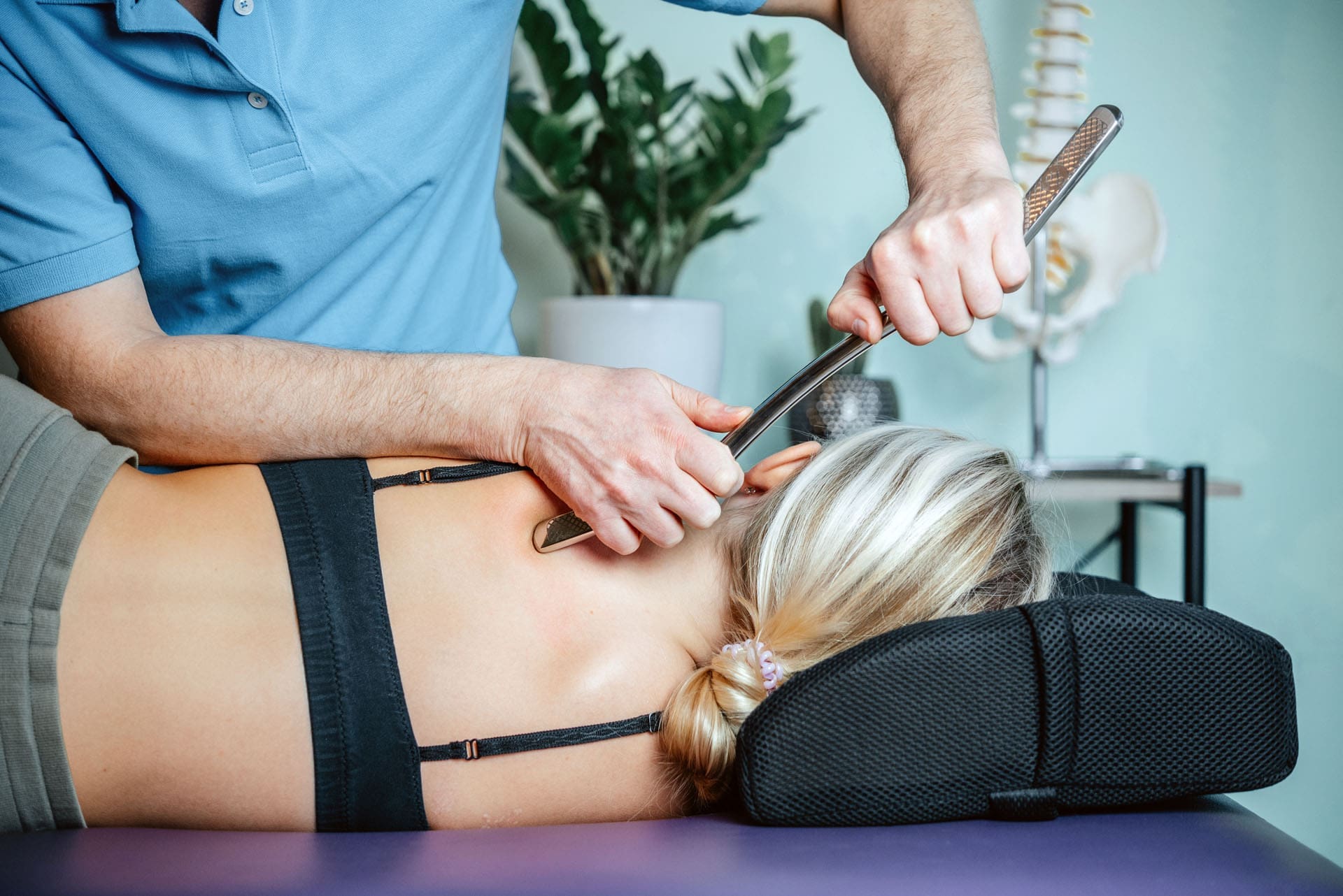
ChiroMed’s Integrative Chiropractic Approach
At ChiroMed, care treats the whole body as connected. Led by Dr. Alexander Jimenez, DC, APRN, FNP-BC, the team uses chiropractic adjustments, nurse practitioner services, naturopathy, rehab, nutrition, and acupuncture. This evidence-based method, inspired by functional medicine, targets root causes like muscle imbalances or nerve issues (ChiroMed – Integrated Medicine, n.d.).
Dr. Jimenez’s clinical work shows that small spinal misalignments accumulate in athletes, leading to poor form and injuries. His approach restores alignment for better nerve flow and movement.
Tools at ChiroMed include:
Adjustments: To correct spinal and joint alignment, reducing nerve pressure.
Soft tissue therapy: Massage and tools to heal muscles and reduce scars.
Rehab exercises: To build strength and balance for safe play.
Holistic support: Nutrition and recovery tips to boost healing.
This helps with sports injuries like those in softball, promoting faster recovery without drugs (Push as Rx, n.d.).
Benefits for Fastpitch Players at ChiroMed
Athletes see real gains. Shoulder strains heal in half the time. Pitches get faster with better body mechanics. Ankles strengthen after sprains. Reports show less pain, more flexibility, and fewer missed practices (Southern California University of Health Sciences, n.d.).
Dr. Jimenez notes that softball players often ignore early signs, which can lead to more serious issues. ChiroMed’s personalized plans help them return stronger and more confident.
Preventing Injuries with ChiroMed’s Help
Stopping problems before they start is key. ChiroMed offers check-ups to spot tight spots early. Programs include:
Warm-ups tailored to pitching
Exercises for core and hips
Mechanics training to protect arms
Rest guidelines based on pitch counts
Teams that use this stay healthier throughout the season.
Why Choose ChiroMed for Softball Recovery
Fastpitch demands resilience, but injuries can stop progress. ChiroMed’s integrative chiropractic care in El Paso offers a natural way to heal, perform better, and prevent setbacks. Visit https://chiromed.com/ to learn more and get back in the game stronger.
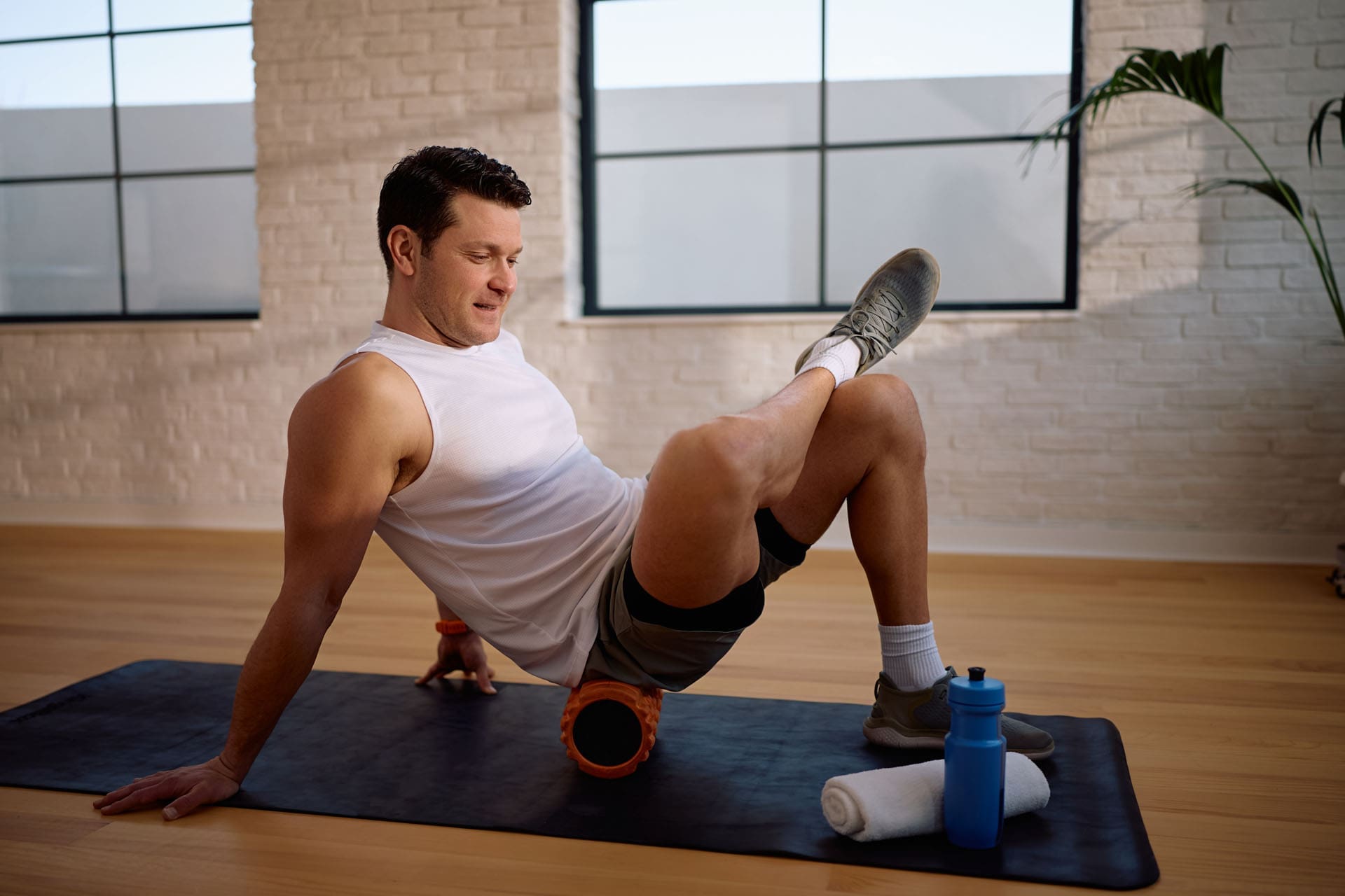
Tennis balls, foam rollers, and calf-release techniques are effective for self-massage.
Sciatica is not a single diagnosis. It is a symptom pattern—often burning, sharp, or “electric” pain that can start in the low back or buttock and travel down the leg. Some people also feel tingling or numbness. At ChiroMed, sciatica is usually discussed as a signal problem: something is irritating or sensitizing nerve tissues, and the body reacts with pain, tightness, and protective muscle guarding. (ChiroMed, 2025a)
Self-massage can be a helpful tool because many sciatica flare-ups include tight muscles and trigger points in the low back, glutes, hips, and the piriformis muscle. When these tissues tighten, they can increase pressure around sensitive areas and prolong symptoms. (Healthline, 2021)
Below is a practical, safe, “easy-to-follow” self-massage plan that matches what many people use at home—tennis balls, foam rollers, and gentle calf work—plus how these tools often fit into a whole-person chiropractic plan at ChiroMed. (ChiroMed, 2025b)
The #1 Safety Rule: Stay in the “Hurts Good” Zone
Self-massage should feel like helpful pressure, not intense pain.
Use this simple rule:
0–3 out of 10 discomfort: okay (“hurts good”)
4–10 out of 10: too much (back off)
Sharp zaps, burning, or increasing numbness: stop right away
This approach aligns with common guidance for piriformis massage: start gently, avoid excessive pressure, and discontinue if symptoms worsen. (Healthline, 2021)
Important: Do not apply intense pressure directly over the area where you feel the “electric line” of sciatic symptoms. The goal is to release muscle tension around the area, not to crush a nerve. (HSS, 2024)
Why These Areas Matter for Sciatica
Most effective self-massage targets the “hot spots” that commonly tighten during sciatica:
Low back muscles (especially near the pelvis)
Glutes (buttock muscles)
Piriformis (a deep hip muscle)
The calf is often tight due to compensation or referred pain.
Piriformis tension is a common reason people feel buttock pain and leg symptoms that look like sciatica. (Healthline, 2021)
Tool 1: Tennis Ball Release for Glutes and Piriformis
A tennis ball is useful because it can quickly locate a tight point. Many sciatica massage guides recommend using a ball to target trigger points in the gluteal/piriformis area. (Massage Chair Store, 2021; Healthline, 2021)
How to do it (simple floor method)
Sit on the floor and place a tennis ball under one buttock.
Lean your body weight slightly into the ball.
Slowly roll a few inches until you find a tender spot.
Apply light pressure for 20–45 seconds; breathe slowly.
Move off the spot and repeat 2–4 times.
Switch sides if needed.
What you want to feel:
Dull ache, pressure, warmth = okay What you do not want:
If you experience a sharp shooting pain down your leg or an increase in numbness, you should stop immediately (Healthline, 2021).
“Peanut” trick (two tennis balls)
Some people tape two balls together (or put them in a sock) to create a “peanut.” This can help you apply pressure on both sides of soft tissue—not the spine. (Massage Chair Store, 2021)
Tool 2: Foam Roller for Broader, Gentler Pressure
Foam rolling distributes pressure over a larger area than a ball, which may feel safer on sensitive days. It is often used for myofascial release, meaning the application of slow pressure to relax tight muscles and fascia. (Dorsal Health, 2020)
Best foam roller targets for sciatica patterns
Glutes (roll slowly, small range)
Outer hip muscles
Upper hamstrings (avoid behind the knee)
Simple foam rolling dose:
30–60 seconds per area
1–2 rounds total
Keep discomfort ≤3/10 (Dorsal Health, 2020)
Calf Massage for Referred Pain and “Compensation Tightness”
Sciatica symptoms often alter a person’s gait, stance, and leg loading. That can make the calf feel tight, sore, or crampy—even if the main issue is higher up. Gentle calf massage can lower muscle guarding and improve comfort while you work on the hip and low back. (Chicago Pain Control, n.d.)
Simple calf massage (hands-only)
Sit comfortably and support your leg.
Use your thumbs and palms to knead the calf slowly.
Work from mid-calf toward the ankle and back up.
Stop if you feel sharp nerve sensations.
Add Heat to Improve Results
Heat can relax tissues and make self-massage feel more effective. Many home-care routines for sciatica recommend applying heat before soft-tissue work to reduce muscle guarding. (HSS, 2024)
Try:
Heat 10–15 minutes
Then self-massage for 5–8 minutes
Then a short walk, 3–10 minutes if tolerated
What ChiroMed Often Adds: An Integrative Plan for Long-Term Relief
Self-massage is helpful, but many people need more than symptom relief. A longer-term plan often focuses on **-two goals:
Lower nerve irritation
Fix the mechanical patterns that keep stress on tissues
ChiroMed’s content frequently emphasizes combining chiropractic care, soft-tissue work, and rehabilitation strategies so patients are not merely chasing pain day to day. (ChiroMed, 2025b)
Common integrative elements include:
Focused chiropractic adjustments to improve motion and reduce joint stress (Fremont Chiropractic, n.d.)
Myofascial release/trigger point therapy to calm tight muscles and improve movement (Pinnacle Hill Chiropractic, 2023)
Spinal decompression (when appropriate) to reduce pressure and support disc-related cases (Posture Perfect PH, n.d.)
Rehab and mobility exercises to build stability and reduce flare-ups (Bend Total Body Chiropractic, n.d.)
Clinical perspective reflected in Dr. Alexander Jimenez’s published education
Dr. Alexander Jimenez, DC, APRN, FNP-BC, often describes sciatica care as a “systems” problem—muscles, joints, discs, inflammation, and movement habits can all contribute. His educational posts emphasize combining hands-on care with guided self-care to support function rather than merely providing temporary relief. (Jimenez, n.d.-a; Jimenez, n.d.-b)
When to Stop Self-Massage and Get Evaluated
Seek medical evaluation urgently if you have:
New or worsening leg weakness
Loss of bowel/bladder control
Numbness in the groin/saddle area
Severe pain that is rapidly worsening
These signs may indicate more severe nerve involvement and should not be managed with home massage alone. (ChiroMed, 2026)
A Simple 7-Minute Routine (Daily or During Flares)
Heat: 10 minutes (optional) (HSS, 2024)
Tennis ball glute/piriformis: 2 minutes total (Healthline, 2021)
Foam roll glutes/outer hip: 2 minutes total (Dorsal Health, 2020)
Calf massage: 2 minutes total (Chicago Pain Control, n.d.)
Easy walk: 3–10 minutes if tolerated (ChiroMed, 2025a)
Keep it gentle, stay in the 0–3/10 range, and avoid pressing directly into sharp nerve pain.
Strength, Cardio, Mobility, and Recovery with Essential Warm-Ups and Cool-Downs
In El Paso, TX, establishing a weekly workout regimen requires consideration of the hot desert climate. The sun can be strong, and temperatures often climb high, so it’s smart to plan exercises that fit indoor spaces or early-morning times. An effective routine combines strength training, cardio, mobility work, and rest days to build fitness without injury. This helps people stay active and healthy in a region known for its dry heat and outdoor trails, such as those in the Franklin Mountains. Local gyms and classes make it easy to find options that match your level, whether you’re just starting or have some experience.
Balancing different types of workouts keeps things fun and effective. Strength training builds muscle, cardio boosts heart health, mobility improves how you move, and recovery allows your body to heal. In El Paso, using facilities such as Fit Body Boot Camp or Shanti Yoga can add variety. Fit Body Boot Camp offers 30-minute group classes with high-intensity interval training (HIIT) that burns fat and tones muscles, perfect for beginners with trainer support (Fit Body Boot Camp, n.d.). Shanti Yoga offers classes such as hot yoga, which can be comfortable in the desert air, but they are held indoors to avoid the heat (Fox Lexus of El Paso, n.d.).
For beginners and those at an intermediate level, aim for 3 to 5 workout days each week. This leaves room for rest, which is key in a hot climate where your body needs time to cool off and rebuild. Always include warm-ups and cool-downs to stay safe.
Why Balance Matters in Your Routine
A balanced regimen prevents burnout and helps you see results. Strength days focus on weightlifting or bodyweight exercises to build strength. Cardio could be running, cycling, or brisk walking to improve endurance. Mobility exercises, such as stretching or yoga, keep joints flexible. Recovery includes light walks or full rest. In El Paso, the desert climate means you should stay well hydrated and choose cooler times, such as before 11 a.m. or after 5 p.m. (Ortho El Paso, 2024). This reduces risks like heat stress.
Experts suggest splitting workouts by muscle groups or full-body sessions. For example, upper-body days target the arms, chest, and back, while lower-body days focus on the legs and core (Grinder Gym, n.d.). Mixing in cardio 2-3 times a week keeps your heart strong (Self, n.d.).
Strength Training Benefits: Builds muscle, boosts metabolism, and helps with daily tasks.
Cardio Advantages: Improves lung capacity and burns calories.
Mobility Perks: Reduces stiffness and improves range of motion.
Recovery Importance: Allows muscles to repair and prevents overtraining.
Incorporate local amenities such as Revolution Fitness for Zumba or weight training, which are indoor and air-conditioned (Fox Lexus of El Paso, n.d.).
Sample Weekly Regimen for Beginners
For someone new to fitness, start with 3 days a week. Each session lasts 30-45 minutes, plus warm-up and cool-down. Focus on simple moves.
Monday: Full-Body Strength – Squats, push-ups, rows. Do 2-3 sets of 10-12 reps.
Wednesday: Cardio – Brisk walk or cycle for 20-30 minutes.
Friday: Mobility – Yoga poses like child’s pose and cat-cow.
Rest Days: Tuesday, Thursday, Saturday, and Sunday. Light walks if you feel well.
This setup from sources like Emily Skye Fit builds confidence without overload (Emily Skye Fit, n.d.). In El Paso, try this at Her Gym, a women-only facility with classes such as BODYPUMP for strength (Fox Lexus of El Paso, n.d.).
Warm-ups: Spend 5-10 minutes with dynamic stretches like arm circles or leg swings to get blood flowing (Mayo Clinic, n.d.a). Cool-downs: Static stretches, held for 15-30 seconds, such as touching your toes (Les Mills, n.d.).
Sample Weekly Regimen for Intermediates
If you have some experience, go for 4-5 days. Add more intensity.
Monday: Upper Body Strength – Bench press, pull-ups, shoulder presses. 3 sets of 8-10 reps.
Tuesday: Cardio – Run or HIIT for 30 minutes.
Wednesday: Lower Body Strength – Lunges, deadlifts, and calf raises.
Thursday: Mobility and Core – Planks, twists, and stretches.
Friday: Full-Body or Active Recovery – Light yoga or swimming.
Weekend Rest: Focus on recovery.
This mirrors Health.com’s plans, adjusted for progress (Health.com, n.d.a). In El Paso’s heat, shift outdoor cardio to mornings and drink water every 10-15 minutes (Fusion Workplaces, 2022).
The Role of Warm-Ups and Cool-Downs
Warm-ups prepare your body. They raise heart rate and loosen joints. In the desert, this helps adjust to the heat. Do light cardio, such as walking, then dynamic moves (McPress Mayo Clinic, n.d.).
Warm-Up Tips: 5-10 minutes. Include marching in place and jumping jacks.
Why It Helps: Reduces injury risk by increasing flexibility (Shore Physicians Group, n.d.).
El Paso Twist: In hot weather, start slow to avoid overheating (National Weather Service, n.d.).
Cool-downs bring your heart rate down. Walk slowly, then static stretch. This aids recovery and flexibility (AIM7, n.d.).
Cool-Down Ideas: Hold stretches for major muscle groups such as the hamstrings and quadriceps.
Benefits: Prevents stiffness and promotes blood flow (Westwood Fitness, n.d.a).
Local Advice: After Pure Barre workouts, stretch to recover from toning classes (Fox Lexus of El Paso, n.d.).
Skipping these can lead to sprains or tendonitis (Shore Physicians Group, n.d.).
Adapting to El Paso’s Desert Climate
El Paso’s dry heat means special care. Exercise indoors at facilities such as Fusion Fitness Studio, which offers cycling classes (Fox Lexus of El Paso, n.d.). For outdoor hikes, hike early on trails and acclimate slowly, starting with 10-15 minutes (DesertUSA, n.d.).
Hydration: Drink water often; add electrolytes in hot weather (Zaca, n.d.).
Indoor Options: Use gyms for air-conditioned sessions.
Local spots from Yelp include Planet Fitness (4 stars, affordable), OrangeTheory Fitness (4.5 stars, HIIT), and CrossFit gyms for variety.
Enhancing with Integrative Chiropractic
Integrative chiropractic optimizes routines by fixing imbalances. It improves coordination, posture, and mobility to prevent injuries. Dr. Alexander Jimenez, DC, APRN, FNP-BC, notes that adjustments restore joint function, improve stability, reduce pain, and enhance performance (Jimenez, n.d.a.). His work at Injury Medical Clinic uses functional medicine to address root causes, such as in sports rehabilitation (Jimenez, n.d.b).
Neuromuscular Benefits: Enhances signal transmission for better moves.
Posture Correction: Fixes misalignments to avoid strain.
Mobility Gains: Restores range for full workouts.
Injury Prevention: Athlete-specific plans.
In El Paso, combine with routines for max results (Mountain Movement Center, n.d.).
Putting It All Together
A weekly regimen in El Paso should be balanced, safe, and fun. Use local amenities, adapt to the climate, and include chiropractic for optimization. Start slow, listen to your body, and track progress.
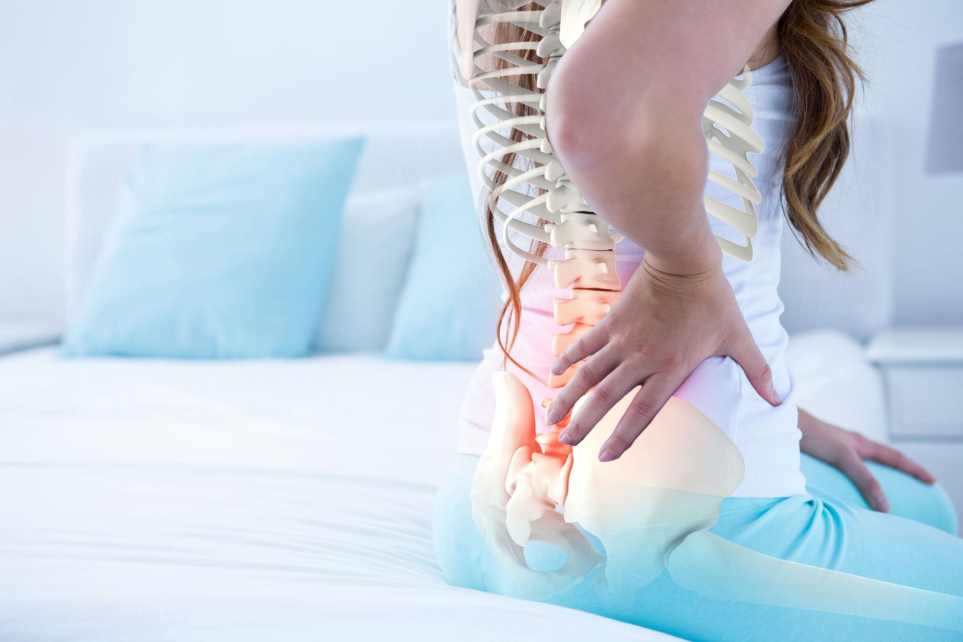
Car accidents are common in El Paso, Texas. Busy roads like I-10 and US-54 see many crashes each year. A single injury can significantly alter your daily routine. But what if you had a health issue before the accident? People often worry whether a new crash can worsen an old injury. And if it does, can they still get money for medical bills and other costs?
This question comes up a lot. Pre-existing conditions include back pain, arthritis, or stress-related issues. A car accident can worsen their pain. In Texas, the law protects you. You can receive compensation for the extra injury caused by the crash. The same rule applies in El Paso. Local rules follow the state. However, insurance companies might try to decline. They could blame all your pain on the old condition.
You have options. The “eggshell skull rule” is important. It means the person who caused the accident is responsible for all damages. This rule applies even if your past health problem put you at a higher risk. This rule is based on prior court decisions. It ensures fair help for everyone (Smith & Hassler, n.d.; Reyes Browne Law, n.d.).
In this article, we explain it all. What is the rule? How do you establish your case? What compensation can you get? What are the options for finding care in El Paso? We focus on ChiroMed – Integrated Medicine. This local clinic helps with these issues. Led by Dr. Alexander Jimenez, it offers care for accident injuries.
El Paso has unique problems. Border traffic and hot weather can increase accident risk. But support is here. Keep reading to learn.
What Is a Pre-Existing Condition?
A pre-existing condition is any health problem from before the accident. It might be from long ago or recent. Examples are:
Examples include back or neck pain resulting from previous falls.
Arthritis in joints.
Diabetes or heart problems.
Anxiety or other mental health issues.
Healed breaks that left weak spots.
These don’t block your claim. But they add challenges. The accident must cause additional harm or worsen the existing harm. This is aggravation (Gutierrez Law Firm, n.d.a).
Aggravation means the problem gets bad for good. It doesn’t return to normal. For instance, mild back pain becomes constant after the crash. That’s aggravation. A brief flare-up might not count as much (Reyes Browne Law, n.d.).
Doctors check your old and new records. They see the differences. In El Paso, clinics like ChiroMed do this well. They understand how accidents affect the body.
This matters because insurers want to pay less. Insurers might argue that your pain is due to pre-existing conditions. But Texas law disagrees. You get help for what the accident caused (GDL Law Firm, n.d.).
The Eggshell Skull Rule Explained
The eggshell skull rule is central to Texas law. Also known as the thin-skull or eggshell plaintiff rule. The rule originated in common law through judges’ rulings.
It works like this. Think of someone with a skull as thin as an eggshell. A light hits it. A normal person might be okay. The rule says the hitter pays for the whole break. They can’t say they didn’t know it was thin. They accept the victim as is (Amtz Law, n.d.; Abraham, Watkins, Nichols, Agosto, Aziz, & Stogner, n.d.).
In crashes, it covers pre-existing conditions. If you had weak bones from a condition, and the accident breaks them easily, the at-fault driver pays fully. This applies even if your condition made the accident worse (Smith & Hassler, n.d.).
Texas uses this in courts. Jury guides include it. This is evident in cases such as Leitch v. Hornsby (1996). Juries give money for the worsening, not the full old condition. The focus is solely on the additional damage (BHW Law Firm, n.d.; GTA Law, n.d.).
There’s also the crumbling skull rule. This rule applies to conditions that deteriorate over time, such as disc problems. The at-fault party pays only for the crash’s extra effect. This policy does not apply to natural changes (Smith & Hassler, n.d.).
In El Paso, this helps many. The area has older people with health concerns. Crashes often involve fast trucks. Injuries can be severe. The rule safeguards you (Reyes Browne Law, n.d.).
Experts advise honesty. Share all with your doctor and lawyer. It strengthens your case (No Bull Law, n.d.; STL Injury Law, n.d.).
Can You Get Compensation in El Paso, TX?
Yes. El Paso uses Texas laws. If the crash worsens your condition, you can get money. You can receive compensation for bills, lost pay, pain, and other related expenses.
Recoverable items include:
New medical expenses: Visits, therapy, or surgery due to the worsening.
Pain and suffering: Added hurt from the aggravation.
Lost income: Missed work due to the new severity.
Future treatment: Long-term care needs.
Life quality loss: Inability to enjoy activities or do tasks.
Amounts depend on the extent of worsening and evidence. Small cases get less; lasting ones more (Siegfried & Jensen, n.d.; Gutierrez Law Firm, n.d.b).
Insurers may resist. Claim it’s not crash-related. But solid proof wins. El Paso lawyers know this. They deal with local cases.
You have two years to file, per statute of limitations (Gutierrez Law Firm, n.d.b; No Bull Law, n.d.).
How Insurers Handle These Claims
Insurers aim to cut costs. They review your history. They may claim that all pain is pre-existing or inevitable.
Tactics they use:
Ask for a complete medical history, then use it inappropriately.
Use their doctors to deny aggravation.
Push fast, low offers.
Blame age or natural wear.
Avoid traps. Get a lawyer. They protect you. Handle records, share only essentials (Eckell, Sparks, Levy, Monte, Sloane, Matthews, & Auslander, n.d.; Romanow Law Group, n.d.).
In El Paso, insurers understand local judges. Strong cases lead to better settlements.
Proving the Aggravation
Proof is crucial. Show that the accident caused the deterioration.
Ways to prove it:
Collect records: Pre- and post-accident. Compare changes.
Visit doctors soon: Describe the old issue and new symptoms.
Do imaging: X-rays and MRIs to reveal differences.
Journal daily: Track pain and limitations.
Expert testimony: Doctors explain crash impact.
Accident evidence: Reports, photos, and statements.
El Paso clinics like ChiroMed use tests to document patient care. Builds strong claims (ChiroMed – Integrated Medicine, n.d.).
Be credible. Honesty is key. Dishonesty hurts (Gage Mathers, n.d.).
Importance of Medical Care in El Paso at ChiroMed
See a doctor quickly after a crash. Pain might start later. In El Paso, ChiroMed – Integrated Medicine specializes in this.
They document aggravation. Start recovery. Supports your claim.
ChiroMed is at 11860 Vista Del Sol Dr, Suite 128, El Paso, TX 79936. The clinic is led by Dr. Alexander Jimenez, who holds the titles of DC, APRN, and FNP-BC. Dr. Alexander Jimenez, DC, APRN, FNP-BC, leads the clinic and boasts over 30 years of experience. The clinic integrates chiropractic, functional medicine, and more.
They treat auto accidents and personal injuries. The clinic employs a holistic approach to address the underlying causes of injuries. Services include chiropractic adjustments, rehab, nutrition, and acupuncture.
Dr. Jimenez notes that accidents often exacerbate existing issues. For example, whiplash can exacerbate prior neck pain. Similarly, accidents can aggravate pre-existing back problems. Symptoms increase: pain, numbness.
They use advanced tools, including digital X-rays and nerve studies. Show pre- and post-changes.
Treatments: Non-surgical. Adjustments, decompression, therapy. Custom plans. We provide comprehensive documentation for both insurers and lawyers (ChiroMed – Integrated Medicine, n.d.).
Other services help too. These services are effective for treating soft-tissue injuries and chronic pain. Reduce inflammation and improve mobility (Foundation Chiropractic Clinic, n.d.; Hurst Clinic, n.d.; Concord Chiropractic, n.d.).
Pick specialists. They get auto injuries. Better than regular doctors. Provide claim reports (Comprehensive Accident and Injury Center, n.d.; Your Back in Line, n.d.).
In El Paso, ChiroMed handles local factors. Like heat aggravating pain.
What to Do After an Accident
Move fast. Safeguard your claim.
Steps to take:
Contact police: Obtain report.
Photograph everything: Area, vehicles, and wounds.
Collect witness details.
Seek medical care; discuss prior conditions.
Avoid solo insurer talks.
Engage a lawyer: They manage.
Record all: Expenses, pain notes.
Effective in El Paso. Help is nearby (STL Injury Law, n.d.; Spektor Law, n.d.).
Conclusion
Accidents in El Paso are stressful. This is particularly challenging when pre-existing conditions are involved. Texas law, under the eggshell skull rule, provides compensation. Prove with evidence. Get care at ChiroMed. Don’t let insurers prevail. Seek assistance. Recovery is possible.
Optimal joint movement is essential for living an active, comfortable life. It’s defined as the ability to move a joint through its full, anatomically intended range of motion (ROM) in a smooth, coordinated, and pain-free way. This is often known as high-quality mobility, blending flexibility with active control to support daily activities and sports performance (Anschutz Medical Campus, n.d.). At ChiroMed Integrated Medicine in El Paso, TX, we understand how crucial this is. Our holistic approach combines chiropractic care, rehabilitation, and nutrition to help restore and maintain optimal joint function.
When joint balance is disrupted by injury or a sedentary lifestyle, mobility decreases, leading to compensatory movements elsewhere. This can create a chain of issues, like back pain from stiff hips. Optimal joint movement means joints move through their natural ROM smoothly, efficiently, and without pain. It balances mobility (active movement) and stability (joint control), enabling muscles, ligaments, and tendons to function effectively. At ChiroMed, our integrative chiropractic care uses spinal adjustments, soft tissue therapy, and movement guidance to restore function, reduce inflammation, and improve neuromuscular coordination (Mainstay Medical, n.d.).
By enhancing joint mobility, strengthening muscles, and optimizing nervous system pathways, our comprehensive methods at ChiroMed help you move with ease and efficiency and reduce your risk of injury. Located at 11860 Vista Del Sol Dr, Suite 128, El Paso, TX, we’ve provided superior expertise since 1996, with a focus on patient-centered care.
Understanding Range of Motion at ChiroMed
Range of motion (ROM) measures how far a joint can move. For instance, a normal knee bends from 0 to 135 degrees, and a shoulder reaches 180 degrees overhead (Verywell Health, 2023a). At ChiroMed, we assess ROM to tailor treatments for better daily function.
Here are typical ROM values for key joints:
Neck: Flexion 50 degrees, extension 60 degrees, rotation 80 degrees per side (Physiopedia, n.d.a).
Our team at ChiroMed uses tools such as goniometers to capture precise measurements, ensuring personalized treatment plans.
Balancing Mobility and Stability with ChiroMed’s Approach
Mobility allows free movement, while stability provides control. At ChiroMed, we follow a joint-by-joint approach: ankles and hips prioritize mobility, while knees and the lower back emphasize stability (Motus Physio, n.d.). Imbalances can cause pain, but our rehabilitation services address them.
Common Imbalances: Hip stiffness causing back strain, or unstable shoulders affecting the neck.
Our acupuncture and naturopathy complement chiropractic adjustments for optimal balance.
How Injuries and Sedentary Lifestyles Affect Joints – Insights from ChiroMed
Injuries cause scar tissue, limiting ROM, while prolonged sitting tightens muscles (Dr. OngKeeLeong, n.d.). This leads to compensation, such as overusing the back due to poor hip mobility (Physical Therapy FitMJC, n.d.).
At ChiroMed, we see this in patients with auto accidents or sports injuries. Our team, led by Dr. Alex Jimenez, DC, APRN, FNP-BC, uses muscle energy techniques (MET) to address imbalances, restore gait, and prevent chronic pain. Prolonged immobility worsens issues, but our rehab breaks the cycle (Frozen Shoulder Clinic, n.d.).
Key Benefits of Optimal Joint Movement at ChiroMed
Good joint movement enhances life quality. At ChiroMed, patients report:
Everyday Ease: Simpler tasks like reaching or walking (OneStep, n.d.).
Sports Edge: Greater power and agility (Activ Therapy, n.d.).
Injury Avoidance: Stronger joints handle stress (Anschutz Medical Campus, n.d.).
Pain Management: Less arthritis discomfort through lubrication (Arthritis Foundation, n.d.).
Improved Gait: Better balance and health (Baliston, n.d.).
Dr. Alex Jimenez, with over 25 years of experience in chiropractic and physical therapy, observes mobility loss due to poor lifestyle choices. At ChiroMed, he treats sciatica and hip pain with adjustments and MET to restore ROM quickly.
His blog covers how gait affects joints and the use of functional medicine for inflammation. Patients regain activity post-treatment for back or knee issues. Dr. Jimenez links gut health to joint health and offers detox programs.
Team members like Helen Wilmore (massage) and Kristina Castle (PT) enhance care.
Joint Movement in Daily Activities – ChiroMed Tips
In walking, joints coordinate: ankles flex, knees bend (Physiopedia, n.d.c). Limited ROM causes issues, but ChiroMed’s warm-ups and footwear advice help.
Addressing Specific Joint Challenges at ChiroMed
Shoulders are mobile but unstable (Indy Spine, n.d.). Knees need functional ROM (The GO Knee, n.d.). We treat frozen shoulder with therapy (Frozen Shoulder Clinic, n.d.).
The Kinetic Chain in ChiroMed’s Holistic View
Body parts move together; one imbalance affects all (OMassageT, n.d.). ChiroMed ensures chain-wide mobility and stability (ACE Fitness, n.d.a).
Components of Movement Health at ChiroMed
We address flexibility, strength, and coordination (Stretch Affect, n.d.), creating custom plans.
Conclusion: Partner with ChiroMed for Optimal Mobility
Optimal joint movement powers a vibrant life. At ChiroMed Integrated Medicine, our blend of chiropractic, rehab, and nutrition restores it. Reach out to us at +1 (915) 412-6680 or visit https://chiromed.com/ to begin your journey. Achieve pain-free movement today with the help of experts like Dr. Jimenez.
Signs, Symptoms, and Holistic Care Options at ChiroMed
Digestive problems can affect anyone, from mild stomach aches to more serious issues that impact daily life. Many people aren’t sure whether to see their primary care doctor or a specialist such as a gastroenterologist. At ChiroMed – Integrated Medicine in El Paso, TX, we believe in a holistic approach that combines traditional care with natural therapies to address the root causes of gut health concerns. This article explains when to see a primary care physician (PCP) versus a gastroenterologist, key warning signs, and how integrative services, such as those at ChiroMed, can support your digestive wellness. Whether you’re dealing with heartburn or chronic pain, understanding your options can lead to better health outcomes.
The Roles of Primary Care Physicians and Gastroenterologists
Primary care physicians, such as family doctors, manage routine health needs and can treat common digestive complaints. They might recommend simple fixes like changing your diet or taking over-the-counter remedies (Verywell Health, 2023). If issues persist, they can refer you to experts.
Gastroenterologists specialize in the digestive tract, including the stomach, intestines, liver, and pancreas. They complete additional training to use tools such as endoscopies to ensure accurate diagnoses (Advocate Health, n.d.). Seeing a specialist often results in better management of complex conditions, reducing the need for hospital visits (Gastro1, n.d.).
At ChiroMed, Dr. Alex Jimenez, a board-certified Doctor of Chiropractic and Family Nurse Practitioner, notes that many digestive issues stem from imbalances that PCPs may initially overlook. His integrated approach combines chiropractic adjustments with functional medicine to support gut health (Jimenez, n.d.).
Starting with a Primary Care Physician for Mild Digestive Issues
For short-term or mild problems, begin with your PCP. These can often be resolved without specialist input, saving time and resources.
Common situations for PCP visits include:
A short bout of stomach flu with temporary vomiting or diarrhea.
Mild heartburn triggered by certain foods.
Occasional constipation due to stress or travel.
Basic abdominal pains that resolve quickly (IDCC Health, n.d.).
Your PCP can:
Review your symptoms and history.
Perform simple tests, such as blood or stool analysis.
Suggest lifestyle adjustments, such as increasing water intake or fiber-rich foods.
Prescribe basic medications for relief (IWC Primary Care, n.d.).
Acute symptoms—those that start suddenly but aren’t intense— are usually manageable by PCPs (Texas Specialty Clinic, n.d.). If you’re unsure, starting here allows you to request a referral if needed.
Recognizing When to Consult a Gastroenterologist
For ongoing, severe, or recurring symptoms, especially if you’re over 45, a gastroenterologist is recommended. They manage chronic conditions and perform procedures such as colonoscopies (Houston Methodist, 2022).
Gastroenterologists provide advanced care for conditions such as Crohn’s disease and liver conditions, offering treatments that PCPs may not specialize in (Gastro1, n.d.).
Key symptoms warranting a specialist:
Trouble swallowing, which might indicate esophageal problems (Virtua, n.d.).
Constant belly pain that lingers.
Blood in your stool or rectal bleeding, possibly from hemorrhoids or something more serious (Rush, n.d.).
Sudden weight loss without trying.
Long-lasting diarrhea or constipation (Oshi Health, n.d.).
Heartburn that doesn’t respond to usual treatments.
Skin or eyes turning yellow (jaundice).
Unusual bloating or gas.
Changes in bowel movements, such as thinner stools.
Family history of digestive cancers (Unio Specialty Care, n.d.).
Blood in stool may indicate cancer, but early detection through specialized tests significantly improves survival rates (Houston Methodist, 2022; Havranek, n.d.).
Dr. Jimenez at ChiroMed notes that digestive disorders are often linked to spinal misalignments affecting nerve function. He recommends specialist consults alongside holistic therapies for comprehensive care (Jimenez, 2017).
What to Do If You’re Not Sure About Your Symptoms
If symptoms confuse you, consult your PCP first. They can evaluate and, if necessary, refer, often required by insurance (IDCC Health, n.d.).
Dr. Jimenez emphasizes that PCPs play a vital role but benefit from collaborating with integrative experts, such as those at ChiroMed, to gain holistic insights (Jimenez, 2017).
Holistic Support for Digestive Health at ChiroMed
At ChiroMed – Integrated Medicine, located in El Paso, TX, we offer a blend of conventional and alternative therapies to tackle digestive issues from the ground up. Our team, led by Dr. Alex Jimenez, focuses on personalized plans that include chiropractic care, nutrition counseling, and functional medicine (ChiroMed, n.d.).
Nurse practitioners at ChiroMed, specializing in integrative medicine, examine causes such as nutrient deficiencies, stress, and poor sleep. They order tests such as microbiome analysis and create tailored nutrition plans (Rupa Health, n.d.).
Our integrative chiropractors target:
Gut-brain connection: Adjusting spinal alignments to improve nerve signals for better digestion.
Manual therapies: Using visceral manipulation to reduce abdominal tension and boost gut movement.
Lifestyle guidance: Recommending anti-inflammatory diets and supplements for gut healing (Tru Healers, n.d.).
ChiroMed addresses viscerosomatic disturbances, in which spinal issues affect organs such as the stomach. Our services include acupuncture and rehab to enhance overall wellness (ChiroMed, n.d.).
Dr. Jimenez, with over 30 years of experience, uses evidence-based methods to treat conditions like IBS through nutrition and adjustments. Patients at ChiroMed report improved digestion without relying solely on medications (LinkedIn, n.d.).
Integrative care at ChiroMed complements medical treatments, promoting long-term health through natural means (Integrative Behavioral, n.d.).
Common Digestive Issues and How ChiroMed Can Help
Many digestive issues are preventable through lifestyle changes. Acid reflux, for example, often stems from diet and can be managed with smaller meals (Providence Medical Partners, n.d.).
Other frequent concerns:
IBS: Involves cramps and irregular bowels; ChiroMed uses stress reduction and diet plans.
Diarrhea: From infections; hydration and probiotics are key.
Celiac disease: Gluten avoidance; functional testing at ChiroMed identifies sensitivities (Providence Medical Partners, n.d.).
For those over 45, colonoscopies are crucial for polyp removal (Nuvance Health, n.d.). At ChiroMed, we support pre- and post-screening care with holistic therapies.
Preparing for Your Healthcare Visit
Track symptoms, diet, and family history before any appointment (Havranek, n.d.). At ChiroMed, our initial consultations involve thorough assessments to build custom plans.
Don’t delay seeking help—early intervention prevents complications. Visit ChiroMed for integrated support that addresses the whole body.
In conclusion, PCPs handle mild issues, while gastroenterologists tackle complex ones. For holistic options, ChiroMed provides expert care in El Paso, focusing on natural healing for digestive health.
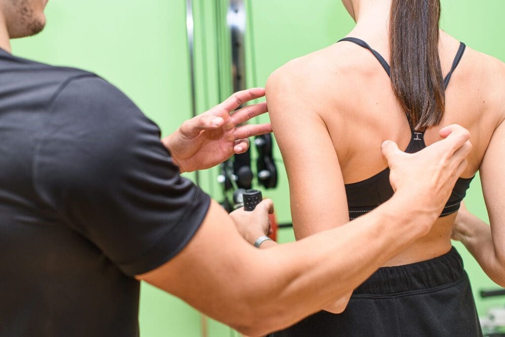
A woman, assisted by a trainer, performs shoulder and back exercises during a beginner gym workout.
Simple, Safe, and Athletic
Starting a gym routine can feel confusing because there are so many workouts online. For beginners who want “sports training” (not just bodybuilding), the goal is simple: build a foundation of strength, movement quality, and conditioning—without getting hurt or burning out.
A beginner-friendly sports training plan usually works best as a 3-day-per-week full-body program, built around compound movements (squat, hinge, push, pull, carry) plus core stability and low-impact cardio. This structure appears in many beginner training guides because it provides enough practice to improve while still leaving recovery time for your body to adapt. (Planet Fitness, 2019/2025; Under Armour, n.d.; Mikolo, 2024).
Below is a practical sports training plan you can follow for 4–8 weeks, along with tips on technique, progression, recovery, and how integrative chiropractic care can support your training and help you move better.
What “Sports Training” Means for a Beginner
For beginners, sports training is not about maxing out or doing complicated drills. It’s about learning to produce force safely and efficiently, in patterns that show up in real life and sport:
A full-body approach is especially helpful early on, because you practice these patterns more often without needing long workouts. Many beginner gym plans also recommend starting with simple machines or stable variations so you can learn form safely (Planet Fitness, 2018/2025; 10 Fitness, 2025).
The “3 Rules” That Make a Beginner Plan Actually Work
1) Keep it simple and repeatable
You want a plan you can do even when you’re tired or busy. If the workout has 25 exercises, it won’t last.
2) Train hard enough, not maximal
Most sets should feel like you could do 2–3 more reps with proper form. That’s how you build strength without turning every day into a recovery problem (Squatwolf, n.d.).
3) Progress slowly on purpose
The beginner’s “secret” is consistency. Small weekly increases add up fast.
Friday: Full-Body Workout A (next week start with B)
On non-lifting days, add low-impact cardio and mobility (e.g., walking, biking, rowing, or an incline treadmill) (Planet Fitness, 2019; Under Armour, n.d.; Mikolo, 2024).
Warm-Up (8–12 Minutes)
A good warm-up raises your body temperature, wakes up your joints, and teaches your body the positions you need.
Step 1: Easy cardio (3–5 minutes)
Treadmill walk (flat or slight incline)
Bike
Rower
Planet Fitness highlights that beginner cardio doesn’t need to be extreme—simple options work (Planet Fitness, 2019).
The Beginner Sports Training Gym Program (3 Days/Week)
Reps, Sets, and Rest (Simple Standards)
Most strength moves: 3 sets of 8–12 reps
Core holds: 3 sets of 20–40 seconds
Rest: 60–90 seconds between sets (longer if needed)
This aligns with the common beginner recommendation to use moderate rep ranges that build skill and strength together (Mikolo, 2024; 10 Fitness, 2025).
Workout A (Full Body Foundation)
1) Squat pattern (choose one)
Goblet squat (dumbbell) or
Leg press (machine)
3 sets x 8–12
2) Push pattern (choose one)
Incline push-up (hands on bench) or
Chest press machine
3 sets x 8–12
3) Pull pattern (choose one)
Seated row machine or
Dumbbell row (bench-supported)
3 sets x 8–12
4) Hinge pattern (choose one)
Romanian deadlift with dumbbells (light) or
Hip hinge with cable pull-through
3 sets x 8–12
5) Core stability
Plank 3 x 20–40 seconds
6) Conditioning finisher (optional)
Rower: 6 minutes, easy steady pace or
Incline treadmill walk: 8–12 minutes
Planet Fitness and other beginner guides commonly use incline walking, machines, and simple cardio finishers because they’re easy to scale (Planet Fitness, 2025; 10 Fitness, 2025).
Workout B (Full Body Athletic Balance)
1) Lunge/single-leg pattern
Reverse lunge (bodyweight or light dumbbells) or
Step-ups
3 sets x 8 reps each leg
2) Overhead or vertical push (beginner-friendly)
Dumbbell shoulder press (light) or
Shoulder press machine
3 sets x 8–12
3) Vertical pull
Lat pulldown machine 3 sets x 8–12
4) Glute + posterior chain
Glute bridge or hip thrust (bodyweight or light weight)
3 sets x 10–12
5) Anti-rotation core (beginner sports core)
Pallof press (cable/band) 3 sets x 10 each side
6) Easy aerobic
Bike or elliptical 10–15 minutes conversational pace
This “movement-pattern” setup is common in beginner athletic plans because it builds total-body strength and stability without needing complicated programming (Mikolo, 2024; Under Armour, n.d.).
How Heavy Should You Lift?
A beginner-friendly rule that works:
Pick a weight you can lift for 8–12 reps with correct form
The last 2–3 reps feel challenging, but you could still do 1–2 more reps if you had to
If you can easily do 15+ reps, it’s probably time to increase the weight slightly
That “difficult but manageable” guideline is widely recommended for safe progression (Squatwolf, n.d.).
Progression Plan (So You Keep Improving)
Use a simple progression method for 4–8 weeks:
Week-to-week progression
Option A (reps first): Keep the same weight and add 1 rep per set until you reach the top of the range (12 reps). Then increase weight slightly and go back to 8 reps.
Option B (small weight jumps): If the form is stable, add 2.5–5 lb per dumbbell (or the smallest machine increase) when you can complete all sets cleanly.
What to track
Exercise
Weight used
Reps completed
How it felt (easy/moderate/hard)
Beginner Cardio That Supports Sports Performance (Without Beating You Up)
A common beginner mistake is going too hard on cardio too soon. Instead, use low-impact cardio that builds your base and helps recovery:
Good beginner options
Incline treadmill walking
Rowing machine
Stationary bike
Elliptical
Brisk outdoor walking
Planet Fitness emphasizes beginner-friendly cardio options and the importance of gradually building cardiovascular endurance (Planet Fitness, 2019; Planet Fitness, 2025).
Simple cardio plan
2–3 days/week
15–25 minutes
You should be able to talk in short sentences
Recovery Essentials (Where Beginners Actually Get Results)
Training breaks muscle down. Recovery is where your body rebuilds.
Active recovery examples
Light walking
Mobility work
Gentle cycling
Stretching sessions
Sanford Sports highlights that recovery helps you regenerate and avoid overtraining, and that active recovery can be a smart part of the week (Sanford Sports, 2024).
Basic recovery checklist
Sleep: aim for consistent, restful sleep
Protein: include protein at most meals
Hydration: steady intake throughout the day
Easy movement on rest days
Integrative Chiropractic Care Helps Beginners Train Better
A smart beginner program is not only about exercises—it’s about movement quality. Integrative chiropractic care (when done responsibly and paired with exercise) often focuses on improving joint motion, reducing pain triggers, and correcting movement compensation patterns.
How chiropractic fits into beginner sports training
1) Injury prevention through movement checks Functional movement evaluations can reveal weak links (hip control, ankle stiffness, and shoulder restriction) before they lead to injury. This is a central theme in Dr. Alexander Jimenez’s integrative sports injury education and movement-focused approach (Jimenez, 2026; PushAsRx, 2026).
2) Mobility and joint mechanics Better mobility can help you hit safer positions in squats, hinges, and presses. Dr. Jimenez’s clinical content on integrated chiropractic and NP care frequently emphasizes joint mobility, balance, coordination, and reduced risk of re-injury as practical athletic goals (Jimenez, 2026).
3) Recovery support (especially when you’re sore or stiff) Many chiropractic and sports rehab sources describe combining manual care with exercise to help patients restore function and return to activity (Team Elite Chiropractic, 2022).
Before or after workouts: what’s better?
There isn’t one universal rule, but many clinics describe two common patterns:
Before training: focus on mobility, joint mechanics, and movement quality
After training: focus on reducing stiffness and supporting recovery
Some chiropractic guidance suggests that getting adjusted before exercise may help movement feel smoother, while post-workout care may help with soreness and relaxation (Atlas Total Health, 2022).
Practical beginner tip: If you’re starting out and you tend to get sore easily, schedule chiropractic visits on lighter training days or rest days so you can feel the changes without rushing back into heavy lifting.
Corrective Exercises: The “Bridge” Between Treatment and Training
Corrective exercises are simple drills that restore balance and improve movement patterns. They are often used when someone has tight areas, weak stabilizers, or poor control (Asheville Medical Massage, 2025).
Examples that pair well with beginner lifting
Glute bridges (glute activation)
Bird dogs (core + spine control)
Dead bugs (core bracing)
Wall angels (posture + shoulder mobility)
Cat-cow (spinal mobility)
Many chiropractic exercise lists include similar basics because they reinforce better posture and better movement options (Elevate to Life, n.d.; Team Elite Chiropractic, 2022).
Beginner Mistakes to Avoid (So You Don’t Quit)
1) Going too hard in week one Soreness is normal, but crushing yourself makes consistency harder. Planet Fitness beginner guidance commonly encourages starting with manageable sessions and learning equipment first (Planet Fitness, 2025).
2) Skipping the warm-up A short warm-up improves performance and helps you move better that day.
3) Changing the plan every workout Beginners improve faster by repeating key patterns.
4) Ignoring form for heavier weight The fastest path is controlled reps, full ranges you own, and slow progression.
A Simple 4-Week “Ramp Up” Example
If you want a very clear starting path:
Week 1
Do Workout A and B with light weights
Keep cardio easy
Focus on learning movement
Week 2
Add 1–2 reps per set or a small weight increase
Add one extra 10-minute cardio session if energy is good
Week 3
Increase weight slightly on 1–2 main lifts
Keep form strict
Week 4
Keep building reps/weight gradually
Deload if needed (reduce weights by ~10–15% for a week if you feel beat up)
Under Armour’s beginner schedule also supports the idea of only a few strength days weekly with rest days built in (Under Armour, n.d.).
Safety Notes (Especially for Beginners Who Want Sports Performance)
Stop and get checked if you have:
Sharp pain, numbness, tingling, or weakness
Joint swelling that doesn’t settle
Pain that changes your walking pattern
Symptoms after a recent injury that are getting worse
If you’re under chiropractic or medical care, your training plan should align with your exam findings and current tolerance.
Bottom Line: The Best Beginner Sports Training Plan Is the One You Repeat
A recommended sports training gym workout for beginners is:
3 full-body strength days per week
Built around squat + hinge + push + pull + core
Supported by low-impact cardio
Protected by recovery days
Improved by movement assessments and corrective exercise
Enhanced by integrative chiropractic strategies that help restore mobility, reduce compensation, and support training consistency (Jimenez, 2026; PushAsRx, 2026).
If you want the simplest next step: start with the workouts above for 4 weeks, track your progress, and adjust slowly.
Back and shoulder pain and stress-relief treatment.
ChiroMed Integrated Medicine
Stress is everywhere in our busy lives, but you can fight back and feel better. At ChiroMed Integrated Medicine in El Paso, TX, we know how stress can build up and harm your health. The good news is, yes, there is a way to detox from stress. This means lowering cortisol, your body’s main stress hormone, and helping your nervous system relax. By using simple habits and professional help, you can shift from a tense “fight-or-flight” state to a calm “rest-and-digest” mode. In this article, we’ll explain stress detox, why it’s important, and easy ways to do it. We’ll highlight how our team at ChiroMed, led by Dr. Alex Jimenez, uses integrative chiropractic care, nutrition, and more to help patients in El Paso reduce stress and restore balance.
Stress detox is like giving your body a break from constant pressure. When stressed, your body releases cortisol to handle short-term threats, but chronic stress keeps levels high, causing issues such as poor sleep, anxiety, and pain (Healthline, n.d.). At ChiroMed, we’ve helped people since 1996 with holistic care that targets these problems. Dr. Alex Jimenez, our Doctor of Chiropractic and Family Nurse Practitioner, sees how stress causes tight muscles and spine issues in his patients. Our clinic at 11860 Vista Del Sol Dr, Suite 128, offers personalized plans combining chiropractic adjustments, naturopathic medicine, and nutrition to relieve tension and reduce cortisol levels (ChiroMed, n.d.).
Understanding Stress Buildup and the Need for Detox
Your nervous system has two parts: the sympathetic for action and the parasympathetic for rest. Chronic stress locks you in sympathetic mode, leading to shallow breaths, muscle knots, and misaligned spines (Henry Ford Health, 2025a). Detoxing helps the body shift into rest mode for healing.
Common signs of high stress Include Constant fatigue, frequent colds, and tension headaches.
Advantages of detoxing: Improved energy, better sleep, and stronger immunity.
The process: It reduces cortisol and supports organs like the liver and kidneys to clear stress toxins (Recover Well Studio, n.d.).
Experts recommend starting small. Mindfulness, like meditation, can lower stress and tiredness (Recover Well Studio, n.d.). At ChiroMed, we integrate these with our treatments for full results.
Everyday Habits to Reduce Cortisol Levels
Simple daily changes can make a big impact on stress detox. At ChiroMed, we guide patients through these habits as part of our holistic approach.
Exercise as a Stress Buster
Physical activity is key to burning off stress. Try 30 to 50 minutes of walking, yoga, or light jogging daily. It releases endorphins, natural mood boosters that counter cortisol (Mayo Clinic, n.d.). Our rehabilitation services at ChiroMed include tailored exercise plans to improve movement and reduce tension.
Activities to start with: A quick walk, swimming, or home yoga.
How it aids detox: Boosts circulation to help your body flush toxins.
Our advice: Combine with our physical therapy for safe, effective routines.
Dr. Jimenez often pairs exercise with adjustments to help El Paso patients with stress-related pain (ChiroMed, n.d.).
Prioritize Quality Sleep
Sleep lets your body recharge. Get 7 to 8 hours of sleep nightly to reduce cortisol. Bad sleep fuels a stress cycle (Henry Ford Health, 2025a). At ChiroMed, our naturopathic services assess sleep issues and recommend natural remedies.
Better sleep habits: Stick to a consistent schedule, dim the lights, and avoid caffeine late in the day.
Detox benefits: Deep sleep clears brain toxins from daily stress.
Clinic tips: Use our nutrition counseling for sleep-friendly diets.
Patients at ChiroMed report better rest after our integrative plans (ChiroMed, n.d.).
Practice Meditation and Breathing Exercises
These tools calm you quickly. Meditation focuses your mind, reducing stress. Deep breathing slows your heart and activates rest mode (Goop, n.d.). We teach these at ChiroMed alongside acupuncture for deeper relaxation.
Simple breath technique: Breathe in for 4, hold for 4, out for 4—repeat for 5 minutes.
Meditation starters: Free apps for guided sessions.
Proven effects: Can reduce cortisol by 20% with practice.
Our team uses these in conjunction with chiropractic care to free up blocked energy (Abundant Life Chiropractor, n.d.).
Nutrition and Hydration for Effective Detox
Fuel your body right to handle stress. At ChiroMed, our nutrition counseling creates plans that support detox organs.
Choose vitamin-packed foods like fruits, veggies, and grains. Cut sugar and caffeine to avoid cortisol spikes (Healthline, n.d.). Drink 8 glasses of water daily to aid toxin removal.
Top anti-stress foods: Bananas, nuts, and a bit of dark chocolate (Addiction Center, n.d.a).
Sample meals: Veggie stir-fry with lean protein.
Supplement options: We recommend omega-3s or magnesium after checks.
Dr. Jimenez’s functional medicine at ChiroMed supports detoxification through nutrient-rich diets (DCLabs, n.d.).
Benefits of Nature Time
Outdoor time naturally lowers stress. It drops cortisol and lifts spirits (NatureMed, n.d.). Just 20 minutes in nature promotes rest mode.
Ideas to try: Park walks, gardening, or picnics.
Science behind it: Sunlight increases vitamin D for stress fighting.
Routine building: Schedule weekly outings.
This complements our holistic care at ChiroMed for emotional balance (The Plymouth House, n.d.).
Establishing Work and Digital Boundaries
Non-stop work and screens raise stress. Set limits, such as no emails after hours (Monterey Premier, n.d.). Our wellness plans at ChiroMed include tips for balance.
Tech detox steps: Silence alerts and limit apps.
Work rules: Take breaks and delegate tasks.
Quick cleanse: A tech-free day with reading or hobbies (Local Care Force, n.d.).
This helps reset from acute stress (AdventHealth, n.d.).
Chiropractic Care at ChiroMed for Stress Relief
Chiropractic is central to stress detox at ChiroMed. Adjustments correct spinal misalignments caused by tension, easing nerve pressure and promoting relaxation (Henry Ford Health, 2025b).
Our techniques release muscles and improve flow (Rodgers Stein Chiropractic, n.d.a). Dr. Jimenez, with certifications in multiple states, treats stress-linked anxiety with gentle methods (ChiroMed, n.d.).
Key benefits: Eases pain and boosts mood.
Integrated options: Add massage or acupuncture.
Supporting data: Enhances the nervous system for lower cortisol (North Bay Spine and Rehab, n.d.).
As a nurse practitioner, Dr. Jimenez offers full care (Dallas Accident and Injury Rehab, n.d.).
Our Integrative Health Approaches
At ChiroMed, we blend therapies for the best results. Our team includes chiropractors, physical therapists, and naturopaths (Psychology Today, 2025).
We address root causes using functional medicine, assessing hormones and recommending changes (ChiroMed, n.d.). This builds vitality (RU Well Adjusted, n.d.).
Therapy combinations: Acupuncture for pain (My Evolve Chiropractor, n.d.).
Ongoing perks: Greater stress resistance (Addiction Center, n.d.b).
Sustain detox with lasting habits. Laughter releases oxytocin against stress (Mayo Clinic, n.d.). Try music or pets for relief (MHP Colorado, n.d.).
Build-up strategies: Journal, socialize, or learn skills (CDC, n.d.a).
Avoid mistakes: Avoid unhealthy coping, such as overeating.
Monitor changes: Track feelings weekly.
Our holistic approach at ChiroMed prevents stress from returning (ChiroMed, n.d.).
Try a One-Day Stress Reset
For fast relief, follow this plan: morning exercise, healthy eating, meditation, nature, no tech, and a relaxing end (Goop, n.d.).
AM routine: Breathe and walk.
PM activities: Light meal and outdoors.
Night wind-down: Book and bed.
Incorporate into our programs at ChiroMed (AdventHealth, n.d.).
Wrapping Up Stress Detox with ChiroMed
You can detox from stress with our support and habits at ChiroMed. From exercise to chiropractic, we lower cortisol and restore peace. Dr. Jimenez and our team in El Paso are ready to guide you. Contact us at (915) 850-0900 or visit for a consultation.
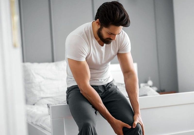
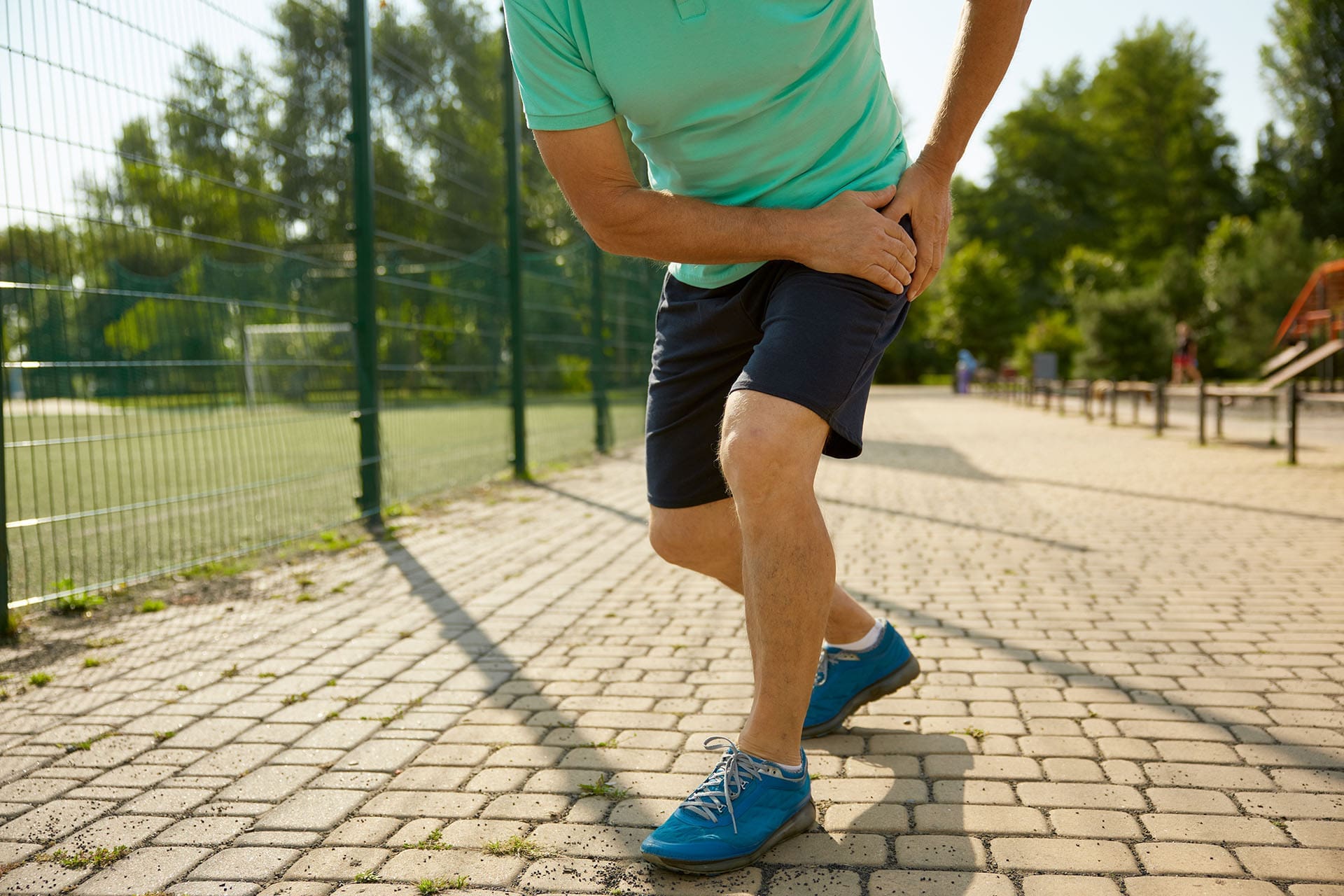
The front of your hip and upper leg holds a powerful group of muscles called the anterior hip and leg muscles. These muscles lift your knee, bend you forward at the waist, straighten your knee, and keep your pelvis steady so you can walk, run, climb stairs, or stand up from a chair without falling. They do a lot of work every day, which is why they sometimes experience soreness, tightness, or injury. At ChiroMed Integrated Medicine in El Paso, Texas, our team sees this problem all the time. We use gentle chiropractic care, nurse practitioner services, rehabilitation exercises, and nutrition support to identify the underlying cause of your pain and help your body heal naturally.
Sitting for hours at work or school shortens these muscles. Running, soccer, or quick direction changes can strain them. When they get out of balance, pain shows up in the front of the hip or down the thigh. The good news? You don’t have to live with it. Dr. Alexander Jimenez, DC, APRN, FNP-BC, and the ChiroMed team create personalized plans that address the root cause rather than just masking pain.
What Exactly Are the Anterior Hip and Leg Muscles?
These muscles sit in the front compartment of your thigh. They start near your lower back and pelvis and run down to your knee. Blood flows to them through the femoral artery, and the femoral nerve tells them when to move.
Here is a simple list of the main muscles:
Iliopsoas (psoas major + iliacus) – The strongest hip flexor. It pulls your knee up toward your chest.
Rectus femoris – Part of the quadriceps. It bends the hip and straightens the knee simultaneously.
Vastus medialis, vastus intermedius, vastus lateralis – The other three quadriceps muscles. They mainly straighten your knee and keep your kneecap in place.
Sartorius – The longest muscle in the body. It helps you cross your legs and rotate your thigh.
Pectineus – A small muscle that pulls your leg toward the middle of your body.
These muscles work as a team. When you take a step, the iliopsoas lifts your leg, and the quadriceps lock your knee so you can push off the ground.
Everyday Jobs These Muscles Do
Think about your day:
Walking to class or work
Getting out of bed
Climbing stairs
Kicking a soccer ball
Standing up after sitting
Each of those movements engages the anterior hip muscles. In sports, they work even harder. Runners use them thousands of times per run. Soccer players sprint and change direction quickly. Cyclists keep them bent for hours. When muscles become tired or tight, they experience pain.
Why Do These Muscles Hurt So Often?
Pain usually stems from two major problems: prolonged sitting and repetitive stress.
Sitting Too Much
Desks, cars, and couches keep your hips bent. The iliopsoas and rectus femoris stay short and tight. When you finally stand up, they feel stiff and pull on your lower back. Over time, this creates a cycle of pain that spreads to your knee or groin.
Overuse in Sports or Work
Sudden stops, starts, or kicks can strain the muscles or tendons. Common injuries include:
Hip flexor strain – A tear in the iliopsoas or rectus femoris from sprinting or kicking.
Iliopsoas tendinopathy – Irritation where the tendon attaches to the bone.
Bursitis – Inflammation of the fluid sac that cushions the tendon.
Muscle imbalance – Weak glutes or core makes the front muscles work overtime.
Dr. Alexander Jimenez has treated hundreds of these cases in El Paso. He explains that many patients arrive with tight hip flexors and weak stabilizers. Once we lengthen tight muscles and strengthen weak ones, pain decreases quickly.
Other Common Causes
Poor posture
Weak core
Previous ankle or knee injuries that change how you walk
Carrying extra weight
Not warming up before exercise
How ChiroMed’s Integrative Approach Fixes the Problem
At ChiroMed, we don’t just adjust your back and send you home. We look at the whole picture—spine, hips, muscles, nerves, and even nutrition.
Step-by-Step Care at ChiroMed
Detailed Exam Dr. Jimenez checks your posture, hip range of motion, muscle strength, and nerve function. We use gentle tests to see exactly which muscle is tight or weak.
Chiropractic Adjustments: Gentle moves realign your pelvis and lower back. This relieves pressure on the hip flexors, allowing them to relax.
Soft-Tissue Therapy Massage therapists and physical therapists release knots in the iliopsoas and quadriceps. We use tools and hands-on work to break up scar tissue.
Rehabilitation Exercises: Our physical therapists teach you safe stretching and strengthening exercises. We start slow and build up so you don’t get hurt again.
Nurse Practitioner Support: If needed, our APRNs can order imaging, prescribe short-term anti-inflammatory medications, or screen for other health issues, such as low vitamin D, that may slow healing.
Nutrition and Lifestyle Coaching Anti-inflammatory foods and proper hydration help muscles recover faster.
Dr. Jimenez often says, “The hip is only as strong as the core and the opposite glute.” That’s why we always work the entire muscle chain, not just the sore spot.
Simple Exercises You Can Do at Home
Do these daily to keep your anterior hip muscles healthy.
Hip Flexor Stretch: Kneel on your right knee. Push your hips forward until you feel a gentle stretch in the front of your right hip. Hold for 30 seconds, switch sides. Do it 3 times.
Glute Bridge: Lie on your back, feet flat. Lift your hips up, squeeze your glutes, and hold for 5 seconds. Lower slowly. 10 reps.
Wall Sit: Slide your back down the wall until your knees are bent 90 degrees. Hold for 20–30 seconds. Builds quadriceps strength.
Bird-Dog: On hands and knees, reach one arm forward and the opposite leg back. Hold for 5 seconds. 10 reps on each side. Strengthens your core so the hip flexors don’t have to work alone.
Prevention Tips from the ChiroMed Team
Stand up and walk every 30 minutes
Stretch your hip flexors before and after exercise
Strengthen your glutes and core 3 times a week
Wear supportive shoes
Warm up before sports
Stay at a healthy weight
Real Results from Real Patients
Patients tell us the same thing: “I can finally walk without limping,” or “My knee pain is gone because my hips finally move right.” Dr. Jimenez’s combination of chiropractic care, functional medicine, and rehabilitation helps people return to work, sports, and family life faster.
Ready to Feel Better?
If the front of your hip or thigh hurts, don’t wait. Call ChiroMed today at (915) 850-0900 or visit chiromed.com to schedule your exam. We are located at 11860 Vista Del Sol Dr, Suite 128, El Paso, TX 79936. Let our integrated team—led by Dr. Alexander Jimenez—help you move freely again.
Back pain is a big issue for many people. In the United States, up to 80% of adults deal with low back pain at some point in their lives. This is one of the leading reasons for missed work and doctor visits. People often seek ways to manage pain, identify treatments, and modify daily habits to maintain health. In El Paso, Texas, where many folks are active, work in factories, or drive a lot, questions about back pain are common, too. Issues like sciatica, herniated discs, and spinal stenosis show up often due to local lifestyles. This article examines key questions about back health, such as wearing backpacks safely, treatment options, and tips for relief. It draws on authoritative sources to provide clear answers.
Is It Okay to Wear a Backpack?
Yes, wearing a backpack is fine if done right. Backpacks can help carry items without much strain, but improper use can harm your back. Heavy or poorly fitted packs can cause muscle fatigue, bad posture, and even chronic pain. They do not cause scoliosis, a spinal curvature that affects approximately 4% of people, typically beginning in adolescence (Scoliosis SOS, n.d.). Instead, overloads lead to slouching, shoulder pain, and lower back issues.
To spread weight and avoid strain:
Keep the pack light: Aim for no more than 10-15% of your body weight. For a 150-pound person, that’s up to 22.5 pounds.
Use both straps: Adjust them so the pack sits in the middle of your back, not sagging low. This keeps the weight even.
Pack smart: Put heavy items at the bottom and close to your back. Clean out extras often.
Add support: Look for packs with padded straps, a back panel, and a waist strap to share the load with your hips.
Lift correctly: Bend your knees, not your back, when lifting it.
These steps reduce risks like nerve irritation or misalignment (Ireland Clinic, n.d.). In El Paso, where people carry work tools or drive with bags, even weight helps prevent daily aches.
Spinal Health in the US: Dealing with Chronic Back Pain
Back pain hits hard in the US. It costs over $100 billion a year in health care, matching spending on cancer or diabetes (NCBI, 2023). Most cases are due to muscle strains, but others are due to disc problems, arthritis, or stenosis. Pain can be sharp, dull, or burning, and it often limits daily life. For adults over 50, risks rise with age, poor sleep, or extra weight (NCOA, n.d.). Bad sleep worsens pain by boosting inflammation and slowing healing.
People often inquire about treatment options, specifically whether to choose surgery or conservative care. Most start with non-surgical options. These include physical therapy, meds, and lifestyle changes. Surgery is indicated for severe cases, such as when nerves are pinched or there’s weakness (Mayo Clinic Health System, n.d.). Questions to ask your doctor:
What causes my pain?
Do I need imaging tests such as X-rays or MRIs?
What are my options besides surgery?
How long until I feel better?
Conservative care works for many. Exercise strengthens the core, which supports the spine (UC Davis Health, 2025). Quitting smoking helps too, as it harms spinal tissues and raises surgery risks by up to 50% (UMass Memorial Health, n.d.). For long-term relief, prioritize weight control and daily physical activity. This cuts costs, as back pain leads to high medical bills.
Back Health in El Paso, Texas: Local Issues and Choices
In El Paso, back pain is often associated with active lifestyles, factory work, or long drives. Sciatica is common, characterized by radiating leg pain due to nerve compression. It affects the legs, causing numbness or weakness (EP Manual Physical Therapy, n.d.). Spinal stenosis, a narrowing of the spinal canal, causes cramping or tingling that worsens with walking. Herniated discs, bulges, and compressed nerves, leading to pain that’s hard to ignore (Chiro Desert, n.d.). Accidents compound this, with injuries accumulating over time.
People here ask about chiropractic vs. orthopedic surgery. Chiropractors use adjustments to align the spine and relieve pressure, without medication or incision (Bone & Joint, n.d.). They are used to treat mild to moderate pain resulting from strains or poor posture. Orthopedists manage severe cases, such as those requiring surgery for stenosis or disc disease. They use scans and may do injections or fusions (Monitto Chiro, n.d.). Choose based on your issue: chiropractic for non-invasive relief, surgery if nerves are at risk.
Dr. Alexander Jimenez, a local expert in El Paso, notes that back pain often links to inflammation or imbalances. He employs integrative care, including adjustments and nutrition, to address root causes (Dr. Alex Jimenez, n.d.). For sciatica, he suggests checking for gut issues or metabolic problems. His approach is effective for treating accident-related injuries, using X-rays and exercises for recovery. Patients experience less pain and improved mobility with his plans.
For herniated discs or sciatica:
Try decompression: It stretches the spine to retract discs and boost healing (Right Way Chiro, n.d.).
Avoid hazardous movements: Skip back bends or heavy lifts with stenosis (Orthobiologics Associates, n.d.).
See a pro: If pain lasts weeks or causes leg weakness, get checked.
Costs matter in El Paso. Chiropractic visits cost $50- $ 200 and are often covered by insurance, such as Medicare (Healthgrades, n.d.). Check your plan for limits.
Everyday Changes to Avoid Injury and Get Relief
Small habits prevent back pain. Stay active with low-impact activities such as swimming or walking. Strengthen your core for spine support (My Spine Doc, 2026). Good posture cuts strain: Sit with feet flat, screen at eye level.
Tips for daily life:
At work: Use lumbar support, stand every hour, and wear comfortable shoes.
Lifting: Bend knees; keep items close, and do not twist.
Sleep: Side position with knees bent eases stenosis.
Diet: Calcium and vitamin D build strong bones (Jefferson Health, n.d.).
For relief, try ice or heat, stretches, or over-the-counter meds. If pain spreads or includes numbness, see a doctor (Nebraska Medicine, n.d.). In the long term, these changes reduce care costs and improve quality of life.
In the US and El Paso, back health means knowing risks and acting early. Put prevention first with everything from treatment picks to safe backpacks. Consult professionals like Dr. Jimenez for tailored advice. With appropriate steps, most find relief and remain active.