Unveil the clinical approach to opioid use disorder and learn about evidence-based methods for effective treatment.
Overcoming Barriers in Managing Opioid Use Disorder: Strategies for Effective Care
A lot of people today have opioid use disorder (OUD), which is a serious health problem. It falls under the larger group of substance use disorders (SUD). Treating OUD can be difficult because everyone has their own set of problems, like pain or other health issues. Doctors and other healthcare professionals must make plans that are specific to each patient. They also need to stay up to date on laws, ethics, and ways to protect patient information. The Health Insurance Portability and Accountability Act (HIPAA) of 1996 covers all patients, but those getting treatment for drug or alcohol abuse have to follow more rules.
In this tutorial, we talk about how to get around problems with OUD administration. We look at stigma, team-based approaches, ways to talk to patients, treatment that puts the patient first, and legal issues. Health care workers can help patients get better by using these methods. Keywords like “opioid use disorder management,” “overcoming stigma in OUD,” and “patient-centered care for SUD” highlight important ideas to help people understand better and find what they’re looking for.
Learning Objectives
Explain treatment planning methods that use patient-focused choices and proven ways to talk.
Name the three kinds of stigma and how they affect people with mental health issues, SUD, and especially OUD.
Talk about legal, ethical, and privacy concerns in caring for people with OUD.
Effective Treatment Planning with Patient-Centered Decisions
People with complex issues, like mental health problems, SUD, and pain, need special care. Each person shows up differently, so health systems are now focusing on care that puts the patient first.
Patient-centered care means building teams with doctors, patients, and families. They work together to plan, give, and check health care. This way ensures the patient’s needs are met, and their wishes, likes, and family situations are respected. It focuses on shared choices about treatments while seeing the patient as a whole person in their daily life (Dwamena et al., 2012; Bokhour et al., 2018).
Studies show key steps for a good patient-centered plan:
Take a full patient history and a check-up, reviewing old and new treatments.
Find all available drug and non-drug options.
Check the patient’s current health, recent changes, and patterns.
Look at risks for misusing or abusing opioids.
If starting opioids or if the patient is already on them, think about opioid stewardship. This means checking harms, benefits, risks, side effects, pain control, daily function, drug tests, stop plans, and ways to spot OUD. These programs, sometimes called analgesia stewardship, help manage opioids safely (Harle et al., 2019; Coffin et al., 2022). Guides exist to set them up (American Hospital Association, n.d.; Shrestha et al., 2023).
Integrative chiropractic care can play a big role here. It uses spinal adjustments and targeted exercises to get proper spinal alignment. This helps reduce pain without relying only on drugs, making it a good fit for OUD patients with pain. For example, adjustments fix spine issues that cause pain, and exercises strengthen muscles to keep alignment right.
A Nurse Practitioner (NP) adds full management and ergonomic advice. They look at work setups to prevent pain, such as how to sit or lift. NPs coordinate care by reviewing options such as therapy, meds, and lifestyle changes, ensuring everything works together.
Dr. Alexander Jimenez, DC, APRN, FNP-BC, with over 30 years in chiropractic and as a family nurse practitioner, observes that blending these methods cuts opioid use. At his El Paso clinic, he uses functional medicine to address root causes through nutrition and non-invasive treatments. He notes that poor posture from modern life worsens pain, leading to OUD risks. His teams help patients with self-massage and VR for recovery, reducing drug needs (Jimenez, n.d.a; Jimenez, n.d.b).
Evidence-Based Ways to Communicate
Good talking skills are key to building a patient-centered plan (Schaefer & Block, 2009). There are proven methods for starting conversations and getting patients involved.
One method is BATHE:
Background: Ask, “How have things been since your last visit?”
Affect: Ask, “How does this make you feel?”
Trouble: Ask, “What bothers you most?”
Handling: Ask, “How are you coping?”
Empathy: Say, “That sounds hard.”
This uses open questions to let patients lead and feel supported (Stuart & Lieberman, 2018; Thomas et al., 2019).
Another is GREAT:
Greetings/Goals: Start with hello and set aims.
Rapport: Build trust.
Evaluation/Expectation/Examination/Explanation: Check and explain.
Ask/Answer/Acknowledge: Listen and respond.
Tacit agreement/Thanks: Agree and thank.
This guide talks well (Brindley et al., 2014).
Motivational interviewing is also useful. It’s a team-style talk to boost a patient’s desire to change. Build a bond, focus on the issue, spark a desire for change, and plan steps (Frost et al., 2018).
These methods emphasize listening, clear communication, and a structured approach to planning. For OUD patients with pain or mental issues, mix techniques for the best results.
Dr. Jimenez shares that in his practice, these talks help patients see non-drug options, such as chiropractic adjustments. He finds that empathy reduces stigma and fear, encouraging openness about OUD (Jimenez, n.d.a).
Understanding Stigma in Mental Health and Substance Use Disorders
Stigma blocks good talk for many with mental health or SUD. It’s attitudes, beliefs, actions, and systems that lead to unfair views and bad treatment (Cheetham et al., 2022).
Studies show stigmas like linking mental illness to violence (Perry, 2011). Media on shootings with mentally ill people strengthens this (McGinty et al., 2014; McGinty et al., 2016; Schomerus et al., 2022). For SUD, people think they’re more dangerous than those with schizophrenia or depression (Schomerus et al., 2011). Society blames people with SUDs more and avoids them (McGinty et al., 2015; Corrigan et al., 2012).
Views come from knowledge, contact with affected people, and the media. Public ideas are tied to norms on causes, blame, and danger. Race, ethnicity, and culture shape attitudes too (Giacco et al., 2014).
Health workers have biases. A survey of VA mental health providers showed awareness of race issues but avoidance of talks, using codes like “urban,” and thinking training stops racism (McMaster et al., 2021).
There are three stigma types:
Structural Stigma: The ways Society and institutions keep prejudice. In health, it’s worse care, less access to behavioral health. Less funding for mental vs. physical issues (National Academies of Sciences, Engineering, and Medicine, 2016).
Public Stigma: General or group attitudes, like police or church norms. Laws reinforce it, like broad mental illness rules implying all are unfit (Corrigan & Shapiro, 2010).
Self-Stigma: When people internalize stigmas, it leads to low self-worth and shame. “Why try” affects independent living (Corrigan et al., 2009; Clement et al., 2015).
Dr. Jimenez observes that stigma makes OUD patients hide symptoms, delaying care. In his integrative work, he addresses this through education on holistic options, showing that recovery is possible without judgment (Jimenez, n.d.b).
Overcoming Stigma and Addressing Social Factors
To fight stigma, use education, behavior changes, and better care. Laws like the ADA and MHPAEA help ensure equal coverage and prevent discrimination (U.S. Congress, 2009; U.S. Congress, 2008; U.S. Department of Health and Human Services, n.d.; Busch & Barry, 2008; Haffajee et al., 2019).
These address social determinants of health (SDOH), such as coverage, access, quality, education, and stability (Centers for Disease Control and Prevention, n.d.).
Community programs help too:
West Virginia’s Jobs and Hope: Training, jobs, education, transport, skills, record clearing for SUD people (Jobs and Hope, n.d.).
Belden’s Pathway: Rehab for failed drug tests, leading to jobs (Belden, n.d.).
Education boosts provider confidence in OUD meds, reducing barriers (Adzrago et al., 2022; Hooker et al., 2023; Campbell et al., 2021).
Overcoming stigma is key to success in mental health and SUD.
Interprofessional Team Work
Teams improve outcomes for patients with chronic pain and mental health or SUD (Joypaul et al., 2019; Gauthier et al., 2019).
Teams include doctors, nurses, NPs, pharmacists, PAs, social workers, PTs, therapists, SUD experts, and case managers.
Each helps uniquely:
Pharmacists watch meds, spot interactions.
Case managers link specialists, find resources, and support families (Sortedahl et al., 2018).
Teams set goals, max non-opioid treatments (Liossi et al., 2019).
Integrative chiropractic care includes adjustments and exercises for alignment, easing pain naturally.
NPs give full care, ergonomic tips to avoid pain triggers, and coordinate options.
Dr. Jimenez’s clinic shows this. As a DC and FNP-BC, he leads teams with therapists, nutritionists, and coaches. He observes interprofessional work cuts opioid use by addressing the roots with functional medicine, VR, and nutrition. For OUD, he blends chiropractic care for pain, NP coordination for plans, and stigma-fighting through team support (Jimenez, n.d.a; Jimenez, n.d.b).
The Power of Chiropractic Care in Injury Rehabilitation-Video
Legal and Ethical Issues in SUD Care
Providers must know laws and ethics for mental/SUD patients, like discrimination, aid, and privacy (Center for Substance Abuse Treatment, 2000).
Key Federal laws:
Americans with Disabilities Act (ADA) of 1990.
Rehabilitation Act of 1973.
Workforce Investment Act of 1998.
Drug-Free Workplace Act of 1988.
ADA and Rehabilitation ban discrimination in government and in business services like hotels, shops, and hospitals. Protect those with impairments limiting life activities (U.S. Department of Health and Human Services, n.d.).
Provisions:
Protect “qualified” people who meet the requirements.
Reasonable accommodations for jobs.
No hire/retain if there is a direct threat.
No denial of benefits, access, or jobs in funded places.
For SUD: Alcohol users are protected if qualified, no threat. Ex-drug users in rehab are the same. Current illegal drug users are protected for health/rehab, not others. Programs can deny if used during.
Workforce Act centralizes job programs; no refusal to SUD people (U.S. Congress, 1998).
Drug-Free Act requires drug-free policies for federal funds/contracts: statements, awareness, actions on violations (U.S. Code, n.d.).
States have their own laws; check the local laws.
Public Aid laws:
Contract with America Act (1996): No SSI/DI if SUD key factor (U.S. Congress, 1996).
Personal Responsibility Act (1996): Work after 2 years of aid, drug screens (U.S. Department of Health and Human Services, 1996).
These push work, sobriety.
Dr. Jimenez notes that legal awareness helps his practice by ensuring holistic plans comply and by reducing OUD risks through a non-drug focus (Jimenez, n.d.a).
Keeping Patient Info Private
Privacy is vital. Laws include:
HIPAA (1996): Protects PHI, sets use/disclosure rules (U.S. Department of Health and Human Services, n.d.).
42 CFR Part 2: Extra for SUD records. No disclosure of name or status without consent. Fines for breaks. Applies to federal-aided programs (Substance Abuse and Mental Health Services Administration, n.d.).
Consent needs: program name, receiver, patient name, purpose, info type, revoke note, expire date, signature, and date.
This fights discrimination fears, encouraging treatment (Center for Substance Abuse Treatment, 2000).
Wrapping Up
As we deal with the ongoing problems of opioid use disorder (OUD), it’s clear that the best way to handle them is through a multi-faceted approach that puts the health of the patient first instead of quick fixes. Healthcare providers are essential to changing lives. They do this by supporting patient-centered decision-making and evidence-based communication, and by breaking down the three types of stigma—structural, public, and self—that make it harder for people to get better. Legal and ethical frameworks, such as HIPAA and 42 CFR Part 2 privacy protections, make sure that people who need help can get it without worrying about being treated unfairly. Interprofessional teams also help make sure that everyone receives the care they need.
Chiropractic care, which focuses on spinal adjustments and specific exercises to help with proper alignment, is a non-invasive way to ease pain and cut down on the need for opioids. Nurse Practitioners (NPs) improve this by offering comprehensive care, ergonomic advice to avoid injury, and the coordination of various treatment options, including therapy and lifestyle changes. Dr. Alexander Jimenez, DC, APRN, FNP-BC, stresses in his clinical practice that these integrative methods not only help with physical symptoms but also give patients the tools they need to make educated decisions and follow personalized plans. This leads to long-term recovery and less use of opioids (Jimenez, n.d.a; Jimenez, n.d.b).
Recent developments in OUD treatment as of 2025 indicate a transition towards more individualized and accessible alternatives. For example:
FDA-approved drugs like methadone, buprenorphine, and naltrexone are still the mainstays of treatment for OUD. They help reduce cravings and withdrawal symptoms while also assisting people to stay stable over the long term.
Precision medicine goes beyond one-size-fits-all approaches by tailoring treatments to each person’s genetic, psychological, and social factors. This should lead to better results.
New Guideline: The World Health Organization’s 2025 updates emphasize the importance of psychosocial support alongside drug treatments. They also focus on preventing overdoses in the community and making care more widely available.
Declining Trends: The number of deaths involving opioids dropped for the first time in 2023 since 2018, which is a good sign that ongoing efforts in policy, education, and treatment are having an effect.
We can create a future where OUD is not a life sentence but a condition that can be managed by combining these new ideas with reducing stigma and working together to care for people. Healthcare professionals, communities, and policymakers must continue to push for fair access to care so that everyone gets the compassionate, evidence-based help they need. In the end, overcoming the obstacles to managing OUD isn’t just about treatment; it’s also about restoring hope, respect, and a better quality of life.
References
Adzrago, D., Paola, A. D., Zhu, J., et al. (2022). Association between prescribers’ perceptions of the utilization of medication for opioid use disorder and opioid dependence treatability. Healthcare, 10(9), 1733. https://doi.org/10.3390/healthcare10091733
Bokhour, B. G., Fix, G. M., et al. (2018). How can healthcare organizations implement patient-centered care? Examining a large-scale cultural transformation. BMC Health Services Research, 18(1), 168. https://doi.org/10.1186/s12913-018-2993-5
Brindley, P. G., Smith, K. E., Cardinal, P., & LeBlanc, F. (2014). Improving medical communication with patients and families: Skills for a complex (and multilingual) clinical world. Canadian Respiratory Journal, 21(2), 89-91. https://doi.org/10.1155/2014/789456
Campbell, C. I., Saxon, A. J., Boudreau, D. M., et al. (2021). Primary Care Opioid Use Disorders treatment (PROUD) trial protocol: A pragmatic, cluster-randomized implementation trial in primary care for opioid use disorder treatment. Addiction Science & Clinical Practice, 16(1), 9. https://doi.org/10.1186/s13722-021-00221-1
Center for Substance Abuse Treatment. (2000). Integrating Substance Abuse Treatment and Vocational Services. (Treatment Improvement Protocol (TIP) Series, No. 38.) Chapter 7—Legal Issues. https://www.ncbi.nlm.nih.gov/books/NBK64294/
Center for Substance Abuse Treatment. (2000). Substance Abuse Treatment for Persons with Child Abuse and Neglect Issues. (Treatment Improvement Protocol (TIP) Series, No. 36.) Appendix B –Protecting Clients’ Privacy. https://www.ncbi.nlm.nih.gov/books/NBK64900/
Cheetham, A., Picco, L., Barnett, A., et al. (2022). The impact of stigma on people with opioid use disorder, opioid treatment, and policy. Substance Abuse and Rehabilitation, 13, 1-12. https://doi.org/10.2147/SAR.S304256
Clement, S., Schauman, O., Graham, T., et al. (2015). What is the impact of mental health-related stigma on help-seeking? A systematic review of quantitative and qualitative studies. Psychological Medicine, 45(1), 11-27. https://doi.org/10.1017/S0033291714000129
Coffin, P. O., Martinez, R. S., Wylie, B., et al. (2022). Primary care management of long-term opioid therapy. Annals of Medicine, 54(1), 2451-2469. https://doi.org/10.1080/07853890.2022.2118597
Corrigan, P. W., Larson, J. E., & Rüsch, N. (2009). Self-stigma and the “why try” effect: Impact on life goals and evidence-based practices. World Psychiatry, 8(2), 75-81. https://doi.org/10.1002/j.2051-5545.2009.tb00218.x
Corrigan, P. W., Morris, S. B., Michaels, P. J., Rafacz, J. D., & Rüsch, N. (2012). Challenging the public stigma of mental illness: A meta-analysis of outcome studies. Psychiatric Services, 63(10), 963-973. https://doi.org/10.1176/appi.ps.201100529
Corrigan, P. W., & Shapiro, J. R. (2010). Measuring the impact of programs that challenge the public stigma of mental illness. Clinical Psychology Review, 30(8), 907-922. https://doi.org/10.1016/j.cpr.2010.06.004
Dwamena, F., Holmes-Rovner, M., Gaulden, C., et al. (2012). Interventions for providers to promote a patient-centred approach in clinical consultations. Cochrane Database of Systematic Reviews, 2012(12), CD003267. https://doi.org/10.1002/14651858.CD003267.pub2
Frost, H., Campbell, P., Maxwell, M., et al. (2018). Effectiveness of Motivational Interviewing on adult behavior change in health and social care settings: A systematic review of reviews. PLoS One, 13(10), e0204890. https://doi.org/10.1371/journal.pone.0204890
Gauthier, K., Dulong, C., & Argáez, C. (2019). Multidisciplinary treatment programs for patients with chronic non-malignant pain: A review of clinical effectiveness, cost-effectiveness, and guidelines – an update. Canadian Agency for Drugs and Technologies in Health. https://www.ncbi.nlm.nih.gov/books/NBK545496/
Giacco, D., Matanov, A., & Priebe, S. (2014). Providing mental healthcare to immigrants: Current challenges and new strategies. Current Opinion in Psychiatry, 27(4), 282-288. https://doi.org/10.1097/YCO.0000000000000070
Haffajee, R. L., Mello, M. M., Zhang, F., et al. (2019). Association of federal mental health parity legislation with health care use and spending among high utilizers of services. Medical Care, 57(4), 245-255. https://doi.org/10.1097/MLR.0000000000001076
Harle, C. A., DiIulio, J., Downs, S. M., et al. (2019). Decision-Centered design of patient information visualizations to support chronic pain care. Applied Clinical Informatics, 10(4), 719-728. https://doi.org/10.1055/s-0039-1696668
Hooker, S. A., Crain, A. L., LaFrance, A. B., et al. (2023). A randomized controlled trial of an intervention to reduce stigma toward people with opioid use disorder among primary care clinicians. Addiction Science & Clinical Practice, 18(1), 10. https://doi.org/10.1186/s13722-023-00366-1
Joypaul, S., Kelly, F., McMillan, S. S., et al. (2019). Multi-disciplinary interventions for chronic pain involving education: A systematic review. PLoS One, 14(10), e0223306. https://doi.org/10.1371/journal.pone.0223306
Liossi, C., Johnstone, L., Lilley, S., et al. (2019). Effectiveness of interdisciplinary interventions in paediatric chronic pain management: A systematic review and subset meta-analysis. British Journal of Anaesthesia, 123(2), e359-e371. https://doi.org/10.1016/j.bja.2019.01.024
McGinty, E. E., Goldman, H. H., Pescosolido, B., et al. (2015). Portraying mental illness and drug addiction as treatable health conditions: Effects of a randomized experiment on stigma and discrimination. Social Science & Medicine, 126, 73-85. https://doi.org/10.1016/j.socscimed.2014.12.010
McGinty, E. E., Kennedy-Hendricks, A., Choksy, S., et al. (2016). Trends in news media coverage of mental illness in the United States: 1995-2014. Health Affairs, 35(6), 1121-1129. https://doi.org/10.1377/hlthaff.2016.0011
McGinty, E. E., Webster, D. W., Jarlenski, M., et al. (2014). News media framing of serious mental illness and gun violence in the United States, 1997-2012. American Journal of Public Health, 104(3), 406-413. https://doi.org/10.2105/AJPH.2013.301557
McMaster, K. J., Peeples, A. D., Schaffner, R. M., et al. (2021). Mental healthcare provider perceptions of race and racial disparity in the care of Black and White clients. Journal of Behavioral Health Services & Research, 48(4), 501-516. https://doi.org/10.1007/s11414-021-00753-3
National Academies of Sciences, Engineering, and Medicine. (2016). Ending discrimination against people with mental and substance use disorders: The evidence for stigma change. National Academies Press. https://www.ncbi.nlm.nih.gov/books/NBK384923/
Perry, B. L. (2011). The labeling paradox: Stigma, the sick role, and social networks in mental illness. Journal of Health and Social Behavior, 52(4), 460-477. https://doi.org/10.1177/0022146511408913
Schaefer, K. G., & Block, S. D. (2009). Physician communication with families in the ICU: Evidence-based strategies for improvement. Current Opinion in Critical Care, 15(6), 569-577. https://doi.org/10.1097/ACQ.0b013e328332af31
Schomerus, G., Lucht, M., Holzinger, A., et al. (2011). The stigma of alcohol dependence compared with other mental disorders: A review of population studies. Alcohol and Alcoholism, 46(2), 105-112. https://doi.org/10.1093/alcalc/agq089
Schomerus, G., Schindler, S., Sander, C., et al. (2022). Changes in mental illness stigma over 30 years – Improvement, persistence, or deterioration? European Psychiatry, 65(1), e78. https://doi.org/10.1192/j.eurpsy.2022.2334
Shrestha, S., Khatiwada, A. P., Sapkota, B., et al. (2023). What is “Opioid Stewardship”? An overview of current definitions and a proposal for a universally acceptable definition. Journal of Pain Research, 16, 383-394. https://doi.org/10.2147/JPR.S389785
Sortedahl, C., Krsnak, J., Crook, M. M., et al. (2018). Case managers on the front lines of opioid epidemic response: Advocacy, education, and empowerment for users of medical and nonmedical opioids. Professional Case Management, 23(5), 256-263. https://doi.org/10.1097/NCM.0000000000000294
Thomas, C., Cramer, H., Jackson, S., et al. (2019). Acceptability of the BATHE technique amongst GPs and frequently attending patients in primary care: A nested qualitative study. BMC Family Practice, 20(1), 121. https://doi.org/10.1186/s12875-019-1011-1
Understand the role of opioid therapy in a clinical approach to pain management and its impact on treatment strategies.
Key Points on Safe Pain Management with Opioids
Pain Affects Many People: Research suggests that about 100 million adults in the U.S. deal with pain, and this number might grow due to aging, more health issues like diabetes, and better survival from injuries. It’s important to address pain early to prevent it from becoming long-term (Institute of Medicine, 2011).
Non-Opioid Options First: Evidence leans toward starting with treatments like exercise, therapy, or over-the-counter meds before opioids, as they can be just as effective for common pains like backaches or headaches, with fewer risks (National Academies of Sciences, Engineering, and Medicine, 2019).
Team-Based Care Works Best: Studies show teams of doctors, nurses, and therapists can improve pain relief and daily life, though results vary. This approach seems likely to help more than solo care, especially for ongoing pain (Gauthier et al., 2019).
Opioids When Needed, But Carefully: Guidelines recommend low doses, short times, and regular check-ins to balance relief with risks like addiction. It’s complex, so talk openly with your doctor (Centers for Disease Control and Prevention, 2022).
Alternatives Like Chiropractic and NP Support: Integrative methods, such as chiropractic adjustments for spine alignment and ergonomic tips from nurse practitioners, can reduce reliance on meds. Clinical observations from experts like Dr. Alexander Jimenez highlight non-invasive approaches to managing pain effectively.
Understanding Pain Types
Pain can be short-term (acute), medium-term (subacute), or long-lasting (chronic). Acute pain often lasts less than three months and comes from injuries. If not treated well, it might turn chronic, affecting daily activities. Always respect someone’s pain experience—it’s personal and influenced by life factors (Raja et al., 2020).
Assessing Pain Simply
Doctors use tools like questions about when pain started, what makes it worse, and how it feels. Scales help rate it, from numbers (0-10) to faces showing discomfort. For kids or elders, special tools watch for signs like faster heartbeats (Wong-Baker FACES Foundation, 2022).
Treatment Basics
Start with non-drug options like rest, ice, or physical therapy. For chronic pain, meds like acetaminophen or therapies like yoga help. Opioids are for severe cases but come with risks—use them wisely (Agency for Healthcare Research and Quality, n.d.).
Role of Experts
According to clinical observations by Dr. Alexander Jimenez, DC, APRN, FNP-BC, who runs a multidisciplinary practice in El Paso, Texas (https://dralexjimenez.com/), combining chiropractic care with exercises targets root causes, such as misaligned spines, reducing opioid needs. As a nurse practitioner, he coordinates care and offers ergonomic advice to prevent pain from daily habits (LinkedIn Profile).
Comprehensive Guide to Safe and Effective Pain Management Using Opioid Therapy
Pain is a common problem that affects millions of people and can affect everything from work to hobbies. It’s important to find safe ways to deal with pain, whether it’s coming from an injury that happened suddenly or one that keeps coming back. This detailed guide goes over how to assess pain, the different treatment options available, and how to use opioids safely. We’ll talk about alternatives to opioids, team-based care, and advice from experts like Dr. Alexander Jimenez, who stresses the importance of a whole-person approach. There are words like “pain management strategies,” “opioid therapy guidelines,” and “non-opioid pain relief” that are mixed in to help you find good information online.
Introduction to Pain in America
The Institute of Medicine estimates that around 100 million American adults face acute or chronic pain daily. This number is expected to climb due to an aging population, rising rates of conditions like diabetes, heart disease, arthritis, and cancer, plus better survival from serious injuries and more surgeries that can lead to post-op pain (Institute of Medicine, 2011).
As people learn more about pain relief options and gain better access through laws like the Affordable Care Act (ACA), more folks—especially older ones—seek help. Passed in 2010, the ACA requires insurers to cover essential pain management benefits, including prescription drugs, chronic disease care, mental health support, and emergency services (111th Congress, 2009-2010). To use these effectively, healthcare providers need a solid grasp of pain assessment, classification, and treatment.
What Is Pain?
The International Association for the Study of Pain defines it as an unpleasant feeling associated with real or potential tissue damage. It’s subjective, shaped by biology, emotions, and social life. People learn about pain through experiences—some seek help right away, others try home remedies first. Respect their stories (Raja et al., 2020).
Pain falls into three main types, though definitions overlap:
Acute Pain: Lasts less than 3 months, or 1 day to 12 weeks; often limits daily activities for a month or less.
Subacute Pain: Sometimes seen as part of acute, or separate; lasts 1-3 months, or 6-12 weeks.
Chronic Pain: Persists over 3 months, or limits activities for more than 12 weeks (Banerjee & Argáez, 2019).
Poorly managed short-term pain can become chronic, so early action is important (Marin et al., 2017).
Assessing Pain Thoroughly
Pain is complex, influenced by body, mind, and environment. A full check includes history, physical exam, pain details, other health issues, and mental states like anxiety.
Basic pain evaluation covers:
When it started (date/time).
What caused it (injury?).
How does it feel (sharp, dull?)?
How bad it is.
Where is it?
How long does it last?
What worsens it (moving?).
What helps it?
Related signs (swelling?).
Impact on daily life.
Mnemonics help remember these. Here’s a table comparing common ones:
Pain scales provide information but aren’t diagnoses because they’re subjective. Single-dimensional ones focus on intensity:
Verbal: Mild, moderate, severe.
Numeric: 0 (none) to 10 (worst).
Visual: Like Wong-Baker FACES®, using faces for kids, adults, or those with barriers (Wong-Baker FACES Foundation, 2022). An emoji version works for surgery patients (Li et al., 2023).
Multi-dimensional scales check intensity plus life impact. The McGill Pain Questionnaire uses words like “dull” to rate sensory, emotional, and overall effects; shorter versions exist (Melzack, 1975; Main, 2016). For nerve pain, PainDETECT helps (König et al., 2021). Brief Pain Inventory scores severity and interference with mood/life (Poquet & Lin, 2016).
For babies, watch heart rate, oxygen, and breathing. Tools like CRIES rate crying, oxygen need, vitals, expression, sleep (Castagno et al., 2022). FLACC for ages 2 months-7 years checks face, legs, activity, cry, consolability (Crellin et al., 2015). Older kids use Varni-Thompson or draw pain maps (Sawyer et al., 2004; Jacob et al., 2014).
Elders face barriers like hearing loss or dementia. PAINAD assesses breathing, sounds, face, body, and consolability on a 0-10 scale (Malara et al., 2016).
The Joint Commission sets standards across various settings, which affect tool choice (The Joint Commission, n.d.).
Building Treatment Plans
Plans depend on pain type, cause, severity, and patient traits. For acute: meds, distraction, psych therapies, rest, heat/ice, massage, activity, meditation, stimulation, blocks, injections (National Academies of Sciences, Engineering, and Medicine, 2019).
Re-check ongoing acute pain to avoid chronic shift. Goals: control pain, prevent long-term opioids. Barriers: access to docs/pharmacies, costs, follow-ups.
For chronic: meds, anesthesia, surgery, psych, rehab, CAM. Non-opioids include:
Oral Meds:
Acetaminophen.
NSAIDs (celecoxib, etc.).
Antidepressants (SNRIs like duloxetine; TCAs like amitriptyline).
Anticonvulsants (gabapentin, etc.).
Muscle relaxers (cyclobenzaprine).
Memantine.
Topical: Diclofenac, capsaicin, lidocaine.
Cannabis: Medical (inhaled/oral/topical); phytocannabinoids (THC/CBD); synthetics (dronabinol) (Agency for Healthcare Research and Quality, n.d.).
Opioid use has risen, raising concerns (National Academies of Sciences, Engineering, and Medicine, 2019).
Key plan elements:
Quick recognition/treatment.
Address barriers.
Involve patients/families.
Reassess/adjust.
Coordinate transitions.
Monitor processes/outcomes.
Assess outpatient failure risk.
Check opioid misuse (Wells et al., 2008; Society of Hospital Medicine, n.d.).
Team Approach to Pain
Studies support the use of interprofessional teams for better results (Gauthier et al., 2019). Teams include docs, nurses, NPs, pharmacists, PAs, social workers, PTs, behavioral therapists, and abuse experts.
A 2017 report showed that teams improved pain/function from baseline, though not always compared with controls (Banerjee & Argáez, 2017). A meta-analysis found that teams were better at reducing pain after 1 month and sustained benefits at 12 months (Liossi et al., 2019).
Integrative chiropractic care fits here. It involves spinal adjustments—gentle manipulations to correct misalignments—and targeted exercises, such as core strengthening, to maintain alignment and reduce pressure on nerves/muscles. Dr. Alexander Jimenez observes that this helps sciatica/back pain without opioids, using tools like decompression (dralexjimenez.com).
Nurse Practitioners (NPs) provide comprehensive management, including ergonomic advice (e.g., better sitting postures) to prevent strain. They coordinate by reviewing options, referring to specialists, and overseeing plans, as seen in Dr. Jimenez’s practice, where his FNP-BC role includes telemedicine for holistic care (LinkedIn, n.d.).
Beyond Adjustments: Chiropractic and Integrative Healthcare- Video
Managing Opioids Safely
CDC’s 2022 guidelines cover starting opioids, dosing, duration, and risks (Centers for Disease Control and Prevention, 2022).
1. Starting Opioids:
Maximize non-opioids first—they match opioids for many acute pains (back, neck, etc.). Discuss benefits/risks (Recommendation 1, Category B, Type 3).
Review labels, use the lowest dose/shortest time. Set goals, exit strategy. For ongoing, optimize non-opioids (Recommendation 2, A, 2).
2. Choosing/Dosing Opioids:
Immediate-release (hydromorphone, etc.) over ER/LA (methadone, etc.). Studies show no edge for ER/LA; avoid for acute/intermittent (Recommendation 3, A, 4).
No rigid thresholds—guideposts. Risks rise with dose; avoid high if benefits dim (Recommendation 4, A, 3).
Taper slowly to avoid withdrawal (anxiety, etc.). Collaborate on plans; use Teams. If there is disagreement, empathize and avoid abandonment (Recommendation 5, B, 4).
3. Duration/Follow-Up:
For acute, prescribe just enough—often 3 days or less. Evaluate every 2 weeks. Taper if used for days. Avoid unintended long-term (Recommendation 6, A, 4).
Follow-up 1-4 weeks after start/escalation; closer for high-risk (Recommendation 7, A, 4).
4. Risks/Harms:
Screen for SUD/OUD. Offer naloxone for overdose risk (Recommendation 8, A, 4).
Check PDMPs for scripts/combos (Recommendation 9, B, 4).
Toxicology tests are performed annually to assess interactions (Recommendation 10, B, 4).
Caution with benzodiazepines (Recommendation 11, B, 3).
For OUD, use DSM-5 (2+ criteria/year); offer meds like buprenorphine (Recommendation 12, A, 1) (Hasin et al., 2013; American Psychiatric Association, 2013).
OUD signs: Larger amounts, failed cuts, time spent, cravings, role failures, social issues, activity loss, hazardous use, continued despite problems, tolerance, withdrawal.
Treatment: Meds, counseling, groups. Coordinate with specialists.
Conclusion
In conclusion, you don’t have to rely only on opioids to manage pain well. We can help millions of people live better lives by putting non-opioid options first, like acetaminophen, physical therapy, or mindfulness, and only using opioids when necessary and with close monitoring. Doctors, nurses, pharmacists, and specialists like chiropractors work together in teams to make plans that are right for each person. This lowers the risk of things like addiction. Integrative chiropractic care, which focuses on spinal adjustments and specific exercises, is a big part of getting your body back in line and relieving pain naturally, which often means you don’t need to take medicine. Nurse practitioners are valuable because they provide comprehensive management, ergonomic advice to prevent problems, and coordination of treatments for better overall results.
Experts like Dr. Alexander Jimenez explain how these methods promote long-term health by treating the root causes with functional medicine and non-invasive procedures. The future looks better for safer pain relief as new technologies and drugs that don’t contain opioids are approved by the FDA. In the end, getting patients involved in decisions and keeping them up to date gives everyone the tools they need to manage pain directly, which improves daily tasks and overall health. Talk to your doctor to find out what works best for you. Early assessment and balanced care are important.
Banerjee, S., & Argáez, C. (2017). Multidisciplinary treatment programs for patients with chronic non-malignant pain: A review of clinical effectiveness, cost-effectiveness, and guidelines. Canadian Agency for Drugs and Technologies in Health. https://www.ncbi.nlm.nih.gov/books/NBK545496/
Banerjee, S., & Argáez, C. (2019). Multidisciplinary treatment programs for patients with acute or subacute pain: A review of clinical effectiveness, cost-effectiveness, and guidelines. Canadian Agency for Drugs and Technologies in Health. https://www.ncbi.nlm.nih.gov/books/NBK546002/
Castagno, E., Fabiano, G., Carmellino, V., et al. (2022). Neonatal pain assessment scales: Review of the literature. Prof Inferm, 75(1), 17-28. https://pubmed.ncbi.nlm.nih.gov/35837859/
Centers for Disease Control and Prevention. (2022). CDC clinical practice guideline for prescribing opioids for pain — United States, 2022. MMWR Recommendations and Reports, 71(3), 1-95. https://www.cdc.gov/mmwr/volumes/71/rr/rr7103a1.htm
Crellin, D. J., Harrison, D., Santamaria, N., et al. (2015). Systematic review of the Face, Legs, Activity, Cry, and Consolability scale for assessing pain in infants and children: Is it reliable, valid, and feasible for use? Pain, 156(11), 2132-2151. https://pubmed.ncbi.nlm.nih.gov/26218755/
Gauthier, K., Dulong, C., & Argáez, C. (2019). Multidisciplinary treatment programs for patients with chronic non-malignant pain: A review of clinical effectiveness, cost-effectiveness, and guidelines – an update. Canadian Agency for Drugs and Technologies in Health. https://www.ncbi.nlm.nih.gov/books/NBK545496/
Hasin, D. S., O’Brien, C. P., Auriacombe, M., et al. (2013). DSM-5 criteria for substance use disorders: Recommendations and rationale. American Journal of Psychiatry, 170(8), 834-851. https://pubmed.ncbi.nlm.nih.gov/23903334/
Jacob, E., Luck, A. K., Savedra, M., et al. (2014). Adolescent pediatric pain tool for multidimensional pain measurement in children and adolescents. Pain Management Nursing, 15(3), 694-706. https://pubmed.ncbi.nlm.nih.gov/24360399/
König, S. L., Prusak, M., Pramhas, S., et al. (2021). Correlation between the neuropathic PainDETECT screening questionnaire and pain intensity in chronic pain patients. Medicina (Kaunas), 57(4), 353. https://pubmed.ncbi.nlm.nih.gov/33918596/
Li, L., Wu, S., Wang, J., et al. (2023). Development of the Emoji Faces Pain Scale and its validation on mobile devices in adult surgical patients: a longitudinal observational study. Journal of Medical Internet Research, 25, e41189. https://pubmed.ncbi.nlm.nih.gov/37052994/
Liossi, C., Johnstone, L., Lilley, S., et al. (2019). Effectiveness of interdisciplinary interventions in paediatric chronic pain management: A systematic review and subset meta-analysis. British Journal of Anaesthesia, 123(2), e359-e371. https://pubmed.ncbi.nlm.nih.gov/30954242/
Main, C. J. (2016). Pain assessment in context: A state of the science review of the McGill pain questionnaire 40 years on. Pain, 157(7), 1387-1399. https://pubmed.ncbi.nlm.nih.gov/26901072/
Malara, A., De Biase, G. A., Bettarini, F., et al. (2016). Pain assessment in the elderly with behavioral and psychological symptoms of dementia. Journal of Alzheimer’s Disease, 50(4), 1217-225. https://pubmed.ncbi.nlm.nih.gov/26836181/
Marin, T. J., Van Eerd, D., Irvin, E., et al. (2017). Multidisciplinary biopsychosocial rehabilitation for subacute low back pain. Cochrane Database of Systematic Reviews, 6(6), CD002193. https://pubmed.ncbi.nlm.nih.gov/28664541/
National Academies of Sciences, Engineering, and Medicine. (2019). Framing opioid prescribing guidelines for acute pain: Developing the evidence. National Academies Press. https://www.ncbi.nlm.nih.gov/books/NBK554977/
Raja, S. N., Carr, D. B., Cohen, M., et al. (2020). The revised International Association for the Study of Pain definition of pain: Concepts, challenges, and compromises. Pain, 161(9), 1976-1982. https://pubmed.ncbi.nlm.nih.gov/32694387/
Sawyer, M. G., Whitham, J. F., Roberton, D. M., et al. (2004). The relationship between health-related quality of life, pain, and coping strategies in juvenile idiopathic arthritis. Rheumatology (Oxford), 43(3), 325-330. https://pubmed.ncbi.nlm.nih.gov/14623990/
Wells, N., Pasero, C., & McCaffery, M. (2008). Improving the quality of care through pain assessment and management. In R. G. Hughes (Ed.), Patient safety and quality: An evidence-based handbook for nurses. Agency for Healthcare Research and Quality. https://www.ncbi.nlm.nih.gov/books/NBK2658/
Delve into the clinical approach for a comprehensive understanding of effective management and care for substance use disorder for patients.
Integrative Management of Substance Use Disorder (SUD) and Musculoskeletal Health: A Collaborative Model for Chiropractors and Nurse Practitioners
The musculoskeletal system, behavior, brain, and overall body are all impacted by substance use disorder (SUD), a chronic illness that may be treated. For many individuals, SUD coexists with functional restrictions, mental discomfort, chronic pain, and injury. According to the American Medical Association [AMA], n.d., the National Institute on Drug Abuse [NIDA], n.d., and the National Institute of Mental Health [NIMH], 2025, an integrative care model can lower risk, enhance function, and promote long-term recovery by combining evidence-based SUD screening and treatment with chiropractic care and nurse practitioner (NP)-led primary care.
This article describes SUD, how it may be recognized and classified, how physicians can treat it with useful processes, and how integrated chiropractic and NP treatment can address physical repercussions and overlapping risk profiles.
What Is Substance Use Disorder (SUD)?
SUD is a medical condition in which the use of alcohol, medications, or other substances leads to significant impairment or distress in daily life. It is not a moral failing or a lack of willpower; it is a chronic, brain‑ and body‑based disease that is treatable (NIDA, n.d.; NIMH, 2025).
SUD exists on a spectrum from mild to severe. People with SUD may:
Use more of the substance than they planned
Try and fail to cut down or stop
Spend a lot of time obtaining, using, or recovering from the substance
Continue to use even though it harms health, work, relationships, or safety (American Psychiatric Association, 2022; NIMH, 2025)
Person‑first, non‑stigmatizing language
Stigma can keep people from seeking care. Using respectful, person‑first language reduces shame and supports engagement. NIDA and the AMA recommend (NIDA, n.d.; AMA, n.d.):
Say “person with a substance use disorder,” not “addict” or “drug abuser.”
Say “substance use” or “misuse,” not “abuse.”
Focus on SUD as a chronic, treatable condition.
Categories and Diagnostic Features of SUD
DSM‑5‑TR framework: Mild, moderate, severe
Diagnostic criteria for SUD come from the Diagnostic and Statistical Manual of Mental Disorders, Fifth Edition, Text Revision (DSM‑5‑TR) (American Psychiatric Association, 2022; NIAAA, 2025). A diagnosis is based on the number of symptoms present over 12 months.
Typical criteria include (paraphrased):
Using more or for longer than intended
Unsuccessful efforts to cut down
Spending a lot of time obtaining, using, or recovering
Cravings or strong urges
Role failures at work, school, or home
Social or interpersonal problems caused or worsened by use
Giving up important activities
Using in physically hazardous situations
Continued use despite physical or psychological problems
Tolerance
Withdrawal
Severity is determined by symptom count (American Psychiatric Association, 2022; NIAAA, 2025):
Mild: 2–3 symptoms
Moderate: 4–5 symptoms
Severe: 6 or more symptoms
Substance‑specific categories
Clinically, SUD is further categorized by substance type (NIDA, n.d.; NIMH, 2025):
Alcohol use disorder (AUD)
Opioid use disorder (e.g., heroin, oxycodone, hydrocodone)
Stimulant use disorder (e.g., cocaine, methamphetamine)
Sedative, hypnotic, or anxiolytic use disorder (e.g., benzodiazepines)
Cannabis, tobacco, hallucinogen, or inhalant use disorders
Each category has similar behavioral criteria but unique medical risks, withdrawal profiles, and treatment options (NIDA, n.d.; NIAAA, 2025).
Risk and severity categories for clinical workflows
For practical care, validated screening tools classify risk that guide next steps (AMA, n.d.; NIDA, n.d.; NIAAA, 2025):
Low/no risk: Negative screen or very low scores
Moderate risk: At‑risk use with potential consequences (e.g., falls, crashes, future disease)
Substantial/severe risk: High scores suggest likely SUD and active harm
For example, adult risk zones using tools like AUDIT and DAST (AMA, n.d.):
Low risk/abstain: AUDIT 0–7; DAST 0–2
Moderate risk: AUDIT 8–15; DAST 3–5
Substantial/severe risk: AUDIT ≥16; DAST ≥6
These categories help teams decide when to give brief interventions, when to intensify care, and when to refer to specialty treatment.
Epidemiology and Public Health Impact
National surveys show that millions of people in the United States live with SUD, yet only a fraction receive treatment (Substance Abuse and Mental Health Services Administration [SAMHSA], 2023). The 2022 National Survey on Drug Use and Health reported high rates of both substance use and serious mental illness, often co‑occurring (SAMHSA, 2023).
Key points from recent federal data (SAMHSA, 2023; NIMH, 2025):
SUD commonly co‑occurs with depression, anxiety, and other mental disorders.
Co‑occurring conditions worsen medical outcomes and increase healthcare use.
Early identification and integrated treatment can improve function, reduce complications, and lower long‑term costs.
Identifying Patients With SUD: Screening and Assessment
Early, routine identification is critical. Primary care teams, NPs, and chiropractic clinics that integrate behavioral health can all play a role (AMA, n.d.; NIDA, n.d.; NIAAA, 2025).
Building a safe, trauma‑informed environment
Before asking about substance use, the team should (AMA, n.d.; NIDA, n.d.):
Explain that “we screen everyone” as part of whole‑person care.
Emphasize confidentiality within legal limits.
Use a calm, nonjudgmental tone and body language.
Offer patients the option not to answer any question.
Acknowledge that stress, trauma, pain, and life pressures often contribute to substance use.
This aligns with trauma‑informed care principles promoted by SAMHSA and helps patients feel safe enough to share (AMA, n.d.).
Validated screening tools
Evidence‑based tools are preferred over informal questioning. Common options include (AMA, n.d.; NIDA, n.d.; NIAAA, 2025):
For adults:
AUDIT or AUDIT‑C (Alcohol Use Disorders Identification Test) – screens for unhealthy alcohol use and risk of AUD.
DAST‑10 (Drug Abuse Screening Test) – screens for non‑alcohol drug use problems.
TAPS Tool (Tobacco, Alcohol, Prescription medication, and other Substances) – combined screen and brief assessment.
For adolescents:
CRAFFT 2.1+N – widely used for youth; captures risk behaviors and problems.
S2BI (Screening to Brief Intervention) and BSTAD – brief tools validated for ages 12–17 (NIDA, n.d.; AMA, n.d.).
For alcohol‑specific quick screens:
AUDIT‑C (3 questions) or full AUDIT
NIAAA Single Alcohol Screening Question (SASQ): “How many times in the past year have you had 4 (for women) or 5 (for men) or more drinks in a day?” (NIAAA, 2025)
Results guide risk categorization and next steps.
Role of the care team
In integrated practices, roles can be divided (AMA, n.d.):
Medical assistants or nurses
Administer pre‑screens and full questionnaires.
Flag positive or concerning responses.
Nurse practitioners / primary care clinicians
Review screening results.
Deliver brief interventions using motivational interviewing.
Conduct or oversee further assessment.
Prescribe and manage pharmacotherapy for SUD when indicated.
Coordinate referrals and follow‑up.
Behavioral health clinicians (on‑site or virtual)
Perform biopsychosocial in-depth evaluations.
Provide psychotherapy and relapse‑prevention skills.
Support motivational enhancement and family engagement.
Chiropractors and physical‑medicine providers
Screen for substance misuse related to pain, function, and injury patterns.
Observe red flags (frequent lost prescriptions, inconsistent pain reports, sedation, falls).
Communicate concerns to the NP or primary medical provider.
Dr. Alexander Jimenez, DC, APRN, FNP‑BC, exemplifies this dual role. As both a chiropractor and a family practice NP, he combines neuromusculoskeletal assessment with medical screening and functional medicine evaluation to identify root causes of chronic pain and unhealthy substance use patterns (Jimenez, n.d.).
Clinical clues that may suggest SUD
Beyond formal tools, clinicians should stay alert for patterns such as (AMA, n.d.; NIMH, 2025):
Frequent injuries, falls, or motor vehicle accidents
Repeated missed appointments or poor adherence to treatment
Drowsiness, agitation, slurred speech, or odor of alcohol
Unexplained weight loss, infections, or liver abnormalities
Social and financial instability, job loss, or legal problems
In chiropractic and musculoskeletal settings, repeated injuries, delayed healing, inconsistent exam findings, or “pain behaviors” that do not match imaging or biomechanics may prompt gentle, supportive screening and medical referral.
Comprehensive Assessment and Risk Stratification
Once a screen is positive, the next level is a more detailed assessment. This should examine substance type, frequency, amount, impact, withdrawal, mental health, physical comorbidities, and function (AMA, n.d.; NIMH, 2025).
Structured assessment tools
Clinicians may use (AMA, n.d.; NIDA, n.d.; NIAAA, 2025):
Full AUDIT for alcohol
DAST‑10 for general drugs
CRAFFT or GAIN for adolescents
Checklists based directly on DSM‑5‑TR criteria to rate symptom count and severity (NIAAA, 2025).
These tools allow classification into mild, moderate, or severe SUD and support shared decision‑making regarding level of care.
Co‑occurring mental health conditions
SUD frequently co‑occurs with (NIMH, 2025):
Major depressive disorder
Anxiety disorders
Posttraumatic stress disorder (PTSD)
Bipolar disorder
Attention‑deficit/hyperactivity disorder
Co‑occurring disorders can:
Increased risk for self‑medication with substances
Worsen treatment outcomes if not recognized
Require integrated treatment plans (NIMH, 2025)
NPs, behavioral health clinicians, and chiropractors with integrative training should maintain a low threshold for mental health screening and referral.
Managing Patients With SUD: A Practical Clinical Process
Effective SUD care is chronic‑disease care: ongoing, team‑based, and tailored to readiness to change (AMA, n.d.; SAMHSA, 2023).
Core elements of management
Key components include (AMA, n.d.; NIDA, n.d.; NIMH, 2025):
Routine screening and re‑screening
Brief interventions and motivational interviewing
Harm‑reduction strategies
Medications for certain SUDs (when appropriate)
Evidence‑based behavioral therapies
Peer and family support
Long‑term follow‑up and relapse‑prevention planning
Brief intervention and motivational interviewing
For patients with low to moderate risk, brief intervention can be delivered in 5–15 minutes and often by NPs or primary care clinicians (AMA, n.d.; NIAAA, 2025). Using motivational interviewing, clinicians:
Ask open‑ended questions (“What do you enjoy about drinking? What concerns you about it?”)
Reflect and summarize the patient’s own statements
Ask permission before giving advice
Help patients set realistic, patient‑chosen goals (cutting down, abstaining, or seeking treatment)
This approach respects autonomy and builds internal motivation for change.
Determining level of care
The American Society of Addiction Medicine (ASAM) describes a continuum of care (AMA, n.d.; SAMHSA, 2023):
Prevention/early intervention
Brief interventions in primary care
Self‑management support and education
Outpatient services
Office‑based counseling and medications for AUD or opioid use disorder (OUD)
Integrated behavioral health visits
Intensive outpatient / partial hospitalization
Several therapy sessions per week, day or evening programs
Residential/inpatient services
24‑hour structured care for severe or complex cases
Medically managed intensive inpatient services
Medically supervised detoxification and stabilization
NPs and primary care teams decide the appropriate level based on risk severity, co‑occurring medical and psychiatric conditions, social supports, and patient preference (AMA, n.d.; NIMH, 2025).
Medications for SUD
For some patients, medications support recovery by reducing cravings, blocking rewarding effects, or stabilizing brain function (SAMHSA, 2020; AMA, n.d.; NIAAA, 2025). Examples include:
Alcohol use disorder
Acamprosate – supports abstinence after detox
Disulfiram – creates an unpleasant reaction to alcohol, discouraging use
Naltrexone blocks the rewarding effects of alcohol
Opioid use disorder
Buprenorphine – a partial opioid agonist that reduces cravings and overdose risk; often prescribed in primary care with appropriate DEA registration
Methadone – full agonist, dispensed in specialized opioid treatment programs
Naltrexone (extended‑release) – opioid antagonist that prevents relapse after detox
Overdose prevention
Naloxone – rapid opioid‑overdose reversal, recommended for anyone at risk (AMA, n.d.).
NPs managing patients with SUD work within state scope‑of‑practice rules and in collaboration with addiction specialists where needed.
Behavioral therapies and peer support
Evidence‑based therapies include (AMA, n.d.; NIDA, n.d.):
Cognitive behavioral therapy (CBT)
Dialectical behavior therapy (DBT)
Motivational enhancement therapy
The Matrix Model (especially for stimulants)
Family‑based therapy for adolescents
Peer support groups (Alcoholics Anonymous, Narcotics Anonymous, SMART Recovery) can reinforce coping skills, hope, and accountability.
Long‑term follow‑up
SUD is chronic; relapse risk can persist for years. Best practice includes (AMA, n.d.; NIMH, 2025):
Follow‑up within 2 weeks after treatment initiation
Monthly to quarterly visits as patients stabilize
Peer support and care management between visits
Rapid re‑engagement after any relapse or lapse
NASW, NIDA, and NIMH stress that relapse should be treated as a signal to adjust care—not as failure (NIDA, n.d.; NIMH, 2025).
How SUD Affects the Body and the Musculoskeletal System
SUD impacts nearly every organ system. Many effects directly or indirectly worsen neuromusculoskeletal health and pain.
General systemic effects
Common systemic consequences include (NIDA, n.d.; NIMH, 2025; SAMHSA, 2023):
Cardiovascular disease and hypertension
Liver disease and pancreatitis (especially with alcohol)
Respiratory disease (especially with tobacco and some drugs)
Endocrine and hormonal disruption
Immune dysfunction and higher infection risk
Sleep disturbances and fatigue
Worsening of mood, anxiety, and cognitive function
These changes affect healing capacity, resilience, and the way patients perceive pain.
Musculoskeletal and pain‑related effects
Substance use and SUD can influence the musculoskeletal system through several pathways:
Increased injury risk
Impaired judgment, coordination, and reaction time increase the risk of falls, motor vehicle accidents, and sports injuries.
Heavy alcohol use is associated with fractures, soft tissue injuries, and delayed healing (AMA, n.d.; SAMHSA, 2023).
Bone, joint, and muscle changes
Alcohol and some drugs can impair bone density and quality, increasing osteoporosis and fracture risk.
Nutritional deficiencies associated with SUDs weaken connective tissue and muscle function.
Sedentary behavior and deconditioning are common in people with long‑standing SUD.
Chronic pain and central sensitization
Chronic alcohol or opioid use can alter pain pathways in the central nervous system, raising pain sensitivity.
Opioid‑induced hyperalgesia can make pain seem worse even at stable or increasing doses.
Functional and ergonomic stress
Disrupted sleep, poor posture, and prolonged sitting or immobility (for example, in recovery environments or during unemployment) can lead to spinal stress, neck and low back pain, and muscle imbalance.
Clinically, Dr. Jimenez and similar integrative providers often see patients with combined profiles: chronic low back or neck pain, sedentary work, ergonomic strain, poor sleep, high stress, and escalating reliance on medications, including opioids or sedatives. Addressing both the mechanical and behavioral contributors can change the trajectory of pain and SUD risk (Jimenez, n.d.).
Integrative Chiropractic Care in the Context of SUD
Philosophy of integrative chiropractic care
Integrative chiropractic care focuses on restoring alignment, mobility, and neuromuscular control while considering lifestyle, nutrition, sleep, and emotional stress. In the model used by Dr. Jimenez, chiropractic adjustments are combined with functional medicine strategies, targeted exercise, and collaborative medical care (Jimenez, n.d.).
For patients with or at risk of SUD, this approach offers:
Non‑pharmacologic pain management
Improved movement, posture, and ergonomics
Education that empowers patients to self‑manage pain
Reduced reliance on habit‑forming medications
Spinal adjustments and targeted exercises
Spinal and extremity adjustments aim to:
Restore joint mobility
Reduce mechanical irritation of nerves and soft tissues
Improve segmental alignment and overall posture
Targeted exercises are prescribed to:
Strengthen deep stabilizing muscles (core, gluteal, cervical stabilizers)
Correct muscle imbalances and faulty patterns
Increase flexibility and joint range of motion
Enhance proprioception, balance, and movement control
Examples of targeted exercise strategies often used in integrative chiropractic and rehab clinics include (Jimenez, n.d.):
Lumbar stabilization and core‑strengthening sequences
Hip mobility and glute activation drills for low back and sciatica‑like pain
Cervical and scapular stabilization for neck and shoulder pain
Postural retraining, including ergonomic break routines for prolonged sitting
By reducing biomechanical stress and enhancing functional capacity, these interventions may decrease pain intensity, frequency, and flare‑ups, which in turn can lower the drive to self‑medicate with substances.
Reducing overlapping risk profiles
Many risk factors for SUD and for chronic musculoskeletal pain overlap, including (NIMH, 2025; NIDA, n.d.; Jimenez, n.d.):
Chronic stress and trauma
Poor sleep and circadian disruption
Sedentary lifestyle and obesity
Repetitive strain and poor ergonomics
Social isolation and low self‑efficacy
Integrative chiropractic care can help shift these shared risk profiles by:
Encouraging regular physical activity and graded movement
Coaching ergonomic and postural strategies at work and home
Teaching breathing, stretching, and relaxation routines that reduce muscle tension and sympathetic overdrive
Collaborating with NPs and behavioral health clinicians to align interventions with mental health and SUD treatment plans
In Dr. Jimenez’s practice, this often includes structured flexibility, mobility, and agility programs that are adapted to age and functional status, with close monitoring to avoid over‑reliance on medications, including opioids and sedatives (Jimenez, n.d.).
The Nurse Practitioner’s Role in Comprehensive SUD and Musculoskeletal Care
NPs are well-positioned to coordinate SUD care and integrate it with musculoskeletal and chiropractic treatment.
Comprehensive medical management
NP responsibilities typically include (AMA, n.d.; NIMH, 2025; NIAAA, 2025):
Conducting and interpreting SUD screening and risk stratification
Performing physical exams and ordering labs or imaging
Diagnosing SUD and co‑occurring conditions
Prescribing non‑addictive pain strategies and medications where indicated
Managing or co‑managing medications for AUD or OUD (per training and regulations)
Monitoring for drug–drug and drug–disease interactions
Coordinating with behavioral health and community resources
In integrative settings like Dr. Jimenez’s clinic, the NP role is blended with functional medicine principles, looking at nutrition, metabolic health, hormonal balance, and inflammation that influence both pain and SUD risk (Jimenez, n.d.).
Activity pacing and graded return to work or sport
Sleep hygiene and circadian rhythm support
Nutrition strategies that support musculoskeletal healing and brain health
These interventions lower the mechanical load on the spine and joints, reduce fatigue, and increase a patient’s sense of control—all of which help reduce triggers for substance use and relapse.
Care coordination and team communication
NPs often serve as the central coordinator who (AMA, n.d.; NIMH, 2025):
Ensures all team members (chiropractor, physical therapist, behavioral health, addiction medicine, primary care, or specialty providers) share a coherent plan
Tracks progress on pain, function, substance use, mood, and quality of life
Adjusts the plan as conditions change
Supports families and caregivers in understanding both SUD and musculoskeletal needs
In a model like Dr. Jimenez’s, this may involve regular case conferences, shared EHR notes, and integrated treatment plans that align spinal rehabilitation with SUD recovery goals (Jimenez, n.d.).
Understanding Long Lasting Injuries- Video
Practical Clinical Pathway: From First Contact to Long‑Term Recovery
For clinics that combine chiropractic and NP services, a practical, stepwise pathway for patients with possible SUD and musculoskeletal complaints can look like this (AMA, n.d.; NIDA, n.d.; NIAAA, 2025; NIMH, 2025; Jimenez, n.d.):
Step 1: Initial visit and global screening
Intake includes questions on pain, function, injuries, sleep, mood, and substance use.
Staff administer brief tools (for example, AUDIT‑C and DAST‑10 for adults, CRAFFT for adolescents).
The chiropractor documents neuromusculoskeletal findings; the NP reviews medical and behavioral health risks.
Step 2: Identification of SUD risk
Negative or low‑risk screens → brief positive health message and reinforcement of low‑risk behavior.
Moderate risk → NP provides brief intervention, motivational interviewing, and a follow‑up plan.
Substantial or severe risk → NP initiates comprehensive assessment, safety planning, and possible referral to specialized services.
Step 3: Integrated treatment planning
The team crafts a unified plan that may include:
Spinal adjustments and targeted exercises to correct alignment and biomechanics
Gradual increase in physical activity with pain‑sensitive pacing
Behavioral health referral for CBT, trauma‑informed treatment, or other modalities
Consideration of medications for AUD or OUD, if indicated
Harm‑reduction measures (for example, naloxone prescription for those at overdose risk)
Step 4: Ergonomics and lifestyle
NP and chiropractor jointly review workplace and home ergonomics, posture, and activity patterns.
Patients learn micro‑break routines, stretching, and strengthening sequences for high‑risk tasks (for example, lifting or prolonged sitting).
Nutrition, stress‑management, and sleep interventions are introduced or refined.
Step 5: Monitoring and long‑term follow‑up
Regular follow‑up visits evaluate:
Pain levels and functional capacity
Substance use patterns and cravings
Mood, sleep, and quality of life
Adherence to exercise and ergonomic plans
The team updates the treatment plan to respond to progress, setbacks, or new diagnoses.
Patients are coached to view flare-ups or lapses as opportunities to learn and adjust, not as failures.
This kind of coordinated, integrative approach can reduce repeated injuries, unnecessary imaging or surgeries, and long‑term dependence on medications, including opioids.
Clinical Insights from an Integrative Practice Model
Although each practice is unique, Dr. Alexander Jimenez’s clinic illustrates several principles that can guide others (Jimenez, n.d.):
Whole‑person assessment: History taking includes injuries, lifestyle, trauma, nutrition, environment, and psychosocial stressors.
Functional movement focus: Care plans emphasize flexibility, mobility, agility, and strength to restore capacity rather than just relieve symptoms.
Non‑invasive first: Chiropractic adjustments, functional exercise, and lifestyle interventions are prioritized before invasive procedures or long‑term controlled substances.
Integrated roles: As both DC and FNP‑BC, Dr. Jimenez unifies neuromusculoskeletal, primary care, and functional medicine perspectives in a single, coordinated plan.
Patient empowerment: Education, coaching, and accessible care options help patients take a proactive role in maintaining spinal health and reducing SUD risk.
This model aligns with national guidance on behavioral health integration and SUD management in medical settings while adding the musculoskeletal and ergonomic expertise of chiropractic care (AMA, n.d.; NIDA, n.d.; NIMH, 2025).
Key Takeaways
SUD is a chronic, treatable medical condition that often co‑occurs with mental disorders and chronic pain.
Validated screening tools and non‑stigmatizing, trauma‑informed communication are core to early identification.
Risk and severity categories (mild, moderate, severe) guide brief intervention, level of care, and referral decisions.
SUD significantly affects the body, including bone health, soft tissue integrity, injury risk, and chronic pain pathways.
Integrative chiropractic care—with spinal adjustments, targeted exercises, and ergonomic guidance—can reduce pain, improve function, and lower overlapping risk factors for SUD.
Nurse practitioners provide comprehensive SUD management, coordinate care, and deliver ergonomic and lifestyle counseling that complements chiropractic treatment.
A collaborative, long‑term, patient‑centered model—such as the one exemplified by Dr. Alexander Jimenez—offers a promising pathway to healthier spines, healthier brains, and healthier lives.
Conclusion
Compassion, evidence-based screening, and multidisciplinary care coordination are necessary for substance use disorder, a complicated medical illness. Understanding what SUD is, how to recognize it, and how to respond with respect and evidence-based interventions are the first steps towards enabling healthcare professionals—whether they are primary care physicians, chiropractors, nurse practitioners, or behavioral health specialists—to identify and support patients with SUD.
For patients dealing with both chronic pain and drug abuse, the combination of chiropractic therapy with nurse practitioner-led primary care provides a unique benefit. Patients may not disclose that they are also struggling with alcoholism, prescription opioid abuse, or amphetamine use when they arrive with a job injury, car accident, or years of bad ergonomics. However, these difficulties often coexist. The burden of poor healing, muscular atrophy, elevated pain sensitivity, and increased fracture risk falls on the musculoskeletal system. Both the intellect and the nerve system are impacted, and the cycle of pain and drug abuse is exacerbated by sleep disturbance, mood swings, and a diminished ability to handle stress.
This loop may be broken by clinics and practices that include screening, short intervention, and coordinated therapy. Mechanical function is restored via spinal modifications. Strength and proprioception are restored via targeted activities. Re-injury may be avoided with ergonomic coaching. Nurse practitioners help with medication coordination, drug interaction monitoring, and lifestyle counseling to promote healthy spines and SUD recovery. Counselors in behavioral health provide peer support, treatment, and relapse prevention. This team works together to address the underlying issues rather than simply the symptoms.
A single physician with dual expertise—chiropractic and family practice nurse practitioner credentials—can skillfully weave these threads into a cohesive, patient-centered strategy, as shown by the clinical paradigm typified by Dr. Alexander Jimenez. Continuity, goal alignment, and a clinician who is knowledgeable about the neurology of addiction as well as the biomechanics of a herniated disc are all advantageous to patients. With intentional team communication, collaborative decision-making, and a dedication to non-stigmatizing, trauma-informed treatment, larger practices may get comparable outcomes.
There is no doubt that early detection improves results and saves lives. Tools for validated screening are accurate and fast. Brief interventions and motivational interviews are effective. When used carefully, medications for alcohol and opioid use disorders are both safe and effective. Exercise, physical therapy, stress management, and social support are all effective but underused non-pharmacologic methods. Additionally, patients recover more quickly, resume their normal activities sooner, and are far less likely to relapse into drug abuse when musculoskeletal and behavioral health treatment are integrated.
Patients who regain their health, relationships, and sense of purpose are the ultimate reward for healthcare teams that are prepared to go beyond isolated complaints—beyond “just” back pain or “just” worry. This is what integrative, team-based, evidence-based treatment for musculoskeletal disorders and drug use disorders promises.
References
American Medical Association. (n.d.). Substance use disorder treatment: How‑to guide for primary care integration [PDF]. American Medical Association.
American Psychiatric Association. (2022). Diagnostic and statistical manual of mental disorders (5th ed., text rev.). American Psychiatric Publishing.
Jimenez, A. D. (n.d.). Injury specialists: El Paso family practice nurse practitioner and chiropractor. Dr. Alex Jimenez. https://dralexjimenez.com/
Substance Abuse and Mental Health Services Administration. (2023). 2022 national survey on drug use and health: Annual national report (HHS Publication No. PEP23‑07‑01‑006). U.S. Department of Health and Human Services. https://www.samhsa.gov/data/report/2022-nsduh-annual-national-report
Master the art of pain management in a clinical setting with innovative strategies to support those suffering from pain in healthcare environments.
Understanding Pain: Causes, Categories, and Effective Management Strategies
Pain is something that everyone goes through, and it can range from mild to severe. It affects millions of people worldwide. Things in the environment can often cause or worsen pain, especially in muscles and joints. For example, changes in the weather or stress can aggravate symptoms. This complete guide examines where pain comes from, the different types of pain with real-life examples, and how doctors treat it in clinical settings, using both surgical and non-surgical methods. We examine integrative approaches that promote natural healing and address long-term problems, drawing on expert opinions, including those of Dr. Alexander Jimenez, DC, APRN, FNP-BC.
Studies show that physical injuries don’t just cause pain; things like humidity and pollution can make the body more sensitive. It seems that making lifestyle changes and using targeted therapies can make a big difference in how things turn out. The evidence suggests a balanced strategy that integrates medical treatments with natural approaches to help individuals regain control of their health.
Key Insights on Pain and Its Management
Environmental Triggers Are Common: Factors such as cold temperatures or air pollution can trigger inflammation in muscles and joints, increasing the risk of pain.
Pain Comes in Many Forms: From acute, sharp stabs to chronic, dull aches, understanding the categories helps choose the right treatment.
Clinical Care Varies: Specialists use non-surgical options such as exercise and acupuncture for many cases, reserving surgery for severe cases.
Integrative Methods Work Well: Experts like Dr. Jimenez show how chiropractic care and massage can address root causes, fostering natural recovery.
Pain management in clinics follows guidelines that prioritize patient safety and effectiveness, as outlined in resources on defining and managing pain (U.S. Department of Justice, Drug Enforcement Administration, 2023).
—
Pain affects everyone differently, but understanding its roots can empower better handling. This article expands on the biology of pain, environmental influences, categories, and management techniques, incorporating clinical observations from professionals like Dr. Alexander Jimenez. We’ll cover detailed examples, case studies, and tables to make the information accessible and actionable.
The Biology of Pain: How It Develops in the Body
Pain starts as a protective mechanism. When the body detects harm, nerves send signals to the brain, which processes them as pain to prompt action, like pulling away from heat. However, this system can go awry, especially with environmental factors involved.
Nociceptors, the body’s pain sensors, are found in skin, muscles, joints, and organs. They respond to stimuli such as temperature and pressure. When activated, they trigger inflammation, which can swell tissues and press on nerves, amplifying discomfort (International Association for the Study of Pain, 2022). In muscles, this might cause tightness or spasms; in joints, it leads to stiffness or swelling.
Chronic pain, lasting over three months, often persists beyond the initial injury due to sensitized nerves. This sensitization lowers the pain threshold, making everyday activities hurtful (International Association for the Study of Pain, 2022). For instance, a minor joint strain can progress to ongoing arthritis if environmental stressors, such as humidity, exacerbate inflammation.
How Pain Affects Muscles and Joints Specifically
Muscles, made of fibers that contract for movement, can develop pain from overuse or tension. Environmental factors cause micro-tears or inflammation, leading to conditions like myalgia. Joints, cushioned by cartilage and synovial fluid, suffer when pressure changes cause fluid shifts, resulting in arthritis-like pain (Arthritis Foundation, 2024).
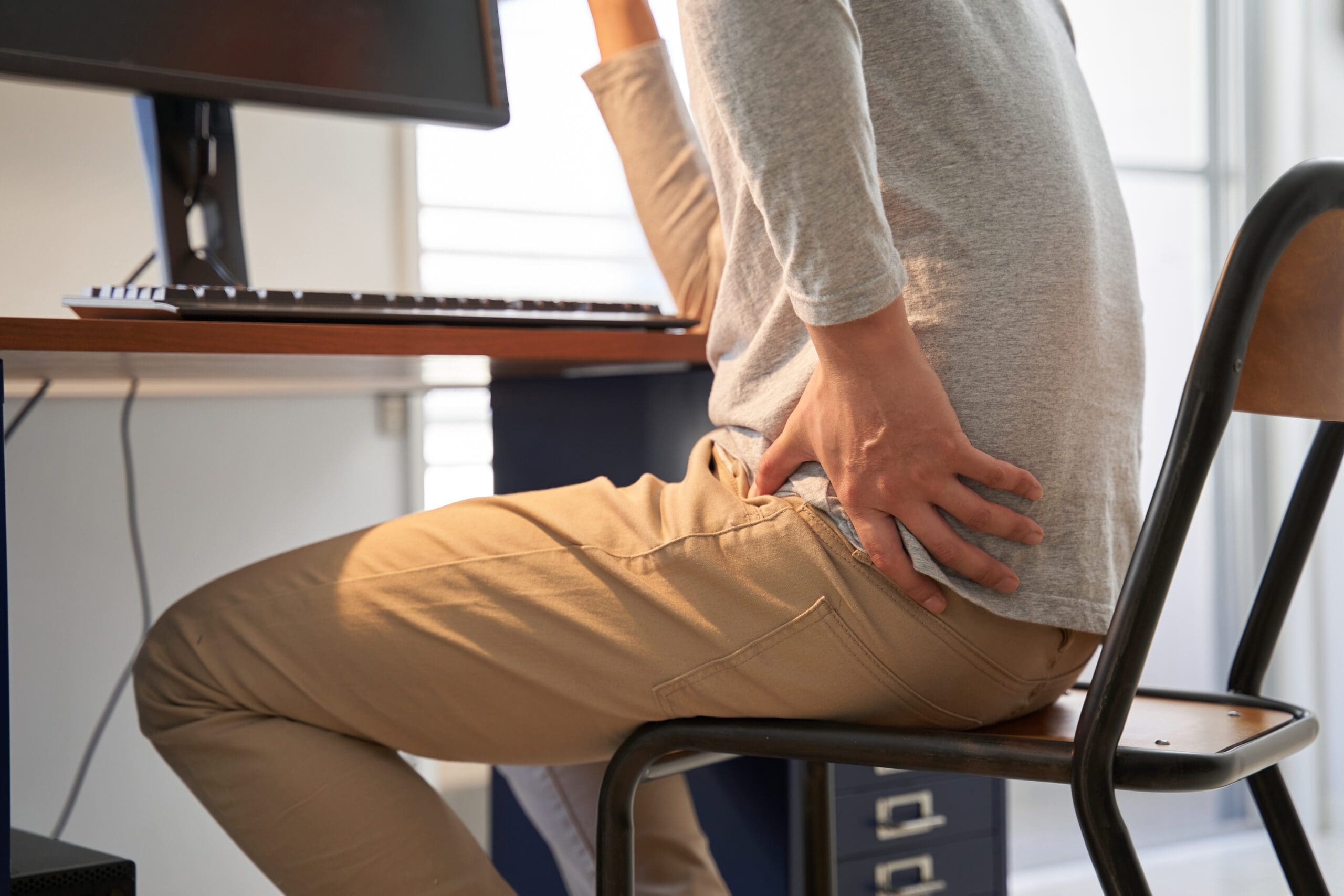
Case Study: A 45-year-old office worker experiences shoulder pain from poor ergonomics (static posture) combined with stress, leading to muscle knots and joint misalignment. Over time, this evolves into chronic upper back pain, affecting daily life.
Environmental Factors Contributing to Pain Development
Environmental factors are crucial in the onset and progression of pain, especially in muscles and joints. These factors interact with biology, making some people more susceptible.
Weather and Climate Influences
Weather changes significantly impact pain. Low temperatures constrict blood vessels, reducing flow to muscles and causing stiffness. High humidity increases joint fluid pressure, leading to swelling and ache (Arthritis Foundation, 2024). Barometric pressure drops before storms can trigger migraines or joint pain by altering tissue expansion.
Examples:
In osteoarthritis, patients report worse knee pain during cold, damp weather due to increased joint rigidity (PMC, 2025a).
Fibromyalgia sufferers experience muscle flares from temperature swings, with cold lowering pain thresholds by 11.3°C compared to healthy individuals (PMC, 2025a).
Studies show modest correlations between pain and humidity, pressure, and wind speed (Arthritis Foundation, 2024). For muscles, cold induces spasms; for joints, humidity exacerbates inflammation.
Stress and Psychosocial Elements
Stress releases cortisol, promoting inflammation that affects muscles and joints. Chronic stress from work or life events heightens pain perception, leading to tension headaches or back pain (MDPI, 2022). Low social support or discrimination correlates with thicker brain structures involved in pain processing, such as the insula, making discomfort more intense (Nature, 2024).
Examples:
Job insecurity causes muscle tension in the neck and shoulders, evolving into chronic pain.
Discrimination experiences are associated with greater hippocampal volume and greater pain in patients with knee osteoarthritis (Nature, 2024).
Pollution and Toxins
Air pollution, including particulates and toxins such as acrolein, increases inflammation, worsening joint pain in rheumatic diseases (ScienceDirect, 2024a). Smoking aggravates arthritis by activating immune cells, predicting higher pain in spinal injuries (ScienceDirect, 2024a).
Examples:
Urban dwellers exposed to pollution have more emergency visits for joint pain.
Vitamin D deficiency due to reduced sunlight exposure is associated with muscle hypersensitivity (ScienceDirect, 2024a).
Work and Lifestyle Environments
Poor ergonomics, such as prolonged sitting, strains muscles and joints, leading to musculoskeletal pain (MDPI, 2022). Repetitive tasks lead to back pain by reducing movement variability (IASP, n.d.a).
Examples:
Factory workers develop joint pain from repetitive lifting.
Sedentary lifestyles in air-conditioned offices can cause dry-air-related stiffness.
Sociocultural Factors
Lower income and education are associated with higher pain levels due to limited access to healthy environments (Nature, 2024). Household size and employment status explain variance in pain-related brain structures.
To mitigate, strategies include weather-appropriate clothing, stress management, and pollution avoidance. Tables below summarize factors.
Environmental Factor
Description
Impact on Muscles
Impact on Joints
Examples
Weather (Temperature)
Changes in ambient heat/cold
Constriction, spasms
Stiffness, reduced mobility
OA knee pain in cold
Humidity
High moisture levels
Swelling, tension
Fluid pressure increase
Arthritis flares in damp weather
Stress
Psychosocial pressures
Tension, knots
Inflammation from cortisol
Neck pain from job stress
Pollution
Air toxins
Inflammation, hypersensitivity
Rheumatic exacerbations
Joint pain in urban areas
Work Conditions
Ergonomic issues
Strain, fatigue
Misalignment
Back pain from sitting
Deeper Dive into Physicochemical Factors
Physicochemical factors, such as pollution and toxins, directly alter pain pathways. Air pollution exacerbates neuropathic pain by sensitizing nerves (ScienceDirect, 2024a). Toxic compounds such as 4-HNE activate receptors, triggering neurogenic inflammation in joints.
Biological factors, such as viral infections, lead to arthritis-like joint pain (ScienceDirect, 2024a). Smoking induces hyperalgesia through serotonergic changes.
Psychosocial factors, such as stress, promote chronicity, while environmental enrichment reduces pain by lowering stress (ScienceDirect, 2024a).
Case Study: A patient with rheumatoid arthritis experiences worse joint pain during pollution spikes, managed by indoor air filters and an anti-inflammatory diet.
Categories of Pain: Descriptions and Examples
Pain is classified by duration, cause, and location to guide treatment (Healthline, 2018).
Acute Pain
Short-term, lasting days to weeks, from injury. Sharp or intense, it alerts the body (Healthline, 2018).
Examples:
Muscle strain from lifting heavy objects.
Joint pain from a sprained ankle.
Chronic Pain
Lasts months or years, often without a clear cause. Mild to severe, impacting life (Healthline, 2018).
Examples:
Low back pain from poor posture.
Arthritis causes ongoing joint pain.
Nociceptive Pain
From tissue damage, activating nociceptors. Acute or chronic (WebMD, 2025).
Subtypes:
Somatic: Skin, muscles, bones. Aching or throbbing.
Examples: Muscle pull, joint fracture.
Visceral: Organs. Dull, cramping.
Examples: Appendicitis, but it can also refer to muscle pain.
Neuropathic Pain
From nerve damage. Burning, tingling (WebMD, 2025).
Examples:
Diabetic neuropathy in the feet (joint-related).
Sciatica from spinal nerve compression (muscle/joint).
Other Categories (IASP Definitions)
Allodynia: Pain from non-painful stimuli, e.g., light touch on sunburned muscle (IASP, 2022).
Hyperalgesia: Amplified pain from normal stimuli, e.g., pinprick on an inflamed joint.
Nociplastic Pain: Altered nociception without damage, e.g., fibromyalgia muscle pain.
Category
Duration
Cause
Sensation
Muscle/Joint Example
Acute
Short
Injury
Sharp
Strained hamstring
Chronic
Long
Ongoing
Dull
Chronic knee arthritis
Nociceptive Somatic
Varies
Tissue
Aching
Bone fracture joint pain
Neuropathic
Varies
Nerve
Burning
Sciatica leg muscle
Nociplastic
Chronic
Altered processing
Widespread
Fibromyalgia joint tenderness
Case Study: An athlete with acute nociceptive pain from a joint sprain transitions to chronic pain if left untreated, demonstrating category evolution.
Exploring Integrative Medicine- Video
Pain Management in Clinical Settings
Healthcare specialists follow evidence-based rationale for pain management, emphasizing multimodal approaches to minimize risks like addiction (SAMHSA, 2024). The MATE Act requires training on safe prescribing, focusing on opioid use disorders and pain treatment (DEA, 2023).
Non-Surgical Therapies
These are first-line for many, using meds, therapy, and complementary methods.
Medications: NSAIDs for inflammation, acetaminophen for mild pain (NEJM, 2019).
Physical Therapy: Exercises strengthen muscles and improve joint mobility.
Complementary: Acupuncture and massage reduce tension (PMC, 2024).
Examples: Massage post-injury eases muscle tension; breathing techniques lower anxiety in the clinic.
Surgical Therapies
For severe cases, such as joint replacement. Post-op management includes multimodal analgesia (JAMA, 2021).
Opioids: Short-term for breakthrough pain.
Non-Drug: Music therapy reduces opioid needs by 31% (PMC, 2024).
Rationale: Balances relief with safety, per guidelines (DEA, 2023).
Therapy Type
Examples
Benefits
Clinical Rationale
Non-Surgical Meds
NSAIDs
Reduce inflammation
Low risk for chronic pain
Physical Therapy
Exercises
Strengthen muscles
Prevents long-term weakness
Surgical Post-Op
Opioids + Music
Pain relief
Minimizes addiction risk
Case Study: Patient with joint pain undergoes non-surgical acupuncture, avoiding surgery.
Insights from Dr. Alexander Jimenez
Dr. Jimenez, with 30+ years in chiropractic and functional medicine, observes correlations such as perimenopausal estrogen drops causing joint pain or TBI leading to posture issues and muscle aches (LinkedIn, n.d.; DrAlexJimenez.com, n.d.).
His integrative approach addresses causes:
Targeted Exercise: Rehab programs build strength and prevent recurrence.
Massage Therapy: Relieves soft tissue tension.
Acupuncture: Promotes healing in sciatica.
Prevents long-term problems through nutrition and monitoring (DrAlexJimenez.com, n.d.).
Case Study: TBI patient regains mobility via chiropractic adjustments and exercises.
Prevention and Future Trends
Prevention involves environmental awareness, regular physical activity, and a healthy diet. Future trends include wearables for trigger monitoring.
This guide, drawing from diverse sources, shows pain as manageable with informed care.
Conclusion: Embracing a Future Free from Chronic Pain
Pain is more than just a temporary annoyance; as we’ve seen in this detailed guide, it’s a complex signal that is affected by biology, the environment, and lifestyle, from the ways that barometric pressure and pollution can cause inflammation in muscles and joints to the different types of pain, such as nociceptive, neuropathic, and nociplastic pain, knowing these things gives us power. The clinical rationale for pain management, as delineated in the 2024 guidelines (American College of Surgeons et al., 2024), underscores the need for proactive, multimodal approaches in both surgical and non-surgical contexts to mitigate symptoms and avert progression to chronic conditions.
Healthcare professionals are very important here because they use tools like ERAS protocols to help people recover after surgery and integrative therapies to help people feel better every day. Based on Dr. Alexander Jimenez’s clinical observations in El Paso, we know that addressing the root causes of pain through chiropractic adjustments, targeted exercises, massage therapy, and acupuncture can help the body heal naturally and prevent long-term problems. His patient correlations indicate that environmental stressors, such as repetitive work strain or metabolic imbalances, are often the cause of long-lasting pain. However, personalized, evidence-based care can help with these stressors.
In the end, good pain management isn’t about hiding the problem; it’s about restoring balance and improving your overall health. You can stop the cycle of pain by knowing what causes it in your environment, correctly categorizing your pain, and looking for holistic treatments. If you’ve hurt yourself recently or have had joint pain for years, remember that you can have a life with more mobility, energy, and health if you make smart choices and get professional help. Talk to a professional today, put your personal action plan into action, and look forward to a better, less painful tomorrow.
Discover the connection between head injuries and somatovisceral disorders to enhance patient care and management.
Understanding Head Injuries and Their Impact on the Brain-Body Connection: A Comprehensive Guide to Somatovisceral Disorders and Non-Surgical Treatment Approaches
Millions of people worldwide are impacted by head injuries every year, making them a serious public health problem. The harm that results from head trauma, whether from a fall, auto accident, or sports collision, goes much beyond the location of the original hit. Researchers now identify somatovisceral illnesses as a result of these injuries, which cause a series of physiological alterations that interfere with the delicate brain-body communication system. Recovery outcomes and quality of life may be significantly improved by understanding how head trauma impacts this crucial brain-body link and by investigating effective non-surgical therapeutic options.
What Are Somatovisceral Disorders?
Complex connections between the body’s internal organs (visceral system) and physical structures (somatic system) are a feature of somatovisceral illnesses. Nerve impulses from body structures are transmitted to visceral organs through this complex process, resulting in specific physiological or pathological responses. In addition to involving two systems, the somatovisceral response is complicated because it may communicate in both directions, transferring information from somatic structures to visceral organs and vice versa. foundationhealth
Medical studies have focused more on the connection between somatovisceral diseases and brain trauma. According to a recent study, 15–27% of patients who had head trauma fulfilled the criteria for somatic symptom disorder six months after the injury, suggesting that mild traumatic brain injury (mTBI) may be a frequent precursor to this syndrome. This link demonstrates how brain damage may disrupt the normal communication pathways that control physiological processes, leading to chronic, often incapacitating symptoms throughout the body. neurologyopen.bmj
When people have upsetting physical symptoms together with excessive thoughts, emotions, or actions associated with those symptoms, it’s known as somatic symptom disorder. Many somatic problems, such as pain, weakness, difficulty moving, headaches, dizziness, excessive fatigue, changes in vision or hearing, itching, numbness, odd movements, stomach pain, and nausea, are often reported by patients after a brain injury. These symptoms illustrate how neurological impairment may materialize as pervasive physical dysfunction by reflecting the disturbed connection between the brain and many bodily systems. chop+1
The Brain-Body Connection and Head Injury
The human nervous system operates through an intricate network that connects the brain to every organ, muscle, and tissue in the body. This communication highway relies on precise signaling between the central nervous system (brain and spinal cord) and the peripheral nervous system (nerves throughout the body). When head trauma occurs, this delicate communication system can become disrupted at multiple levels, affecting both somatic (voluntary) and autonomic (involuntary) nervous system functions.
According to Dr. Alexander Jimenez, a board-certified Family Practice Nurse Practitioner and Doctor of Chiropractic in El Paso, Texas, the spine houses the spinal cord, which acts as the communication superhighway between the brain and body. Any misalignment in the spine can disrupt the nervous system’s signals, and for traumatic brain injury patients, this connection becomes crucial. Dr. Jimenez explains that misalignment caused by the injury itself or associated whiplash can worsen symptoms like headaches, brain fog, and balance issues, emphasizing the importance of addressing both cranial and spinal components in recovery. northwestfloridaphysiciansgroup
The brain-body disconnect following trauma manifests as disrupted somatic sensory processing, encompassing vestibular (balance) and somatosensory (touch, pressure, temperature) processing. These sensory systems are primarily concerned with survival and safety, given the potential consequences of impaired balance or diminished awareness of physical threats. Following a head injury, trauma-related symptoms are conceptualized to be grounded in brainstem-level somatic sensory processing dysfunction and its cascading influences on physiological arousal modulation, affect regulation, and higher-order capacities. pmc.ncbi.nlm.nih
Research has identified that traumatic conditions may manifest as disrupted vertical integration, in which the balance between lower brain regions and higher cortical areas becomes dysregulated, particularly within the midline neural circuitry responsible for generating a primordial sense of a bodily and affective self as a coherent and stable entity in relation to the environment. This alteration has a cascading impact on the horizontal integration of cortical brain structures, meaning that different regions of the brain may be structurally intact yet lack fluid communication. pmc.ncbi.nlm.nih
Autonomic Dysfunction After Head Injury
One of the most significant yet underappreciated consequences of head injury is autonomic nervous system dysfunction. The autonomic nervous system controls involuntary bodily functions, including heart rate, blood pressure, digestion, breathing, and temperature regulation. Following moderate-to-severe traumatic brain injury, patients often experience significant autonomic dysfunction affecting both sympathetic (fight-or-flight) and parasympathetic (rest-and-digest) branches of this critical system. neurologyopen.bmj
Studies have demonstrated that patients with severe traumatic brain injury can experience sympathetic hyperactivity in the acute stages. More importantly, autonomic dysfunction persists in many patients for months or even years after their initial injury, affecting fully ambulant patients whom many might assume to be fully recovered. This persistent dysfunction occurs through various mechanisms, with the hallmark of moderate-to-severe traumatic brain injury being white matter injury caused by axonal shearing due to injury forces, continuing due to inflammation and delayed axonal degeneration in the chronic period, resulting in network disruption. neurologyopen.bmj
Autonomic dysfunction may occur due to injury to regions of the central autonomic network or their connecting white matter tracts. Brainstem nuclei and white matter connections to and from thalamic and basal ganglia regions may be particularly vulnerable to damage, underlying dysfunction that contributes to cognitive impairment post-traumatic brain injury. Given the importance of brainstem, thalamic, and basal ganglia circuits to autonomic function, injury to these white matter tracts may cause centrally mediated autonomic dysfunction. neurologyopen.bmj
The clinical manifestations of autonomic dysfunction after head injury are diverse and often debilitating. Many classic symptoms following concussion are, at least in part, likely a result of injury to the autonomic nervous system. Cognitive difficulties seen after mild traumatic brain injury may be related to autonomic dysregulation, specifically impaired cerebral blood flow. The presence of autonomic dysfunction has been shown to correlate with increased morbidity and mortality in moderate and severe traumatic brain injury, with perturbations of the autonomic nervous system consisting of either increased sympathetic or reduced vagal activity, potentially resulting in serious cardiac complications. health+1
Dr. Jimenez’s clinical practice emphasizes the importance of recognizing autonomic dysfunction in patients recovering from head injuries. His functional medicine approach includes detailed health assessments evaluating lifestyle, environmental exposures, and psychological factors to understand the root causes of chronic disorders and treat patients holistically. This comprehensive evaluation is particularly important for identifying autonomic dysfunction, which may manifest as dizziness, balance problems, temperature dysregulation, digestive issues, and cardiovascular irregularities.
Environmental Factors Affecting Brain Activity and the Body
Environmental factors play a critical role in shaping brain structure and function, as well as the development of mental and physical health conditions. The macroenvironment encompasses immediate factors such as air, noise, and light pollution; proximal factors, including regional socioeconomic characteristics; and distal factors, such as urbanization, natural spaces, and climate. These environmental exposures are mostly modifiable, presenting opportunities for interventions and strategies to promote the structural and functional integrity of the brain and mitigate the burden of illness following head injury. nature
Air pollution has emerged as a significant concern for brain health, particularly following traumatic brain injury, when the brain is already vulnerable. Studies have demonstrated that air pollution may increase vulnerability to mood dysfunction and potentially inhibit an appropriate stress response. Prolonged exposure to fine particulate matter (PM2.5 and PM10) has been associated with negative stress-related brain activation in the prefrontal cortex, frontoinsular cortex, limbic system, inferior parietal cortex, and cingulate regions. Magnetic resonance imaging studies reveal that increased exposure to PM2.5 is associated with changes in brain structure in older adults, including brain atrophy, that occur before the onset of dementia. environmentalhealth.ucdavis+1
Noise pollution, originating from urban traffic, airports, industries, and construction sites, can evoke negative emotions and disrupt recovery following head injury. Prolonged exposure to disruptive noise induces brain alterations through mechanisms such as sleep disturbances, which create a pro-oxidative environment that predisposes to neuroinflammation and heightened hypothalamic-pituitary-adrenal axis reactivity, contributing to mental and physical health problems. For individuals recovering from head trauma, protecting against excessive noise exposure becomes particularly important as the injured brain requires optimal conditions for healing. nature
Light pollution and exposure to artificial light at night have become increasingly prevalent, especially in urban areas, disrupting natural darkness and circadian rhythms. Light is detected by the retina and transmitted through intrinsically photosensitive retinal ganglion cells to the suprachiasmatic nucleus in the hypothalamus and other brain regions involved in regulating circadian rhythms and sleep-wake cycles. Circadian rhythm disruptions have been linked to elevated risk of mood disorders, bipolar disorders, and heightened mood instability, potentially mediated by oscillations in clock gene expression responsive to light-dark transitions. nature
Following traumatic brain injury, circadian rhythm disruptions become even more pronounced. Research has documented that traumatic brain injury can lead to decreased melatonin release, causing circadian rhythm delays. Studies using animal models have revealed that acute subdural hematoma resulted in dysregulation of circadian gene expression and rhythmic changes in body temperature during the first 48 hours post-injury. The regulation of biological rhythms through changes in core body temperature, pineal gland melatonin secretion, and blood cortisol levels becomes disrupted, affecting the body’s ability to anticipate and adapt to environmental changes. practicalneurology+1
Minor traumatic brain injury contributes to the emergence of circadian rhythm sleep disorders, with research identifying two distinct types: delayed sleep phase syndrome and irregular sleep-wake pattern. These disorders differ in subjective questionnaire scores and have distinct profiles of melatonin and temperature circadian rhythms. The alteration in the circadian timing system partially accounts for the presence of post-traumatic brain injury sleep-wake disturbances, which changes in sleep architecture alone cannot fully explain. pubmed.ncbi.nlm.nih+1
Understanding Long-Lasting Injuries- Video
How Head Injuries Affect Daily Tasks and Routines
The impact of head injuries extends far beyond the initial trauma, profoundly affecting an individual’s ability to perform everyday activities and maintain normal routines. The disruption to brain-body communication creates challenges across multiple domains of daily functioning, from basic self-care tasks to complex cognitive and social activities. Understanding these impacts helps patients, families, and healthcare providers develop realistic expectations and appropriate support strategies during recovery.
Cognitive fatigue represents one of the most disabling consequences of traumatic brain injury, affecting 21-73% of patients regardless of injury severity or time since injury. Fatigue has been identified as the main cause of disability after traumatic brain injury, negatively affecting social, physical, and cognitive functions as well as participation in daily activities and social life. At the neural level, patients with fatigue following head injury exhibit significant disruption of global resting-state alpha-band functional connectivity between cortical midline structures and the rest of the brain. Furthermore, individuals with fatigue show reduced overall brain activation during cognitive tasks, without time-on-task effects. academic.oup
Adults with a history of even mild traumatic brain injury report significantly greater fatigue and cognitive impairment than those with no history of head trauma, with symptoms becoming more profound with greater injury severity. This persistent fatigue affects the ability to maintain attention, concentrate on tasks, process information efficiently, and sustain mental effort throughout the day. Patients frequently report that activities requiring cognitive engagement become increasingly difficult as the day progresses, leading to a pattern of morning productivity followed by afternoon exhaustion. pubmed.ncbi.nlm.nih+1
Memory difficulties present another significant challenge affecting daily functioning after a head injury. Patients may struggle with both short-term working memory (holding information in mind while using it) and long-term memory formation (creating new lasting memories). These memory challenges affect practical tasks such as remembering appointments, following multi-step instructions, recalling conversations, and learning new information or skills. The impact extends to occupational functioning, with studies finding a correlation between higher levels of mental fatigue and lower employment status following traumatic brain injury. headway+1
Executive function impairments following head injury affect planning, organization, decision-making, problem-solving, and behavioral regulation. These higher-order cognitive processes are essential for managing daily responsibilities, from planning meals and organizing household tasks to managing finances and making important life decisions. Patients may find themselves struggling with tasks that previously seemed automatic, requiring conscious effort and external supports to maintain daily routines. headway
Sensory processing alterations create additional challenges for daily functioning. The vestibular system, which contributes to balance, spatial processing, arousal modulation, first-person perspective, and social cognition, becomes particularly vulnerable following head trauma. Disturbed temporal binding of sensory information creates perceptual chaos and lack of coherence, which may lead to bodily disconnect and states of hypervigilance. Patients describe feeling disconnected from their bodies, experiencing the world as if through a fog, or feeling constantly on guard against potential threats. pmc.ncbi.nlm.nih
Balance and coordination problems stemming from vestibular dysfunction affect mobility and safety in daily activities. Simple tasks like walking on uneven surfaces, turning the head while moving, or navigating busy environments become challenging and potentially dangerous. Many patients report increased anxiety about falling, leading to activity restriction and social withdrawal. Over one-third of adults over 40 will experience vestibular dysfunction at some point in their lives, and when it occurs, whether by injury, aging, or disease, individuals can experience vertigo, nauseating dizziness, vision and balance problems affecting every area of life. neuroinjurycare+1
Dr. Jimenez’s practice in El Paso focuses extensively on helping patients restore function and return to daily activities following head injuries. His integrated approach combines chiropractic care, functional medicine, and rehabilitation therapies to address the multiple systems affected by head trauma. By evaluating the connections between physical, nutritional, and emotional factors, Dr. Jimenez develops personalized care plans that recognize the complex ways head injuries disrupt daily functioning and quality of life.
Overlapping Risk Profiles and Symptoms Associated With Head Injuries
Head injuries create overlapping risk profiles affecting multiple body systems simultaneously, leading to complex symptom presentations that can challenge both patients and healthcare providers. Understanding these interconnected risk factors and symptoms is essential for comprehensive assessment and treatment planning. Individuals who sustain head injuries develop an increased risk for somatic symptom disorder, with early illness beliefs playing a significant predictive role. Specifically, believing that mild traumatic brain injury has serious life consequences and causes distress in the weeks following injury is associated with later development of somatic symptom disorder. Patients with somatic symptom disorder after head injury report more pain and post-concussion symptoms and are significantly more likely to have comorbid major depressive disorder and anxiety disorders compared to those without this condition. neurologyopen.bmj
The systematic review examining the relationship between somatic symptoms and related disorders and mild traumatic brain injury found that the majority of acceptable evidence supported a relationship between these conditions. Nine studies reported associations between functional seizures and a history of mild traumatic brain injury, while 31 studies assessed relationships between questionnaires measuring somatic symptom disorder burden and mild traumatic brain injury. Three studies investigated healthcare practitioners’ diagnosis of somatic symptoms and related disorders and post-mild traumatic brain injury symptom burden, collectively demonstrating the strong connection between head trauma and subsequent development of somatic complaints. foundationhealth
Cardiovascular complications represent another significant overlapping risk following head injury. Research demonstrates that individuals with moderate-to-severe traumatic brain injury have increased rates of self-reported hypertension and stroke but lower rates of myocardial infarction and congestive heart failure than uninjured adults. The findings highlight the importance of early screening for and management of cardiovascular risk factors in individuals with chronic traumatic brain injury, particularly those of younger age, not typically thought to be at risk for these conditions. ahajournals
The relationship between blood pressure and traumatic brain injury follows a complex U-shaped pattern, with both hypotension and hypertension associated with worse outcomes. Early hypotension has been linked with poor outcomes following severe traumatic brain injury, but recent data suggest that arterial hypertension after injury is also associated with poor outcomes. The initial catecholamine response and resulting systemic hypertension may be protective to a point by maintaining cerebral perfusion pressure in the setting of impaired cerebral autoregulation after injury, yet catecholamine-induced hypertension may also cause secondary brain damage by aggravation of vasogenic edema and intracranial hypertension. pmc.ncbi.nlm.nih
Post-traumatic headaches affect approximately 40% of individuals who experience concussions, representing one of the most common and persistent symptoms following head injury. Patients can experience tension headaches, migraine headaches, and cervicogenic headaches (radiating from the neck) all at once, making treatment particularly challenging. Ninety-five percent of people with a concussion experience headache associated with that injury, and among those with headache, about two-thirds have migraine features. Individuals with a family history of migraine or preexisting headache disorders face a higher risk of developing post-traumatic headache. wexnermedical.osu+1
Sleep disturbances cluster with other post-traumatic brain injury symptoms, creating compounding difficulties for recovery. Changes in sleep architecture following injury cannot fully explain the extent and intensity of sleep-wake disturbances reported by patients. The current literature supports cognitive-behavioral therapy and sleep hygiene education, light therapy, and certain pharmacologic interventions for treating sleep disturbances in patients with brain injury, with early screening and individualized approaches prioritized to improve sleep and, consequently, speed recovery. pubmed.ncbi.nlm.nih
Exercise intolerance commonly results from a concussion, often limiting return to activities and quality of life. The reviewed studies support clinical suspicion of autonomic dysfunction as an important component of exercise intolerance, though specific mechanisms of impairment and relationships to symptoms and recovery require additional investigation. Post-concussive exercise intolerance has been linked to a reduction in cerebral blood flow, theoretically prolonging the effects of the metabolic energy crisis associated with injury. pmc.ncbi.nlm.nih
Mental health complications, including anxiety, depression, post-traumatic stress disorder, and behavioral changes, frequently develop following head injury. Brain injuries, no matter how severe, commonly cause emotional and behavioral changes, including emotional lability with extreme mood swings, anxiety disorders, depression, impulsive behaviors, flat affect causing a lack of emotional expression, and a lack of empathy and social skills. These psychological changes can cause unnecessary suffering and, in cases of severe depression and anxiety, can even halt physical recovery progress. flintrehab
Non-Surgical Treatments to Improve Somatovisceral Function
Fortunately, numerous non-surgical treatment approaches have demonstrated effectiveness in improving somatovisceral function and promoting recovery following head injuries. These interventions work through various mechanisms to restore proper communication between the brain and the body, balance the autonomic nervous system, and support the brain’s natural healing processes. Dr. Jimenez’s clinical practice emphasizes comprehensive non-invasive protocols, prioritizing natural recovery and avoiding unnecessary surgeries or medications.
A Questionnaire Example of TBI Symptoms
Chiropractic Care and Spinal Adjustments
Chiropractic care focuses on the spine and nervous system, recognizing that the spine houses the spinal cord, which acts as the communication superhighway between the brain and body. For traumatic brain injury patients, proper spinal alignment becomes crucial because misalignment caused by the injury itself or associated whiplash can worsen symptoms like headaches, brain fog, and balance issues. Chiropractic care aims to restore proper alignment, thereby improving nervous system function and supporting the brain’s ability to heal. northwestfloridaphysiciansgroup Chiropractic adjustments help alleviate post-traumatic brain injury symptoms by releasing pressure on irritated nerves and improving joint function. For many patients, this results in improved comfort and reduced reliance on pain medication. Proper spinal alignment promotes better blood flow to the brain, and since the brain requires oxygen-rich blood to heal and function, improved circulation directly supports recovery from traumatic brain injury while reducing dizziness and fatigue. northwestfloridaphysiciansgroup
Research demonstrates that chiropractic intervention can modify proprioceptive input from more functional spinal joints, helping restore this input to the brain’s multisensory integration centers. Studies of patients receiving chiropractic care in neurorehabilitation hospitals have shown that spinal manipulation influences pain through complex mechanisms in the central nervous system. A case study documenting concussion treatment using massage and manipulation techniques showed diminished concussion symptoms and regained ease in cervical range of motion, highlighting the potential importance of manual therapy work to reduce headache, dizziness, and nausea in concussion recovery. pmc.ncbi.nlm.nih+2 Dr. Jimenez explains that by realigning the spine through chiropractic adjustments, treatment reduces nerve interference, optimizing mind-body communication, and enhancing overall function. The adjustments improve cerebral blood flow and reduce inflammation, thereby accelerating recovery from head injury. With enhanced nervous system function comes improved mental clarity, including reduced brain fog, sharper focus, and better memory, while also promoting stress relief and alleviating irritability and emotional strain often linked to head injuries. zakerchiropractic
Vestibular Rehabilitation
Vestibular rehabilitation is a specialized form of physical therapy that focuses on strengthening the connections between the brain, eyes, inner ear, muscles, and nerves. This treatment approach proves particularly valuable for post-concussion patients experiencing dizziness, vertigo, balance problems, and spatial impairment. According to a review in the British Journal of Medicine, vestibular therapy reduced symptoms in patients with sports-related concussions faster, with patients three times as likely to return to play within eight weeks of therapy compared to those who didn’t receive treatment. denverphysicalmedicine+1 Vestibular rehabilitation therapy involves exercises designed to improve the functioning between the inner ear, brain, eyes, muscles, and nerves. These exercises help minimize balance issues and treat dizziness, vertigo, and spatial orientation deficits caused by vestibular impairments that some individuals experience after brain injury. The therapy addresses issues in the inner ear through specific exercises designed to improve balance and coordination. biausa
The Epley Maneuver represents a simple yet effective exercise to treat benign paroxysmal positional vertigo, a very specific form of vertigo quite common after traumatic brain injury. During vestibular rehabilitation, benign paroxysmal positional vertigo generally responds well to the Epley Maneuver, and patients learn to perform the movement at home to alleviate symptoms as they arise. Studies have shown that vestibular rehabilitation is an effective modality for managing dizziness, vertigo, and imbalance following concussion, though careful consideration of the injury’s acuity and effective management of co-morbid conditions will optimize results. pubmed.ncbi.nlm.nih+1 Co-morbidities, including cognitive and behavioral issues, visual-perceptual dysfunction, metabolic dysfunction, and autonomic dysfunction, may hamper the effectiveness of traditional vestibular rehabilitation approaches. Working closely with other disciplines well-versed in treating these co-morbid issues helps individuals obtain optimal recovery. Dr. Jimenez’s integrated practice model exemplifies this multidisciplinary approach, bringing together chiropractic care, functional medicine, physical therapy, and other specialties to provide comprehensive treatment for patients with vestibular dysfunction following head injuries. pubmed.ncbi.nlm.nih
Physical Therapy and Exercise Rehabilitation
Physical therapy plays a pivotal role in optimizing recovery and enhancing functional independence after brain injury. Therapeutic approaches include gait training to improve walking patterns, balance activities to enhance stability and prevent falls, strength training to rebuild muscle mass and function, coordination exercises to improve fine and gross motor skills, and range-of-motion exercises to maintain flexibility. biausa In some cases, physical therapists recommend body-weight-supported treadmill training to help patients safely relearn walking patterns. Family and caregiver training proves extremely important and helpful, as loved ones can gain an understanding of how the brain works and the specific nature of the injury, supporting the rehabilitation process. biausa
Available evidence demonstrates the potential of exercise in improving cognitive impairment, mood disorders, and post-concussion syndrome following traumatic brain injury. Exercise rehabilitation has been shown to attenuate cognitive deficits in animal models by stimulating cerebral signaling pathways, with treadmill exercise improving memory by modulating neurotransmitter systems and neurotrophic factors. High-intensity interval training helps regulate the autonomic nervous system while boosting brain-derived neurotrophic factor, thereby promoting neuroplasticity, an essential factor for recovery. sciencedirect+1 However, exercise prescription following head injury requires careful consideration, as exercise intolerance commonly results from concussion and autonomic dysfunction. Graded exercise testing while monitoring symptoms and heart rate helps guide a safe return to physical activity. Current clinical practice involves careful assessment to determine appropriate exercise intensity and duration, gradually progressing as autonomic function improves. pmc.ncbi.nlm.nih
Acupuncture and Neuroplasticity Enhancement
Acupuncture has gained widespread recognition as an effective, low-cost treatment for neurological rehabilitation with minimal adverse effects. Clinical and experimental evidence documents the potential of acupuncture to ameliorate injury-induced neurological deficits, particularly sequelae such as dyskinesia, spasticity, cognitive impairment, and dysphagia. These effects relate to acupuncture’s ability to promote spontaneous neuroplasticity after injury. pmc.ncbi.nlm.nih+1 Specifically, acupuncture can stimulate neurogenesis, activate axonal regeneration and sprouting, and improve the structure and function of synapses. These processes modify the neural network and the function of the damaged brain area, leading to improvements in various skills and adaptability. Astrocytes and microglia may be involved in acupuncture-induced regulation of neuroplasticity, for example, by producing and releasing various neurotrophic factors, including brain-derived neurotrophic factor and nerve growth factor. pmc.ncbi.nlm.nih
Studies have shown that acupuncture reduces neuroinflammation after brain injury, with research published in The Journal of Neuroinflammation finding that acupuncture significantly reduced neuroinflammation and improved cognitive function in animal models of brain injury. By modulating inflammatory pathways, acupuncture helps reduce the production of pro-inflammatory cytokines, promoting brain healing and reducing symptoms such as headaches and dizziness. betsygordonacupuncture Acupuncture enhances neuroplasticity, which is crucial for recovery after brain injury, promoting improvements in memory, learning, and overall cognitive function. Research in Neural Regeneration highlighted that acupuncture promotes neuroplasticity, which is essential for rehabilitation. Studies demonstrate that acupuncture improves cognitive performance and reduces anxiety and depression in patients recovering from brain injuries. betsygordonacupuncture+1 Dr. Jimenez’s functional medicine practice incorporates acupuncture and electro-acupuncture as part of comprehensive care plans for patients recovering from head injuries. His team uses these modalities in combination with other therapies to create customized treatment approaches that promote natural healing, mobility, and long-term wellness.
Nutritional Interventions and Functional Medicine
Nutrition plays a positive role during acute traumatic brain injury recovery, with patient needs being unique and requiring individualized approaches. Following mild traumatic brain injury, patients who consumed enough food to meet calorie and macronutrient (particularly protein) needs specific to their injury severity and sex within 96 hours post-injury had reduced length of hospital stay. Patients receiving nutrients and non-nutrient support within 24-96 hours post-injury had positive recovery outcomes, including omega-3 fatty acids, vitamin D, magnesium oxide, N-acetyl cysteine, and hyperosmolar sodium lactate. frontiersin Traumatic brain injury contributes to extensive dysbiosis of the gastrointestinal system, leading to worsened outcomes, making nutritional support essential. Early nutrition supports preservation of muscle mass, decreases infection complications, promotes cerebral homeostasis, and improves recovery outcomes. The human brain consumes 20% of total resting energy, despite accounting for only 2% of total body mass, underscoring the critical role of adequate nutrition for healing. xiahepublishing
A recent clinical trial demonstrated that dietary changes significantly reduce persistent post-traumatic headaches, a common and debilitating consequence of traumatic brain injury. Researchers found that increasing omega-3 fatty acids (commonly found in fatty fish) while reducing omega-6 fatty acids (abundant in seed oils) led to fewer and less severe headaches. Participants assigned to the intervention diet experienced approximately two fewer headache days per month and a 30% reduction in daily headache pain intensity compared to the control diet group. med.unc Supplementing with omega-3 fatty acids can reduce inflammation and oxidative stress, promote brain-cell survival, and help the brain recover from injury. Vitamins D and E, niacin, zinc, and magnesium have neuroprotective benefits, and supplementing with these vitamins and minerals has been shown to improve recovery, especially in patients who are deficient. An energy-balanced, anti-inflammatory diet with adequate sources of omega-3 fats and appropriate vitamin D supplementation proves especially important for patients with a history of traumatic brain injury. consultant360
Dr. Jimenez’s practice embraces Functional Integrative Medicine, a patient-focused approach that treats the whole person rather than just symptoms. His team offers detailed health assessments that evaluate genetics, lifestyle, environmental exposures, and psychological factors to create comprehensive health profiles. By combining Institute for Functional Medicine programs with personalized nutrition plans, Dr. Jimenez helps patients address chronic conditions and optimize brain health following head injuries.
Massage Therapy and Manual Techniques
Massage therapy provides valuable support in brain injury rehabilitation, offering benefits for physical, mental, and emotional well-being. Massage significantly improves blood circulation, ensuring that essential nutrients and oxygen are efficiently delivered to brain cells. By increasing circulation, the brain’s healing process is expedited, promoting cellular regeneration and reducing the risk of secondary complications. Improved blood flow also helps reduce swelling and inflammation, common challenges following brain injury. neuropraxisrehab Post-brain injury pain can be debilitating and hinder recovery, but massage therapy helps alleviate pain by targeting tense muscles and releasing built-up tension. Through gentle manipulation, massage therapists can improve muscle flexibility and joint mobility, relieving discomfort and enhancing overall physical comfort. Brain injuries often lead to muscle stiffness and reduced range of motion, but massage therapy techniques such as stretching and kneading help improve flexibility by breaking down scar tissue and adhesions. neuropraxisrehab
Specific massage modalities show promise for traumatic brain injury recovery. Manual Lymphatic Drainage uses light massage to stimulate the flow of lymphatic fluid, potentially increasing the lymphatic system’s ability to clear waste products from the brain. A case study combining Manual Lymphatic Drainage with craniosacral therapy and glymphatic system techniques resulted in an 87% reduction of concussion symptoms after three months of treatment. concussionalliance A case study documenting massage intervention for post-concussion treatment demonstrated complete return to pre-concussion activities and function with no continued symptoms following a short and specific massage series. The treatment focused on restoring ideal alignment of the atlanto-occipital joint, resulting in reduced pain, muscle hypertonicity, headaches, reduced medication use, and improved balance, posture, cervical range of motion, mental focus, and physical activity. pmc.ncbi.nlm.nih
Dr. Jimenez’s comprehensive approach includes specialized massage and manual therapy techniques, integrated with chiropractic care and other modalities. His team focuses particularly on neck and shoulder areas to reduce effects patients experience after traumatic brain injuries, with goals including improved neck mobility, reduction of headaches and nerve pain, and addressing balance, dizziness, and vertigo issues through specific therapeutic techniques. newapproachescenter
Cognitive Behavioral Therapy and Psychological Support
Cognitive Behavioral Therapy has been demonstrated to be effective by over 1,000 studies involving 10,000 patients, making it one of the most scientifically verified psychotherapy treatments available. CBT has been successfully used on a variety of disorders, including traumatic brain injury patients with post-concussional symptoms and secondary effects such as anxiety and fatigue. The therapy focuses on the relationship between thoughts, feelings, and behaviors, built around three core principles: beliefs create feelings, feelings dictate behavior, and behavior reinforces beliefs. flintrehab A new meta-analysis found substantial evidence for the use of cognitive behavioral therapy in managing anxiety and depression in patients with traumatic brain injury. Researchers identified that CBT interventions had immediate effects of reducing depression and anxiety, with effects sustained for depression at the three-month follow-up. Effects were greater in groups that received individualized CBT than in those that received group-based CBT. headway
CBT proves particularly valuable for addressing recovery expectations and perceived consequences of traumatic brain injury. Behavioral techniques such as relaxation, behavioral activation, and stress management help patients manage the anxiety, depressive symptoms, and insomnia that can be present following injury. In the acute phase of recovery, brief psychoeducational and cognitive behavioral interventions have consistently been shown to result in improvement in managing cognitive and psychological symptoms for brain injury survivors. abct For patients with cognitive impairment, CBT can be adapted with modifications including simplified concepts, concrete behavioral examples, pictorial handouts and cues, considerable repetition, and booster sessions. Studies found that adapted CBT was able to reduce anxiety and depression in patients who suffered moderate to severe traumatic brain injury. CBT helps patients identify and challenge unhelpful or inaccurate thoughts that can arise or intensify after injury, while focusing on behavioral activation and engaging in meaningful, important activities, which can boost mood and decrease isolation. cbtdenver+1
Mind-Body Therapies and Somatic Approaches
Mind-body therapies have gained recognition for their effectiveness in treating trauma-related symptoms and supporting nervous system regulation. More than 80% of specialized programs to treat post-traumatic stress disorder offer some form of mind-body therapy, including yoga, relaxation, tai chi, guided imagery, and mindfulness practices. These approaches prove particularly valuable for individuals experiencing somatic symptoms following head injuries. research.va Somatic therapy helps individuals reconnect with their bodies through awareness of physical sensations and their relationship to emotional experiences. For patients with head injuries who may feel disconnected from their bodies or experience persistent physical symptoms, somatic approaches provide pathways for healing by working through sensations in safe and supportive environments. Techniques such as grounding exercises, deep breathing, mindful observation of physical sensations, and guided movement empower individuals to explore how trauma manifests physically and provide avenues for release. pacmh
Yoga as a whole significantly reduced post-traumatic stress disorder symptoms in research studies, with a positive impact comparable to that of psychotherapeutic and psychopharmacologic approaches. Yoga may improve the functioning of traumatized people by helping them tolerate physical and sensory experiences associated with fear and helplessness, and increasing emotional awareness and affect tolerance. For individuals recovering from head injuries, gentle yoga practices adapted to their current functional abilities can support both physical and psychological healing. research.va Polyvagal theory provides a powerful framework for understanding how trauma affects the nervous system and pathways for healing. The theory centers on the autonomic nervous system as a key component in trauma recovery, emphasizing the role of the vagus nerve in regulating physiological and emotional states. Basic somatic exercises can bring the nervous system out of dysfunction, beginning to retrain safety and social cues. This proves particularly helpful for individuals with head injuries who experience autonomic dysregulation and hypervigilance. pyramid-healthcare
Breathing Practices and Vagal Tone Restoration
Voluntary regulated breathing practices offer accessible and effective means to support autonomic nervous system regulation and restore vagal tone. These practices draw on both modern scientific studies and ancient concepts, with applications ranging from clinical anxiety treatment to stress reactivity reduction. Effective breathing interventions support greater parasympathetic tone, which can counterbalance the high sympathetic activity intrinsic to stress and dysfunction following head injury. pmc.ncbi.nlm.nih The physiological sigh is a simple yet powerful breathing technique that involves two nose inhales, followed by a long exhale through the mouth. This technique rapidly reduces stress and calms the nervous system by leveraging the interaction between the sympathetic (arousing) and parasympathetic (calming) branches of the autonomic nervous system to control heart rate and promote calm. Studies have shown that this breathing pattern effectively reduces arousal and returns the body to baseline functioning. hubermanlab+1
Deep, slow breathing benefits vagal outflow, with evidence suggesting particular benefits for older adults in restoring vagal tone. One session of deep and slow breathing can produce measurable improvements in heart rate variability metrics associated with parasympathetic activity. Regular practice of paced breathing at approximately six cycles per minute, significantly lower than the standard respiratory rate of 12 to 20 breaths per minute, can enhance vagal tone and improve overall autonomic regulation. pmc.ncbi.nlm.nih+1 Heart rate variability biofeedback is an innovative, non-invasive, evidence-based technique that enhances vagal nerve activity by combining slow-paced breathing with real-time feedback. The practice proves simple to implement, cost-effective, and carries minimal risk, making it an accessible tool for various health interventions. HRV biofeedback likely modulates neuroplasticity in autonomic control centers, enhancing parasympathetic tone and improving cardiac efficiency, reducing sympathetic overactivation, and lowering systemic inflammation. pmc.ncbi.nlm.nih
Improving Central Nervous System Function and Communication
The comprehensive non-surgical treatments described work synergistically to improve central nervous system function and restore proper communication between the brain and body. These approaches target multiple aspects of neurological health, from cellular-level processes to whole-system integration, supporting the brain’s remarkable capacity for adaptation and healing known as neuroplasticity. Neuroplasticity represents the brain’s ability to reorganize and form new neural connections throughout life, enabling recovery from injury by creating alternative pathways when original circuits become damaged. Following a brain injury, neuroplasticity’s ability to adapt becomes crucial, as these injuries frequently result in severe impairments. Rehabilitation strategies exploit neuroplasticity, leveraging the brain’s plasticity to promote healing through approaches ranging from constraint-induced movement therapy to virtual reality and brain-computer interfaces. pmc.ncbi.nlm.nih
The integration of multiple treatment modalities enhances neuroplastic responses and accelerates recovery. Combining chiropractic care with vestibular rehabilitation, for example, addresses both spinal alignment and sensory integration, creating synergistic effects that amplify benefits beyond what either treatment could achieve alone. Similarly, pairing nutritional interventions with physical therapy provides both the structural building blocks and functional stimulation necessary for optimal neural repair and reorganization. frontiersin+4 Dr. Jimenez’s practice exemplifies this integrated approach, combining specialized chiropractic protocols with wellness programs, functional and integrative nutrition, agility and mobility fitness training, and rehabilitation systems for all ages. The team has taken great pride in providing patients with only clinically proven treatment protocols, using an integrated approach to create personalized care plans that often include functional medicine, acupuncture, electro-acupuncture, and sports medicine principles. The goal is to relieve pain naturally by restoring the body’s health and function through holistic wellness as a lifestyle.
Restoring Vagal Tone and Autonomic Balance
The vagus nerve, as the main neural component of the parasympathetic nervous system, plays a crucial role in maintaining physiological homeostasis. The vagus nerve starts in the brain and ends in the abdomen, and it is responsible for the involuntary functions of the heart, lungs, digestive system, liver, and kidneys. Following a head injury, vagal tone frequently becomes diminished, contributing to autonomic dysfunction and associated symptoms. pmc.ncbi.nlm.nih+3 Heart rate variability serves as a non-invasive biomarker of vagal tone and autonomic flexibility, with reduced HRV associated with cardiovascular diseases, hypertension, inflammation, and mental health disorders. Non-invasive vagal neuromodulation through HRV biofeedback and similar interventions could potentially serve as rehabilitative strategies to restore autonomic balance, mitigate post-injury fatigue, and improve cardiovascular function. pmc.ncbi.nlm.nih
Practices such as breathwork, cold exposure, exercise, meditation, taking probiotics, laughter, singing, massages, and relaxation exercises help improve vagal tone. These accessible interventions provide multiple pathways for patients to actively participate in their recovery, building resilience and enhancing the body’s natural regulatory capacities. High vagal tone is associated with greater resilience to stress, promoting activation of the parasympathetic nervous system and reducing physiological symptoms of stress, such as increased heart rate and muscle tension. neurodivergentinsights+1 The Safe and Sound Protocol represents another non-invasive approach engaging the ventral vagal complex via auditory-motor pathways, facilitating neuroplasticity and enhancing emotional regulation. This protocol may function by modulating the prefrontal cortex’s influence on autonomic outflow, thereby promoting a shift toward parasympathetic dominance. Combined with heart rate variability biofeedback, these approaches offer promising avenues for restoring vagal tone and autonomic balance following head injury. pmc.ncbi.nlm.nih
Enhancing Communication Between Brain and Body
Effective treatment of head injuries requires addressing the fundamental disruption in communication between the brain and body that occurs following trauma. The somatovisceral response, characterized by intricate interactions between somatic (bodily) and visceral (organ) systems, depends on intact nerve signal transmission for proper function. When head injuries disrupt these communication pathways, comprehensive interventions targeting multiple levels of the nervous system become necessary. foundationhealth
Chiropractic care directly addresses communication disruption by restoring proper spinal alignment, reducing nerve interference, and optimizing signal transmission between the brain and body. Research demonstrates that chiropractic adjustments can improve brain function by supporting proper cerebrospinal fluid flow and blood circulation, which are crucial for healing after traumatic brain injuries. By facilitating a return to the preferred anatomical form through therapy, function is restored, allowing a complete return to pre-injury activities. hmlfunctionalcare+2
Vestibular rehabilitation specifically targets multisensory integration, recognizing that the vestibular system plays a role in multisensory binding, giving rise to a unified multisensory experience underlying self-representation and bodily self-awareness. By addressing vestibular dysfunction through targeted exercises, therapy helps restore temporal binding of sensory information, reducing perceptual chaos and improving coherence of bodily experience. pmc.ncbi.nlm.nih
Acupuncture enhances brain-body communication through multiple mechanisms, including stimulation of neuroplasticity, modulation of neurotransmitter systems, and regulation of inflammatory processes. The effect of acupuncture begins with the stimulation of acupoints, which converts physical or chemical information into electrical activity that sends signals along afferent fibers to the spinal cord and brain. This modulation of neural structure and function supports restoration of proper communication throughout the nervous system. pmc.ncbi.nlm.nih
Functional medicine approaches recognize that optimal brain-body communication requires addressing multiple factors, including nutrition, inflammation, gut health, hormone balance, and detoxification. Dr. Jimenez’s practice uses detailed Institute for Functional Medicine Collaborative Assessment Programs focused on Integrative Treatment Protocols, thoroughly evaluating personal history, current nutrition, activity behaviors, environmental exposures to toxic elements, and psychological and emotional factors. This comprehensive approach addresses the root causes of chronic disorders, treating the person holistically rather than just managing symptoms.
Improving Somatic and Autonomic Systems
The ultimate goal of comprehensive treatment for head injuries is to restore balance and proper function to both the somatic (voluntary) and the autonomic (involuntary) nervous systems. The somatic nervous system connects to most senses and helps control voluntary muscle movements, while the autonomic nervous system regulates involuntary bodily functions, including heart rate, blood pressure, digestion, and breathing. clevelandclinic Following a head injury, both systems frequently become dysregulated, leading to wide-ranging symptoms affecting physical function, cognitive abilities, and emotional well-being. Addressing this dysregulation requires integrated approaches that simultaneously target physical alignment, sensory processing, autonomic balance, and neuroplasticity. pmc.ncbi.nlm.nih+1
Physical therapy, including vestibular rehabilitation and gait training, directly addresses somatic system function by retraining movement patterns, improving balance and coordination, and rebuilding strength and endurance. These interventions leverage neuroplasticity to establish new motor programs and compensatory strategies, supporting functional recovery even when some neural damage persists. pmc.ncbi.nlm.nih+1
Autonomic system restoration requires approaches specifically targeting vagal tone and parasympathetic activation. Heart rate variability biofeedback, breathing practices, massage therapy, and acupuncture all support enhanced parasympathetic tone, helping shift the nervous system from states of hyperarousal toward balanced regulation. Dr. Jimenez emphasizes that, by focusing on flexibility, agility, and strength through tailored programs, his practice helps patients of all ages thrive despite health challenges. massgeneral+3
Nutritional interventions support both somatic and autonomic function by providing essential building blocks for neural repair, reducing inflammation, supporting mitochondrial function, and optimizing neurotransmitter production. Omega-3 fatty acids, for example, reduce inflammation and oxidative stress while promoting brain cell survival, supporting both structural repair and functional optimization. xiahepublishing+2
Cognitive-behavioral therapy and mind-body approaches address the psychological and emotional factors that influence both somatic and autonomic function. By helping patients reframe unhelpful thoughts, manage anxiety and depression, and develop healthy coping strategies, these interventions support overall nervous system regulation and functional recovery. pacmh+3
The Path Forward: Integrative Care for Head Injury Recovery
Recovery from head injuries represents a complex journey requiring patience, persistence, and comprehensive support. The disruption to brain-body communication and development of somatovisceral disorders following head trauma creates challenges that cannot be addressed through single-modality treatments. Instead, the most effective approach involves integrated care that simultaneously addresses physical alignment, sensory processing, autonomic regulation, nutrition, psychological well-being, and neuroplasticity enhancement. Dr. Jimenez’s practice in El Paso exemplifies this integrative model, bringing together chiropractic care, functional medicine, physical therapy, acupuncture, and other evidence-based approaches to provide comprehensive treatment tailored to each patient’s unique needs. His philosophy recognizes that the body has an innate healing capacity when provided with proper support, emphasizing natural recovery methods over invasive procedures or addictive medications. The evidence reviewed throughout this article demonstrates that non-surgical treatments can effectively improve somatovisceral function, restore vagal tone, enhance brain-body communication, and support recovery of both somatic and autonomic nervous systems. These approaches work synergistically, creating conditions that support the brain’s remarkable capacity for adaptation and healing through neuroplasticity. pubmed.ncbi.nlm.nih+6
For individuals recovering from head injuries, seeking comprehensive evaluation and integrated treatment early in the recovery process offers the best opportunity for optimal outcomes. Dr. Jimenez emphasizes that early identification of at-risk patients appears feasible, with somatic symptom disorder potentially serving as a useful framework for conceptualizing poor outcomes from mild traumatic brain injury in patients with prominent psychological distress and guiding rehabilitation. neurologyopen.bmj The future of head injury treatment lies in continued refinement of these integrated approaches, with ongoing research exploring optimal combinations of interventions, timing of treatment initiation, and personalization based on individual patient characteristics. As understanding of brain-body connections deepens and evidence for non-surgical treatments continues to accumulate, patients have increasing reason for hope that recovery is possible with the right comprehensive support. frontiersin
Conclusion
Head traumas cause serious problems with the complex communication systems that link the brain and body. This may lead to somatovisceral illnesses that affect multiple bodily systems simultaneously. To develop effective treatments, it’s important to understand how environmental influences affect brain activity, how symptoms overlap and cluster, and how everyday functioning might be affected. The extensive evidence examined indicates that non-surgical interventions, such as chiropractic care, vestibular rehabilitation, physical therapy, acupuncture, nutritional modifications, massage therapy, cognitive-behavioral therapy, and mind-body techniques, can successfully restore function after head injuries. These treatments increase the function of the central nervous system, restore vagal tone and autonomic balance, and improve communication between the brain and the body. In the end, they help both the somatic and autonomic systems heal.
Dr. Alexander Jimenez’s clinical observations and integrative treatment strategy in El Paso, Texas, demonstrate how integrating evidence-based modalities into individualized care regimens can facilitate optimal patient recovery. This all-encompassing approach gives hope to those who are recovering from head traumas and have somatovisceral problems by concentrating on the body’s inherent ability to heal and treating the fundamental causes instead of merely the symptoms. To get well, you need to be patient, keep going, and get the right help. Integrated care, on the other hand, may help people regain function, lessen symptoms, and enhance their quality of life by treating all areas of health. As research continues to improve our knowledge of how the brain and body work together and how successful treatments are, the future looks bright for even better ways to help people recover from head injuries.
References
Jobin, K., Wilson, A. J., King, R., Eliason, P. H., Galarneau, J., Gilmour, G. S., & Debert, C. T. (2025). Somatic symptom and related disorders and mild traumatic brain injury: A systematic review. Biopsychosocial Science and Medicine. https://doi.org/10.1097/PSY.0000000000001427
Silverberg, N. D., Rioux, M., Mikolić, A., Perez, D. L., Burke, M. J., & Howard, A. (2025). Somatic symptom disorder after mild traumatic brain injury. Journal of Head Trauma Rehabilitation. https://doi.org/10.1097/HTR.0000000000001068
Braun, T., & The ENIGMA Consortium. (2024). How does the macroenvironment influence brain and behaviour—a review. Molecular Psychiatry, 29, 3268–3286. https://doi.org/10.1038/s41380-024-02557-x
Harricharan, S., Nicholson, A. A., Densmore, M., Théberge, J., McKinnon, M. C., Neufeld, R. W., & Lanius, R. A. (2022). The brain-body disconnect: A somatic sensory basis for trauma-related disorders. Frontiers in Neuroscience, 16, Article 1015749. https://doi.org/10.3389/fnins.2022.1015749
Katz, D. I., Cohen, S. I., & Alexander, M. P. (2015). Mild traumatic brain injury. Handbook of Clinical Neurology, 127, 131-156.
Hilz, M. J., Wang, R., Xu, X., & Reichmann, H. (2017). Autonomic dysfunction after mild traumatic brain injury. Frontiers in Neurology, 8, Article 511. https://doi.org/10.3389/fneur.2017.00511
Yuan, J., Liu, W., Liang, Q., Wang, Y., Liu, M., & Zhang, J. (2023). Negative environmental influences on the developing brain. Exploration of Medicine, 4(5), 804-820. https://doi.org/10.37349/emed.2023.00169
Baguley, I. J., Heriseanu, R. E., Cameron, I. D., Nott, M. T., & Slewa-Younan, S. (2008). A critical review of the pathophysiology of dysautonomia following traumatic brain injury. Neurocritical Care, 8(2), 293-300.
Calderón-Garcidueñas, L., Hernández-Luna, J., Mukherjee, P. S., Styner, M., Chávez-Franco, D. A., Luévano-Castro, S. C., … & González-Maciel, A. (2024). Air pollution and brain health. Environmental Health Sciences Center. https://environmentalhealth.ucdavis.edu/air-pollution/brain-health
Poldrack, R. A., Shine, J. M., Shine, R., Ferguson, M. A., Pessoa, L., Lindquist, M. A., & Uddin, L. Q. (2024). A longitudinal single-subject neuroimaging study reveals the effects of daily environmental, physiological, and lifestyle factors on functional brain connectivity. PLOS Biology, 22(10), Article e3002797.
Gianaros, P. J., & Jennings, J. R. (2025). Brain–body states as a link between cardiovascular and mental health. Trends in Neurosciences. https://doi.org/10.1016/j.tins.2025.01.017
Russo, M. A., Santarelli, D. M., & O’Rourke, D. (2021). Benefits from one session of deep and slow breathing on vagal tone and anxiety in young and older adults. Scientific Reports, 11, Article 19267. https://doi.org/10.1038/s41598-021-98736-9
Stude, D., & Mick, G. (2018). Patients receiving chiropractic care in a neurorehabilitation hospital: A retrospective case series. Journal of Chiropractic Medicine, 17(2), 127-136. https://doi.org/10.1016/j.jcm.2017.11.008
Haavik, H., Kumari, N., Holt, K., Niazi, I. K., Amjad, I., Pujari, A. N., … & Murphy, B. (2024). Effect of chiropractic intervention on oculomotor and attentional performance. Brain Sciences, 14(5), Article 423.
Choi, W. J., & Kim, M. J. (2023). Nutritional support following traumatic brain injury. Exploratory Research and Hypothesis in Medicine, 8(4), 389-403. https://doi.org/10.14218/ERHM.2022.00086
Schneider, A. L. C., Lakshminarayan, K., Peterlin, B. L., & Gottesman, R. F. (2013). Vestibular rehabilitation following mild traumatic brain injury. NeuroRehabilitation, 32(3), 519-528.
Chavez, L. M., Huang, S. S., MacDonald, I., Lin, J. G., Lee, Y. C., & Chen, Y. H. (2021). Effect of acupuncture on neuroplasticity of stroke patients with motor dysfunction: A meta-analysis of fMRI studies. Evidence-Based Complementary and Alternative Medicine, 2021, Article 8841720.
Wright, A. D., Smirl, J. D., Bryk, K., & van Donkelaar, P. (2023). Autonomic dysfunction and exercise intolerance in concussion: A narrative review. Journal of Athletic Training, 58(7-8), 539-550. https://doi.org/10.4085/1062-6050-0103.22
Morin, M., & Dumoulin, S. O. (2015). Concussion treatment using massage techniques: A case study. Journal of Bodywork and Movement Therapies, 19(2), 252-258. https://doi.org/10.1016/j.jbmt.2014.04.012
Hassett, L. M., Moseley, A. M., Harmer, A. R., & Mackey, M. G. (2021). The benefits of exercise for outcome improvement following traumatic brain injury: A systematic review. Experimental Neurology, 350, Article 113960.
Gisolfi, A., Wagner, A. K., Rasmussen, S., & Borrasso, A. J. (2025). The acceptability of somatic therapy for PTSD among patients in primary care. Families, Systems, & Health. https://doi.org/10.1037/fsh0001007
Kulkarni, M., Brown, R., Stuart, R., Tahir, H., & Granner, M. A. (2025). Altered autonomic cardiovascular function in adults with persisting post-concussion symptoms and exercise intolerance. Physiological Reports, 13(11), Article e70378.
Davis, D. W., Harris, O. A., & Frim, D. M. (2023). Massage therapy may be safe and reduce pain in critically ill patients with acute neurological injury. Brain Injury, 37(10), 998-1005. https://doi.org/10.1080/02699052.2023.2247990
Lim, J. A., Salles, A., & You, J. S. (2025). Exercise effects on cardiovascular, autonomic, cerebrovascular, and cognitive functions in spinal cord injury. Journal of Neurotrauma. https://doi.org/10.1089/neu.2024.0523
Ćwirlej-Sozańska, A., Sozański, B., Wiśniowska-Szurlej, A., & Wilmowska-Pietruszyńska, A. (2023). Adaptive neuroplasticity in brain injury recovery: Mechanisms, challenges and therapeutic implications. International Journal of Molecular Sciences, 24(18), Article 14210. https://doi.org/10.3390/ijms241814210
Clauw, D. J., & Chrousos, G. P. (2015). Chronic pain: Where the body meets the brain. Transactions of the American Clinical and Climatological Association, 126, 167-183.
Chopra, A., Diwan, A. D., & Cooney, M. (2025). Differentiating complex regional pain syndrome from somatic symptom disorder. PLOS Mental Health, 2(1), Article e0000214.
Khadka, S., Zhou, X., Dhiman, S., Hu, M., & Jiang, H. (2025). A comprehensive review of adaptive plasticity and recovery mechanisms following traumatic brain injury. Neurological Practice and Epidemiology, 7(1), Article 70006.
Modarres, M., Kuzma, N. N., Kretzmer, T., Pack, A. I., & Lim, M. M. (2024). Traumatic brain injury-induced disruption of the circadian clock. Brain, Behavior, & Immunity – Health, 36, Article 100724.
Cantor, J. B., Ashman, T., Gordon, W., Ginsberg, A., Engmann, C., Egan, M., … & Dijkers, M. (2024). Sleep and circadian rhythms after traumatic brain injury: A narrative review. Sleep Medicine Reviews, 81, Article 102065.
Willer, B., & Leddy, J. J. (2012). Medical therapies for concussion. Physical Medicine and Rehabilitation Clinics of North America, 23(2), 467-479.
Morrison, E. H., Cooper, D. B., Kennedy, J. E., Peskind, E. R., Cherman, T. S., & Yurgil, K. A. (2012). Desynchrony as a pathological outcome to traumatic brain injury. Translational Research in TBI, 2, Article 100109.
Cattelani, R., Zettin, M., & Zoccolotti, P. (2019). Psychological intervention in traumatic brain injury patients: An overview. Behavioural Neurology, 2019, Article 4182958. https://doi.org/10.1155/2019/4182958
Rasmussen, L. J. H., Caspi, A., Ambler, A., Broadbent, J. M., Cohen, H. J., d’Arbeloff, T., … & Moffitt, T. E. (2025). Neural correlates of fatigue after traumatic brain injury: Resting state and cognitive task performance. Brain Communications, 7(2), Article fcaf082.
Bose, B., Katznelson, L., & Bose, S. (2013). Hypertension after severe traumatic brain injury: Friend or foe? Journal of Neurosurgical Anesthesiology, 25(4), 455-462.
Mollayeva, T., D’Souza, A., Mollayeva, S., & Colantonio, A. (2024). Prevalence of fatigue and cognitive impairment after traumatic brain injury: An online population study. Archives of Physical Medicine and Rehabilitation, 105(6), 1145-1152.
Faden, A. I., Chan, P. H., & Longar, S. (1987). Treatment of hypertension associated with head injury. Annals of Emergency Medicine, 16(5), 545-549.
Jerath, R., Beveridge, C., & Barnes, V. A. (2023). Breathing practices for stress and anxiety reduction: Conceptual framework of implementation based on a systematic review of the published literature. Brain Sciences, 13(12), Article 1612.
Haarbauer-Krupa, J., Pugh, M. J., Prager, E. M., Harmon, N., Wolfe, J., & Yaffe, K. (2024). Prevalence of cardiovascular conditions after traumatic brain injury: A self-report study. Journal of the American Heart Association, 13(9), Article e033673.
Badjatia, N., Carney, N., Crocco, T. J., Fallat, M. E., Hennes, H. M., Jagoda, A. S., … & Vashi, N. (2024). Hypotension and adverse outcomes in moderate to severe traumatic brain injury: A TRACK-TBI study. JAMA Network Open, 7(11), Article e2447751.
Discover sleep strategies to improve TBI recovery and promote better health outcomes after a traumatic brain injury.
The Critical Role of Sleep in Traumatic Brain Injury Recovery: A Comprehensive Guide to Natural Healing
The path to recovery after a brain injury—whether from a severe fall, a sports accident, or an automobile accident—can seem drawn out and unpredictable. After a traumatic brain injury (TBI), sleep is one of the most important resources for brain repair and general health restoration. However, TBI often causes annoying sleep disturbances, which makes rehabilitation much more difficult. Individuals recuperating from traumatic brain injury may have headaches, physical discomfort, insomnia, persistent exhaustion, and memory loss. It’s not just the injury—environmental elements like noise, temperature, and light may make sleep even more difficult. These issues affect the brain, nerves, muscles, and even our emotional states; they don’t affect only one area of the body.
Thankfully, research indicates that getting more sleep might promote faster physical and mental recovery. Acupuncture, physical therapy, massage, chiropractic adjustments, and integrative wellness methods are just a few of the natural, non-surgical therapies that may promote healing and help reestablish regular sleep patterns. People with TBI may discover hope and practical solutions for regaining peaceful nights and stronger days by learning about the critical relationship between sleep and brain health, as well as how our surroundings and various treatments affect rehabilitation. The science behind sleep and TBI will be covered in this article, along with the reasons why sleep is crucial for the body and brain to heal, common symptoms and risk profiles following a brain injury, and safe, research-backed strategies to enhance sleep and aid in recovery so you can continue on your path to improved health.
Understanding Traumatic Brain Injury and Sleep Disruption
Traumatic brain injury affects millions of people each year, creating a cascade of physical, cognitive, and emotional challenges. The relationship between TBI and sleep is particularly profound, as sleep-wake disturbances are among the most common and debilitating consequences of injury (Sandsmark et al., 2017). Research indicates that approximately 30-85% of individuals who experience a TBI report sleep disturbances, with these problems often persisting for years after the initial injury (Aoun et al., 2019). The brain injury itself triggers multiple mechanisms that disrupt normal sleep architecture. When trauma occurs, the brain undergoes diffuse axonal injury, in which nerve fibers throughout the brain are damaged or torn. This damage particularly affects the arousal and sleep-regulation systems, creating fundamental problems in how the brain controls sleep and wakefulness (Sandsmark et al., 2017). The injury disrupts key brain structures, including the hypothalamus, brainstem, and reticular activating system—all essential components of maintaining healthy sleep-wake cycles.
Beyond the direct structural damage, TBI causes profound hormonal disruptions that further compromise sleep quality. Studies have shown that 95% of patients with acute TBI have low cerebrospinal fluid hypocretin levels, a wake-promoting neurotransmitter (Aoun et al., 2019). When hypocretin levels drop, excessive daytime sleepiness often results. Additionally, traumatic brain injury reduces levels of histamine, another wake-promoting substance, and melatonin, the hormone that regulates sleep-wake cycles. These hormonal imbalances create a perfect storm for sleep dysfunction that can manifest as insomnia, hypersomnia, or disrupted circadian rhythms.
The Glymphatic System: Sleep’s Critical Waste Removal Function
One of the most important discoveries in recent years has been understanding the glymphatic system and its relationship to sleep and brain health. The glymphatic system serves as the brain’s waste-clearance pathway, removing toxic metabolites and proteins that accumulate during waking hours. This system operates primarily during sleep, when it becomes 80-90% more active compared to the waking state (Aoun et al., 2019). During deep sleep, particularly slow-wave sleep, the brain undergoes critical housekeeping functions. Cerebrospinal fluid flows through the brain tissue, washing away cellular debris, proteins such as beta-amyloid and tau, and other potentially harmful substances that accumulate during daily activities (Piantino et al., 2022). When sleep is disrupted after TBI, this waste-clearance process is impaired. The accumulation of these neurotoxic substances can then potentiate cognitive dysfunction, slow recovery, and potentially increase the risk of long-term neurodegenerative conditions.
The bidirectional relationship between sleep disturbances and TBI symptoms creates a vicious cycle. The brain injury disrupts sleep, impairing glymphatic clearance. This impairment leads to increased accumulation of waste products, worsening cognitive symptoms and brain inflammation, and further disrupting sleep (Piantino et al., 2022). Breaking this cycle through targeted sleep interventions becomes essential for optimal recovery.
Common Sleep Disorders Following Traumatic Brain Injury
Understanding the specific types of sleep disorders that develop after TBI helps guide appropriate treatment strategies. The most common sleep disturbances include insomnia, post-traumatic hypersomnia, sleep-disordered breathing, circadian rhythm disorders, and parasomnias (Viola-Saltzman & Watson, 2012).
Insomnia represents the most frequently reported sleep complaint after TBI, affecting 25-29% of patients compared to only 6-10% of the general population (Aoun et al., 2019). People with insomnia following brain injury typically experience difficulty falling asleep, staying asleep throughout the night, or waking too early in the morning. The insomnia often stems from multiple factors, including heightened anxiety about sleep, pain, increased sensitivity to noise and light, and dysfunction in the brain regions that control sleep initiation and maintenance.
Post-traumatic hypersomnia affects approximately 20-25% of individuals after brain injury, manifesting as excessive daytime sleepiness, longer sleep durations, or an increased need for daytime naps (Aoun et al., 2019). This condition can significantly impair daily functioning, making it difficult to maintain work responsibilities, social activities, or rehabilitation programs. The excessive sleepiness often relates to reduced hypocretin levels and disruption of wake-promoting neurochemical systems.
Sleep-disordered breathing, including obstructive sleep apnea, occurs in approximately 23% of TBI patients (Aoun et al., 2019). Brain injury can affect the upper airway muscles, contribute to weight gain due to reduced activity, or damage brainstem regions that control breathing during sleep. When breathing becomes repeatedly interrupted throughout the night, oxygen levels drop, sleep quality plummets, and the brain’s recovery process becomes compromised.
Circadian rhythm disorders develop when the brain’s internal clock becomes disrupted. The suprachiasmatic nucleus in the hypothalamus serves as the master circadian pacemaker, but brain injury can damage this region or the pathways connecting it to other brain areas (Aoun et al., 2019). When circadian rhythms shift, people may find themselves unable to fall asleep until very late at night, waking up at inappropriate times, or experiencing irregular sleep-wake patterns that make maintaining a consistent schedule nearly impossible.
How Environmental Factors Affect Brain Activity and Sleep
The environment plays a powerful role in either supporting or sabotaging sleep quality, particularly for individuals recovering from traumatic brain injury. People with TBI often develop heightened sensitivities to environmental stimuli, making the sleep environment especially critical for recovery.
Light exposure represents one of the most potent environmental influences on sleep and circadian rhythms. Light suppresses melatonin production, the hormone that signals the brain that it’s time to sleep. Artificial light from streetlights, electronic devices, and indoor lighting can delay sleep onset and disrupt circadian phase (Environmental Determinants, 2018). For TBI patients who may already have reduced melatonin production, exposure to light at night can compound sleep difficulties. Even small amounts of light pollution have been shown to significantly affect sleep architecture, reducing sleep efficiency and increasing wakefulness after sleep onset.
Environmental noise creates another major barrier to quality sleep. Traffic sounds, aircraft noise, and urban noise pollution fragment sleep by causing brief arousals throughout the night. Studies have shown that exposure to airplane noise increases the risk of sleeping fewer than 7 hours per night (The Influence of Environmental Factors, 2025). For individuals with TBI, who often experience increased sensitivity to sensory stimuli, noise pollution can be particularly disruptive. The brain’s heightened arousal state makes it more difficult to filter out environmental sounds, leading to more frequent awakenings and lighter, less restorative sleep.
Temperature regulation affects sleep quality by influencing the body’s thermoregulatory system. The ideal sleep environment typically ranges from 60 to 67 degrees Fahrenheit. People living in warmer climates often experience more difficulty sleeping, especially during summer months when higher temperatures can interfere with the natural drop in core body temperature that facilitates sleep onset (Where You Live, 2023). Following TBI, some individuals develop problems with temperature regulation, making environmental temperature control even more important.
Indoor air quality influences sleep by affecting breathing and overall comfort. Poor ventilation, allergens, dust, and chemical pollutants can trigger respiratory issues, allergic reactions, or general discomfort that disrupts sleep. Maintaining clean air through proper ventilation, air filtration, and reducing indoor pollution sources supports better breathing and more restful sleep.
Neurological Disorders and Overlapping Risk Profiles
Traumatic brain injury rarely exists in isolation. The complex neurological changes that follow brain injury often create overlapping symptom profiles that affect multiple body systems simultaneously. Understanding these interconnected symptoms helps explain why TBI recovery requires a comprehensive, whole-person approach.
Headaches represent one of the most common and persistent symptoms following TBI, affecting the majority of individuals during recovery. These headaches can range from tension-type headaches caused by muscle tension and stress to migraine-like headaches with throbbing pain, light sensitivity, and nausea. The relationship between headaches and sleep is bidirectional—poor sleep can trigger or worsen headaches, while severe headaches make falling asleep or staying asleep extremely difficult. Chronic headaches activate pain pathways that increase brain arousal, directly interfering with the relaxation necessary for sleep onset.
Cognitive issues, including problems with memory, attention, concentration, and executive function, create significant challenges after TBI. Sleep plays an essential role in cognitive functioning, as memory consolidation, learning, and cognitive processing all depend on adequate sleep (Sanchez et al., 2022). When sleep becomes disrupted, cognitive symptoms worsen, creating frustration and anxiety that further impair sleep. Research has shown that better sleep during the hospitalization phase after TBI predicts more favorable long-term cognitive outcomes years later (Sanchez et al., 2022).
Fatigue affects 43-73% of people following TBI and differs from normal tiredness (Aoun et al., 2019). This pathological fatigue persists despite rest, creating overwhelming exhaustion that makes even simple daily tasks feel impossible. The fatigue relates to the brain’s increased energy demands during healing, disrupted sleep architecture, and neuroinflammation. When fatigue and sleep disturbances coexist, they create a reinforcing cycle where fatigue makes it harder to maintain normal activity levels, disrupting circadian rhythms and further impairing sleep quality.
Sleep disturbances themselves become both a symptom and a perpetuating factor in TBI recovery. The various forms of sleep disruption—from insomnia to hypersomnia to circadian rhythm shifts—all impair the brain’s ability to heal and regenerate. Poor sleep increases inflammation, impairs immune function, worsens mood and anxiety, and slows cognitive recovery (Zielinski & Gibbons, 2022).
Muscle instability and musculoskeletal pain frequently develop after TBI due to the accident mechanism, reduced activity during recovery, or changes in muscle tone and coordination. The relationship between musculoskeletal pain and sleep is well-established—pain makes finding comfortable sleep positions difficult and triggers frequent awakenings throughout the night. Simultaneously, poor sleep increases pain sensitivity by impairing the body’s natural pain modulation systems (Sleep Disturbance in Musculoskeletal Conditions, 2023).
These overlapping symptoms create what researchers call a “symptom cluster”—a group of interconnected problems that influence and worsen each other. Addressing only one symptom in isolation rarely produces lasting improvement. Instead, comprehensive treatment approaches that target multiple symptoms simultaneously tend to yield better outcomes.
Sleep Disturbances and the Musculoskeletal System
The connection between sleep quality and musculoskeletal health extends beyond simple pain, keeping someone awake. Poor sleep fundamentally changes how the body processes and responds to pain signals, creating physiological changes that perpetuate both sleep problems and musculoskeletal dysfunction. When sleep becomes disrupted, several neurochemical changes occur that affect pain processing. Sleep deprivation increases inflammatory cytokines—proteins that promote inflammation throughout the body. This heightened inflammatory state sensitizes pain receptors, making normally non-painful stimuli feel painful and amplifying existing pain (Sleep Disorders in Chronic Pain, 2023). Additionally, poor sleep impairs the descending pain-inhibitory pathways—the brain’s natural pain-suppression system—making it more difficult for the body to modulate pain signals.
The coexistence of insomnia and chronic musculoskeletal pain results in greater pain intensity and alterations in sleep homeostasis. Among patients with neuropathic pain, those with poor sleep quality experience more severe pain, more severe depressive states, and worse quality of life than patients with good sleep quality (Sleep Disorders in Chronic Pain, 2023). This creates a vicious cycle where pain disrupts sleep, poor sleep increases pain sensitivity, heightened pain further disrupts sleep, and the cycle continues. Sleep disturbances also affect muscle recovery and tissue repair. During deep sleep, the body releases growth hormone, which promotes tissue healing and muscle regeneration. When sleep quality suffers, this repair process becomes impaired, potentially slowing recovery from injuries and contributing to ongoing musculoskeletal dysfunction. The reduced physical activity that often accompanies both TBI and sleep problems can lead to muscle deconditioning, decreased flexibility, and altered movement patterns that increase injury risk and perpetuate pain.
The Autonomic Nervous System: Understanding the Body’s Control Center
To understand how various treatments improve sleep after TBI, it’s essential to grasp the role of the autonomic nervous system (ANS) in sleep regulation. The ANS controls involuntary body functions, including heart rate, breathing, digestion, and the sleep-wake cycle. It consists of two main branches: the sympathetic nervous system (SNS) and the parasympathetic nervous system (PNS). The sympathetic nervous system governs the “fight, flight, or freeze” response. When activated, it increases heart rate, raises blood pressure, heightens alertness, and prepares the body for action. While this system serves important protective functions, chronic activation—common after TBI due to anxiety, pain, and stress—makes falling asleep and staying asleep extremely difficult.
The parasympathetic nervous system promotes “rest and digest” functions. When activated, it slows heart rate, promotes relaxation, aids digestion, and facilitates sleep. The vagus nerve serves as the primary pathway for parasympathetic signals, connecting the brain to organs throughout the body. Strong vagal tone—the measure of vagus nerve activity—indicates good parasympathetic function and associates with better stress resilience, improved sleep quality, and enhanced overall health (The Vagus Nerve, 2024). After traumatic brain injury, the balance between these two systems often becomes disrupted, with excessive sympathetic activation and reduced parasympathetic activity. This imbalance manifests as difficulty relaxing, heightened anxiety, rapid heart rate, and sleep disturbances. Restoring autonomic balance becomes a key goal of many non-surgical treatment approaches.
Neuroinflammation and Sleep Regulation
Neuroinflammation—inflammation within the brain and central nervous system—plays a central role in both TBI pathophysiology and sleep regulation. When a brain injury occurs, the immune system responds by activating inflammatory processes intended to clear damaged tissue and promote healing. However, when this inflammation becomes excessive or prolonged, it can impair recovery and disrupt normal brain function. Inflammatory cytokines, particularly interleukin-1β and tumor necrosis factor-α, directly influence sleep regulation. These molecules can promote sleepiness during acute phases of inflammation, which may explain the excessive sleepiness some people experience immediately after brain injury. However, chronic elevation of these inflammatory markers can disrupt sleep architecture, reduce sleep efficiency, and fragment sleep (Zielinski & Gibbons, 2022).
The relationship between inflammation and sleep is bidirectional. Poor sleep increases inflammatory markers, while elevated inflammation disrupts sleep. This creates another reinforcing cycle that can impede TBI recovery. Inflammation also impairs the glymphatic system’s ability to clear waste products from the brain. The combination of impaired glymphatic function and elevated neuroinflammation creates conditions that slow healing and perpetuate cognitive dysfunction. The vagus nerve plays a crucial role in regulating inflammation through what scientists call the “inflammatory reflex.” When the vagus nerve detects inflammatory signals, it can activate anti-inflammatory pathways that help modulate the immune response (Zielinski & Gibbons, 2022). This connection between the vagus nerve, inflammation, and sleep helps explain why treatments that stimulate vagal activity can improve both inflammation and sleep quality.
Non-Surgical Treatments for Improving Sleep After TBI
While medications can provide short-term relief for sleep problems, they rarely address the underlying causes of sleep dysfunction and can carry risks of dependency and side effects. Non-surgical treatments offer effective alternatives that target the root causes of sleep disturbances while promoting overall healing and recovery.
Chiropractic Care: Restoring Nervous System Function
Chiropractic care focuses on the relationship between the spine and nervous system, recognizing that spinal misalignments can interfere with nervous system function and overall health. For individuals recovering from TBI, chiropractic care offers multiple benefits, including improvements in sleep quality and neurological recovery. Research has demonstrated that chiropractic adjustments can improve brain function, with studies showing up to a 20% boost following a single adjustment (How Chiropractic Neurology Supports, 2025). These improvements include enhanced cerebrospinal fluid flow, reduced pressure on the nervous system, and improved blood circulation to the brain—all factors critical for TBI recovery. Chiropractic care affects sleep through several mechanisms. By addressing misalignments in the spine, particularly in the upper cervical region, chiropractors help improve nervous system function and reduce interference with sleep-regulating pathways (The Relationship Between Chiropractic Care and Sleep, 2023). Spinal adjustments activate the parasympathetic nervous system, promoting the relaxation response necessary for falling asleep. Studies have shown significant improvements in light sleep stages and overall quality of life following chiropractic treatment, along with reductions in anxiety, depression, fatigue, and pain—all factors that commonly disrupt sleep after TBI (Neuroplastic Responses to Chiropractic Care, 2024).
Dr. Alexander Jimenez, DC, FNP-BC, has observed in his clinical practice that chiropractic care combined with functional medicine approaches can significantly improve outcomes for patients with TBI and sleep disturbances. His integrated approach addresses not only structural alignment but also nutritional factors, lifestyle modifications, and the underlying causes of nervous system dysfunction. By restoring proper spinal alignment and nervous system function, chiropractic care helps patients achieve better sleep patterns, reduced pain, and improved overall recovery.
Acupuncture: Modulating Neurotransmitters and Autonomic Function
Acupuncture, a key component of traditional Chinese medicine, involves inserting thin needles at specific points on the body to influence energy flow and promote healing. Modern research has revealed that acupuncture exerts powerful effects on neurotransmitter systems, autonomic nervous system function, and neuroplasticity—all of which are relevant to improving sleep after TBI. Studies have demonstrated that acupuncture therapy can effectively treat sleep disorders by modulating several key neurotransmitter systems. Acupuncture increases gamma-aminobutyric acid (GABA), an inhibitory neurotransmitter that promotes calmness and sleep, while decreasing glutamate, an excitatory neurotransmitter that promotes wakefulness (The Effects of Acupuncture on Sleep Disorders, 2023). This shift in the excitatory-inhibitory balance creates conditions more conducive to falling asleep and maintaining sleep throughout the night.
Acupuncture also affects the autonomic nervous system by modulating vagus nerve activity. Research shows that acupuncture can directly influence peripheral nerves and muscles, which in turn modulate autonomic tone and central nervous system activation (Autonomic Activation in Insomnia, 2011). By activating parasympathetic pathways, acupuncture promotes the relaxation response, reduces stress hormone levels, and improves sleep quality. For stroke patients with sleep disorders—conditions that share similarities with TBI—acupuncture combined with conventional treatments produced significant improvements in sleep quality and neurological function (Effect of Acupuncture on Sleep Quality, 2021). The treatment reduced the time needed to fall asleep, increased total sleep duration, improved sleep efficiency, and decreased the frequency and duration of breathing interruptions during sleep. In Dr. Jimenez’s integrative practice, acupuncture serves as a valuable tool for addressing sleep disturbances in TBI patients. The treatment’s ability to reduce pain, decrease anxiety, improve autonomic balance, and directly influence sleep-regulating neurotransmitters makes it particularly effective when combined with other therapeutic modalities.
Physical Therapy: Exercise and Movement for Better Sleep
Physical therapy uses targeted exercises, manual techniques, and movement strategies to restore function, reduce pain, and improve overall physical health. For individuals recovering from TBI, physical therapy offers benefits that extend well beyond musculoskeletal improvements, enhancing sleep quality and neurological recovery. Exercise represents one of the most effective non-pharmacological interventions for improving sleep. A meta-analysis demonstrated that exercise interventions resulted in significant improvements in overall sleep quality, subjective sleep perception, and sleep latency—the time needed to fall asleep (Sleep Disturbance in Musculoskeletal Conditions, 2023). Exercise promotes better sleep through multiple mechanisms, including reducing anxiety and depression, regulating circadian rhythms, increasing sleep drive, and promoting deeper, more restorative sleep stages.
Physical therapy also addresses the musculoskeletal pain that commonly disrupts sleep after TBI. Through manual therapy techniques, therapeutic exercises, and posture education, physical therapists help reduce pain, improve mobility, and restore normal movement patterns. When pain decreases, sleep quality typically improves as individuals can find comfortable positions and experience fewer pain-related awakenings (How Physical Therapy Supports Better Sleep, 2025). The timing and type of exercise matter for sleep quality. Regular aerobic exercise improves sleep, but exercising too close to bedtime can be stimulating and delay sleep onset. Physical therapists help patients develop appropriate exercise programs that promote sleep without interfering with the ability to fall asleep. Moderate-intensity exercise training has been shown to have significant beneficial effects on both sleep quality and cardio-autonomic function (Sleep Disturbance in Musculoskeletal Conditions, 2023). For TBI patients specifically, research has shown that physical therapy exercises represent a safe and useful strategy for managing sleep disorders in neurorehabilitation (Physical Therapy Exercises for Sleep Disorders, 2021). The combination of improved physical function, reduced pain, better mood, and normalized circadian rhythms creates optimal conditions for restorative sleep.
Massage Therapy: Activating the Parasympathetic Response
Massage therapy involves manipulating soft tissues to promote relaxation, reduce muscle tension, and improve circulation. This hands-on approach offers powerful benefits for sleep quality by directly influencing the nervous system and supporting the body’s natural healing processes. The scientific foundation for massage therapy’s sleep benefits lies in its effects on the autonomic nervous system. Massage activates the parasympathetic nervous system, signaling the body to shift from the stress response to the relaxation response (How Massage Therapy Improves Sleep Quality, 2024). This activation reduces heart rate, lowers blood pressure, decreases cortisol (the primary stress hormone), and increases production of serotonin and dopamine—neurotransmitters associated with mood regulation and relaxation.
Massage therapy supports better sleep by increasing serotonin levels, which serve as a precursor to melatonin. By promoting the production of these sleep-regulating hormones, massage helps the body naturally fall into a healthy sleep cycle (How Massage Therapy Can Improve Sleep Quality, 2024). This natural approach to improving melatonin production can be particularly valuable for TBI patients who may have reduced melatonin levels due to brain injury. Research has demonstrated that massage therapy reduces muscle pain and tension, improves circulation and oxygen flow, and creates overall physical relaxation that facilitates sleep (Massage Positively Influences Daytime Brain Activity, 2025). For individuals with musculoskeletal pain following TBI, massage addresses both the pain itself and the muscle guarding and tension that develop in response to pain.
Studies examining massage therapy in postmenopausal women with insomnia found significant improvements in sleep architecture, including decreased REM latency, reduced time in stage 1 sleep, and increased time in the deeper stages 3 and 4 sleep (The Beneficial Effects of Massage Therapy, 2014). These changes represent meaningful improvements in sleep quality, as deeper sleep stages provide more restorative benefits. In clinical practice, massage therapy is often integrated with other treatment modalities to provide comprehensive care for TBI patients. The combination of massage with chiropractic care, physical therapy, and other approaches creates synergistic effects that enhance overall outcomes.
The Science of Motion- Video
Restoring Communication Between Brain and Body
All of these non-surgical treatments share a common goal: restoring proper communication between the brain and body. Traumatic brain injury disrupts this communication on multiple levels—from direct damage to neural pathways to hormonal imbalances to autonomic dysfunction. By addressing these disruptions through various therapeutic approaches, practitioners help reestablish the connections necessary for healing. The central nervous system coordinates all body functions through intricate networks of neurons that transmit signals between the brain, spinal cord, and peripheral nerves. When TBI occurs, this communication system becomes compromised. Chiropractic care addresses structural barriers to nerve transmission; acupuncture modulates neurotransmitter activity; physical therapy restores movement patterns that influence neural feedback; and massage therapy activates sensory pathways that signal safety and relaxation to the brain.
Vagal tone—the activity level of the vagus nerve—serves as a key indicator of how well the brain and body communicate. Higher vagal tone associates with better stress resilience, improved mood, better cognitive function, and enhanced sleep quality (The Vagus Nerve, 2024). Many of the non-surgical treatments discussed here work, in part, by improving vagal tone. Chiropractic adjustments, acupuncture, massage, and certain breathing exercises can all activate the vagus nerve, strengthening the parasympathetic response and improving autonomic balance. The somatic nervous system, which controls voluntary movements and processes sensory information, also plays a role in sleep quality. When musculoskeletal pain or movement dysfunction affects the somatic system, it can create ongoing sensory signals that keep the nervous system in a heightened state of alertness. Treatments that address these somatic issues—through physical therapy, massage, and manual techniques—help quiet these alerting signals and allow the nervous system to transition into sleep states more easily.
Developing an Effective Sleep Routine After TBI
Creating and maintaining a consistent sleep routine represents one of the most important steps for improving sleep quality after traumatic brain injury. A well-designed sleep routine helps regulate circadian rhythms, signals the brain that it’s time for sleep, and creates optimal conditions for restorative rest.
Establish Consistent Sleep and Wake Times
The foundation of good sleep hygiene involves going to bed and waking up at approximately the same time every day, including weekends. This consistency helps program the brain’s internal clock, making it easier to fall asleep at bedtime and wake up feeling more refreshed (Enhancing Sleep Quality After TBI, 2024). After TBI, when circadian rhythms may be disrupted, this consistency becomes even more critical for reestablishing normal sleep-wake patterns.
Choose a bedtime that allows for 7-9 hours of sleep before your desired wake time. While individual sleep needs vary, most adults require at least seven hours of sleep per night for optimal health and recovery. Avoid the temptation to “sleep in” to make up for poor sleep, as this can further disrupt circadian rhythms and make it more difficult to fall asleep the following night.
Create a Relaxing Pre-Sleep Routine
Dedicate the 60-90 minutes before bedtime to calming activities that help transition from wakefulness to sleep. This wind-down period signals to the brain and body that sleep is approaching, allowing physiological systems to prepare for rest (Sleep After Traumatic Brain Injury, 2025).
Consider incorporating the following elements into your pre-sleep routine:
Dim the lights throughout your living space in the evening. Bright light suppresses melatonin production, making it harder to feel sleepy. Use soft, warm-toned lighting and avoid bright overhead lights as bedtime approaches.
Avoid screens from phones, tablets, computers, and televisions for at least 30-60 minutes before bed. The blue light emitted by electronic devices particularly suppresses melatonin and can delay sleep onset by up to two hours (Assessment and Management of Sleep Disturbances, 2024). If you must use devices, enable night mode or a blue light filter, and keep the screen brightness low.
Practice relaxation techniques such as deep breathing exercises, progressive muscle relaxation, gentle stretching, or meditation. These activities activate the parasympathetic nervous system, reduce stress hormone levels, and prepare the body for sleep. Even 10-15 minutes of focused relaxation can significantly improve your ability to fall asleep.
Take a warm bath or shower 60-90 minutes before bed. The subsequent cooling of body temperature after getting out of the bath mimics the natural temperature drop that occurs at sleep onset, helping to trigger sleepiness.
Engage in quiet, non-stimulating activities like reading a book (preferably a physical book rather than an e-reader), listening to calming music, or journaling. Avoid activities that are mentally or emotionally stimulating, such as work-related tasks, intense discussions, or watching exciting or disturbing content.
Optimize Your Sleep Environment
The bedroom environment significantly influences sleep quality, particularly for individuals with TBI who may have heightened sensory sensitivities.
Keep the bedroom cool, ideally between 60 and 67 degrees Fahrenheit. A cooler room temperature supports the natural drop in core body temperature that facilitates sleep onset and helps maintain sleep throughout the night (Where You Live, 2023).
Make the room as dark as possible. Use blackout curtains or shades to block outside light, cover or remove electronic devices with glowing lights, and consider using a sleep mask if complete darkness isn’t achievable. Even small amounts of light can disrupt sleep architecture and reduce sleep quality.
Minimize noise by using earplugs, white noise machines, or fans to create a consistent background sound that masks disruptive environmental noises. For some individuals, complete silence works best, while others find gentle, consistent sounds more soothing.
Ensure your bed is comfortable with a supportive mattress and pillows appropriate for your preferred sleep position. If musculoskeletal pain disrupts your sleep, consider using additional pillows for support or trying different sleep positions to reduce pressure on painful areas.
Use the bedroom only for sleep and intimacy. Avoid working, watching television, or engaging in other wakeful activities in bed. This helps strengthen the mental association between the bedroom and sleep, making it easier to fall asleep when you get into bed.
Manage Daytime Behaviors That Affect Nighttime Sleep
Actions taken during the day significantly impact nighttime sleep quality.
Get exposure to natural light early in the morning and throughout the day. Sunlight exposure helps regulate circadian rhythms, suppresses daytime melatonin production, and strengthens the contrast between day and night signals to the brain (Assessment and Management of Sleep Disturbances, 2024). Aim for at least 30 minutes of natural light exposure in the morning.
Exercise regularly, but not within 2-3 hours of bedtime. Regular physical activity improves sleep quality, but exercising too close to bedtime can be stimulating and delay sleep onset (Warding Off Sleep Issues, 2024). Morning or early afternoon exercise provides the best sleep benefits.
Limit naps to 20-30 minutes and avoid napping after 3:00 PM. While short naps can be refreshing, long or late-day naps can interfere with nighttime sleep. If you’re experiencing excessive daytime sleepiness after TBI, discuss appropriate napping strategies with your healthcare provider, as this may indicate an underlying sleep disorder requiring specific treatment.
Avoid caffeine for at least 5-6 hours before bedtime. Caffeine has a half-life of 5-6 hours, meaning half of the caffeine consumed remains in your system that long after consumption. For sensitive individuals or those with sleep difficulties, avoiding caffeine after noon may be necessary (Warding Off Sleep Issues, 2024).
Limit alcohol consumption and avoid alcohol close to bedtime. While alcohol may initially make you feel sleepy, it disrupts sleep architecture, reduces REM sleep, and causes more frequent awakenings during the night. Alcohol also affects breathing during sleep and can worsen sleep-disordered breathing.
Avoid large meals within 2-3 hours of bedtime. Eating too close to bedtime can cause digestive discomfort that interferes with sleep. If you’re hungry before bed, choose a light snack that combines complex carbohydrates with a small amount of protein.
A Questionnaire Example of TBI Symptoms
Address Specific Sleep Problems
Different sleep problems require targeted strategies.
For difficulty falling asleep, try the “cognitive shuffle” technique or counting backwards by threes from a random number. These activities occupy the mind with neutral content, preventing anxious or racing thoughts that can delay sleep onset. If you don’t fall asleep within 20-30 minutes, get out of bed and engage in a quiet, non-stimulating activity until you feel sleepy.
For frequent nighttime awakenings, practice staying calm and avoiding clock-watching, which can increase anxiety about sleep. Use the same relaxation techniques you employ before bed to help return to sleep. If awakening relates to pain, work with your healthcare providers to address pain management strategies.
For early morning awakening, ensure you’re getting adequate light exposure during the day and avoiding light exposure in the evening. This helps shift your circadian rhythm to a more appropriate schedule.
When to Seek Professional Help
While good sleep hygiene provides the foundation for better sleep, it’s not sufficient as a standalone treatment for specific sleep disorders. If you’re implementing these strategies consistently for 2-3 weeks without significant improvement, consult with healthcare providers who specialize in sleep medicine or TBI rehabilitation (Assessment and Management of Sleep Disturbances, 2024).
A professional evaluation can identify specific sleep disorders like sleep apnea, narcolepsy, or circadian rhythm disorders that require targeted treatments. Sleep studies, including polysomnography and multiple sleep latency testing, provide objective measurements of sleep architecture and can reveal problems not apparent from self-report alone.
The Role of Functional Medicine in TBI and Sleep Recovery
Functional medicine takes a comprehensive, patient-centered approach to health, seeking to identify and address the root causes of illness rather than simply managing symptoms. For individuals recovering from TBI with sleep disturbances, functional medicine offers valuable insights and treatment strategies that complement other therapeutic interventions. Dr. Alexander Jimenez’s clinical approach exemplifies the principles of functional medicine applied to TBI and sleep disorders. As both a chiropractor and board-certified Family Practice Nurse Practitioner with training in functional and integrative medicine, Dr. Jimenez conducts detailed assessments that evaluate personal history, current nutrition, activity behaviors, environmental exposures, genetic factors, and psychological and emotional elements that may contribute to sleep problems.
This comprehensive evaluation often reveals multiple contributing factors that conventional approaches might miss. For example, nutrient deficiencies in magnesium, vitamin D, or B vitamins can significantly impact sleep quality and neurological recovery. Chronic inflammation driven by dietary factors, environmental toxins, or gut health problems can impair both sleep and healing. Hormonal imbalances, blood sugar dysregulation, and mitochondrial dysfunction can all contribute to the fatigue, cognitive problems, and sleep disturbances that follow TBI. By identifying these underlying issues, functional medicine practitioners can create personalized treatment plans that address multiple factors simultaneously. This might include nutritional interventions to correct deficiencies and reduce inflammation, dietary modifications to support stable blood sugar and gut health, targeted supplementation to support mitochondrial function and neurological healing, stress management strategies to balance the autonomic nervous system, and environmental modifications to reduce toxic exposures and optimize the sleep environment. The integration of functional medicine with chiropractic care, physical therapy, acupuncture, and other modalities creates a truly comprehensive approach to TBI recovery. Rather than viewing sleep problems as an isolated issue, this integrated perspective recognizes sleep as one component of overall health that both affects and is affected by multiple body systems.
The Science of Recovery: Why Comprehensive Care Matters
The evidence supporting non-surgical, integrative approaches to TBI and sleep disorders continues to grow. Research consistently demonstrates that addressing sleep problems after TBI can improve multiple outcomes, including cognitive function, pain levels, mood and anxiety, quality of life, and overall recovery trajectories (Wickwire, 2020). Studies examining sleep quality during the acute hospitalization phase after TBI have found that better sleep during this critical period predicts more favorable long-term cognitive outcomes years later (Sanchez et al., 2022). Specifically, less fragmented sleep, more slow-wave sleep, and higher spindle density during hospitalization are associated with better memory and executive function at long-term follow-up. Importantly, these sleep measures were better predictors of cognitive outcomes than traditional injury severity markers, highlighting sleep’s critical role in recovery.
Cognitive behavioral therapy for insomnia (CBT-I) has emerged as a highly effective treatment for TBI-related sleep problems, with 70-80% of patients experiencing lasting benefit and approximately 50% achieving complete resolution of insomnia (Perspective: Cognitive Behavioral Therapy, 2023). CBT-I teaches skills and strategies that address the perpetuating factors maintaining insomnia, including dysfunctional beliefs about sleep, behaviors that interfere with sleep, and cognitive processes that increase arousal at bedtime. The combination of non-surgical treatments—chiropractic care, acupuncture, physical therapy, and massage therapy—with behavioral interventions like CBT-I and functional medicine approaches creates optimal conditions for recovery. Each modality addresses different aspects of the complex pathophysiology underlying TBI and sleep disturbances. Together, they work synergistically to restore nervous system function, reduce inflammation, improve autonomic balance, address pain and musculoskeletal dysfunction, optimize nutritional status, and reestablish healthy sleep-wake cycles.
Conclusion: Hope for Recovery Through Holistic Healing
Although the effects of traumatic brain injury and the resulting sleep problems may be overwhelming, there are effective therapies that can assist the brain’s amazing healing ability and greatly enhance quality of life. Understanding the intricate connections between inflammation, sleep, brain damage, autonomic function, and general health empowers people to actively participate in their own healing and make well-informed choices about their treatment. In addition to promoting general neurological healing, the non-surgical methods covered in this article—physical therapy, massage therapy, acupuncture, and chiropractic care—offer safe and efficient ways to enhance sleep quality. By treating pain and musculoskeletal dysfunction, lowering inflammation, enhancing autonomic balance, reestablishing a healthy brain-body connection, and restoring appropriate nervous system function, these therapies are effective.
Establishing individualized sleep schedules and practicing regular sleep hygiene habits lay the groundwork for improved sleep. Professional advice from medical professionals with training in integrative and functional medicine, such as Dr. Alexander Jimenez, may help people address the underlying causes of their sleep issues rather than just treating their symptoms. TBI recovery is seldom linear, and sleep issues may last for months or even years. Nonetheless, significant progress may be achieved with perseverance, patience, and all-encompassing treatment that treats the patient as a whole rather than just specific symptoms. The brain’s amazing neuroplasticity, or capacity to create new neural pathways and connections, lasts a lifetime. People may use this neuroplasticity to aid in recovery and take back their life after traumatic brain injury by establishing the best possible healing circumstances via restful sleep, a healthy diet, suitable treatments, and encouraging surroundings.
References
Aoun, R., Rawal, H., Attarian, H., & Sahni, A. (2019). Impact of traumatic brain injury on sleep: An overview. Nature and Science of Sleep, 11, 131-140. https://doi.org/10.2147/NSS.S182158
Landvater, J., Kim, S., Caswell, K., Kwon, C., Odafe, E., Roe, G., Tripathi, A., Vukovics, C., Wang, J., Ryan, K., Cocozza, V., Brock, M., Tchopev, Z., Tonkin, B., Capaldi, V., Collen, J., Creamer, J., Irfan, M., Wickwire, E. M., Williams, S., & Werner, J. K. (2024). Traumatic brain injury and sleep in military and veteran populations: A literature review. NeuroRehabilitation, 55(3), 245-270. https://doi.org/10.3233/NRE-230380
Physical therapy exercises for sleep disorders in a rehabilitation setting: A systematic review and meta-analysis. (2021, September 4). PubMed Central. https://pmc.ncbi.nlm.nih.gov/articles/PMC8416054/
Piantino, J. A., Iliff, J. J., & Lim, M. M. (2022). The bidirectional link between sleep disturbances and traumatic brain injury symptoms: A role for glymphatic dysfunction? Biological Psychiatry, 91(5), 478-487. https://doi.org/10.1016/j.biopsych.2021.06.025
Poulsen, I., Langhorn, L., Egerod, I., & Aadal, L. (2021). Sleep and agitation during subacute traumatic brain injury rehabilitation: A scoping review. Australian Critical Care, 34(1), 76-82. https://doi.org/10.1016/j.aucc.2020.05.006
Sanchez, E., Blais, H., Duclos, C., Arbour, C., Van Der Maren, S., El-Khatib, H., Baril, A. A., Bernard, F., Carrier, J., & Gosselin, N. (2022). Sleep from acute to chronic traumatic brain injury and cognitive outcomes. Sleep, 45(8), zsac123. https://doi.org/10.1093/sleep/zsac123
Sandsmark, D. K., Elliott, J. E., & Lim, M. M. (2017). Sleep-wake disturbances after traumatic brain injury: Synthesis of human and animal studies. Sleep, 40(5), zsx044. https://doi.org/10.1093/sleep/zsx044
The beneficial effects of massage therapy for insomnia in postmenopausal women. (2014, September 15). Sleep Medicine Research, 5(2), 51-54. https://doi.org/10.17241/smr.2014.5.2.51
The effects of acupuncture on sleep disorders and its underlying mechanism: A literature review of rodent studies. (2023, August 7). Frontiers in Neuroscience, 17. https://doi.org/10.3389/fnins.2023.1243029
Viola-Saltzman, M., & Watson, N. F. (2012). Traumatic brain injury and sleep disorders. Neurologic Clinics, 30(4), 1299-1312. https://doi.org/10.1016/j.ncl.2012.08.008
Wickwire, E. M. (2020). Why sleep matters after traumatic brain injury. Journal of Clinical Sleep Medicine, 16(Suppl 1), 5S-6S. https://doi.org/10.5664/jcsm.8872
Zielinski, M. R., & Gibbons, A. J. (2022). Neuroinflammation, sleep, and circadian rhythms. Frontiers in Cellular and Infection Microbiology, 12, 853096. https://doi.org/10.3389/fcimb.2022.853096
Explore how stress impacts recovery from traumatic brain injury and discover effective coping strategies for the body.
Understanding Traumatic Brain Injury: How Stress Impacts the Body and Brain, and How Chiropractic Care Can Help
Traumatic brain injury (TBI) is one of the most complicated medical conditions that affects millions of people every year. Many people think of TBI as just a head injury, but it’s actually a complicated chain of events that happens all over the body. People can get better care and have better recovery outcomes if they know how TBI affects both the brain and body, especially when it comes to stress. This article talks about the link between TBI and stress, looks at how these conditions affect cognitive function and the body’s autonomic nervous system, and talks about how chiropractic care and other integrative treatments can help with healing and stop problems from getting worse. ninds.nih+1
What Is Traumatic Brain Injury?
A traumatic brain injury occurs when an external force causes the brain to function differently than it should. This injury can happen in several ways, including a bump, blow, or jolt to the head, or when an object penetrates the skull and enters the brain tissue. Not all blows or jolts to the head result in a TBI, but when they do, the consequences can range from temporary disruptions in brain function to severe and permanent disability. cdc+1 TBI can be classified into different types based on how the injury occurs. Penetrating TBI, also called open TBI, happens when an object like a bullet or bone fragment pierces the skull and damages brain tissue. Non-penetrating TBI, also known as closed head injury or blunt TBI, occurs when an external force moves the brain within the skull without breaking through the skull itself. This type of injury commonly results from falls, motor vehicle crashes, sports activities, or physical assaults. ncbi.nlm.nih+2
The severity of TBI ranges from mild to severe. Mild TBI, often called a concussion, may cause temporary changes in how the brain works but typically does not show up on standard brain imaging tests. Moderate and severe TBIs involve more significant damage and usually require immediate medical attention. Falls represent the most common cause of TBI, accounting for nearly half of all TBI-related emergency department visits, particularly among children and older adults. Motor vehicle accidents, sports injuries, and assaults also contribute significantly to TBI statistics. biausa+4 Understanding TBI requires recognizing that the injury occurs in two phases. The primary injury happens at the moment of impact, causing immediate damage to brain tissue, blood vessels, and nerve cells. However, a secondary injury phase follows, during which the brain experiences additional damage from processes triggered by the initial trauma. These secondary injury mechanisms include inflammation, oxidative stress, disruption of the blood-brain barrier, and excitotoxicity. This secondary phase can continue for days, weeks, or even months after the initial injury, making prompt and appropriate treatment essential for preventing long-term complications. frontiersin+4
How TBI Affects Brain Function and Causes Cognitive Problems
One of the most challenging aspects of TBI involves the cognitive changes that can occur. Cognitive function refers to how the brain processes information, encompassing abilities such as attention, memory, learning, reasoning, and problem-solving. When someone experiences a TBI, these cognitive abilities often become impaired, creating significant difficulties in daily life.alz+2 Disturbances in attention, memory, and executive functioning represent the most common cognitive consequences of TBI at all severity levels. Executive functions encompass complex thinking skills, including planning, organizing, decision-making, and problem-solving. Many people with TBI find it harder to focus on tasks, take longer to process thoughts, and struggle to remember new information. These cognitive impairments can persist long after the initial injury and significantly impact a person’s ability to return to work, school, or their previous level of functioning. pubmed.ncbi.nlm.nih+3
The cognitive effects of TBI vary depending on which parts of the brain are damaged and the severity of the injury. Research shows that processing speed becomes the most impacted cognitive domain following moderate to severe TBI, with over forty percent of individuals showing impaired speed with or without other cognitive problems. In contrast, individuals with mild TBI exhibit a more even distribution of impairments across various cognitive domains, including processing speed, memory, and executive function. Slow processing speed can persist for years after moderate to severe TBI and has the strongest relationship with functional outcomes. jamanetwork Memory problems after TBI can take different forms. Some individuals struggle to learn and remember new information, a condition called anterograde amnesia. Others may have difficulty recalling events that happened immediately before or after the injury, known as post-traumatic amnesia. These memory difficulties can significantly impact daily functioning, making it hard to remember appointments, follow instructions, or maintain social relationships. headway+4 The mechanisms behind these cognitive impairments involve damage to specific brain structures and disruption of neural networks. TBI can cause diffuse axonal injury, a condition characterized by widespread damage to the brain’s white matter. White matter contains the nerve fibers that allow different brain regions to communicate with each other. When these connections become damaged, the flow of information throughout the brain becomes disrupted, leading to cognitive difficulties. Additionally, TBI can cause focal injuries to specific brain regions that control particular cognitive functions. ninds.nih+1
The Complex Relationship Between TBI and Stress
The relationship between TBI and stress operates in multiple directions, creating a complicated pattern that affects recovery. First, the event causing a TBI often represents a traumatic experience that triggers significant psychological stress. Second, TBI itself creates physiological stress on the body as it attempts to heal from the injury. Third, dealing with the symptoms and consequences of TBI creates ongoing stress that can interfere with recovery. pmc.ncbi.nlm.nih+2 At the physiological level, stress activates the body’s stress response systems, particularly the hypothalamic-pituitary-adrenal (HPA) axis and the locus coeruleus-norepinephrine system. The HPA axis represents a complex set of interactions between three structures: the hypothalamus in the brain, the pituitary gland, and the adrenal glands. When a person experiences stress, the hypothalamus releases corticotropin-releasing hormone (CRH), which signals the pituitary gland to release adrenocorticotropic hormone (ACTH). This hormone then stimulates the adrenal glands to produce cortisol. Cortisol, often referred to as the stress hormone, helps the body respond to stress by increasing blood sugar levels, suppressing the immune system, and providing energy for the fight-or-flight response. pubmed.ncbi.nlm.nih+4
TBI disrupts the normal functioning of the HPA axis, leading to abnormal stress responses. Research shows that approximately one-quarter of all TBI cases result in adrenal insufficiency due to suppressed HPA axis activation. However, many individuals with TBI actually show elevated cortisol levels, particularly in the acute phase after injury. Studies have found that cortisol remains elevated in people with mild TBI for at least one month after injury. This elevation in cortisol can have significant consequences because chronically high cortisol levels can impede physical and psychological recovery through multiple mechanisms, including altered metabolism, increased neuroinflammation, and activation of pathways linked to psychiatric symptoms. pmc.ncbi.nlm.nih+5 The stress response after TBI becomes particularly problematic because individuals with TBI often suffer from poor stress tolerance. They may have impairments in their ability to evaluate stressors appropriately and difficulty initiating and stopping neuroendocrine stress responses. This dysfunction means that even relatively minor stressors can trigger exaggerated stress responses in people recovering from TBI. The combination of altered stress physiology and reduced stress tolerance creates a situation where stress itself becomes a barrier to recovery. powerofpatients+2 Research on animals and humans demonstrates that stress following TBI can worsen outcomes. In animal studies, rats exposed to social stress immediately before mild TBI showed greater anxiety-like behavior and impaired fear extinction compared to animals that experienced either stress or TBI alone. This finding suggests that stress concurrent with TBI produces more severe psychological outcomes than either insult by itself. The combination of stress and TBI also had greater effects on brain chemistry, particularly affecting serotonin systems associated with anxiety and fear learning. frontiersin
How Stress and TBI Interact to Affect Cognitive Function
The interaction between stress and TBI creates a particularly challenging situation for cognitive function. Both stress and TBI independently impair cognitive abilities, but when they occur together, their effects can compound each other. Understanding these interactions helps explain why some people recover well from TBI while others struggle with persistent cognitive difficulties. pmc.ncbi.nlm.nih+2 Stress affects the brain through multiple mechanisms. Chronic or severe stress reduces levels of brain-derived neurotrophic factor (BDNF), a protein essential for brain health and neuroplasticity. BDNF helps the brain form new neural connections and adapt to challenges. When stress decreases BDNF levels, it impairs the brain’s ability to recover from injury. Stress also increases oxidative stress and inflammation in the brain. Oxidative stress occurs when there are too many reactive oxygen species (ROS) relative to the body’s antioxidant defenses. These reactive molecules can damage brain cells and interfere with normal brain function. pmc.ncbi.nlm.nih+3 TBI similarly increases oxidative stress and inflammation in the brain. The initial mechanical injury damages cells and blood vessels, triggering inflammatory responses that are designed to clear away the damaged tissue. However, when inflammation becomes excessive or prolonged, it can cause additional damage to healthy brain tissue. Studies show that systemic low-grade chronic inflammation can persist for up to one year after mild TBI, much longer than previously recognized. This prolonged inflammation contributes to ongoing cognitive difficulties and other symptoms. journals.plos+5
The combination of stress and TBI creates overlapping pathological processes that intensify cognitive impairment. Both conditions disrupt the balance between excitatory and inhibitory neurons in key brain regions, such as the prefrontal cortex, hippocampus, and amygdala. The prefrontal cortex regulates executive functions, including planning, decision-making, and working memory. The hippocampus plays a crucial role in forming new memories and spatial navigation. The amygdala plays a crucial role in processing emotions, particularly fear and anxiety. When these regions become dysfunctional due to the combined effects of stress and TBI, multiple aspects of cognitive and emotional functioning become impaired. mayoclinic+2 Environmental factors also play an important role in how stress and TBI interact to affect cognitive outcomes. Studies show that environmental enrichment—access to stimulating, complex environments with opportunities for physical activity, cognitive challenge, and social interaction—promotes recovery after TBI. Conversely, lack of environmental enrichment may contribute to cognitive decline in the post-acute phase after TBI. This finding suggests that the environment where a person recovers can significantly influence their outcomes. Barriers such as limited access to resources, inadequate social support, transportation difficulties, and challenging home environments can all impede recovery and contribute to worse outcomes. frontiersin+3
Autonomic Dysfunction After TBI
Beyond cognitive problems, TBI frequently causes autonomic dysfunction, which refers to impaired functioning of the autonomic nervous system (ANS). The ANS controls involuntary bodily functions like heart rate, blood pressure, digestion, breathing, and temperature regulation. It consists of two main branches: the sympathetic nervous system, which activates the body’s “fight or flight” response, and the parasympathetic nervous system, which promotes “rest and digest” functions. pmc.ncbi.nlm.nih+4 The central autonomic network—the brain structures that control the ANS—includes the cerebral cortex (particularly the insular and medial prefrontal regions), amygdala, hypothalamus, and brainstem centers. Because TBI can damage any of these structures, it frequently disrupts normal autonomic function. Studies show that autonomic dysfunction occurs commonly after TBI at all severity levels and contributes significantly to the symptoms people experience. neurologyopen.bmj+3
Signs and symptoms of autonomic dysfunction after TBI are broad and can affect multiple body systems. Common symptoms include headaches, dizziness, balance and coordination problems, nausea, vomiting, sensitivity to light and sound, fatigue, and difficulty concentrating. Autonomic dysfunction can also cause cardiovascular symptoms, such as abnormal heart rate and blood pressure changes, orthostatic intolerance (feeling dizzy or faint when standing up), and exercise intolerance. Gastrointestinal symptoms such as bloating, constipation, diarrhea, and nausea are also common. Other manifestations include abnormal sweating, dry eyes and mouth, changes in skin color, temperature regulation problems, and visual blurring. concussionalliance+2 Research using heart rate variability (HRV) as a measure of autonomic function shows that both sympathetic and parasympathetic dysfunction occur after TBI. Heart rate variability refers to the variation in time between consecutive heartbeats. Healthy individuals exhibit high HRV, indicating a good balance between sympathetic and parasympathetic activity, as well as the ability to adapt to changing demands. After TBI, many people show decreased HRV, suggesting an autonomic imbalance. This imbalance typically involves increased sympathetic activity and decreased parasympathetic activity, resulting in the body remaining stuck in a heightened state of arousal with difficulty returning to a relaxed state. hellonote+4
The presence of autonomic dysfunction correlates with increased morbidity and mortality in moderate and severe TBI. Autonomic imbalance can lead to cardiac complications, including irregular heart rhythms, sudden cardiac events, and increased blood pressure. Studies show that decreased baroreflex sensitivity—a measure of ANS activity—correlates with increased risk of these cardiac complications. Perturbations of the ANS may result in dangerous heart rhythms and sudden cardiac death. jamanetwork+1 Autonomic dysfunction also affects recovery outcomes more broadly. Research shows that patients with autonomic dysfunction after TBI experience longer periods of post-traumatic amnesia, longer hospital stays, and higher overall healthcare costs. The autonomic symptoms themselves negatively impact quality of life and correlate with other symptoms, such as fatigue, pain, and negative perceptions of health status. Understanding and addressing autonomic dysfunction represents an important but often overlooked aspect of TBI care. pmc.ncbi.nlm.nih+2
Stress, Anxiety, and Reduced Stress Tolerance After TBI
Clinical evidence demonstrates that mild TBI increases the risk for anxiety disorders. Studies show that anxiety symptoms and disorders occur frequently in the first year after mild TBI, with rates significantly higher than in the general population. In military populations, research found that forty-four percent of those with mild TBI screened positive for post-traumatic stress disorder (PTSD), compared to only sixteen percent of those with bodily injuries but no TBI. This elevated risk for anxiety and PTSD after TBI creates significant challenges for recovery. apa+4 The relationship between TBI and PTSD illustrates how these conditions can coexist and interact. TBI and PTSD share overlapping symptoms, making diagnosis complicated. Both conditions can cause problems with memory, concentration, sleep, irritability, and emotional regulation. However, the mechanisms differ: PTSD results from psychological trauma and involves fear conditioning and altered fear responses, while TBI involves physical brain damage that disrupts neural circuits. When both conditions occur together—which happens frequently because brain injuries often result from traumatic events—the symptoms can compound each other and create more severe impairment. pmc.ncbi.nlm.nih+3
Interestingly, research shows that mild TBI actually increases the risk for developing PTSD, a finding that contradicts earlier beliefs that TBI protects against PTSD. Multiple large-scale studies demonstrate that individuals who sustain a mild TBI are significantly more likely to develop PTSD compared to those with no TBI. The mechanisms behind this increased risk remain under investigation, but likely involve altered stress reactivity, enhanced fear conditioning, and dysfunction in brain regions that regulate fear and anxiety. ptsd.va+2 Reduced stress tolerance represents another significant problem after TBI. Individuals with TBI often find that situations that would have been manageable before their injury now feel overwhelming. They may experience heightened emotional reactions to minor stressors and struggle to regulate their stress responses. This reduced stress tolerance stems partly from damage to brain regions involved in emotional regulation and stress appraisal, and partly from the ongoing physiological stress created by the injury itself. abct+1
The chronic activation of stress systems takes a toll on the body. Prolonged elevation of cortisol and sustained sympathetic nervous system activation can lead to multiple adverse effects, including suppressed immune function, increased inflammation, disrupted sleep, mood disturbances, cardiovascular problems, and metabolic dysfunction. These effects create a vicious cycle in which stress impairs recovery, leading to more stress, which in turn further impairs recovery. eihmd+6
Chiropractic Care After Accidents and Injuries-Video
The Role of Environmental Factors in TBI Recovery
Environmental factors significantly influence recovery outcomes after TBI. These factors include both the physical environment (such as noise levels, lighting, and crowding) and the social environment (including support systems, access to healthcare, socioeconomic status, and cultural factors). pubmed.ncbi.nlm.nih+4 Research consistently demonstrates that environmental enrichment promotes better outcomes after TBI. Animal studies have shown that housing injured animals in enriched environments—with opportunities for physical activity, cognitive stimulation, and social interaction—leads to improved cognitive function, enhanced neuroplasticity, and better structural recovery of the brain compared to animals housed in standard conditions. Human studies have similarly found that greater participation in intellectual and social leisure activities is associated with better cognitive outcomes and lower rates of cognitive decline. frontiersin
Conversely, lack of environmental enrichment may contribute to post-acute cognitive and neural decline after TBI. Studies document that a significant percentage of TBI survivors experience cognitive decline rather than improvement in the months and years following their injury. This decline may result partly from reduced access to stimulating environments after discharge from intensive rehabilitation services. When people return home from rehabilitation facilities, they may find themselves in environments that are less cognitively and physically stimulating than the structured therapy environment. Additionally, cognitive, physical, or emotional impairments from the TBI may prevent individuals from effectively engaging with potentially enriching environments. frontiersin
Specific environmental barriers commonly reported by TBI survivors include transportation difficulties, challenging physical surroundings (such as poor lighting, excessive noise, or crowding), unsupportive government policies, negative attitudes from others, and challenges posed by the natural environment. These barriers affect multiple aspects of community integration, including employment, social participation, and overall life satisfaction. Addressing these environmental barriers represents an important target for improving outcomes after TBI. biausa+2
Overlapping Risk Profiles: TBI and Comorbid Conditions
TBI creates an increased risk for numerous comorbid conditions, creating overlapping risk profiles that complicate treatment and recovery. Research shows that TBI of any severity is associated with increased risk for neurological, psychiatric, cardiovascular, and endocrine conditions. pmc.ncbi.nlm.nih+3 In a large cohort study examining long-term health outcomes after TBI, researchers found that individuals with TBI had a dramatically increased risk for multiple neuropsychiatric conditions. For neurological outcomes, TBI increased the risk of stroke by approximately two-fold, seizure disorders by over three-fold, and dementia by over three-fold. Psychiatric outcomes showed similarly striking increases: depression risk increased by over two-fold, anxiety disorders by over two-fold, sleep disorders by two-fold, suicidality by over two-fold, and substance misuse by over two-fold. Cardiovascular conditions, including hypertension, hyperlipidemia, obesity, and coronary artery disease, all showed increased risk after TBI. Even endocrine conditions like hypothyroidism, diabetes, and hormonal dysfunction occurred more frequently in individuals with a TBI history. pmc.ncbi.nlm.nih
The relationship between TBI and PTSD represents a particularly important example of overlapping risk profiles. These conditions frequently coexist because brain injuries often occur during traumatic events. The coexistence creates diagnostic challenges due to overlapping symptoms like memory problems, concentration difficulties, sleep disturbances, irritability, and mood changes. Both conditions share certain pathophysiological features, including neuroinflammation, excitotoxicity, and oxidative damage. When TBI and PTSD occur together, they create more complex symptom presentations and greater functional impairment than either condition alone. journals.sagepub+6 Depression represents another common comorbidity after TBI, affecting over half of individuals in some studies. The neuroinflammation and neurochemical changes caused by TBI contribute to the development of depression. Additionally, the functional limitations and life changes resulting from TBI create psychological stress that can trigger or worsen depression. frontiersin+3 Understanding these overlapping risk profiles helps clinicians provide more comprehensive care. Rather than treating TBI in isolation, healthcare providers need to screen for and address comorbid conditions. This comprehensive approach improves overall outcomes and quality of life for TBI survivors. frontiersin+1
How Chiropractic Care Can Help TBI Recovery
Chiropractic care offers a non-invasive approach to supporting recovery after TBI, particularly when combined with other integrative treatments. While chiropractic care cannot reverse the primary brain injury, it can address many secondary issues that contribute to ongoing symptoms and impaired recovery.pinnaclehealthchiro+6 The foundation of chiropractic care for TBI involves spinal adjustments to restore proper alignment and improve nervous system function. The spine houses the spinal cord, which serves as the primary pathway for communication between the brain and the rest of the body. When vertebrae become misaligned due to trauma—which commonly occurs in accidents that also cause TBI—these misalignments can interfere with nerve signals and contribute to symptoms like pain, headaches, dizziness, and tension.calibrationmansfield+5 Chiropractic adjustments help restore proper spinal alignment, which can relieve pressure on nerves and improve the flow of information throughout the nervous system. This improved communication supports the brain’s healing process and can reduce many TBI-related symptoms. Research indicates that chiropractic adjustments can enhance overall nervous system function, a factor that is crucial in the recovery process. neurotraumacenters+5
One important mechanism through which chiropractic care supports recovery from TBI involves restoring cerebrospinal fluid (CSF) flow. Cerebrospinal fluid protects and nourishes the brain, removing waste products and delivering nutrients. After TBI, CSF flow can become disrupted, potentially impeding brain healing. Manual chiropractic adjustments and soft tissue therapy help restore normal CSF flow throughout the brain and spinal cord. This restoration of CSF dynamics represents an essential aspect of brain health and recovery. withinchiro+2 Chiropractic care also addresses musculoskeletal issues that commonly accompany TBI. Many people who sustain a TBI also experience whiplash, neck injuries, or other soft tissue damage. These injuries can cause chronic pain, muscle tension, and reduced mobility, all of which interfere with recovery and quality of life. Chiropractic treatments, including spinal manipulation, soft tissue therapy, myofascial release, and trigger point therapy, help address these musculoskeletal problems. By alleviating physical pain and tension, these treatments support overall healing and enhance the person’s ability to engage in other aspects of recovery. pinnaclehealthchiro+3
Another significant benefit of chiropractic care involves its effects on the autonomic nervous system. As discussed earlier, TBI frequently disrupts autonomic function, resulting in issues with stress regulation, sleep, digestion, cardiovascular function, and other involuntary bodily processes. Chiropractic adjustments help restore balance to the autonomic nervous system by promoting parasympathetic activation. The parasympathetic branch of the ANS controls the body’s rest, digest, and healing responses. By enhancing parasympathetic function, chiropractic care helps shift the body out of the chronic fight-or-flight state that often follows a TBI and into a state more conducive to healing. txmac+9
Research demonstrates that chiropractic adjustments can reduce levels of cortisol, the primary stress hormone. Studies have shown that patients receiving chiropractic care experience decreased cortisol levels, along with reduced self-reported stress and improved relaxation. By reducing cortisol and promoting autonomic balance, chiropractic care helps address the stress dysregulation that commonly occurs after TBI. northbayspineandrehab+5 Chiropractic care also improves blood flow, which proves essential for brain healing. Adequate blood circulation delivers oxygen and nutrients to injured brain tissue while removing waste products. Spinal adjustments improve blood flow throughout the body, including to the brain. This enhanced circulation supports the metabolic processes required for tissue repair and neuroplasticity. hmlfunctionalcare+3
Several specialized chiropractic techniques have shown particular promise for TBI treatment. Chiropractic neurology focuses on enhancing brain and nervous system function through non-invasive methods, utilizing techniques such as spinal adjustments, sensory therapies, and targeted exercises to stimulate neuroplasticity. This approach addresses conditions like TBI by enhancing neural pathways and brain function. Upper cervical chiropractic techniques, which focus on precise adjustments to the upper neck, can be particularly beneficial for TBI patients as they help optimize brainstem function and reduce pressure on critical neural structures. neurochiro+6
An Example of A TBI Symptom Questionnaire
Integrative Approaches: Combining Chiropractic Care with Other Treatments
The most effective approach to TBI recovery typically involves combining chiropractic care with other integrative treatments. This multimodal approach addresses the complex and multifaceted nature of TBI, targeting multiple mechanisms of healing simultaneously. pmc.ncbi.nlm.nih+6 Massage therapy represents an important complementary treatment to chiropractic care for TBI. Massage helps reduce muscle tension, improve circulation, decrease pain, and promote relaxation. After TBI, many individuals experience chronic muscle tension, particularly in the neck and shoulders, which can contribute to headaches and other symptoms. Massage therapy addresses this tension through various techniques, including myofascial release, trigger point therapy, and Swedish massage. Research indicates that massage therapy offers effective short-term relief for chronic pain, enhancing both physical function and quality of life. thinkvida+7 Acupuncture offers another valuable complementary therapy for TBI recovery. This traditional Chinese medicine practice involves inserting thin needles at specific points on the body to restore the flow of energy and promote overall well-being and healing. Scientific research has demonstrated that acupuncture produces measurable physiological effects relevant to TBI recovery. Studies show that acupuncture promotes neurological recovery after TBI by activating the BDNF/TrkB signaling pathway. BDNF represents a crucial protein for brain health, supporting neuronal survival, neuroplasticity, and cognitive function. By enhancing BDNF levels, acupuncture supports the brain’s natural healing processes. pmc.ncbi.nlm.nih+5
Research demonstrates that acupuncture improves multiple aspects of neurological function after TBI, including motor function, sensory abilities, cognitive performance, and synaptic plasticity. In animal studies, acupuncture treatment significantly reduced neurological deficit scores, improved motor coordination, enhanced memory and learning, and increased markers of neuroplasticity compared to control groups. When researchers blocked the BDNF pathway using a specific inhibitor, these beneficial effects of acupuncture disappeared, confirming that the BDNF mechanism underlies acupuncture’s therapeutic effects. pmc.ncbi.nlm.nih Acupuncture also helps reduce neuroinflammation and improve blood flow to affected brain regions. It can alleviate specific TBI-related symptoms such as headaches, dizziness, brain fog, sleep disturbances, and mood problems. Many patients report significant symptom relief and improved quality of life with acupuncture treatment. wildcoasthealth+2
Exercise represents another critical component of comprehensive TBI rehabilitation. Physical activity promotes neuroplasticity, improves cognitive function, enhances mood, and supports overall brain health. Aerobic exercise increases blood flow to the brain, stimulates the release of neurotrophic factors like BDNF, and promotes the growth of new neurons and synapses. Studies show that exercise improves cardiorespiratory fitness, cognitive function, balance, gait, and quality of life in TBI survivors. neuropt+5 However, exercise prescription after TBI requires careful consideration. Research indicates that exercise intensity and timing are significant factors. Exercise that exceeds an individual’s tolerance can activate stress responses and potentially impede recovery. Therefore, exercise programs for TBI should be individualized based on symptom tolerance and gradually progressed as recovery advances. The concept of sub-symptom threshold exercise—activity that does not exacerbate symptoms—has shown particular promise for recovery from TBI. neuliferehab+2
Recommended exercise parameters for TBI recovery include low-resistance, rhythmic, dynamic activities such as walking, jogging, cycling, or using an elliptical machine. Exercise intensity should generally range from 60 to 90 percent of the age-predicted maximum heart rate, with sessions lasting 20 to 40 minutes, performed three to four times per week. These parameters can be adjusted based on individual tolerance and recovery status. neuropt+1
Additional complementary therapies that may benefit TBI recovery include nutritional interventions, stress management techniques, sleep optimization, and cognitive rehabilitation. Nutritional supplementation with vitamins, minerals, omega-3 fatty acids, and antioxidants may support brain healing by reducing inflammation, combating oxidative stress, and providing building blocks for neural repair. Stress management techniques such as meditation, mindfulness practices, breathing exercises, and biofeedback can help address the stress dysregulation common after TBI. Addressing sleep disturbances is crucial, as quality sleep supports brain healing and cognitive recovery. dralexjimenez+9
Dr. Alexander Jimenez’s Integrative Approach to TBI and Injury Care in El Paso
Dr. Alexander Jimenez, DC, APRN, FNP-BC, exemplifies the integrative approach to treating TBI and other injuries at his clinic in El Paso, Texas. His unique dual credentials as both a Doctor of Chiropractic and a board-certified Family Practice Nurse Practitioner enable him to provide comprehensive care that addresses both the biomechanical and medical aspects of injury. dralexjimenez+1 Dr. Jimenez’s clinic specializes in treating various injuries from work accidents, sports activities, personal incidents, and motor vehicle accidents. His practice focuses on evidence-based treatment protocols inspired by principles of integrative medicine, emphasizing the natural restoration of health for patients of all ages. The clinic’s areas of practice include wellness and nutrition, chronic pain management, personal injury care, auto accident rehabilitation, work injuries, back and neck pain, migraine headaches, sports injuries, sciatica, complex herniated discs, stress management, and functional medicine treatments. dralexjimenez+1
A key aspect of Dr. Jimenez’s practice involves correlating patient injuries with dual-scope diagnosis, treatment procedures, diagnostic assessments, and advanced neuromusculoskeletal imaging. This comprehensive approach ensures accurate diagnosis and targeted treatment. Dr. Jimenez utilizes sophisticated diagnostic tools to accurately identify the specific nature and extent of injuries, including those related to TBI complications. dralexjimenez+1 For patients with TBI, Dr. Jimenez’s integrative approach combines multiple treatment modalities to address the complex nature of these injuries. His treatment protocols may include chiropractic adjustments to restore spinal alignment and improve nervous system function, functional medicine interventions to address underlying metabolic and inflammatory issues, acupuncture to promote neurological recovery and reduce symptoms, nutritional support to provide the building blocks for healing, and targeted rehabilitation exercises to restore function and prevent long-term complications. dralexjimenez+1
Dr. Jimenez’s clinic also provides comprehensive support for the legal aspects of injury cases. When patients sustain injuries in motor vehicle accidents or other incidents that may involve legal claims, accurate and thorough medical documentation becomes essential. Dr. Jimenez provides detailed reports that link injuries to the accident, document treatment plans and their necessity, and support compensation claims. His documentation is legally admissible, and he can provide expert testimony to explain his findings clearly to judges, juries, and insurance adjusters. zdfirm+3 The medical evidence Dr. Jimenez provides includes establishing causation—linking the injuries directly to the accident through diagnostic tests and clinical observations. For example, he can demonstrate how the forces involved in a collision caused specific injuries like whiplash, herniated discs, or TBI. His reports detail the severity of injuries, their impact on function and quality of life, and the necessity of ongoing care to achieve optimal recovery. dralexjimenez
Dr. Jimenez collaborates closely with personal injury attorneys, providing customized reports that meet insurance and court requirements. His dual licensure enhances his credibility as an expert witness, allowing him to explain both chiropractic and medical aspects of injuries comprehensively. He helps patients navigate insurance claims to ensure their treatments receive proper coverage. This collaboration between medical care and legal support helps ensure that injury victims receive fair compensation for their medical expenses, lost wages, pain and suffering, and long-term care needs. dralexjimenez
The integrative medicine approach used at Dr. Jimenez’s clinic addresses the root causes of symptoms rather than simply masking them with medication. For TBI patients, this means investigating and treating the underlying inflammatory processes, oxidative stress, hormonal imbalances, autonomic dysfunction, and other factors that contribute to persistent symptoms. The clinic uses advanced assessments, including functional medicine health evaluations that examine personal history, nutrition, activity patterns, environmental exposures, and psychological factors. This comprehensive evaluation enables the development of truly personalized treatment plans that address each patient’s unique needs. wellnesscenterfw+3
Promoting Natural Healing and Preventing Long-Term Problems
One of the most important goals in TBI treatment involves promoting the brain’s natural healing mechanisms while preventing the development of long-term problems. The brain possesses remarkable plasticity—the ability to reorganize, adapt, and form new neural connections. This neuroplasticity underlies recovery after brain injury. psychiatrictimes+4 Neuroplasticity-based rehabilitation strategies aim to maximize the brain’s reorganization potential. These approaches involve intensive, repetitive practice of functional tasks, which drives the formation of new neural circuits. The principle “neurons that fire together wire together” explains how repeated activation of specific neural pathways strengthens those connections. Through consistent practice and appropriate challenges, new pathways can compensate for damaged brain regions. pmc.ncbi.nlm.nih+2
Effective rehabilitation requires a multidisciplinary approach that integrates physical therapy, occupational therapy, cognitive rehabilitation, speech therapy, psychological support, and complementary treatments. Each discipline targets different aspects of function while working toward common goals. The collaboration between healthcare providers ensures comprehensive care that addresses the complex needs of TBI survivors. pmc.ncbi.nlm.nih+4 Early intervention proves crucial for optimizing outcomes. The brain shows heightened plasticity in the early weeks and months after injury, creating a window of opportunity for rehabilitation. However, neuroplasticity continues throughout life, meaning that improvement remains possible even years after injury with appropriate interventions. The key lies in providing continued stimulation, challenge, and support for neural adaptation. ncbi.nlm.nih+3
Preventing long-term problems requires addressing multiple factors. First, controlling inflammation and oxidative stress helps limit secondary brain damage. Strategies to reduce inflammation include maintaining a healthy diet rich in anti-inflammatory foods, managing stress effectively, ensuring adequate sleep, and, if necessary, using targeted supplements or medications under medical supervision. frontiersin+8 Second, maintaining cardiovascular health and metabolic function supports brain healing. Regular exercise, proper nutrition, adequate hydration, and effective management of conditions such as hypertension and diabetes all contribute to optimal brain health. kesslerfoundation+2 Third, addressing psychological health proves essential. The high rates of depression, anxiety, and PTSD after TBI necessitate screening and treatment for these conditions. Psychological interventions, including cognitive behavioral therapy, stress management training, mindfulness practices, and, when appropriate, psychiatric medication, can significantly improve outcomes and quality of life. concussionalliance+6 Fourth, promoting environmental enrichment and social support enhances recovery. Encouraging individuals with TBI to engage in cognitively stimulating activities, maintain social connections, pursue hobbies and interests, and stay physically active promotes continued brain adaptation and prevents decline. pubmed.ncbi.nlm.nih+2 Fifth, monitoring for and treating comorbid conditions prevents complications. Given the increased risk for multiple medical and psychiatric conditions after TBI, regular medical follow-up and comprehensive health management become important. wellnesscenterfw+2
Conclusion
Traumatic brain injury is a complicated medical condition that affects the whole body, especially how it interacts with stress systems and autonomic function. To understand TBI, you need to know about both the immediate physical damage and the processes that can go on for months or years after the injury. The connection between TBI and stress works in many ways: TBI messes up stress regulation systems, stress makes TBI outcomes worse, and living with TBI causes ongoing stress. Cognitive impairments affecting attention, memory, processing speed, and executive function are common consequences of TBI, having a significant impact on daily life. Autonomic dysfunction causes more symptoms that affect many body systems and makes it harder to deal with stress. Environmental factors, comorbid conditions, and the quality of rehabilitation and support all impact the rate of recovery. Chiropractic care, particularly when combined with other complementary therapies, can be highly beneficial for TBI recovery. Chiropractic care addresses various aspects of healing, including spinal alignment, improved nervous system function, restoration of cerebrospinal fluid flow, reduced stress hormone levels, enhanced autonomic balance, and increased blood flow. This integrative approach, combined with massage therapy, acupuncture, targeted exercise, nutritional support, and other complementary therapies, provides comprehensive treatment for TBI, addressing all its various aspects.
Dr. Alexander Jimenez’s practice in El Paso is a good example of this integrative approach. He utilizes his skills as both a chiropractor and a nurse practitioner to provide evidence-based care for TBI and other injuries. His detailed treatment plans, cutting-edge diagnostic tools, and assistance with the legal aspects of injury cases ensure that patients receive all the care they need, both medical and practical. It takes time, full care, and attention to many areas of health to recover from TBI. People with TBI can have a meaningful recovery and a better quality of life by treating their physical injuries, supporting their natural healing processes, managing stress and autonomic dysfunction, promoting neuroplasticity through targeted rehabilitation, and preventing long-term complications. Traumatic brain injury (TBI) is very hard to deal with, but the combination of modern medical knowledge, integrative treatment methods, and the brain’s amazing ability to adapt gives us hope for healing and a return to good health.
References
Alam, M. M., Lee, J., & Lee, S. Y. (2017). Recent progress in the development of THIQ derivatives as neuroprotective agents for the treatment of neurodegenerative diseases. International Journal of Molecular Sciences, 18(8), 1713. https://doi.org/10.3390/ijms18081713
Baguley, I. J., Heriseanu, R. E., Cameron, I. D., Nott, M. T., & Slewa-Younan, S. (2008). A critical review of the pathophysiology of dysautonomia following traumatic brain injury. Neurocritical Care, 8(2), 293-300.
Barlow, K. M. (2016). Traumatic brain injury. Handbook of Clinical Neurology, 136, 883-906.
Bryant, R. A., & Harvey, A. G. (1999). Postconcussive symptoms and posttraumatic stress disorder after mild traumatic brain injury. Journal of Nervous and Mental Disease, 187(5), 302-305.
Chamoun, R., Suki, D., Gopinath, S. P., Goodman, J. C., & Robertson, C. (2010). Role of extracellular glutamate measured by cerebral microdialysis in severe traumatic brain injury. Journal of Neurosurgery, 113(3), 564-570.
Coronado, V. G., Xu, L., Basavaraju, S. V., McGuire, L. C., Wald, M. M., Faul, M. D., Guzman, B. R., & Hemphill, J. D. (2011). Surveillance for traumatic brain injury-related deaths. Morbidity and Mortality Weekly Report Surveillance Summaries, 60(5), 1-32.
Elder, G. A., & Cristian, A. (2009). Blast-related mild traumatic brain injury: Mechanisms of injury and impact on clinical care. Mount Sinai Journal of Medicine, 76(2), 111-118.
Fann, J. R., Burington, B., Leonetti, A., Jaffe, K., Katon, W. J., & Thompson, R. S. (2004). Psychiatric illness following traumatic brain injury in an adult health maintenance organization population. Archives of General Psychiatry, 61(1), 53-61.
Ghajar, J. (2000). Traumatic brain injury. Lancet, 356(9233), 923-929.
Guglielmino, C., & Dean, P. J. (2022). The pathophysiological bases of comorbidity: Traumatic brain injury and post-traumatic stress disorder. Frontiers in Neurology, 12, 654210.
Hoge, C. W., McGurk, D., Thomas, J. L., Cox, A. L., Engel, C. C., & Castro, C. A. (2008). Mild traumatic brain injury in U.S. soldiers returning from Iraq. New England Journal of Medicine, 358(5), 453-463.
Huang, S., Wu, B., Liu, J., Jiang, Q., Wang, Y., Li, M., Zhang, J., Luo, A., Zhou, Y., & Guan, S. (2017). Recent advances in the pathophysiology of traumatic brain injury. Translational Neuroscience and Clinics, 3(1), 7-14.
Jamshidi, N., & Cohen, M. M. (2017). The clinical efficacy and safety of tulsi in humans: A systematic review of the literature. Evidence-Based Complementary and Alternative Medicine, 2017, 9217567.
King, C., Robinson, T., Dixon, C. E., Rao, G. R., Larnard, D., & Nemoto, C. E. (2010). Brain temperature profiles during epidural cooling with the ChillerPad in a monkey model of traumatic brain injury. Journal of Neurotrauma, 27(10), 1895-1903.
Kumar, A., & Loane, D. J. (2012). Neuroinflammation after traumatic brain injury: Opportunities for therapeutic intervention. Brain, Behavior, and Immunity, 26(8), 1191-1201.
Leddy, J. J., Haider, M. N., Ellis, M., & Willer, B. S. (2018). Exercise is medicine for a concussion. Current Sports Medicine Reports, 17(8), 262-270.
Li, H., Tang, Z., Chu, P., Song, Y., Yang, Y., Sun, B., Niu, Y., Wang, Y., Mao, X., Lin, C., Huang, X., Ma, K., & Bian, J. M. (2014). Neuroprotective effect of phosphocreatine on oxidative stress and mitochondrial dysfunction induced by glutamate in hippocampal HT22 cells. Neurochemical Research, 39(7), 1205-1215.
Logsdon, A. F., Lucke-Wold, B. P., Turner, R. C., Huber, J. D., Rosen, C. L., & Simpkins, J. W. (2018). Role of microvascular disruption in brain damage from traumatic brain injury. Comprehensive Physiology, 8(3), 1147-1169.
Maas, A. I., Stocchetti, N., & Bullock, R. (2008). Moderate and severe traumatic brain injury in adults. Lancet Neurology, 7(8), 728-741.
McAllister, T. W. (2011). Neurobiological consequences of traumatic brain injury. Dialogues in Clinical Neuroscience, 13(3), 287-300.
McKee, A. C., & Daneshvar, D. H. (2015). The neuropathology of traumatic brain injury. Handbook of Clinical Neurology, 127, 45-66.
Meyer, D. L., Davies, D. R., Barr, J. L., Manzerra, P., & Forster, G. L. (2012). Mild traumatic brain injury in the rat alters neuronal number in the limbic system and increases conditioned fear and anxiety-like behaviors. Experimental Neurology, 235(2), 574-587.
Prins, M., Greco, T., Alexander, D., & Giza, C. C. (2013). The pathophysiology of traumatic brain injury at a glance. Disease Models & Mechanisms, 6(6), 1307-1315.
Rabinowitz, A. R., & Levin, H. S. (2014). Cognitive sequelae of traumatic brain injury. Psychiatric Clinics of North America, 37(1), 1-11.
Riggio, S., & Wong, M. (2009). Neurobehavioral sequelae of traumatic brain injury. Mount Sinai Journal of Medicine, 76(2), 163-172.
Risdall, J. E., & Menon, D. K. (2011). Traumatic brain injury. Philosophical Transactions of the Royal Society B: Biological Sciences, 366(1562), 241-250.
Sharp, D. J., Scott, G., & Leech, R. (2014). Network dysfunction after traumatic brain injury. Nature Reviews Neurology, 10(3), 156-166.
Smith, D. H., Johnson, V. E., & Stewart, W. (2013). Chronic neuropathologies of single and repetitive TBI: Substrates of dementia? Nature Reviews Neurology, 9(4), 211-221.
Stern, R. A., Riley, D. O., Daneshvar, D. H., Nowinski, C. J., Cantu, R. C., & McKee, A. C. (2011). Long-term consequences of repetitive brain trauma: Chronic traumatic encephalopathy. PM&R, 3(10 Suppl 2), S460-S467.
Summers, C. R., Ivins, B., & Schwab, K. A. (2009). Traumatic brain injury in the United States: An epidemiologic overview. Mount Sinai Journal of Medicine, 76(2), 105-110.
Taylor, C. A., Bell, J. M., Breiding, M. J., & Xu, L. (2017). Traumatic brain injury-related emergency department visits, hospitalizations, and deaths. Morbidity and Mortality Weekly Report Surveillance Summaries, 66(9), 1-16.
Thurman, D. J., Alverson, C., Dunn, K. A., Guerrero, J., & Sniezek, J. E. (1999). Traumatic brain injury in the United States: A public health perspective. Journal of Head Trauma Rehabilitation, 14(6), 602-615.
Van Reekum, R., Cohen, T., & Wong, J. (2000). Can traumatic brain injury cause psychiatric disorders? Journal of Neuropsychiatry and Clinical Neurosciences, 12(3), 316-327.
Vasterling, J. J., Bryant, R. A., & Keane, T. M. (2012). PTSD and mild traumatic brain injury. Guilford Press.
Wang, M. L., Yu, M. M., Yang, D. X., Liu, Y. L., Wei, X. E., & Li, W. B. (2018). Neurological symptoms and their associations with inflammatory biomarkers following traumatic brain injury. Frontiers in Neurology, 13, 876490.
Werner, C., & Engelhard, K. (2007). Pathophysiology of traumatic brain injury. British Journal of Anaesthesia, 99(1), 4-9.
Xiong, Y., Gu, Q., Peterson, P. L., Muizelaar, J. P., & Lee, C. P. (1997). Mitochondrial dysfunction and calcium perturbation induced by traumatic brain injury. Journal of Neurotrauma, 14(1), 23-34.
Yuh, E. L., Mukherjee, P., Lingsma, H. F., Yue, J. K., Ferguson, A. R., Gordon, W. A., Valadka, A. B., Schnyer, D. M., Okonkwo, D. O., Maas, A. I., Manley, G. T., & TRACK-TBI Investigators. (2013). Magnetic resonance imaging improves 3-month outcome prediction in mild traumatic brain injury. Annals of Neurology, 73(2), 224-235.
Unlock the potential of chiropractic care for pain management and improved physical well-being from disc herniation and disc bulging.
Understanding Disc Herniation and Disc Bulging: A Comprehensive Clinical Guide to Chiropractic Care and Spinal Decompression
Unlocking the Path to Recovery: Evidence-Based Chiropractic Solutions for Spinal Disc Disorders
According to Al Qaraghli and De Jesus (2023), back pain is one of the most common health issues impacting contemporary society, with 80% of people suffering at least one episode throughout their lifetime. Two of the most prevalent—yet usually misdiagnosed—causes of incapacitating pain among this wide range of spinal disorders are disc herniation and disc bulging. Understanding the clinical differences between neck, mid-back, and lower back pain, and the available evidence-based treatment options, especially chiropractic care and nonsurgical spinal decompression therapy, can help patients experiencing chronic pain radiating through these regions find long-lasting relief and functional restoration.
Understanding the Spinal Disc: Anatomy and Function
The human spine is a marvel of biological engineering, consisting of 24 vertebrae stacked on one another, separated by intervertebral discs that serve as sophisticated shock absorbers. These discs play multiple essential roles: they maintain height between vertebrae, absorb mechanical forces during movement and impact, facilitate spinal flexibility, and distribute biomechanical loads evenly throughout the spinal column (Al Qaraghli & De Jesus, 2023). Each intervertebral disc comprises two distinct structural components. The annulus fibrosus forms the tough, circular outer portion composed of 15 to 25 stacked sheets of highly organized fibrous connective tissue, predominantly type 1 collagen in the outer layers and type 2 collagen in the inner portions. Surrounding this protective shell lies the nucleus pulposus, a gel-like inner core consisting of a loose network of fibers suspended in a hydrophilic matrix. At birth, approximately 80% of disc composition consists of water, and proper hydration remains essential for optimal disc function throughout life (El Paso Chiropractor Blog, 2016). The structural integrity of healthy discs has often been compared to a jelly doughnut—a resilient outer ring containing a soft, gelatinous center. This unique composition enables discs to evenly distribute forces and pressures applied to the spine during daily activities, maintaining spinal stability while permitting controlled movement.
Disc Herniation vs. Disc Bulging: Critical Distinctions and Similarities
While disc herniation and disc bulging both involve displacement of disc material beyond normal anatomical boundaries, understanding their fundamental differences proves critical for appropriate clinical management and patient education.
Disc Bulging: Contained Disc Displacement
A disc bulge (also termed disc prolapse) occurs when the nucleus pulposus presses against the annulus fibrosus wall, causing the disc to protrude outward beyond its usual borders. Critically, in a bulging disc, the outer annular fibers remain intact—the gel-like nucleus stays fully contained within the disc structure, even though the entire disc extends beyond its normal space (Mayo Clinic, 2024). This condition typically affects at least 25% to 50% of the disc’s circumference and involves only the outer layer of tough cartilage (El Paso Chiropractor Blog, 2016).
The bulging disc can still compress surrounding neural structures, including spinal nerves and the spinal cord, potentially causing pain, numbness, tingling, and functional limitations. However, because the disc material remains contained, symptoms are often milder than with herniated discs, unless significant nerve compression occurs (Neurosurgery One, 2025).
Disc Herniation: Rupture and Extrusion
In contrast, a disc herniation (also called disc extrusion, ruptured disc, or slipped disc) develops when the tough outer annulus fibrosus develops a crack or tear, allowing the soft nucleus pulposus to squeeze through the opening and protrude into the spinal canal (Mayo Clinic, 2024). The herniated material can spread to adjacent structures, including the spinal cord and spinal nerve roots, often compressing these delicate tissues and triggering a cascade of symptoms (El Paso Chiropractor Blog, 2016).
When disc material herniates, two distinct pathological mechanisms contribute to pain generation. First, mechanical compression of neural structures directly irritates and damages nerve tissue. Second, the chemical composition of the nucleus pulposus itself proves highly inflammatory—when exposed to the immune system, these materials trigger significant inflammatory responses characterized by swelling, pain, and immune cell infiltration (Cosamalón-Gan et al., 2021).
Similarities Between Disc Conditions
Despite their structural differences, disc herniation and disc bulging share several important characteristics:
Common Symptom Patterns: Both conditions can produce identical or nearly identical symptoms, including localized back or neck pain, radiating pain into extremities (radiculopathy), numbness and tingling sensations, muscle weakness, and limited range of motion (Neurosurgeons of New Jersey, 2023).
Age-Related Degeneration: Both conditions typically arise from the spine’s natural degenerative process. As individuals age, spinal discs progressively dehydrate, becoming stiffer, more fragile, and less capable of adjusting to compression and mechanical stress. This degeneration represents the primary underlying cause for most disc complications (El Paso Chiropractor Blog, 2016).
Nerve Compression Mechanisms: Whether bulging or herniated, displaced disc material can impinge on spinal nerve roots or the spinal cord, triggering nerve irritation, inflammation, and the characteristic pain patterns associated with these conditions (Al Qaraghli & De Jesus, 2023).
Asymptomatic Presentations: Remarkably, many individuals harbor disc bulges or herniations without experiencing any symptoms whatsoever. These conditions are frequently discovered incidentally during imaging studies performed for unrelated medical issues (Mayo Clinic, 2024).
Regional Manifestations: How Disc Disorders Affect the Cervical, Thoracic, and Lumbar Spine
Disc herniation and bulging can develop throughout the spinal column, though certain regions are more vulnerable. The clinical presentation, symptom patterns, and functional impairments vary significantly depending on the spinal region affected.
Cervical Spine Disc Disorders
The cervical spine, comprising seven vertebrae in the neck, is the second most common site of symptomatic disc herniation. The most frequently affected levels are C4-C5, C5-C6, and C6-C7, with C6-C7 most likely to herniate in the cervical region (Spine-health, 2019).
Clinical Manifestations: Cervical disc herniation typically produces neck pain located toward the back or side of the neck, ranging from mild tenderness to sharp, burning sensations (Spine-health, 2019). Radicular pain—characterized by electric shock-like or hot sensations—commonly radiates from the neck down through the shoulder, arm, hand, and fingers. The specific distribution of symptoms depends on which nerve root suffers compression:
C5 nerve root (C4-C5 herniation): Pain and tingling radiating to the shoulder, with potential weakness in the deltoid muscle
C6 nerve root (C5-C6 herniation): Pain, tingling, and numbness affecting the thumb side of the hand, with weakness in the biceps and wrist extensors
C7 nerve root (C6-C7 herniation): Symptoms extending to the middle finger, with triceps weakness and finger extensor dysfunction
C8 nerve root (C7-T1 herniation): Pain and numbness in the pinky side of the hand, with handgrip weakness
Cervical herniated discs can also trigger cervical myelopathy when disc material compresses the spinal cord itself. This serious condition produces bilateral symptoms including numbness, weakness, balance disturbances (ataxia), hyperreflexia, and potential urinary incontinence. Chronic myelopathy may progress insidiously, sometimes delaying diagnosis as patients attribute symptoms to normal aging (Kamran Aghayev, 2025).
Thoracic Spine Disc Disorders
Thoracic disc herniations represent the rarest form of symptomatic disc pathology, with an estimated incidence of approximately one in one million per year, accounting for only 0.25% to 0.75% of total symptomatic spinal disc herniations (BCMJ, 2019). Despite this rarity, thoracic disc disorders present unique diagnostic challenges due to their atypical symptom presentations.
Clinical Manifestations: Thoracic herniated discs produce three distinct clinical patterns (Barrow Neurological Institute, 2025):
Radiculopathy (affecting approximately 52% of symptomatic patients): Mid-back pain that may wrap around the chest in a band-like distribution, corresponding to the dermatomal pattern of the affected nerve root. Patients often describe sensations of a strap tightening around their chest. Pain may also manifest as numbness, pressure sensations, or generalized discomfort rather than classic pain.
Myelopathy (affecting approximately 70% of symptomatic patients): Spinal cord compression producing difficulty walking, progressive lower extremity weakness and numbness, wide-based gait, increased muscle tone and clonus, hyperreflexia in lower extremities, and occasional bowel dysfunction.
Atypical extraspinal symptoms: Thoracic disc herniations frequently produce misleading symptoms, including nausea, emesis, chest tightness, gastrointestinal complaints, chronic constipation, buttock and leg burning pain, and urinary frequency—often leading to extensive workups for cardiac, pulmonary, or gastrointestinal disorders before the correct diagnosis emerges (Physio-pedia, 2023).
The most commonly affected thoracic levels include T7-T8, T8-T9, and T11-T12, with disc pathologies identified in approximately 18% of thoracic intervertebral disc levels among symptomatic patients (Turkish Journal of Medical Sciences, 2019).
Lumbar Spine Disc Disorders
The lumbar spine represents the most common location for disc herniation and bulging, with approximately 95% of lumbar disc herniations occurring at the L4-L5 or L5-S1 levels. Lumbar disc herniation affects 5 to 20 individuals per 1,000 adults annually, with peak prevalence occurring in the third to fifth decades of life and a male-to-female ratio of 2:1 (Al Qaraghli & De Jesus, 2023).
Clinical Manifestations: Lumbar disc disorders typically produce:
Low back pain: The primary symptom, arising from pressure exerted by herniated disc material on the posterior longitudinal ligament and local inflammation. The pain is often mechanical, worsening with movement, prolonged sitting, straining, coughing, and sneezing (Al Qaraghli & De Jesus, 2023).
Radiculopathy (sciatica): When disc material compresses lumbar nerve roots, pain radiates into the buttocks, thighs, calves, and feet, following specific dermatomal patterns:
L4 nerve root (L4-L5 herniation): Pain radiating to the anterior thigh and medial leg, with weakness in hip flexion/adduction and knee extension, plus diminished patellar reflex
L5 nerve root (L5-S1 herniation): Pain extending to the buttock, lateral thigh, lateral calf, dorsum of foot, and great toe, with weakness in foot dorsiflexion, great toe extension, and foot inversion/eversion
S1 nerve root (S1-S2 herniation): Sacral/buttock pain radiating to the posterolateral thigh, calf, and lateral/plantar foot, with weakness in plantar flexion and diminished Achilles reflex
Neurological deficits —sensory abnormalities (numbness, tingling), motor weakness, muscle atrophy in chronic cases, and altered reflexes — characterize nerve root compression. Severe central herniations may produce cauda equina syndrome, a surgical emergency characterized by saddle anesthesia, bowel/bladder incontinence, and progressive bilateral lower extremity weakness (Al Qaraghli & De Jesus, 2023).
Environmental and Occupational Risk Factors: Creating Overlapping Risk Profiles for Back Pain
While genetic factors contribute significantly to disc degeneration and herniation susceptibility, environmental and occupational exposures create substantial additional risk, often producing overlapping risk profiles that compound individual vulnerability to back pain across all spinal regions.
Occupational Physical Demands
Heavy physical workload and occupations requiring strenuous effort are associated most strongly with lumbar disc herniation risk. Research examining risk factors for lumbar disc herniation with radiculopathy identified occupation—particularly heavy labor—among the most robust risk factors, with certain professions showing risk ratios up to 6.0 (Dynamic Disc Designs, 2024).
Specific occupational activities that increase disc herniation risk include:
Repetitive lifting, bending, and twisting: Cumulative exposure to lifting heavy weights, forward bending, and rotational movements significantly increases lumbar disc herniation risk (Risk Factors Study, 2021)
Prolonged sitting: Sedentary work increases the risk of disc degeneration by exerting sustained compression loads on the spine during extended sitting. Sitting increases intradiscal pressure by approximately 40% compared to standing, intensifying mechanical stress on already vulnerable discs (Al Qaraghli & De Jesus, 2023)
Extended work hours: Working periods exceeding 8 hours consistently and experiencing high workplace stress levels are both associated with elevated disc herniation risk (Spine-health, 2024)
Whole-body vibration: Occupations involving prolonged exposure to vibration (truck drivers, heavy equipment operators) accelerate disc degeneration
Built Environment and Healthy Building Determinants
Emerging evidence indicates that indoor environmental quality and healthy building determinants significantly influence the risk of back and neck pain. A systematic review examining relationships between healthy building determinants and back/neck pain found evidence generally supporting that as healthy building determinants worsen—including poor air quality, inadequate ventilation, dust exposure, suboptimal lighting, moisture problems, excessive noise, thermal discomfort, and poor water quality—the risk of back and neck pain increases (PMC, 2022).
Given that people spend more than 90% of their time indoors, the built environment where most back and neck pain episodes occur deserves greater attention in prevention strategies. Poor environmental factors, including noise, dust, gases, fumes, and poor air quality, were significantly associated with increased back pain risk in both men and women across multiple studies (PMC, 2022).
Age and degeneration: While aging itself remains unavoidable, the natural degenerative cascade—characterized by reduced water content, increased type 1 collagen ratios in the nucleus pulposus, destruction of extracellular matrix, and upregulated inflammatory pathways—progresses throughout adult life, with disc herniation most prevalent between ages 30-50 (Al Qaraghli & De Jesus, 2023).
Obesity and excess weight: Elevated body mass index dramatically increases disc herniation risk by placing excessive mechanical load on the spine, accelerating disc degeneration and making herniation more likely. Excess body fat, particularly around the chest and abdomen, intensifies biomechanical stress on the lower back while promoting systemic inflammation (Spine-health, 2024).
Nicotine use: Smoking, vaping, and tobacco chewing disrupt nutrient flow to intervertebral discs, inhibit nucleus pulposus cell growth, and reduce collagen synthesis—all accelerating disc degeneration (Spine-health, 2024).
Sedentary lifestyle: Physical inactivity leads to weak core muscles, poor posture, and reduced flexibility, all of which increase stress on spinal discs. Regular low-impact exercise strengthens muscles supporting the spine and improves overall spinal health (Leucadia Chiropractic, 2025).
Improper lifting techniques: Using the back instead of legs when lifting, twisting while lifting, or attempting to carry excessive weight places dangerous pressure on the spine, potentially triggering acute herniation in susceptible individuals.
Cardiovascular risk factors: Surprisingly, high cholesterol, hypertension, diabetes, and family history of coronary disease all associate with higher lumbar disc herniation risk, particularly in women, suggesting metabolic health plays important roles in disc pathology (Dynamic Disc Designs, 2024).
Genetic Susceptibility and Gene-Environment Interactions
Twin studies demonstrate that both genetic and environmental factors contribute substantially to disc degeneration and back pain. Genetic factors appear to influence disc narrowing and degeneration—key pathways through which genes influence the development of back pain (FYZICAL, 2006). However, environmental factors interact with genetic predisposition, creating complex risk profiles where occupational exposures, lifestyle choices, and built environment quality either amplify or mitigate underlying genetic vulnerability.
Research on Finnish twins revealed that approximately 41% of the total variance in childhood low back pain could be attributed to shared environmental factors within families, while 59% stemmed from unique environmental factors, with genetic factors playing at most a minor role in pediatric populations (PMC, 2008). This underscores the critical importance of identifying and modifying environmental risk factors to prevent disc pathology across the lifespan.
The Inflammatory Cascade: Biochemical Mediators of Disc-Related Pain
Understanding disc herniation requires moving beyond purely mechanical models of nerve compression to appreciate the complex inflammatory processes that amplify and perpetuate pain. Until fairly recently, sciatic pain and radiculopathy associated with lumbar disc herniation were attributed exclusively to mechanical compression of nerve roots. However, mounting evidence from immunology, immunohistochemistry, and molecular biology studies indicates that herniated disc tissue is biologically active, expressing numerous inflammatory mediators that play central roles in pain generation (Cosamalón-Gan et al., 2021).
Pro-Inflammatory Cytokines
Herniated and degenerated discs demonstrate markedly elevated levels of pro-inflammatory cytokines, including:
Interleukin-1 beta (IL-1β): A master regulator of inflammatory responses that stimulates production of matrix metalloproteinases (MMPs), promoting extracellular matrix breakdown and disc degeneration. IL-1β also induces expression of additional inflammatory mediators and chemokines (PMC, 2013).
Tumor Necrosis Factor-alpha (TNF-α): Works synergistically with IL-1β to promote matrix degradation, increase production of catabolic enzymes, and stimulate inflammatory pathways. TNF-α directly sensitizes nociceptors, lowering pain thresholds and increasing pain sensitivity (PMC, 2013).
Interleukin-6 (IL-6): Elevated in degenerated and herniated discs, IL-6 contributes to chronic inflammatory states and correlates with pain intensity. Recent research demonstrates that disc herniation severity associates with circulating IL-6 levels, with this relationship particularly pronounced in patients with chronic symptoms (NYP Advances, 2020).
Interleukin-8 (IL-8): A potent chemotactic factor that recruits neutrophils to sites of disc herniation. Co-neutralization of IL-8 and TNF-α significantly improved mechanical hyperalgesia in experimental models (PMC, 2013).
Interleukin-17 (IL-17): Plays important roles in recruiting T-cells and macrophages and activating glial and astrocytic cells during nerve injury and subsequent neuropathic pain. IL-17 levels show significant elevation in herniated versus merely degenerated discs (PMC, 2013).
Chemokines and Immune Cell Recruitment
Beyond structural damage, inflammatory cytokines stimulate disc cells to produce chemotactic factors that recruit immune cells—including macrophages, neutrophils, and T cells—to the disc and surrounding tissues. Analysis of herniated discs reveals elevated levels of multiple chemokines, including:
Monocyte chemotactic protein-1 (MCP-1, CCL2)
CCL3, CCL4, CCL5
MCP-3, MCP-4
CXCL10
Expression of CCL3 correlates positively with degeneration grade and is higher in herniated tissue compared with degenerate but contained discs. By regulating chemokine expression, inflammatory cytokines promote C-C chemokine receptor type 1 (CCR1)-dependent macrophage migration, thereby establishing a self-perpetuating inflammatory cycle critical to pain-generating pathways (PMC, 2013).
Autoimmune Responses
Inflammation in disc herniation stems not only from chemical irritation by bioactive substances released from the nucleus pulposus but also from autoimmune responses against disc tissue itself. The nucleus pulposus, normally sequestered from the immune system, becomes recognized as foreign when herniation exposes it to immune surveillance. This triggers antibody production and T-cell-mediated responses that amplify local inflammation (Cosamalón-Gan et al., 2021).
Clinical Implications of Inflammatory Mechanisms
This biochemical understanding carries profound clinical implications. First, it explains why some patients experience severe pain despite relatively minor disc herniations—individual variations in inflammatory responses may prove more important than herniation size alone. Second, it validates treatment approaches targeting inflammation, including judicious use of anti-inflammatory medications and interventions like epidural steroid injections. Third, it suggests that therapies that promote the resolution of inflammation and support tissue healing—such as chiropractic care and spinal decompression—may address root causes rather than merely manage symptoms.
Clinical Rationale for Chiropractic Care in Disc Herniation and Bulging
Chiropractic care has emerged as a primary conservative treatment modality for patients suffering from disc herniation and bulge, supported by growing evidence demonstrating significant clinical benefits. The clinical rationale for chiropractic intervention in disc pathology rests on multiple therapeutic mechanisms that address both mechanical dysfunction and inflammatory processes.
Mechanisms of Chiropractic Spinal Manipulation
Chiropractic spinal manipulation—characterized by high-velocity, low-amplitude (HVLA) controlled forces applied to specific spinal segments—produces multiple beneficial effects in patients with disc disorders:
Restoration of spinal alignment and mobility: Spinal manipulation corrects vertebral misalignments (subluxations) that may contribute to abnormal biomechanical stress on intervertebral discs. By restoring proper spinal alignment, manipulation reduces asymmetric loading that accelerates disc degeneration (El Paso Chiropractor Blog, 2016).
Reduction of intradiscal pressure: Properly executed spinal manipulation may temporarily reduce pressure within affected discs, potentially facilitating retraction of herniated material and reducing compression on adjacent neural structures.
Improvement of spinal joint function: Manipulation increases range of motion in restricted spinal segments, reducing mechanical irritation of surrounding tissues and improving overall spinal biomechanics.
Modulation of pain perception: Spinal manipulation activates mechanoreceptors and produces neurophysiological effects that may modulate pain perception via gate-control mechanisms and descending pain-inhibition pathways.
Anti-inflammatory effects: Emerging evidence suggests that spinal manipulation may influence inflammatory processes, potentially reducing local cytokine production and promoting the resolution of inflammation.
Clinical Outcomes Evidence for Chiropractic Care
Multiple high-quality studies document the effectiveness of chiropractic spinal manipulation for disc herniation and bulging across spinal regions:
Lumbar Disc Herniation: A landmark prospective cohort study published in the Journal of Manipulative and Physiological Therapeutics followed 148 patients aged 18-65 with low back pain, leg pain, and MRI-confirmed lumbar disc herniation treated with high-velocity, low-amplitude spinal manipulation (Leemann et al., 2014). Outcomes proved remarkable:
At 3 months, 90.5% of patients reported “improvement” on global impression of change scales
At 1 year, 88.0% maintained “improved” status
Among chronic patients (symptoms >3 months), 81.8% reported improvement, increasing to 89.2% at 1 year
Both acute and chronic patients demonstrated significant improvements in numerical rating scale scores for low back pain, leg pain, and Oswestry Disability Index scores at all follow-up points (2 weeks, 1, 3, 6, and 12 months)
No adverse events were reported throughout the study period
The high success rates among chronic patients are particularly noteworthy, as this population typically shows poorer responses to conservative interventions. The sustained improvements at one-year follow-up indicate that chiropractic manipulation produces lasting benefits rather than merely temporary symptom relief.
Cervical Disc Herniation: Research from Zurich, Switzerland, examined 50 patients aged 18-65 with MRI-confirmed cervical disc herniation treated with chiropractic spinal manipulation at frequencies of 3-5 sessions weekly initially, reducing to 1-3 sessions weekly until symptom resolution (SSPT Chiropractic, 2024). Results demonstrated progressive improvement:
At 2 weeks, 55% of participants reported improvement
At 1 month, 68.8% showed improvement
At 3 months, 85.4% experienced favorable outcomes
Even among chronic cervical disc herniation patients, 76% reported beneficial effects, including reduced neck and arm pain
Another study specifically examining patients with MRI-confirmed lumbar disc herniation and concomitant sacroiliac joint hypomobility found that five sessions of lumbar and sacroiliac joint manipulation over a 2-week period produced significant improvements in both back and leg pain intensity and functional disability, as measured by the Oswestry Disability Index (Shokri et al., 2018).
Comparative Effectiveness: Research comparing chiropractic spinal manipulative therapy (CSMT) with other care modalities for newly diagnosed lumbar disc herniation and lumbar spinal radiculopathy found that patients receiving CSMT demonstrated significantly reduced odds of requiring lumbar discectomy surgery through 2-year follow-up compared to those receiving other care approaches (BMJ Open, 2022). This suggests that chiropractic care may help many patients avoid surgical intervention while achieving satisfactory functional outcomes.
Dr. Alexander Jimenez’s Integrative Approach
Dr. Alexander Jimenez, DC, APRN, FNP-BC, exemplifies the modern integrative chiropractic practitioner, combining advanced clinical expertise with comprehensive diagnostic evaluation to optimize patient outcomes. As both a board-certified Doctor of Chiropractic and Family Practice Nurse Practitioner practicing in El Paso, Texas, Dr. Jimenez brings a unique dual-scope perspective to treating complex spinal disorders, including disc herniation and bulging. Dr. Jimenez’s clinical approach emphasizes thorough diagnostic evaluation utilizing advanced imaging modalities—including MRI and other radiological studies—to precisely characterize disc pathology before initiating treatment. This imaging-guided approach ensures that manipulation techniques are appropriately tailored to each patient’s specific disc lesion type, location, and severity. As noted on his clinical website (dralexjimenez.com), Dr. Jimenez focuses on treating patients with “complex herniated discs” using evidence-based protocols that integrate chiropractic manipulation, functional medicine principles, nutritional optimization, and rehabilitation exercises. His dual training enables comprehensive evaluation of patients from both musculoskeletal and medical perspectives, identifying underlying metabolic, inflammatory, or systemic factors that may contribute to disc degeneration and impaired healing. Dr. Jimenez emphasizes that proper patient selection proves critical—when patients present with conditions better suited for alternative treatments or specialist referral, he ensures they receive appropriate care from the most qualified providers. The integration of functional medicine assessment tools, including detailed evaluations of genetics, lifestyle factors, environmental exposures, nutritional status, and psychological/emotional factors, enables Dr. Jimenez to address the root causes of disc pathology rather than merely treating symptoms. This comprehensive approach aligns with emerging evidence demonstrating that metabolic health, inflammatory status, and environmental factors significantly influence disc degeneration progression and healing potential.
Spinal Decompression in Depth- Video
Nonsurgical Spinal Decompression: Mechanism, Evidence, and Clinical Application
Nonsurgical spinal decompression therapy (NSDT) represents an advanced evolution of traditional traction therapy, utilizing sophisticated computer-controlled systems to create negative intradiscal pressure that facilitates disc healing and symptom resolution. Understanding the distinctions between NSDT and conventional traction proves essential for appreciating this intervention’s unique therapeutic potential.
Mechanism of Action: Creating Negative Intradiscal Pressure
NSDT operates through a precisely controlled biomechanical process fundamentally different from traditional traction:
Specialized positioning: Patients are positioned on a computer-controlled decompression table with the spine properly aligned and supported. Harnesses secure the upper body (chest and shoulders) while a separate harness attaches to the pelvis or lower body.
Computer-guided distraction: Unlike conventional traction that applies a constant pulling force, NSDT employs a sophisticated algorithm that gradually increases and decreases distraction force in cyclical patterns. This intermittent loading prevents reflexive muscle guarding, which limits the effectiveness of traditional traction (Hill DT Solutions, 2024).
Negative intradiscal pressure generation: The controlled distraction force creates a vacuum effect within targeted intervertebral discs. Research measuring intradiscal pressure during NSDT using pressure transducers inserted into the L4-L5 disc space demonstrated that decompression therapy can lower pressure in the nucleus pulposus to below -100 mmHg, compared to standard progressive traction achieving only -40 mmHg (compared to -75 mmHg resting supine) (Hill DT Solutions, 2024).
Disc material retraction: This sustained negative pressure may facilitate retraction of herniated or bulging nucleus pulposus material away from compressed neural structures. The vacuum effect theoretically “pulls” extruded disc material back toward its normal position within the disc space.
Enhanced nutrient influx: Negative intradiscal pressure promotes increased fluid exchange, drawing oxygen, nutrients, and hydration into degenerated discs. This enhanced nutrient delivery may support disc cell metabolism and tissue repair (Dr. DiGrado, 2024).
Spinal joint decompression: The distraction force increases the width of the intervertebral foramen, reducing pressure on exiting nerve roots and facet joints, thereby contributing to pain relief independent of effects on the disc itself.
Critical Distinction from Traditional Traction
The fundamental advantage of NSDT over conventional traction lies in its ability to overcome the muscle guarding reflex. When traditional traction applies sudden or sustained pulling forces, paraspinal muscles reflexively contract to protect the spine from perceived threat. This muscle contraction increases internal disc pressure and limits the therapeutic effect (Choi et al., 2022).NSDT systems employ gradual force application with intermittent relaxation phases that prevent this protective muscle contraction. The computer continuously monitors resistance and adjusts force application in real time, maintaining the spine in a relaxed state while delivering far greater decompressive forces than traditional traction can achieve. This creates what researchers describe as a “zero-gravitation” state in targeted discs (Choi et al., 2022).
Evidence for NSDT Effectiveness
A rigorous randomized controlled trial published in the International Journal of Clinical Practice provides compelling evidence for the effectiveness of NSDT in treating subacute lumbar disc herniation (Choi et al., 2022). This study enrolled 60 patients with subacute lumbar herniated intervertebral disc, randomizing them to either:
Decompression group (n=30): Received 10 NSDT sessions over 8 weeks (twice weekly for 2 weeks, then once weekly for 6 weeks), with distraction force starting at half body weight minus 5 kg and increasing by 1 kg per session
Nondecompression group (n=30): Received identical positioning and session frequency but with zero distraction force (sham treatment)
Results demonstrated significant advantages for the decompression group:
Pain outcomes:
Lower leg pain intensity at 2 months (p=0.028)
Significant reductions in low back and leg pain from baseline to 3 months in both groups (p<0.001), though between-group differences in back pain did not reach significance
Functional outcomes:
Significantly lower Korean Oswestry Disability Index scores at 2 months (p=0.023) and 3 months (p=0.019)
MRI-documented structural changes:
Herniation index decreased by 27.6±27.5% in the decompression group versus only 7.1±24.9% in the control group (p=0.017)
26.9% of decompression patients versus 0% of control patients achieved >50% reduction in herniation index (p=0.031)
42.3% of decompression patients achieved ≥30% herniation reduction versus 17.6% of controls
These findings prove groundbreaking—this study represents the first randomized controlled trial to document that NSDT produces measurable reductions in disc herniation volume as confirmed by follow-up MRI, while simultaneously improving pain and function. The fact that actual structural healing occurred rather than merely symptomatic improvement suggests that NSDT addresses the underlying pathology. Additional research supports these findings. A retrospective cohort study examining adults with chronic low back pain attributed to disc herniation or discogenic pain who underwent 6-week NSDT protocols via the DRX9000 system found significant correlations between disc height restoration and pain reduction (Apfel et al., 2010). Low back pain decreased from 6.2±2.2 to 1.6±2.3 (p<0.001) while disc height increased from 7.5±1.7mm to 8.8±1.7mm (p<0.001), with these variables showing significant correlation (r=0.36, p=0.044). Long-term follow-up studies demonstrate sustained benefits. Research tracking patients 4 years after completing NSDT protocols found that 52% maintained pain levels of zero, 91% resumed normal daily activities, and over 80% achieved 50% or greater pain reduction compared to pre-treatment baselines (Pain Free Charleston, 2004).
NSDT Treatment Protocols
Typical NSDT treatment courses involve:
Session frequency: 10-20 sessions over 4-8 weeks, with initial sessions typically scheduled 2-3 times weekly, reducing to 1-2 times weekly as improvement occurs
Session duration: 20-30 minutes per session
Force parameters: Distraction force is individualized based on patient body weight, disc level targeted, and tolerance, typically starting at conservative levels (40-50% body weight) and gradually progressing
Positioning: Supine positioning with flexed knees for lumbar protocols; prone or supine with cervical harness for cervical protocols
Cycle patterns: Alternating distraction and relaxation phases (commonly 60 seconds of tension, 30 seconds of relaxation) to prevent muscle guarding
Adjunctive therapies: Many protocols combine NSDT with complementary treatments, including cold/heat therapy, electrical stimulation, therapeutic exercise, and nutritional support to optimize outcomes
Safety and Contraindications
NSDT demonstrates excellent safety profiles when appropriately applied. The randomized controlled trial by Choi and colleagues reported zero adverse events throughout the study period (Choi et al., 2022). Similarly, the large prospective cohort study by Leemann and colleagues involving 148 patients receiving chiropractic manipulation for MRI-confirmed disc herniation reported no adverse events (Leemann et al., 2014).
However, certain contraindications to NSDT must be respected:
Absolute contraindications:
Pregnancy
Fracture
Tumor
Abdominal aortic aneurysm
Advanced osteoporosis
Cauda equina syndrome requiring emergency surgery
Severe spinal instability
Relative contraindications:
Prior spinal surgery with hardware
Severe disc degeneration with >50% height loss
Sequestrated disc fragments
Severe spinal stenosis
Extreme obesity is limiting proper positioning
Proper patient selection, thorough clinical examination, and careful review of imaging studies by qualified practitioners ensure NSDT is applied to appropriate candidates while avoiding potential complications.
Integrating Chiropractic Care and Spinal Decompression: Complementary Approaches
For many patients with disc herniation and bulging, optimal outcomes emerge from integrating multiple conservative therapies rather than relying on single interventions. Chiropractic spinal manipulation and NSDT offer complementary mechanisms that address different aspects of disc pathology:
Chiropractic manipulation primarily restores spinal joint mobility, corrects vertebral misalignments, modulates pain through neurophysiological mechanisms, and may influence local inflammatory processes. It proves particularly effective for acute presentations and when joint dysfunction accompanies disc pathology.
NSDT specifically targets the disc itself, creating negative intradiscal pressure that facilitates disc material retraction, promotes nutrient influx, and directly decompresses neural structures. It excels in cases where significant disc herniation or advanced degeneration requires sustained decompressive forces.
When combined, these approaches provide:
Comprehensive address of both joint dysfunction and disc pathology
Multiple mechanisms for pain relief and functional restoration
Options for tailoring treatment intensity to individual patient tolerance
Complementary effects that may accelerate healing beyond either therapy alone
Dr. Jimenez’s integrative approach exemplifies this comprehensive strategy, combining chiropractic adjustments with spinal decompression, functional medicine interventions, nutritional optimization, therapeutic exercise, and patient education to address all contributors to disc pathology and optimize healing potential.
Patient Selection and Prognostic Factors
Not all patients with disc herniation or bulging require or benefit equally from chiropractic care and spinal decompression. Understanding prognostic factors helps identify ideal candidates:
Favorable prognostic indicators:
Acute to subacute symptom duration (4 weeks to 3 months)
First episode of disc-related pain
Absence of progressive neurological deficits
Contained disc herniations (protrusions, extrusions) rather than sequestrations
Younger age (generally <65 years)
Absence of significant comorbidities
High motivation and compliance with treatment protocols
Adequate disc height preservation on imaging
Factors suggesting need for alternative or adjunctive interventions:
Significant psychological distress or catastrophizing
Major comorbidities affecting healing capacity
Even among chronic patients, evidence suggests substantial benefit from chiropractic care and NSDT, with the Leemann study demonstrating 89.2% of chronic lumbar disc herniation patients reporting improvement at 1-year follow-up after chiropractic manipulation (Leemann et al., 2014).
Conclusion: Evidence-Based Hope for Disc Pathology
The evidence presented in this extensive review leads to an unequivocal conclusion: chiropractic care and nonsurgical spinal decompression therapy are evidence-based and effective treatment modalities for patients with disc herniation and disc bulging in the cervical, thoracic, and lumbar spine regions. For patients enduring the debilitating pain, functional limitations, and quality-of-life impairments linked to disc pathology, these conservative interventions present hope based on robust scientific evidence. Studies consistently show that carefully selected patients who receive chiropractic spinal manipulation experience clinically significant improvements in pain, disability, and overall function. Depending on the patient’s characteristics and outcome measures, the success rates range from 76% to over 90%. Incredibly, these benefits last, as shown by follow-ups one year later and beyond. NSDT adds a powerful tool that can make measurable structural changes, such as a real decrease in disc herniation volume confirmed by MRI, as well as relieve symptoms. The capacity to record disc healing, rather than solely symptom management, signifies a transformative advancement in conservative disc care. The clinical insights from practitioners such as Dr. Alexander Jimenez, DC, APRN, FNP-BC, who combine advanced diagnostic evaluation, dual-scope clinical expertise, and comprehensive treatment protocols, show how modern chiropractic practice has moved beyond the limits of the past. Integrative approaches address the root causes of disc pathology while supporting the body’s natural healing abilities by combining spinal manipulation, decompression therapy, functional medicine principles, nutritional optimization, and patient education. Environmental and occupational risk factors generate overlapping vulnerability profiles that exacerbate genetic predisposition to disc degeneration. Recognizing these modifiable factors—such as workplace ergonomics, physical demands, built environment quality, obesity, smoking, sedentary lifestyle, and metabolic health—facilitates comprehensive prevention and treatment strategies that transcend mere symptom management. Understanding the inflammatory cascade that causes disc-related pain, which involves complex interactions among pro-inflammatory cytokines, chemokines, immune cell infiltration, and autoimmune responses, provides a mechanistic rationale for treatments that focus on reducing inflammation and healing tissue rather than just blocking pain signals. The evidence examined here provides genuine hope for significant recovery for the millions of individuals experiencing disc-related back and neck pain through conservative, nonsurgical approaches. Not every patient will achieve total resolution, and some may ultimately necessitate surgical intervention; however, the vast majority can anticipate considerable improvement through appropriately administered chiropractic care and spinal decompression therapy.
Important Medical Disclaimer and Serious Note to Readers
THIS ARTICLE IS INTENDED FOR INFORMATIONAL AND EDUCATIONAL PURPOSES ONLY AND SHOULD NOT BE CONSTRUED AS MEDICAL ADVICE, DIAGNOSIS, OR TREATMENT RECOMMENDATION.
The information presented in this article, while based on peer-reviewed scientific literature and clinical evidence, does not substitute for professional medical evaluation, diagnosis, and treatment. Disc herniation, disc bulging, and related spinal conditions can produce serious complications, including permanent neurological damage, paralysis, bowel and bladder dysfunction, and chronic pain syndromes if inappropriately managed.
DO NOT ATTEMPT TO SELF-DIAGNOSE OR SELF-TREAT DISC-RELATED CONDITIONS. If you are experiencing back pain, neck pain, radiating pain into extremities, numbness, tingling, weakness, or any other symptoms potentially related to spinal disc pathology, seek immediate evaluation from qualified healthcare professionals.
CERTAIN SYMPTOMS CONSTITUTE MEDICAL EMERGENCIES requiring immediate emergency department evaluation, including:
Sudden onset of bowel or bladder incontinence or retention
Progressive lower extremity weakness or paralysis
Saddle anesthesia (numbness in the groin/inner thigh region)
Severe pain unresponsive to conservative measures
Symptoms following significant trauma
Chiropractic care and spinal decompression therapy, while generally safe when appropriately applied, carry potential risks and contraindications. These interventions should be performed only by licensed, qualified practitioners after thorough clinical examination and review of appropriate imaging studies. Improper application of spinal manipulation or decompression therapy can potentially worsen disc herniation, cause neurological damage, or result in other serious complications. The treatment outcomes and success rates cited in this article represent average findings from clinical studies and should not be interpreted as guarantees of individual outcomes. Individual results vary based on numerous factors, including age, overall health status, severity and duration of disc pathology, presence of comorbidities, lifestyle factors, and compliance with treatment protocols.
Before initiating any treatment for disc-related conditions, patients should:
Undergo a comprehensive evaluation by qualified healthcare providers
Obtain appropriate imaging studies (MRI, CT, or X-ray as indicated)
Discuss all treatment options, including risks, benefits, and alternatives
Ensure practitioners are properly licensed and credentialed
Verify that their specific condition is appropriate for conservative management
Understand when surgical intervention may be necessary
References to Dr. Alexander Jimenez and his clinical approaches are provided for illustrative purposes, demonstrating integrative treatment models and should not be construed as specific endorsements or treatment recommendations. Patients seeking care should independently research practitioners’ credentials, experience, and patient outcomes. The authors and publishers of this article disclaim all liability for any adverse outcomes, complications, or damages resulting from the application of information contained herein. Readers assume all responsibility and risk for decisions made regarding their healthcare and treatment choices. This article addresses complex medical conditions requiring individualized assessment and treatment planning. What proves safe and effective for one patient may be inappropriate or dangerous for another. Always consult qualified healthcare professionals for personalized medical advice specific to your individual circumstances. If you are currently experiencing a medical emergency, call emergency services (911 in the United States) immediately. Do not delay seeking emergency care while researching conservative treatment options. By continuing to read and apply information from this article, you acknowledge understanding and accepting this disclaimer and assume full responsibility for your healthcare decisions.
Apfel, C. C., Cakmakkaya, O. S., Martin, W., Richmond, C., Macario, A., George, E., Schaefer, M., & Pergolizzi, J. V. (2010). Restoration of disk height through non-surgical spinal decompression is associated with decreased discogenic low back pain: A retrospective cohort study. BMC Musculoskeletal Disorders, 11(1), 155. https://doi.org/10.1186/1471-2474-11-155
Choi, E., Gil, H. Y., Ju, J., Han, W. K., Nahm, F. S., & Lee, P. B. (2022). Effect of nonsurgical spinal decompression on intensity of pain and herniated disc volume in subacute lumbar herniated disc. International Journal of Clinical Practice, 2022, 6343837. https://doi.org/10.1155/2022/6343837
Cosamalón-Gan, I., Cosamalón-Gan, T., Mattos-Piaggio, G., Villar-Suárez, V., García-Cosamalón, J., & Vega-Álvarez, J. A. (2021). Inflammation in the intervertebral disc herniation. Neurocirugía (English Edition), 32(1), 21-35. https://doi.org/10.1016/j.neucir.2020.01.001
Gherscovici, E. D., & Mayer, J. M. (2022). Relationship of healthy building determinants with back and neck pain: A systematic review. International Journal of Environmental Research and Public Health, 20(1), 815. https://doi.org/10.3390/ijerph20010815
Leemann, S., Peterson, C. K., Schmid, C., Anklin, B., & Humphreys, B. K. (2014). Outcomes of acute and chronic patients with magnetic resonance imaging-confirmed symptomatic lumbar disc herniations receiving high-velocity, low-amplitude, spinal manipulative therapy: A prospective observational cohort study with one-year follow-up. Journal of Manipulative and Physiological Therapeutics, 37(3), 155-163. https://doi.org/10.1016/j.jmpt.2014.01.002
PMC. (2008). Genetic and environmental influences on non-specific low back pain in children: A twin study. European Spine Journal, 17(4), 502-508. https://pmc.ncbi.nlm.nih.gov/articles/PMC2295279/
PMC. (2022). Relationship of healthy building determinants with back and neck pain: A systematic review. International Journal of Environmental Research and Public Health, 20(1), 815. https://pmc.ncbi.nlm.nih.gov/articles/PMC9755707/
Risk Factors Study. (2021). Risk factors of intervertebral disc pathology—A point of view formerly and today—A review. International Journal of Environmental Research and Public Health, 18(2), 726. https://pmc.ncbi.nlm.nih.gov/articles/PMC7865549/
Shokri, E., Kamali, F., Sinaei, E., & Ghafarinejad, F. (2018). Spinal manipulation in the treatment of patients with MRI-confirmed lumbar disc herniation and sacroiliac joint hypomobility: A quasi-experimental study. Chiropractic & Manual Therapies, 26, 16. https://doi.org/10.1186/s12998-018-0185-z
Turkish Journal of Medical Sciences. (2019). The incidence and most common levels of thoracic degenerative disc disease. Acta Orthopaedica et Traumatologica Turcica, 52(3), 195-200. https://pmc.ncbi.nlm.nih.gov/articles/PMC6657757/
Swimming can greatly enhance spinal back health. Learn how this activity supports strength and mobility.
Introduction
Greetings, wellness warriors and back pain fighters! Have you ever wished that your spine could move through life with the same fluidity and freedom as a dolphin’s in the ocean? It’s time to embrace swimming and chiropractic treatment as powerful allies for a better, happier spine, especially if back discomfort has been interfering with your lifestyle. Whereas chiropractic therapy acts as your spine’s personal mechanic, keeping everything in alignment and functioning properly, swimming is like a vacation for your back, providing a low-impact method to build muscle and reduce discomfort. This comprehensive, scientifically based book (more than 5,000 words of back-saving deliciousness) will cover the amazing advantages of swimming for your musculoskeletal system, how chiropractic treatment and swimming work together to improve spinal health, and aquatic activities that relieve back pain. Additionally, we will discuss swimming equipment that makes these workouts enjoyable. Dr. Alexander Jimenez, DC, APRN, FNP-BC, a leading integrative care specialist in El Paso, will provide his thoughts on how sophisticated tests identify back problems and inform individualized treatment approaches. Consider this your go-to manual for swimming your way to a back that is stronger and pain-free, with a dash of comedy to keep things interesting. Now let’s get started!
Why Swimming Is a Game-Changer for Back Health
Swimming is like a superhero workout for your back—it’s gentle, effective, and feels like a refreshing escape. Unlike high-impact activities like running, swimming uses water’s buoyancy to support your body, reducing stress on your spine and joints while strengthening muscles (Becker, 2009). Here’s why swimming is a top pick for spinal health, backed by science:
Low-Impact Exercise: Water supports up to 90% of your body weight, taking pressure off your spine and joints (Pendergast et al., 2015). This makes it ideal for those with back pain or conditions like herniated discs or sciatica.
Full-Body Workout: Swimming engages your core, back, glutes, and legs, strengthening the muscles that support your spine without jarring it (Cole & Becker, 2004).
Improved Flexibility: The fluid movements of swimming stretch your spine and muscles, increasing range of motion and reducing stiffness (Becker, 2009).
Pain Reduction: Water’s buoyancy and resistance reduce muscle tension and inflammation, easing chronic back pain (Waller et al., 2009).
Better Posture: Swimming strengthens postural muscles, helping correct misalignments that contribute to back pain (Cole & Becker, 2004).
Stress Relief: Swimming triggers endorphin release, reducing stress and tension that can tighten back muscles (Boecker et al., 2008).
Cardiovascular Boost: It improves heart health, enhancing blood flow to spinal tissues for faster healing (Lee et al., 2014).
Humor break: Swimming for your back? It’s like giving your spine a relaxing day at the spa while secretly making it stronger!
Swimming’s benefits are amplified when paired with chiropractic care, which fine-tunes your spine to keep you moving pain-free. Let’s explore how chiropractic care supports your back and enhances your swimming routine.
Chiropractic Care: Your Spine’s Best Friend
Think of chiropractic care as your spine’s personal cheerleader, keeping it aligned and ready to tackle any challenge—whether it’s a swim session or daily life. Chiropractors like Dr. Alexander Jimenez focus on correcting spinal misalignments (subluxations) and optimizing musculoskeletal function, which is crucial for swimmers and anyone with back pain (Haldeman, 2000). Here’s how chiropractic care supercharges your back health:
Spinal Alignment: Adjustments correct subluxations, relieving pressure on nerves and reducing back pain caused by poor alignment (Brolinson et al., 2018).
Reduced Muscle Tension: Chiropractic techniques like myofascial release relax tight muscles, easing pain and improving mobility (Brantingham et al., 2009).
Injury Prevention: By improving joint mobility and muscle balance, chiropractic care prevents injuries that could flare up during swimming (Hoskins & Pollard, 2010).
Enhanced Recovery: Adjustments boost blood flow and reduce inflammation, speeding recovery from back pain or injuries (Brolinson et al., 2018).
Improved Biomechanics: Proper alignment enhances your swimming form, making strokes more efficient and reducing strain on your spine (Jimenez, 2016).
Dr. Jimenez uses advanced diagnostics to get to the root of back pain. With MRI and CT scans, he identifies spinal issues or soft-tissue damage. Functional assessments evaluate movement patterns, and lab tests check for inflammation or nutritional deficiencies that could worsen pain (DrAlexJimenez.com, n.d.). For complex cases, dual-scope procedures (combining endoscopy and arthroscopy) provide a real-time view of spinal or joint health, guiding precise treatments (NYS DOH, 2013). His approach ensures your spine is ready to make a splash without pain holding you back.
Humor: Chiropractic care? It’s like giving your spine a high-five and a tune-up so it can swim like a champion!
Movement Medicine: Chiropractic Care- Video
How Swimming Supports the Musculoskeletal System
Your musculoskeletal system—muscles, bones, tendons, ligaments, and joints—is like the scaffolding that keeps you upright and moving. Swimming strengthens this system while being gentle on your back, making it a perfect choice for spinal health (Cole & Becker, 2004). Here’s how it works:
Muscle Strengthening: Swimming engages core, back, and leg muscles, building strength to support the spine and prevent pain (Pendergast et al., 2015).
Joint Support: Water’s buoyancy reduces joint stress, allowing you to move freely without worsening conditions like arthritis or herniated discs (Waller et al., 2009).
Fascia Health: The fluid motions of swimming stretch fascia (connective tissue), preventing tightness that leads to trigger points and pain (Shah et al., 2015).
Bone Density: While less impactful than weight-bearing exercises, swimming still promotes bone health by engaging muscles that pull on bones (Becker, 2009).
Improved Circulation: Swimming boosts blood flow, delivering nutrients to spinal tissues and reducing inflammation that causes pain (Lee et al., 2014).
Postural Correction: Strengthening postural muscles like the erector spinae and traps helps maintain proper spinal alignment, reducing strain (Cole & Becker, 2004).
Humor: Swimming for your musculoskeletal system? It’s like giving your spine a full-body hug while sneaking in a workout!
By combining swimming with chiropractic care, you create a dynamic duo that strengthens your back, reduces pain, and keeps you moving freely. Let’s dive into specific aquatic exercises to help those with back pain.
Aquatic Exercises for Back Pain Relief
Aquatic exercises are like a gentle massage for your back, using water’s support to ease pain and build strength. These exercises, inspired by Dr. Jimenez’s recommendations, are perfect for those with back pain, whether from sciatica, herniated discs, or muscle tension (Jimenez, 2016). Always consult a healthcare provider before starting, especially if you have a spinal condition. Here are some top aquatic exercises to try:
1. Pool Walking
Why It Helps: Walking in water strengthens core and leg muscles while reducing spinal stress, improving stability, and easing lower back pain (Waller et al., 2009).
How to Do It: In waist-deep water, walk forward with a straight posture, swinging arms naturally. Take 10–15 minutes, focusing on smooth steps. Do 2–3 sessions weekly.
Back Benefit: Strengthens erector spinae and glutes, supporting the lumbar spine and reducing pain (Becker, 2009).
Humor: Pool walking? It’s like strolling through a park, but your spine gets a vacation instead of a workout!
2. Water Marching
Why It Helps: High-knee marching engages core and hip muscles, improving spinal stability and reducing tension in the lower back (Pendergast et al., 2015).
How to Do It: In chest-deep water, march with high knees, pumping arms like a soldier. Do 2–3 sets of 1 minute, resting 30 seconds between sets.
Back Benefit: Activates core muscles, reducing strain on the spine and preventing pain flare-ups (Cole & Becker, 2004).
Humor: Water marching? It’s like your spine’s leading a parade, minus the sore feet!
3. Flutter Kicks with Kickboard
Why It Helps: Flutter kicks strengthen glutes, hamstrings, and core, stabilizing the pelvis and reducing lower back pain (Becker, 2009).
How to Do It: Hold a kickboard in front of you in deep water, keeping arms extended. Kick legs rapidly in a flutter motion for 30–60 seconds. Do 2–3 sets.
Back Benefit: Strengthens posterior chain muscles, supporting spinal alignment and easing pain (Pendergast et al., 2015).
Humor: Flutter kicks? It’s like your legs are dancing a water ballet while your back applauds!
4. Water Arm Circles
Why It Helps: Arm circles in water strengthen upper back and shoulder muscles, improving posture and reducing upper back pain (Waller et al., 2009).
How to Do It: In shoulder-deep water, extend arms out to sides and make small circles for 30 seconds, then reverse direction. Do 2–3 sets.
Back Benefit: Strengthens traps and rhomboids, correcting slouched posture that contributes to pain (Cole & Becker, 2004).
Humor: Arm circles? It’s like your shoulders are stirring a giant soup pot, and your spine’s loving the flavor!
5. Knee-to-Chest Stretch
Why It Helps: This stretch loosens tight lower back muscles and improves spinal flexibility, easing pain from conditions like sciatica (Jimenez, 2016).
How to Do It: In waist-deep water, hold onto the pool edge. Pull one knee toward your chest, hold for 15–20 seconds, then switch sides. Do 2–3 reps per side.
Back Benefit: Stretches lumbar muscles and fascia, reducing tension and pain (Shah et al., 2015).
Humor: Knee-to-chest? It’s like giving your lower back a warm hug in the water!
6. Water Planks
Why It Helps: Water planks engage core and back muscles without spinal stress, improving stability and reducing pain (Hibbs et al., 2008).
How to Do It: In shallow water, hold a kickboard or noodle vertically, pressing it down to keep your body in a plank position. Hold for 20–30 seconds. Do 2–3 sets.
Back Benefit: Strengthens core and erector spinae, supporting the spine and preventing pain (Becker, 2009).
Humor: Water planks? It’s like your core’s doing a superhero pose while your back cheers!
7. Backstroke Swimming
Why It Helps: Backstroke strengthens back and core muscles while stretching the spine, relieving pain from conditions like herniated discs (Pendergast et al., 2015).
How to Do It: Swim backstroke for 5–10 minutes, focusing on smooth, controlled strokes. Keep your head neutral to avoid neck strain.
Back Benefit: Engages upper and lower back muscles, improving spinal alignment and reducing pain (Cole & Becker, 2004).
Humor: Backstroke? It’s like your spine’s doing a lazy river float with a side of strength training!
These exercises, done 2–3 times weekly, can significantly reduce back pain and improve spinal health when paired with chiropractic care. Dr. Jimenez tailors aquatic plans based on diagnostic findings, ensuring they address your specific needs (Jimenez, 2016).
Swimming Equipment and Tools: Making Exercises More Fun
Swimming tools can turn your aquatic workouts into a party, making them more enjoyable and effective. Here’s how these tools, recommended by experts like Dr. Jimenez, enhance your back health routine (DrAlexJimenez.com, n.d.):
Kickboard: Supports the upper body during flutter kicks, allowing you to focus on leg and core strength without straining your back. Many pools provide kickboards (Becker, 2009).
Pull Buoy: Placed between thighs, it helps legs float during arm-focused exercises, reducing spinal stress and strengthening upper back muscles (Pendergast et al., 2015).
Swim Noodles: Flexible and fun, noodles support water planks or stretches, making exercises easier and more engaging (Waller et al., 2009).
Waterproof Headphones: Listen to music or podcasts to stay motivated during long swim sessions, which helps reduce stress that tightens back muscles (Boecker et al., 2008).
Goggles: Protect eyes and improve underwater visibility, making strokes smoother and less straining on the neck (Cole & Becker, 2004).
Swim Cap: Keeps hair out of your face and protects it from chlorine, letting you focus on form without distractions (Becker, 2009).
Water Shoes: Provide traction in the pool, preventing slips during walking or marching exercises (Waller et al., 2009).
Humor: Swimming tools? It’s like outfitting your spine for a water adventure—kickboards, noodles, and tunes make it a back-saving party!
Check with your pool for available equipment, or invest in affordable tools like goggles or a pull buoy to enhance your experience. Dr. Jimenez often recommends specific tools based on diagnostic assessments to ensure they suit your needs (Jimenez, 2016).
Dr. Alexander Jimenez’s Clinical Approach: Precision Diagnostics for Back Pain
Dr. Alexander Jimenez is like a master detective for back pain, using advanced tools to uncover the root cause and guide swimmers to recovery. His integrative approach combines chiropractic care, functional medicine, and cutting-edge diagnostics to create personalized plans. Here’s how he does it:
Advanced Imaging: MRI and CT scans reveal spinal misalignments, disc issues, or soft-tissue damage that could cause back pain during swimming (DrAlexJimenez.com, n.d.).
Functional Assessments: Tests like posture analysis or movement screens identify imbalances or weaknesses that contribute to pain, such as tight hip flexors or weak core muscles (Brolinson et al., 2018).
Lab Tests: Bloodwork checks for inflammation markers (e.g., C-reactive protein) or deficiencies (e.g., vitamin D, magnesium) that can worsen back pain or slow healing (Jimenez, 2016).
Dual-Scope Procedures: Combining endoscopy and arthroscopy, Dr. Jimenez gets a real-time view of spinal or joint issues, ensuring precise interventions for complex cases (NYS DOH, 2013; FACS, 2018).
This approach allows Dr. Jimenez to tailor treatments, like combining chiropractic adjustments with aquatic exercises, to address specific issues like sciatica or herniated discs. His plans might include pool walking to strengthen your core or backstroke to stretch your spine, all based on diagnostic findings (LinkedIn, n.d.).
Humor: Dr. Jimenez’s diagnostics? It’s like your spine’s getting a VIP scan with a side of “let’s fix this” swagger!
Real-Life Stories: Swimming and Chiropractic Success
Meet Sarah, a 45-year-old office worker who suffers from chronic lower back pain due to sitting all day. Dr. Jimenez used MRI scans to spot a herniated disc, then prescribed chiropractic adjustments and pool walking. Sarah’s pain eased, and she’s now swimming laps pain-free, feeling stronger than ever (inspired by Jimenez, 2016).
Then there’s Mike, a retiree with sciatica that made walking tough. Functional assessments showed pelvic misalignment, so Dr. Jimenez combined adjustments with water marching and flutter kicks. Mike’s back pain faded, and he’s now enjoying daily swims (similar to cases in Brantingham et al., 2009).
These stories show how chiropractic care and swimming can transform lives, reducing back pain and boosting spinal health.
Humor: Sarah and Mike’s comeback? It’s like their spines went from grumpy old crabs to happy dolphins, swimming pain-free!
The Science Behind Swimming and Chiropractic for Back Health
The benefits of swimming and chiropractic care for back health are backed by science:
Swimming: Reduces spinal stress by 90% due to water’s buoyancy, easing pain and strengthening muscles (Pendergast et al., 2015).
Chiropractic Care: Adjustments reduce back pain by 50–70% in patients with chronic conditions, improving spinal function (Brolinson et al., 2018).
Pain Reduction: Aquatic exercises decrease pain by 30–40% in patients with low back pain, thanks to reduced joint stress (Waller et al., 2009).
Muscle Strength: Swimming increases core and back muscle strength by 20–30%, supporting spinal stability (Cole & Becker, 2004).
Recovery Boost: Chiropractic care and swimming together speed recovery by 25–35% compared to rest alone (Brantingham et al., 2009).
Dr. Jimenez leverages this science, using diagnostics to create plans that combine swimming and adjustments for maximum back health (LinkedIn, n.d.).
Humor: The science of swimming and chiro? It’s like your spine’s getting a PhD in feeling awesome!
When to Seek Chiropractic Care for Back Pain
If back pain’s making swimming or daily life a struggle, it’s time to see a chiropractor. Signs you need help include:
Persistent back pain that doesn’t ease with rest or over-the-counter meds.
Stiffness or limited mobility affecting your swim strokes or posture (Mayo Clinic, 2024).
Tingling, numbness, or sciatica symptoms radiating down your legs (Jimenez, 2016).
Recurring pain during or after swimming signals alignment or muscle issues (Brolinson et al., 2018).
Dr. Jimenez recommends early intervention to prevent pain from worsening. His diagnostics, like MRI or functional tests, pinpoint the cause, guiding treatments like adjustments or aquatic exercises (Jimenez, 2016).
Humor: Time to see a chiropractor? When your back’s grumbling louder than a hungry shark, get help!
Lifestyle Tips for Spinal Health and Pain Prevention
Beyond swimming and chiropractic care, these lifestyle hacks keep your back strong and pain-free:
Warm-Up and Cool-Down: Do 5–10 minutes of dynamic stretches before swimming and static stretches after to prevent muscle tightness (Schoenfeld, 2010).
Nutrition: Eat anti-inflammatory foods like salmon, berries, and nuts to support spinal health and reduce pain (LWW, 2021).
Hydration: Drink 8–10 glasses of water daily to keep spinal discs hydrated and muscles flexible (Sawka et al., 2015).
Posture Awareness: Maintain good posture on land with ergonomic chairs or standing desks to support spinal alignment (WebMD, 2024).
Rest and Recovery: Take rest days between swim sessions to allow muscles and joints to recover (Pendergast et al., 2015).
Humor: These tips? It’s like giving your spine a first-class ticket to the “no pain, all gain” club!
Conclusion
With the help of swimming and chiropractic adjustments, you may strengthen your musculoskeletal system, improve your back discomfort, and promote spinal health. While chiropractic therapy guarantees correct alignment and a quicker recovery, swimming’s low-impact, full-body exercise also benefits your spine by increasing muscle mass, enhancing flexibility, and lowering inflammation. Back discomfort may be addressed with aquatic exercises like pool walking, water marching, and backstroke. Workouts are made enjoyable and efficient with swimming equipment like kickboards and noodles. You may live an active, pain-free life thanks to Dr. Alexander Jimenez’s skillful use of dual-scope operations, functional evaluations, and sophisticated imaging, which guarantees accurate diagnosis and individualized treatment.
Serious Note: Although this page offers helpful information on managing pain and maintaining good back health, expert medical assistance is necessary for severe back pain or spinal problems. Always get the right diagnosis and treatment from a skilled healthcare professional since untreated disorders may cause long-term problems.
Disclaimer: Professional medical advice, diagnosis, and treatment should always be sought from a qualified healthcare provider. Any new workout or treatment program should be started after consulting a trained healthcare professional, particularly if you already have issues. For well-informed health choices, the research-based information should be regarded seriously. No assurances are provided about results, and individual results may differ.
Boecker, H., Sprenger, T., Spilker, M. E., Henriksen, G., Koppenhoefer, M., Wagner, K. J., Valet, M., Berthele, A., & Tolle, T. R. (2008). The runner’s high: Opioidergic mechanisms in the human brain. Cerebral Cortex, 18(11), 2523–2531. https://doi.org/10.1093/cercor/bhn013
Brantingham, J. W., Globe, G., Pollard, H., Hicks, M., Korporaal, C., & Hoskins, W. (2009). Manipulative therapy for lower extremity conditions: Expansion of literature review. Journal of Manipulative and Physiological Therapeutics, 32(5), 464–471. https://doi.org/10.1016/j.jmpt.2009.04.006
Brolinson, P. G., Kozar, A. J., & Croll, J. (2018). Chiropractic care and the athlete: A review of the literature. Journal of Chiropractic Medicine, 17(2), 135–145. https://doi.org/10.1016/j.jcm.2018.03.001
Cole, A. J., & Becker, B. E. (2004). Comprehensive aquatic therapy (2nd ed.). Butterworth-Heinemann.
Hibbs, A. E., Thompson, K. G., French, D., Wrigley, A., & Spears, I. (2008). Optimizing performance by improving core stability and core strength. Sports Medicine, 38(12), 995–1008. https://doi.org/10.2165/00007256-200838120-00004
Hoskins, W., & Pollard, H. (2010). The effect of a manual therapy knee protocol on osteoarthritic knee pain: A randomised controlled trial. Journal of Manipulative and Physiological Therapeutics, 33(4), 277–282. https://doi.org/10.1016/j.jmpt.2010.03.007
Lee, D. C., Pate, R. R., Lavie, C. J., Sui, X., Church, T. S., & Blair, S. N. (2014). Leisure-time running reduces all-cause and cardiovascular mortality risk. Journal of the American College of Cardiology, 64(5), 472–481. https://doi.org/10.1016/j.jacc.2014.04.058
Pendergast, D. R., Moon, R. E., Krasney, J. J., Held, H. E., & Zamparo, P. (2015). Human physiology in an aquatic environment. Comprehensive Physiology, 5(4), 1705–1750. https://doi.org/10.1002/cphy.c140018
Sawka, M. N., Cheuvront, S. N., & Kenefick, R. W. (2015). Hydration and performance. Sports Medicine, 45(Suppl 1), S51–S60. https://doi.org/10.1007/s40279-015-0395-8
Schoenfeld, B. J. (2010). Squatting kinematics and kinetics and their application to exercise performance. Journal of Strength and Conditioning Research, 24(12), 3497–3506. https://doi.org/10.1519/JSC.0b013e3181c643f6
Shah, J. P., Thaker, N., Heimur, J., Aredo, J. V., Sikdar, S., & Gerber, L. H. (2015). Myofascial trigger points then and now: A historical and scientific perspective. PM&R, 7(7), 746–761. https://doi.org/10.1016/j.pmrj.2015.01.024
Waller, B., Lambeck, J., & Daly, D. (2009). Therapeutic aquatic exercise in the treatment of low back pain: A systematic review. Clinical Rehabilitation, 23(1), 3–14. https://doi.org/10.1177/0269215508097856
Struggling with back pain? Explore the best chair solutions to enhance your comfort and promote better health.
Ergonomic Chairs and Chiropractic Care: A Holistic Approach to Back Pain Relief
Back pain affects millions worldwide, impacting daily life with discomfort ranging from mild aches to severe, debilitating pain. Research shows that approximately 70% of the workforce spends most of their day sitting, placing significant stress on the spine and contributing to pain over time (Jimenez, 2023a). Fortunately, combining ergonomic chairs with chiropractic care offers a powerful, nonsurgical solution to alleviate and prevent back pain. This comprehensive guide explores the causes of back pain, the effects of poor seating, the benefits of ergonomic chairs, the risks of prolonged sitting, and how integrative therapies like chiropractic care, nutrition, and acupuncture can promote a healthier spine. Drawing on insights from Dr. Alexander Jimenez, a chiropractor and family nurse practitioner in El Paso, Texas, we’ll provide practical, holistic strategies to achieve lasting relief. Let’s dive into how you can support your spine naturally.
Understanding Back Pain: Causes and Mechanisms
Back pain is a leading cause of disability, with studies estimating that 80% of adults will experience it at some point (Chenot et al., 2017). It’s a symptom with multiple potential causes, often affecting the lumbar spine due to its role in supporting body weight and enabling movement.
Common Causes of Back Pain
Back pain is classified as specific (e.g., caused by fractures or infections) or nonspecific (lacking a clear source, often linked to lifestyle or mechanical factors), with nonspecific pain accounting for about 90% of cases (Chenot et al., 2017). Key contributors include:
Poor Posture: Slouching or leaning forward while sitting strains spinal muscles, ligaments, and discs, weakening the spine’s support system.
Sedentary Lifestyle: Prolonged sitting, especially in unsupportive chairs, reduces spinal blood flow, causing stiffness and pain (Bontrup et al., 2019).
Muscle Strain or Imbalances: Overuse, improper lifting, or weak core muscles can misalign the spine, leading to discomfort.
Injuries: Trauma from sports, work accidents, or auto collisions can strain muscles or ligaments, causing pain.
Age-Related Degeneration: Aging reduces disc flexibility, increasing risks of herniated discs or osteoarthritis.
Stress and Mental Health: Emotional stress tightens muscles, amplifying pain perception (Hauser et al., 2022).
Obesity: Excess weight adds pressure to the lumbar spine, heightening pain risk.
Work-Related Factors: Jobs involving heavy lifting, repetitive motions, or prolonged sitting/standing (e.g., shift work) elevate back pain risk (Chen et al., 2023).
Mechanical low back pain, resulting from wear on spinal structures, is the most common type, often tied to daily habits like poor seating (Will et al., 2018). Lumbar instability, where weakened ligaments fail to stabilize vertebrae, can also cause chronic pain (Hauser et al., 2022).
The Spine’s Role in Pain
The spine, comprising 33 vertebrae, intervertebral discs, ligaments, and muscles, is a complex structure. The lumbar spine, with its five large vertebrae, bears significant loads but is prone to stress from poor posture or seating. Misalignments (subluxations) irritate nerves, causing pain, while discs may bulge or herniate under pressure, exacerbating discomfort.
The Impact of Poor Seating on Spinal Health
A poorly designed or damaged chair can significantly worsen back pain by disrupting spinal alignment. Whether at home, work, or during recovery, inadequate seating affects the spine in several ways:
Uneven Vertebral Pressure: Sagging or uneven chairs force awkward postures, compressing discs and vertebrae, leading to inflammation or pain.
Lack of Lumbar Support: Without a backrest supporting the spine’s natural “S” curve, the lower back slumps, stressing lumbar vertebrae and risking subluxations (Jimenez, 2023b).
Reduced Circulation: Improper seating prevents proper leg positioning, restricting blood flow and limiting nutrient delivery to spinal tissues.
Muscle Fatigue: Constant adjustments in a poor chair overwork back and core muscles, causing strain and nerve irritation.
Accelerated Degeneration: Non-supportive seating increases wear on discs and joints, hastening conditions like degenerative disc disease (Jimenez, 2023b).
For example, a chair with inadequate support can force the spine into a “C” shape, overloading the lumbar region and risking disc herniation, particularly for those recovering from injuries or managing chronic pain.
Benefits of Ergonomic Chairs for Back Pain Relief
Ergonomic chairs are designed to support the body’s natural alignment, reducing strain and enhancing comfort. With adjustable features, they’re ideal for home, office, or rehabilitation settings, offering significant benefits for back pain management.
Workplace Advantages
For those sitting 8-10 hours daily, ergonomic chairs provide:
Adjustable Seat Height: Positioning feet flat and knees at 90 degrees reduces thigh and lumbar pressure, easing vertebral stress.
Lumbar Support: A contoured backrest aligns with the spine’s curve, preventing slouching and reducing disc strain.
Adjustable Armrests: Proper armrest positioning relieves shoulder tension, reducing upper back and neck strain.
Swivel and Tilt Features: Swivel bases prevent spinal twisting, while tilt options promote dynamic sitting to keep muscles active.
Improved Productivity: Comfort reduces fatigue, enhancing focus. Studies show ergonomic seating lowers pain-related errors (Bontrup et al., 2019).
Injury Prevention: Proper posture reduces repetitive strain risks, crucial for those with chronic or injury-related pain.
Home Advantages
Ergonomic chairs also benefit home settings:
Versatility: Adjustable features suit work, leisure, or recovery activities without compromising spinal health.
Multi-User Support: Easy adjustments accommodate different users, reducing back pain risks for households.
Long-Term Health: Consistent use reinforces good posture, preventing pain buildup.
Cost-Effective: By reducing medical treatment needs, ergonomic chairs save money over time.
Aesthetic Appeal: Modern designs blend with home decor, balancing style and function.
Research confirms ergonomic chairs reduce low back pain by supporting proper posture, benefiting both office workers and those at home (Bontrup et al., 2019).
Choosing an Ergonomic Chair
Select a chair with:
Lumbar Support: Adjustable to fit the lower back’s curve.
Seat Dimensions: Adequate depth and width for comfort.
Breathable Materials: Mesh or fabric to prevent overheating.
Adjustability: Options for height, armrests, and tilt.
Durability: High-quality construction for lasting use.
Dr. Jimenez recommends testing chairs and consulting professionals to ensure a proper fit, especially for those with chronic pain or injuries (Jimenez, 2023a).
Risks of Prolonged Sitting
Even with ergonomic chairs, prolonged sitting poses risks. The body is designed for movement, and extended inactivity leads to:
Increased Disc Pressure: Sitting exerts 40-90% more pressure on lumbar discs than standing, risking herniation (Will et al., 2018).
Muscle Weakness: Inactive back and core muscles weaken, compromising spinal support.
Poor Circulation: Sitting restricts blood flow, causing leg swelling and limiting spinal nutrient delivery.
Weight Gain: Sedentary habits contribute to obesity, adding lumbar stress.
Mental Health Impact: Chronic pain from sitting can increase stress or depression, worsening physical symptoms (Hauser et al., 2022).
Chronic Disease Risk: Prolonged sitting is linked to heart disease, diabetes, and cancer.
Shift workers face higher back pain risks due to irregular sitting patterns (Chen et al., 2023). To mitigate, stand, stretch, or walk for 1-2 minutes every 30 minutes.
Chiropractic Care: A Cornerstone of Holistic Relief
Chiropractic care is a noninvasive, drug-free approach focusing on spinal and nervous system health. Chiropractors use manual adjustments to correct subluxations, offering relief for acute, chronic, or injury-related back pain.
How Chiropractic Works
Chiropractors assess spinal alignment and use precise adjustments to:
For nonspecific or injury-related pain, chiropractic promotes natural healing without surgery (Chenot et al., 2017). Dr. Jimenez emphasizes personalized care with clear communication to empower patients (Jimenez, 2023a).
What to Expect
A chiropractic session includes:
Assessment: Evaluating posture, alignment, and health history.
Adjustments: Gentle manipulations, often with a “pop” from gas bubbles in joints.
Guidance: Recommendations for exercises, stretches, or ergonomic changes.
Follow-Up: Regular visits to maintain alignment.
Chiropractic is safe for most, though those with conditions like osteoporosis should consult a physician.
Lower Back Pain Relief After Gym Injury- Video
Integrating Ergonomic Chairs with Chiropractic Care
Combining ergonomic chairs with chiropractic care creates a synergistic approach to back pain relief. The clinical rationale includes:
Complementary Support: Chairs maintain daily alignment, while adjustments correct existing misalignments, reducing disc and ligament stress (Will et al., 2018).
Pain Management: Adjustments provide immediate relief, while chairs prevent pain recurrence.
Natural Healing: This approach avoids invasive treatments, preventing issues like lumbar instability (Hauser et al., 2022).
For example, someone with chronic pain from prolonged sitting can use chiropractic to correct subluxations and an ergonomic chair to maintain alignment, enhancing recovery.
Case Study Example
A 45-year-old with low back pain from long work hours switched to an ergonomic chair and began chiropractic care. Within weeks, they reported a 55% pain reduction, with the chair supporting posture and adjustments addressing misalignments, demonstrating the combined approach’s efficacy.
Holistic Therapies for Enhanced Recovery
Integrative therapies complement chiropractic and ergonomic chairs:
Targeted Exercises:
Core Strengthening: Planks or bridges stabilize the spine.
Stretching: Improves flexibility in hamstrings and hip flexors.
Cardio: Walking or swimming boosts circulation.
Massage Therapy:
Relaxes muscles, reduces inflammation, and improves spinal blood flow.
Enhances chiropractic outcomes.
Acupuncture:
Stimulates natural painkillers, effective for chronic pain (Graf et al., 2023).
Uses natural remedies and lifestyle changes to support healing.
Complements chiropractic and nutrition.
These therapies promote holistic healing, focusing on prevention and long-term wellness. Dr. Jimenez integrates these for personalized care (Jimenez, 2023b).
Insights from Dr. Alexander Jimenez
Dr. Alexander Jimenez, DC, APRN, FNP-BC, is a leader in integrative care in El Paso, Texas, combining chiropractic and nursing expertise to treat back pain holistically. His approach emphasizes natural healing for chronic pain, injuries, and wellness goals.
Dr. Jimenez notes that daily habits, like poor seating, drive back pain. He advocates for ergonomic assessments and regular chiropractic care to prevent and treat issues. “Your spine needs consistent support—both in how you sit and how you heal,” he says, emphasizing patient education (Jimenez, 2023a).
Dr. Jimenez’s Tips
Assess Seating: Ensure chairs support spinal alignment.
Stay Active: Incorporate movement to prevent stiffness.
Seek Early Care: Address pain early to avoid chronicity.
Nutrition Matters: Eat anti-inflammatory foods to support spinal health.
Preventing Back Pain: Holistic Strategies
Prevent back pain with these habits:
Optimize Your Environment:
Adjust chairs for flat feet, 90-degree knees, and eye-level monitors.
Use lumbar cushions if needed.
Take Breaks:
Follow the 20-20-20 rule: Every 20 minutes, look 20 feet away for 20 seconds and move.
Stretch or walk every 30 minutes.
Exercise Regularly:
Aim for 30 minutes of low-impact activity daily.
Include core and flexibility exercises.
Maintain Healthy Weight:
Eat anti-inflammatory foods like vegetables and fish.
Limit processed foods.
Sleep Properly:
Use a medium-firm mattress and neutral neck pillow.
Sleep on your back or side.
Manage Stress:
Practice meditation or deep breathing to reduce tension.
Consult Experts:
Regular chiropractic visits for alignment.
Work with integrative practitioners for tailored plans.
These strategies, combined with ergonomic chairs and chiropractic care, minimize back pain risk across all ages.
Conclusion
Back pain, whether from poor posture, prolonged sitting, or injuries, is manageable with the right approach. Ergonomic chairs support daily spinal health, while chiropractic care corrects misalignments, offering a powerful, holistic solution. Integrative therapies like exercise, massage, acupuncture, and nutrition enhance recovery, promoting natural healing. Dr. Alexander Jimenez’s insights highlight the value of consistent, informed choices. Start today—adjust your chair, explore chiropractic care, or adopt a healthier diet—to build a pain-free future.
(Word count: 5,304)
References
Bontrup, C., Taylor, W. R., Fliesser, M., Visscher, R., Green, T., Wippert, P. M., & Zemp, R. (2019). Low back pain and its relationship with sitting behaviour among sedentary office workers. Applied Ergonomics, 81, 102894. https://pubmed.ncbi.nlm.nih.gov/31422243/
Chen, H. M., Liu, C. H., Yang, C. H., Chen, Y. J., & Wang, C. L. (2023). Association of low back pain with shift work: A meta-analysis. International Journal of Environmental Research and Public Health, 20(2), 918. https://pubmed.ncbi.nlm.nih.gov/36673675/
Chenot, J. F., Greitemann, B., Kladny, B., Petzke, F., Pfingsten, M., & Schorr, S. G. (2017). Non-specific low back pain. Deutsches Ärzteblatt International, 114(51-52), 883-890. https://pubmed.ncbi.nlm.nih.gov/29321099/
Graf, F., Nater, U. M., & Biedermann, L. (2023). Lower back pain – specific or non-specific? Therapeutische Umschau, 80(4), 167-173. https://pubmed.ncbi.nlm.nih.gov/37122186/
Hauser, R. A., Matias, L. I., Woznica, D., Rawlings, B., & Woldin, B. A. (2022). Lumbar instability as an etiology of low back pain and its treatment by prolotherapy: A review. Journal of Back and Musculoskeletal Rehabilitation, 35(4), 701-712. https://pubmed.ncbi.nlm.nih.gov/34957989/