Supine Position Explained: Benefits and Uses
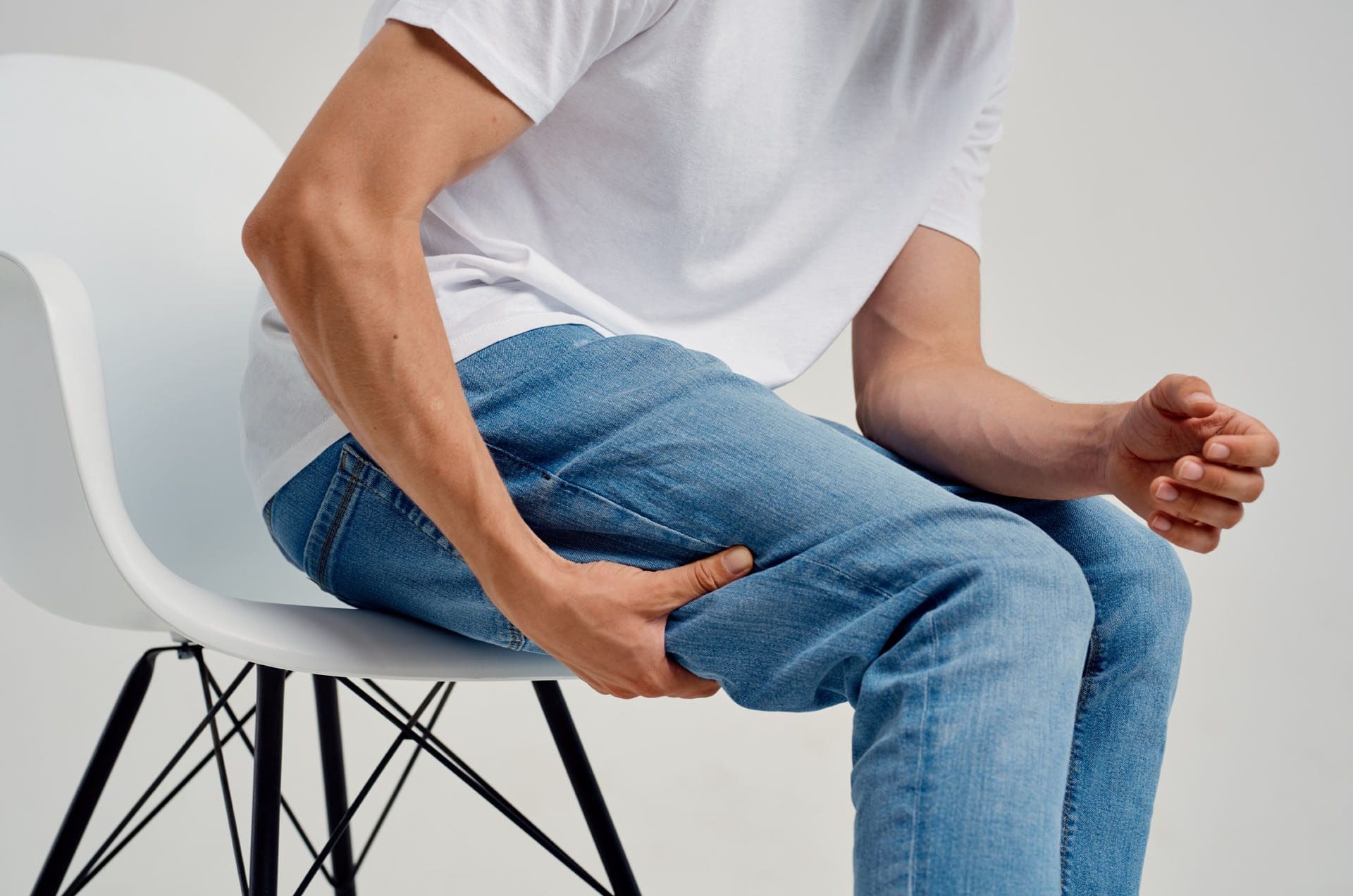
For individuals experiencing back pain, can lying in the supine position help bring relief?
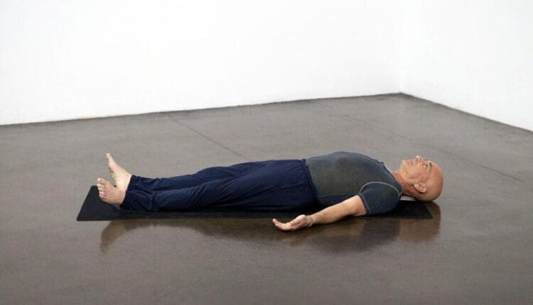
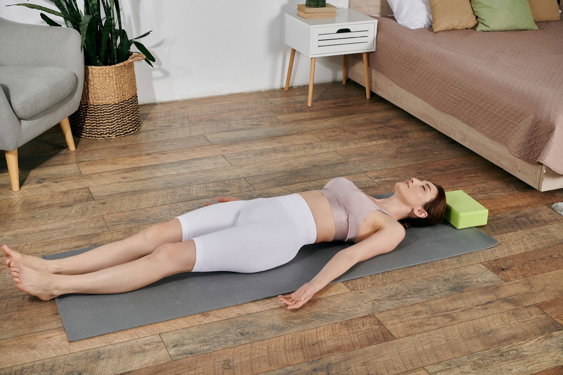
Supine Position
The supine position describes the body’s position when lying on your back with your face up. The individual is flat on their back with no incline, arms at their sides or bent at a 90-degree angle. It’s commonly used in medical settings for examinations, surgeries, and procedures, particularly when access to the anterior/front of the body is needed. It can benefit certain conditions, like helping relieve pain, but exacerbate others, like acid reflux. The term is also used as a modifier for exercises and stretches that begin with the individual on their back. (ScienceDirect Topics, 2009)
Medical Uses
Examinations
- Healthcare providers often use the supine position for physical examinations, including vital signs, palpation of the abdomen, and chest inspection. (Nurse.com, 2024)
Procedures
- It’s also used for various procedures, such as lumbar punctures and injections. (Steris Healthcare, 2025)
Surgery
- Due to its accessibility to the front of the body, the supine position is frequently used for surgeries such as cardiac, abdominal, thoracic, and cranial procedures.
- It allows for easy access to the airway, facilitates anesthesia choices, and can be readily converted to an open procedure if necessary. ScienceDirect Topics, 2009)
Overall Health
Natural Position
- Many individuals naturally fall asleep in the supine position, finding it comfortable and conducive to spinal alignment.
Back Pain Relief
- Sometimes, lying supine with proper support can relieve back pain, particularly in individuals with lumbar spinal issues. (MedicalNewsToday, 2022)
Acid Reflux
- However, the supine position can exacerbate acid reflux, as gravity allows stomach acid to travel up the esophagus. (MedicalNewsToday, 2022)
Sleep Apnea
- The supine position can worsen sleep apnea in some individuals.
Clinical Uses
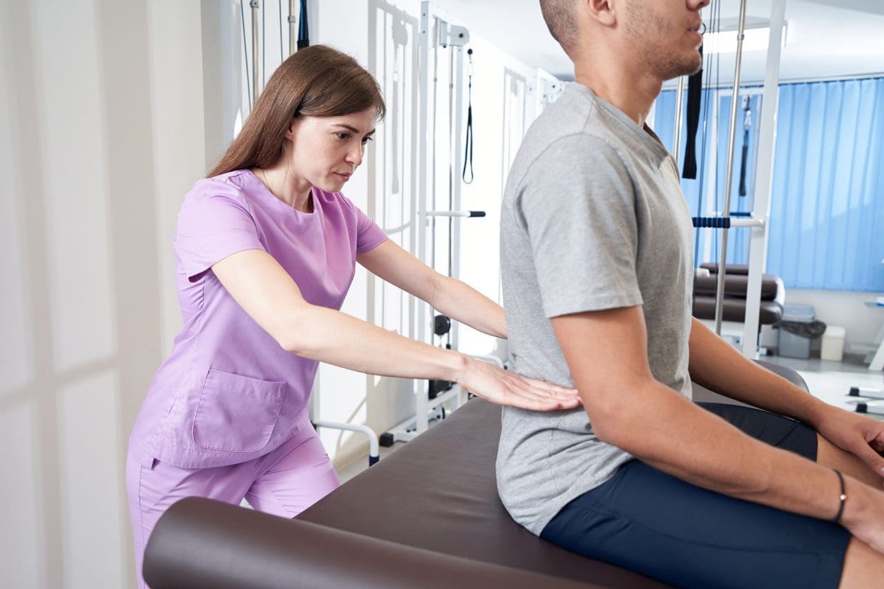
- A physical therapist, trainer, or clinician may use supine to describe positions used for manual therapy or when having the patient do stretching and back exercises as part of a home exercise program.
- If the therapist or personal trainer is training the individual on core stabilization exercises for the first time, the individual will most likely start in the supine position.
- This is because when the body is supine, the muscles have the least work to do to maintain correct posture and position against the force of gravity.
- Many bed exercises begin in this position for rehabilitation.
Back Care
Here are a few recommendations to relieve low back pain using the supine position.
- First, individuals can release the tension in the back by assuming the hook-lying position, a modification of the supine position in which the knees are bent and the feet are resting flat on the floor (Aurora BayCare Medical Center, N.D.).
- Start breathing and relaxing, allowing the tension to drain out of the muscles.
- In a hook-lying position with the fingertips on the lower belly, inhale deeply, then exhale naturally and puff out the remaining air.
- At that point, the fingertips should feel the transverse abdominal muscle engage.
- Release and repeat 10 times.
After the body is warmed up, move on to actual exercise. For example, beginners may be given hip stretches to help relieve back pain. These might be done to maintain or prevent back pain. Individuals can do yoga for their back while in the supine position. As with any exercise program, not all yoga poses involve lying on the back, but many beginners and restorative ones do.
For example, the supine spinal twist involves lying on the back, bending the knees, and gently placing them to one side. The idea is to stay in that position for a few moments—and breathe—to allow the oblique abdominal and back muscles to release.
Variations
Lawn Chair Position
- This variation involves slightly bending the hips and knees and elevating them above the heart, which can help relieve lower back pain.
Frog-Leg Position
- The frog-leg position involves lying on the back with bent knees pushed out to the sides, providing access to the groin and perineum. (Steris Healthcare, 2025)
Injury Medical Chiropractic & Functional Medicine Clinic
Injury Medical Chiropractic and Functional Medicine Clinic works with primary healthcare providers and specialists to develop an optimal health and wellness solution. We focus on what works for you to relieve pain, restore function, and prevent injury. Regarding musculoskeletal pain, specialists like chiropractors, acupuncturists, and massage therapists can help mitigate the pain through spinal adjustments that help the body realign itself. They can also work with other medical professionals to integrate a treatment plan to resolve musculoskeletal issues.
Thoracic Spine Pain
References
ScienceDirect. (2009). Supine Position. Morrey’s The Elbow and Its Disorders (Fourth Edition), 567-577. https://doi.org/https://doi.org/10.1016/B978-1-4160-2902-1.50042-5
Nurse.com. (2024). What Is Supine Position? https://www.nurse.com/nursing-resources/definitions/what-is-supine-position/#:~:text=During%20routine%20physical%20examinations%2C%20the,easier%20to%20perform%20comprehensive%20assessments.
STERIS. (2025). The Complete Guide to Patient Positioning. https://www.steris.com/healthcare/knowledge-center/surgical-equipment/complete-guide-to-patient-positioning#:~:text=The%20most%20common%20position%20used,and%20elevating%20the%20sternal%20notch.
MedicalNewsToday. (2022). What is the supine position? https://www.medicalnewstoday.com/articles/supine-position
Aurora BayCare Medical Center. (N.D.). Lumbar stabilization hooklying position. https://ahc.aurorahealthcare.org/fywb/baycare/x06913bc.pdf