The Vagus Nerve: Body Insights and Benefits Explained
Learn about the vagus nerve in the body and how it plays a crucial role in regulating your bodily functions.
Introduction
Ever wonder whether your body has a secret command center that can calm you down, relieve pain, and lessen tension? In case you missed it, it does! It’s called the vagus nerve, and it’s your body’s superpower nerve! Think of your nervous system as a relaxed, tranquil DJ that plays calming music to keep your body in balance. But if this DJ starts skipping beats, long-term pain, stress, and even aching muscles might spoil the party. This comprehensive study examines the definition of the vagus nerve, its role in the parasympathetic nervous system, and its crucial importance for pain prevention. We will discuss how your body may experience pain due to disruptions to the vagus nerve groove caused by stress, poor eating habits, or simply slouching at your computer. Along with discussing lifestyle strategies to keep the vagus nerve working, we’ll also emphasize how nonsurgical treatments like acupuncture and chiropractic adjustments may enhance the nerve’s calming benefits.
What Is the Vagus Nerve? Your Body’s Superhighway of Calm
Picture the vagus nerve as the ultimate multitasker in your body—a long, winding nerve that’s like a superhighway connecting your brain to almost every major organ. Officially known as the tenth cranial nerve, it’s the longest nerve in your autonomic nervous system, stretching from your brainstem down through your neck, chest, and abdomen, touching your heart, lungs, stomach, and intestines (Cleveland Clinic, 2023). Its name comes from the Latin word for “wandering,” and boy, does it wander! Think of it as your body’s internal GPS, guiding signals to keep your heart rate steady, your digestion smooth, and your stress levels in check.
So, what does this nerve do? The vagus nerve is the star player in the parasympathetic nervous system, the part of your body that says, “Chill out, everything’s cool.” It’s responsible for:
- Heart rate regulation: Slowing your heart rate when you’re relaxed, like when you’re binge-watching your favorite show (Drake & Misha, 2024).
- Digestion: Telling your stomach and intestines to get moving, so you can digest that taco you just ate (Cleveland Clinic, 2023).
- Breathing: Helping you breathe deeply and calmly, like when you’re nailing a yoga pose (Breit et al., 2018).
- Inflammation control: Acting like a firefighter, dousing inflammation to keep your body from going haywire (Bonaz et al., 2016).
- Mood and stress management: Sending signals to your brain to release feel-good chemicals like serotonin, making you feel calm and happy (Breit et al., 2018).
Humor break: The vagus nerve is like the cool aunt who shows up to the family reunion with yoga mats and smoothies, telling everyone to take a deep breath and relax—while secretly keeping the whole party from falling apart!
In short, the vagus nerve is your body’s master regulator, keeping things balanced and preventing chaos. When it’s working well, you feel energized, calm, and pain-free. But when it’s out of tune, it can lead to all sorts of trouble, including body pain. Let’s dive into the parasympathetic system to see how it teams up with the vagus nerve to keep you feeling great.
The Parasympathetic Nervous System: Your Body’s “Rest and Digest” Mode
If the vagus nerve is the DJ, the parasympathetic nervous system (PNS) is the chill lounge where it spins its magic. The PNS is one half of your autonomic nervous system, which controls all the stuff you don’t think about, like breathing, heart rate, and digestion. While the sympathetic nervous system is your “fight or flight” mode—kicking in when you’re running from a bear or stressing about a deadline—the PNS is your “rest and digest” mode, helping you relax, recover, and recharge (Waxenbaum et al., 2023).
The parasympathetic nervous system’s job is to bring your body back to a state of calm after stress. It’s like hitting the reset button after a crazy day. Here’s what it does:
- Slows heart rate: Lowers your heart rate to a relaxed rhythm, saving energy for healing and recovery (Cleveland Clinic, 2023).
- Boosts digestion: Stimulates your gut to break down food and absorb nutrients, so you’re not stuck with that “food baby” feeling (Breit et al., 2018).
- Promotes healing: Encourages tissue repair and reduces inflammation, helping your body bounce back from injuries (Bonaz et al., 2016).
- Calms the mind: Signals your brain to chill out, reducing anxiety and boosting mood (Drake & Misha, 2024).
The vagus nerve is the PNS’s MVP, carrying most of its signals to your organs. When your vagus nerve is firing on all cylinders, it’s like your body’s in a cozy spa day—relaxed, healing, and ready to take on the world. But when the vagus nerve’s “vagal tone” (its strength and efficiency) is low, things can go south, leading to stress, inflammation, and even body pain. Let’s explore what can throw your vagus nerve off its game and how that might lead to aches and pains.
Humor break: The parasympathetic system is like your body’s Netflix-and-chill mode—kicking back, digesting snacks, and telling stress to take a hike!
What Is Vagal Tone, and Why Does It Matter?
Vagal tone is like the signal strength of your vagus nerve—how well it’s communicating with your body to keep things calm and balanced. High vagal tone means your vagus nerve is strong, responsive, and great at keeping your heart rate steady, your digestion smooth, and your stress low. Low vagal tone, on the other hand, is like a weak Wi-Fi signal—your body struggles to stay calm, inflammation spikes, and pain can creep in (Bonaz et al., 2016).
Think of vagal tone as your body’s ability to hit the brakes on stress. When it’s high, you recover quickly from stressful situations, like bouncing back after a tough workout or a heated argument. Low vagal tone means your body stays stuck in “stress mode,” which can mess with your health and lead to pain. So, what can mess with your vagus nerve and its tone? Let’s break it down.
Factors That Affect the Vagus Nerve and Vagal Tone
Your vagus nerve is a sensitive soul—it can get thrown off by a variety of factors, from lifestyle choices to environmental stressors. When vagal tone takes a hit, it can lead to overlapping risk profiles that increase body pain, like back aches, neck stiffness, or even fibromyalgia-like symptoms. Here’s a rundown of the culprits and how they can lead to pain:
1. Chronic Stress
Stress is like kryptonite for your vagus nerve. When you’re constantly stressed—whether from work deadlines, family drama, or scrolling doom-filled news—your sympathetic nervous system (fight or flight) goes into overdrive, suppressing the parasympathetic system and lowering vagal tone (Drake & Misha, 2024). This keeps your body in a state of high alert, spiking stress hormones like cortisol, which can:
- Tightening muscles, leading to neck, shoulder, or back pain (Medical News Today, 2022).
- Increased inflammation makes trigger points (those knotty spots in muscles) more likely to form (Bonaz et al., 2016).
- Disrupted sleep amplifies pain sensitivity and slows recovery (Breit et al., 2018).
Humor: Stress messing with your vagus nerve? It’s like your body’s stuck in a never-ending action movie—tense, twitchy, and ready to ache!
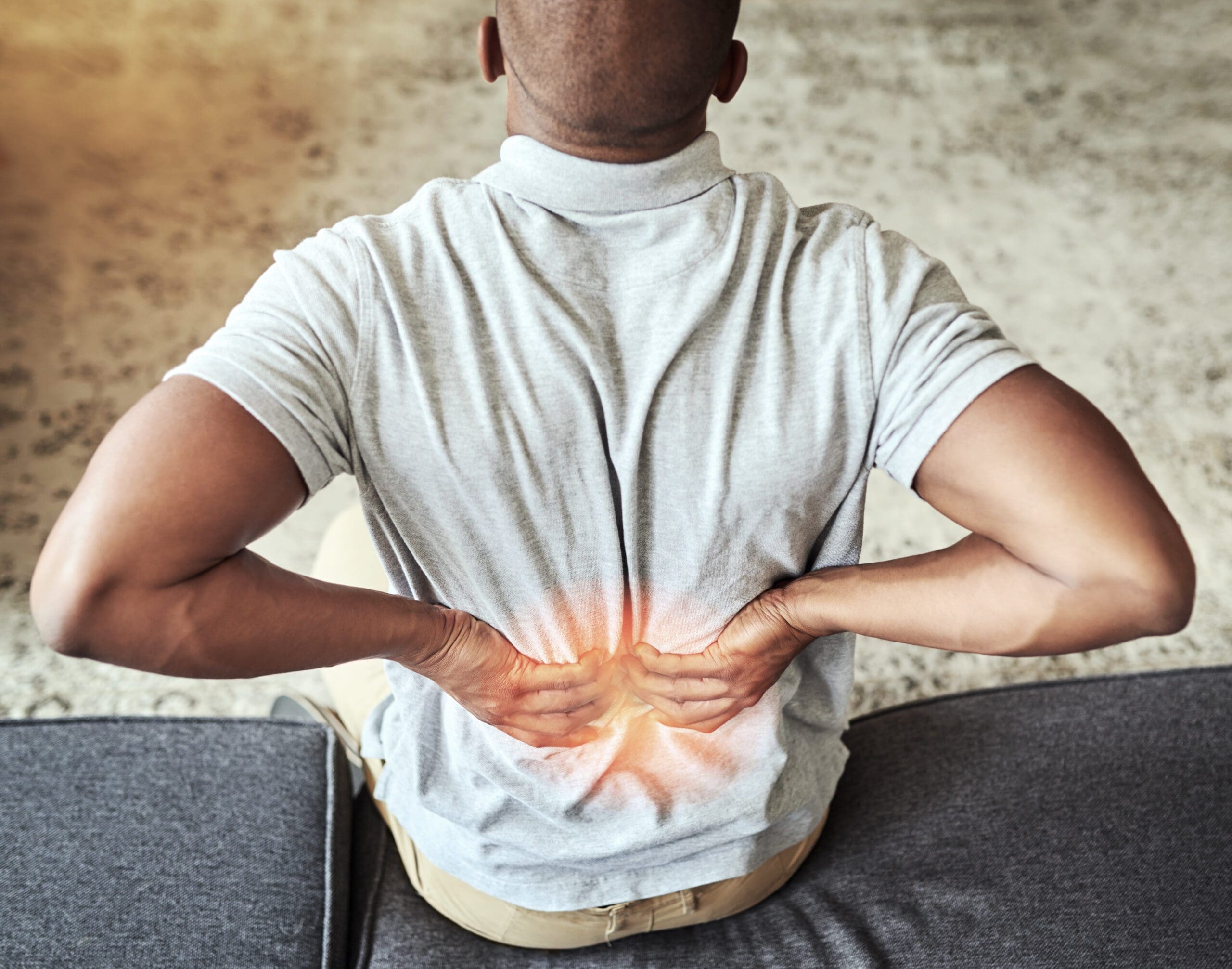
2. Poor Posture
Slouching at your desk or hunching over your phone (hello, text neck!) doesn’t just strain your muscles—it can compress the vagus nerve as it passes through your neck and chest. This can lower vagal tone, reducing its ability to calm your body and manage inflammation (WebMD, 2024). Poor posture also:
- Creates muscle imbalances, leading to pain in your back, shoulders, or hips (Jimenez, 2016).
- Increases tension in the fascia (connective tissue), forming painful trigger points (Shah et al., 2015).
- Disrupts nerve signaling, which can amplify pain perception (StatPearls, 2023a).
Humor: Bad posture? It’s like your vagus nerve is trying to send a text but keeps getting “no signal”—and your muscles are paying the price!
3. Sedentary Lifestyle
Sitting all day or skipping exercise can weaken vagal tone by reducing the stimulation your vagus nerve needs to stay active (Breit et al., 2018). A sedentary lifestyle:
- This condition stiffens muscles and fascia, increasing the risk of painful knots (Healthline, 2024).
- Reduces blood flow, which hampers healing and ramps up inflammation (Bonaz et al., 2016).
- Contributes to stress and poor sleep, creating a vicious cycle of pain and low vagal tone (Medical News Today, 2022).
Humor: Couch potato mode? Your vagus nerve is like, “Get up and move, or I’m taking a nap—and you’re getting aches!”
4. Poor Diet and Nutritional Deficiencies
What you eat matters for your vagus nerve. Diets high in sugar and processed foods spike inflammation, which can suppress vagal tone and make pain worse (LWW, 2021). Deficiencies in key nutrients like:
- Omega-3 fatty acids: Reduce inflammation and support vagal function (Bonaz et al., 2016).
- Vitamin D and magnesium: Essential for nerve health and muscle relaxation (AAPM&R, 2024).
A poor diet can lead to gut issues (like leaky gut), which irritate the vagus nerve and contribute to systemic inflammation, potentially causing body pain (Drake & Misha, 2024).
Humor: Junk food diet? Your vagus nerve is like, “Stop feeding me trash, or I’ll let inflammation throw a pain party!”
5. Environmental Toxins
Pollution, chemicals, and even mold exposure can stress your vagus nerve, lowering its tone and increasing inflammation (ScienceDirect, 2024). This can:
- This can trigger muscle tension and pain, especially in the back or neck (PMC, 2024).
- Disrupt gut health, which the vagus nerve relies on for optimal function (Bonaz et al., 2016).
- Amplify stress responses, making pain feel worse (Breit et al., 2018).
Humor: Toxins bugging your vagus nerve? It’s like your body’s trying to chill in a smoggy city—good luck staying pain-free!
6. Sleep Deprivation
Lack of sleep is a major vagal tone killer. Poor sleep disrupts the parasympathetic system, keeping your body in a stressed state and reducing the vagus nerve’s ability to regulate inflammation (Medical News Today, 2022). This can:
- Increase muscle tension and pain sensitivity (AAPM&R, 2024).
- Slow recovery from injuries, making aches linger (Jimenez, 2016).
- Worsen mood, amplifying the perception of pain (Breit et al., 2018).
Humor: Skimping on sleep? Your vagus nerve is like, “I need my beauty rest, or you’re getting a pain wake-up call!”
7. Physical Trauma or Injury
Injuries like whiplash, falls, or repetitive strain (think typing all day) can irritate the vagus nerve directly or through muscle tension, lowering vagal tone (Jimenez, 2016). This can:
- This condition causes chronic pain in areas like the neck, back, or shoulders (Shah et al., 2015).
- Lead to myofascial pain syndrome, where trigger points form and refer pain elsewhere (StatPearls, 2023a).
- Disrupts nerve signaling, making it harder for the body to calm inflammation (Bonaz et al., 2016).
Humor: Injured your vagus nerve? It’s like accidentally unplugging your body’s chill-out stereo—cue the pain playlist!
These factors—stress, posture, inactivity, diet, toxins, sleep issues, and injuries—create overlapping risk profiles that can weaken vagal tone, ramp up inflammation, and lead to body pain. For example, chronic stress might tighten your neck muscles, while poor posture compresses the vagus nerve, and a bad diet fuels inflammation—boom, you’ve got a recipe for aches and pains! But don’t worry—nonsurgical treatments like chiropractic care and acupuncture can help get your vagus nerve back in the groove.
Chiropractic Care: Boosting Vagus Nerve Function for Pain Relief
Chiropractic care is like a tune-up for your vagus nerve, helping it hit all the right notes to reduce pain and restore balance. By focusing on spinal alignment and muscle tension, chiropractors can stimulate the vagus nerve and improve vagal tone, which calms inflammation and eases body pain (PubMed, 2009). Here’s how it works:
- Spinal Adjustments: Misaligned vertebrae (subluxations) in the neck or upper back can compress the vagus nerve, reducing its function. Gentle chiropractic adjustments realign the spine, relieving pressure and boosting nerve signaling (Integrative Physical Health, 2022). This can reduce pain in areas like the back, neck, or shoulders (Jimenez, 2016).
- Myofascial Release: Chiropractors use soft-tissue techniques to release tight muscles and fascia, which can improve vagal tone by reducing tension around the nerve (Gonstead Chiropractic Center, 2023). This helps with conditions like myofascial pain syndrome, where trigger points cause widespread aches (Shah et al., 2015).
- Reducing Inflammation: By improving nerve function and blood flow, chiropractic care helps the vagus nerve dial down inflammation, a key driver of chronic pain (Bonaz et al., 2016).
- Stress Relief: Adjustments stimulate the parasympathetic system, lowering stress hormones and promoting relaxation, which can ease tension-related pain (Radix Chiro, 2023).
Dr. Alexander Jimenez, with his dual expertise as a chiropractor and nurse practitioner, uses advanced diagnostics to pinpoint how injuries or misalignments affect the vagus nerve. For example, he might use MRI or CT scans to visualize spinal misalignments or soft-tissue damage, functional assessments to evaluate nerve function, or lab tests to check for inflammation markers (DrAlexJimenez.com, n.d.). His approach ensures precise, personalized care that targets the root cause of pain, often linked to vagal dysfunction (Jimenez, 2016).
Humor: Chiropractic care for your vagus nerve? It’s like giving your body’s DJ a new soundboard—suddenly, the pain playlist switches to smooth jazz!
Acupuncture: A Needle-Nudge for Vagus Nerve Stimulation
Acupuncture is another rockstar treatment for boosting vagal tone and easing pain. By inserting tiny needles into specific points on the body, acupuncture stimulates the nervous system, including the vagus nerve, to promote relaxation and healing (LWW, 2021). Here’s how it helps:
- Direct Vagus Nerve Stimulation: Certain acupuncture points, like those in the ear or neck, directly activate the vagus nerve, improving its tone and calming the body (Breit et al., 2018).
- Pain Reduction: Acupuncture releases endorphins and other pain-relieving chemicals, reducing muscle tension and trigger point pain (SE Pain and Spine Care, 2024).
- Inflammation Control: By boosting vagal tone, acupuncture helps the vagus nerve suppress inflammation, easing conditions like myofascial pain or fibromyalgia (Bonaz et al., 2016).
- Stress Management: Acupuncture promotes parasympathetic activity, lowering stress and helping with tension-related pain (Drake & Misha, 2024).
When combined with chiropractic care, acupuncture creates a powerhouse duo for vagus nerve health. Dr. Jimenez often integrates these treatments, using his diagnostic expertise to tailor plans that address both physical and neurological factors contributing to pain (Jimenez, 2016).
Humor: Acupuncture for your vagus nerve? It’s like giving your body’s chill button a gentle poke—pain and stress just melt away!
Dr. Alexander Jimenez’s Clinical Approach: Precision Diagnostics for Pain Relief
Dr. Alexander Jimenez stands out in El Paso for his ability to connect the dots between injuries, vagus nerve dysfunction, and pain. His approach blends chiropractic care, functional medicine, and advanced diagnostics to create personalized treatment plans. Here’s how he does it:
- Advanced Imaging: Using MRI and CT scans, Dr. Jimenez visualizes spinal misalignments or soft-tissue issues (like fascia restrictions) that may compress the vagus nerve, contributing to pain (DrAlexJimenez.com, n.d.).
- Functional Assessments: These tests evaluate how well your nervous system, including the vagus nerve, is functioning. For example, heart rate variability (HRV) tests can measure vagal tone, revealing if low tone is linked to your pain (Breit et al., 2018).
- Lab Tests: Bloodwork can identify inflammation markers or nutritional deficiencies (like low vitamin D or omega-3s) that impair vagal function and fuel pain (Jimenez, 2016).
- Dual-Scope Procedures: Combining endoscopy and arthroscopy, Dr. Jimenez gets a real-time view of joint or tissue damage, ensuring precise interventions that support vagus nerve health (NYS DOH, 2013; FACS, 2018).
This comprehensive approach allows Dr. Jimenez to create tailored plans that not only relieve pain but also boost vagal tone, promoting long-term wellness. For example, a patient with chronic neck pain might get adjustments to free up vagus nerve compression, acupuncture to stimulate it, and nutritional advice to reduce inflammation—all based on precise diagnostics (LinkedIn, n.d.).
Humor: Dr. Jimenez’s diagnostics? It’s like your vagus nerve getting a full-body MRI with a side of “let’s fix this” swagger!
Lifestyle Hacks for Vagus Nerve Health and Pain Prevention
Keeping your vagus nerve happy is like giving your body a daily dose of zen—and it can help prevent pain before it starts. Here are some science-backed lifestyle hacks to boost vagal tone and keep aches at bay:
1. Deep Breathing and Meditation
Slow, deep breathing (like diaphragmatic breathing) directly stimulates the vagus nerve, boosting its tone and calming your body (Breit et al., 2018). Try this:
- Inhale for 4 seconds, hold for 4, exhale for 6. Repeat for 5 minutes daily.
- Apps like Headspace or Calm can guide you through meditation to reduce stress and improve vagal function (Drake & Misha, 2024).
This lowers stress hormones, reduces muscle tension, and prevents pain flare-ups (Medical News Today, 2022).
Humor: Deep breathing for your vagus nerve? It’s like telling your stress to take a long, slow walk off a short pier!
2. Regular Exercise
Moderate exercise, like walking, yoga, or swimming, boosts vagal tone by stimulating the parasympathetic system (Healthline, 2024). Aim for:
- 30 minutes of low-impact activity, 5 days a week.
- Yoga poses like child’s pose or cat-cow to stretch fascia and reduce tension (Mayo Clinic, 2024b).
Exercise improves blood flow, reduces inflammation, and prevents muscle knots that lead to pain (Bonaz et al., 2016).
Humor: Exercise for vagal health? It’s like your vagus nerve hitting the gym—stronger tone, fewer aches!
3. Anti-Inflammatory Diet
Fuel your vagus nerve with foods that fight inflammation:
- Omega-3s: Salmon, walnuts, flaxseeds (LWW, 2021).
- Antioxidants: Berries, spinach, kale (Healthline, 2024).
- Magnesium-rich foods: Nuts, seeds, dark chocolate (AAPM&R, 2024).
Avoid sugar and processed foods, which can inflame your system and weaken vagal tone (Jimenez, 2016).
Humor: Eating for your vagus nerve? It’s like serving your body a gourmet anti-pain smoothie—hold the sugar!
4. Quality Sleep
Aim for 7–9 hours of sleep nightly to support vagal tone and reduce pain sensitivity (Medical News Today, 2022). Tips:
- Create a bedtime routine: no screens 1 hour before bed.
- Use blackout curtains or a sleep mask to improve sleep quality.
Good sleep helps the vagus nerve regulate inflammation and repair tissues (Breit et al., 2018).
Humor: Sleep for vagal health? It’s like giving your vagus nerve a cozy blanket and a lullaby—no pain invited!
5. Posture Correction
Good posture keeps the vagus nerve free from compression. Try:
- Ergonomic chairs or standing desks to avoid slouching.
- Regular posture checks: ears over shoulders, shoulders over hips (WebMD, 2024).
This reduces muscle tension and supports vagal function, preventing pain (Jimenez, 2016).
Humor: Fix your posture? It’s like telling your vagus nerve, “Stand tall, and let’s keep the pain party canceled!”
6. Stress Management
Chronic stress tanks vagal tone, so try:
- Mindfulness practices like journaling or gratitude exercises.
- Hobbies like painting or gardening to relax your mind (Drake & Misha, 2024).
Reducing stress helps the vagus nerve keep inflammation and pain in check (Bonaz et al., 2016).
Humor: Stress management for your vagus nerve? It’s like sending your worries on a one-way trip to Nopeville!
7. Hydration and Detox
Staying hydrated and minimizing toxin exposure supports vagal tone:
- Drink 8–10 glasses of water daily to flush toxins (Healthline, 2024).
- Avoid processed foods and limit exposure to pollutants like cigarette smoke (ScienceDirect, 2024).
This keeps inflammation low and supports the vagus nerve’s anti-pain powers (PMC, 2024).
Humor: Hydrate for vagal health? It’s like giving your vagus nerve a refreshing spa day—toxins out, pain down!
By incorporating these lifestyle hacks, you can boost your vagal tone, reduce inflammation, and prevent body pain. Pairing these with chiropractic care and acupuncture creates a holistic approach to keeping your vagus nerve—and your body—in top shape.
Real-Life Stories: Vagus Nerve and Pain Relief in Action
Let’s meet Lisa, a 40-year-old teacher who was plagued by chronic shoulder pain and tension headaches from grading papers all day. Dr. Jimenez used MRI scans to spot a neck misalignment compressing her vagus nerve, then applied chiropractic adjustments and acupuncture to relieve the pressure. With a tailored plan including deep breathing and an anti-inflammatory diet, Lisa’s pain faded, and she’s back to teaching without wincing (inspired by Jimenez, 2016).
Then there’s Jake, a weekend soccer player with nagging lower back pain. Functional assessments showed low vagal tone from stress and poor posture. Dr. Jimenez combined chiropractic care, yoga stretches, and nutritional tweaks to boost Jake’s vagal tone. Now, Jake’s scoring goals pain-free and sleeping like a champ (similar to cases in PubMed, 2009).
These stories show how stimulating the vagus nerve through integrative care can transform lives, reducing pain and boosting wellness.
Humor: Lisa and Jake’s vagus nerve comeback? It’s like their bodies went from a grumpy cat to a purring kitten—pain-free and happy!
The Science Behind Vagus Nerve Stimulation for Pain Relief
The vagus nerve’s pain-relieving powers are backed by science. It’s part of the cholinergic anti-inflammatory pathway, where it releases acetylcholine to dampen inflammation, a major cause of pain (Bonaz et al., 2016). Studies show:
- High vagal tone is linked to lower pain sensitivity and faster recovery from injuries (Breit et al., 2018).
- Chiropractic adjustments improve vagal tone by reducing spinal stress, easing pain in conditions like myofascial pain syndrome (PubMed, 2009).
- Acupuncture stimulates vagus nerve pathways, reducing inflammation and pain in chronic conditions (LWW, 2021).
- Lifestyle changes like exercise and meditation boost heart rate variability (HRV), a marker of vagal tone, correlating with less pain (Drake & Misha, 2024).
Dr. Jimenez’s approach leverages this science, using diagnostics to identify vagal dysfunction and tailoring treatments to restore balance (LinkedIn, n.d.).
Humor: The science of vagus nerve stimulation? It’s like your body’s got a built-in pain zapper—chiro and acupuncture just flip the switch!
When to Seek Professional Help for Vagus Nerve-Related Pain
If you’re dealing with persistent pain, especially in your neck, back, or shoulders, or if stress, poor sleep, or digestive issues are piling on, it might be time to check in with a pro. Signs your vagus nerve needs help include:
- Chronic pain that doesn’t budge with rest or over-the-counter meds.
- Frequent tension headaches or muscle knots (Mayo Clinic, 2024b).
- Feeling wired but tired, with poor sleep or high stress (Breit et al., 2018).
- Digestive issues like bloating or sluggishness can signal vagal dysfunction (Bonaz et al., 2016).
Dr. Jimenez recommends early intervention to prevent pain from escalating. His diagnostic tools, like MRI scans and HRV tests, can confirm if vagal tone is contributing to your symptoms, guiding a targeted treatment plan (Jimenez, 2016).
Humor: Time to see a pro? When your vagus nerve is sending SOS signals louder than your phone’s low-battery alert—get help!
Conclusion
This in-depth look at the vagus nerve and its role in relieving pain demonstrates its power as a calming force in your body. The vagus nerve is the main part of the parasympathetic nervous system. It controls heart rate, digestion, inflammation, and stress. When it works well, it keeps pain away. Chronic stress, bad posture, being inactive, eating poorly, toxins, sleep problems, and injuries can all lower vagal tone, which can cause inflammation and pain in the body. Chiropractic care and acupuncture, along with lifestyle changes like deep breathing, exercise, and an anti-inflammatory diet, can all help boost vagal tone, lower pain, and improve long-term health. Dr. Alexander Jimenez is an expert in advanced imaging, functional assessments, and dual-scope procedures. This means he can make accurate diagnoses and give each patient the care they need to get better.
Important: This post gives information about the vagus nerve and how it can help with pain management, but if you have chronic pain or think you might have vagal dysfunction, you should see a doctor. Always see a qualified healthcare provider for the right diagnosis and treatment, because problems that aren’t treated can get worse over time.
This article is only meant to give you information; it is not a substitute for professional medical advice, diagnosis, or treatment. Before starting any new treatment or making any changes to your lifestyle, talk to a qualified healthcare provider, especially if you already have health problems. The information is based on research, so you should take it seriously when making health decisions. There are no guarantees about what will happen, and results may be different for each person.
References
- American Academy of Physical Medicine and Rehabilitation. (2024). Myofascial pain | PM&R KnowledgeNow. https://now.aapmr.org/myofascial-pain/
- Bonaz, B., Sinniger, V., & Pellissier, S. (2016). Anti-inflammatory properties of the vagus nerve: Potential therapeutic implications of vagus nerve stimulation. Journal of Inflammation Research, 9, 261–267. https://doi.org/10.2147/JIR.S121135
- Breit, S., Kupferberg, A., Rogler, G., & Hasler, G. (2018). Vagus nerve as modulator of the brain–gut axis in psychiatric and inflammatory disorders. Frontiers in Psychiatry, 9, 44. https://doi.org/10.3389/fpsyt.2018.00044
- Cleveland Clinic. (2023). Vagus nerve: Function, stimulation, and more. https://my.clevelandclinic.org/health/body/22279-vagus-nerve
- Drake, M. J., & Misha, T. (2024). Activate your calm: Harnessing the vagus nerve for health after 40. [PDF]. activate-your-calm-harnessing-the-vagus-nerve-for-health-after-40_67014a8a.pdf
- DrAlexJimenez.com. (n.d.). Injury specialists. https://dralexjimenez.com/
- FACS. (2018). ACS TQIP best practices guidelines in imaging. https://www.facs.org/media/oxdjw5zj/imaging_guidelines.pdf
- Gonstead Chiropractic Center. (2023). How chiropractic can help you overcome myofascial pain syndrome. https://gonsteadchiropracticcenter.com/blog/b/how-chiropractic-can-help-you-overcome-myofascial-pain-syndrome
- Healthline. (2024). Myofascial pain: Treatment, symptoms, causes, and more. https://www.healthline.com/health/myofascial-pain
- Integrative Physical Health. (2022, February). How chiropractic can help with myofascial pain syndrome. http://integrativephysicalhealth.com/how-chiropractic-can-help-with-myofascial-pain-syndrome/
- Jimenez, A. (2016, August). How to ease & treat myofascial pain syndrome. El Paso Chiropractor Blog. https://www.elpasochiropractorblog.com/2016/08/how-to-ease-treat-myofascial-pain.html
- LinkedIn. (n.d.). Dr. Alexander Jimenez, DC, APRN, FNP-BC. https://www.linkedin.com/in/dralexjimenez/
- LWW. (2021). Finding the possible link between physical activities, dietary nutrient-associated conditions on health may too be responsible for the development of MPS …. https://journals.lww.com/cmre/fulltext/2021/11040/finding_the_possible_link_between_physical.4.aspx
- Mayo Clinic. (2024b). Myofascial pain syndrome – Diagnosis and treatment. https://www.mayoclinic.org/diseases-conditions/myofascial-pain-syndrome/diagnosis-treatment/drc-20375450
- Medical News Today. (2022). Myofascial pain syndrome: Causes, symptoms, and treatment. https://www.medicalnewstoday.com/articles/myofascial-pain-syndrome
- NYS DOH. (2013). Advanced Diagnostic Imaging: Background on Use, Patient Safety …. https://www.health.ny.gov/facilities/public_health_and_health_planning_council/meetings/2013-07-17/docs/2013-07-03_adv_diag_imag_backgrnd_papers.pdf
- PMC. (2024). Myofascial pain syndrome: An update on clinical characteristics, etiopathogenesis, diagnosis, and treatment. https://pmc.ncbi.nlm.nih.gov/articles/PMC11998975/
- PubMed. (2009). Chiropractic management of myofascial trigger points and myofascial pain syndrome: A systematic review of the literature. https://pubmed.ncbi.nlm.nih.gov/19121461/
- Radix Chiro. (2023, May). How chiropractic helps with myofascial pain. https://www.radixchiro.com/how-chiropractic-helps-with-myofascial-pain/
- ScienceDirect. (2024). Understanding the Vascular Environment of Myofascial Trigger Points. https://pmc.ncbi.nlm.nih.gov/articles/PMC3493167/
- SE Pain and Spine Care. (2024, March). Exploring Effective Treatment Options for Myofascial Pain Syndrome. https://www.sepainandspinecare.com/exploring-effective-treatment-options-for-myofascial-pain-syndrome/
- Shah, J. P., Thaker, N., Heimur, J., Aredo, J. V., Sikdar, S., & Gerber, L. H. (2015). Myofascial trigger points then and now: A historical and scientific perspective. PM&R, 7(7), 746–761. https://doi.org/10.1016/j.pmrj.2015.01.024
- StatPearls. (2023a). Myofascial pain syndrome. https://www.ncbi.nlm.nih.gov/books/NBK499882/
- Waxenbaum, J. A., Reddy, V., & Varacallo, M. (2023). Anatomy, autonomic nervous system. In StatPearls. https://www.ncbi.nlm.nih.gov/books/NBK539845/
- WebMD. (2024). Myofascial pain syndrome (chronic soft tissue pain). https://www.webmd.com/pain-management/myofascial-pain-syndrome