The Future of Regenerative Medicine: Peptides, Aesthetics, and Integrative Chiropractic Care

Abstract
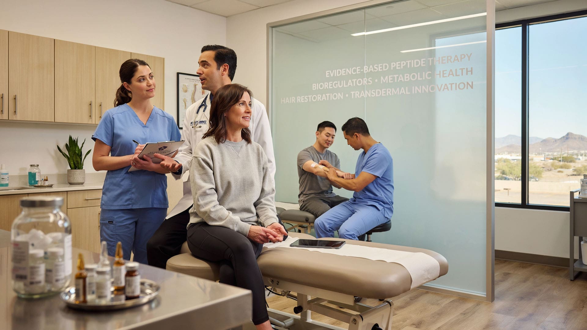
Welcome to our educational portal. I am Dr. Alex Jimenez, and I am honored to guide you through the transformative world of modern, evidence-based wellness. In this comprehensive post, we will explore the cutting-edge applications of peptide therapy and bioregulators, a fascinating frontier that is revolutionizing our approach to metabolic health, weight management, hair restoration, and aesthetic skin rejuvenation. We will examine the intricate physiological mechanisms behind these biological messengers, demystifying how they orchestrate cellular repair and restore homeostasis. This post will highlight specific compounds, including GHK-Cu, BPC-157, SNAP-8, and modern GLP-1 agonists like semaglutide, tirzepatide, and retatrutide. I will also share insights into our breakthrough oral and transdermal delivery systems designed to improve patient access and clinical efficacy.
I will detail how we apply these therapies within a collaborative, multidisciplinary practice. You will learn how my expertise in chiropractic and functional medicine synergizes with the internal medicine oversight of our Medical Director, Dr. Maria Guadalupe Cardenas, MD, to deliver comprehensive personal injury care, rehabilitation, and regenerative treatments. Finally, I will introduce the Peptide Protocol Portal and our AI assistant, Dr. Peppy, resources we developed to ground these therapies in safety and rigorous clinical data. This educational journey aims to empower you with a clear, in-depth understanding of how an integrative care model unlocks the body’s incredible innate potential to heal and thrive.
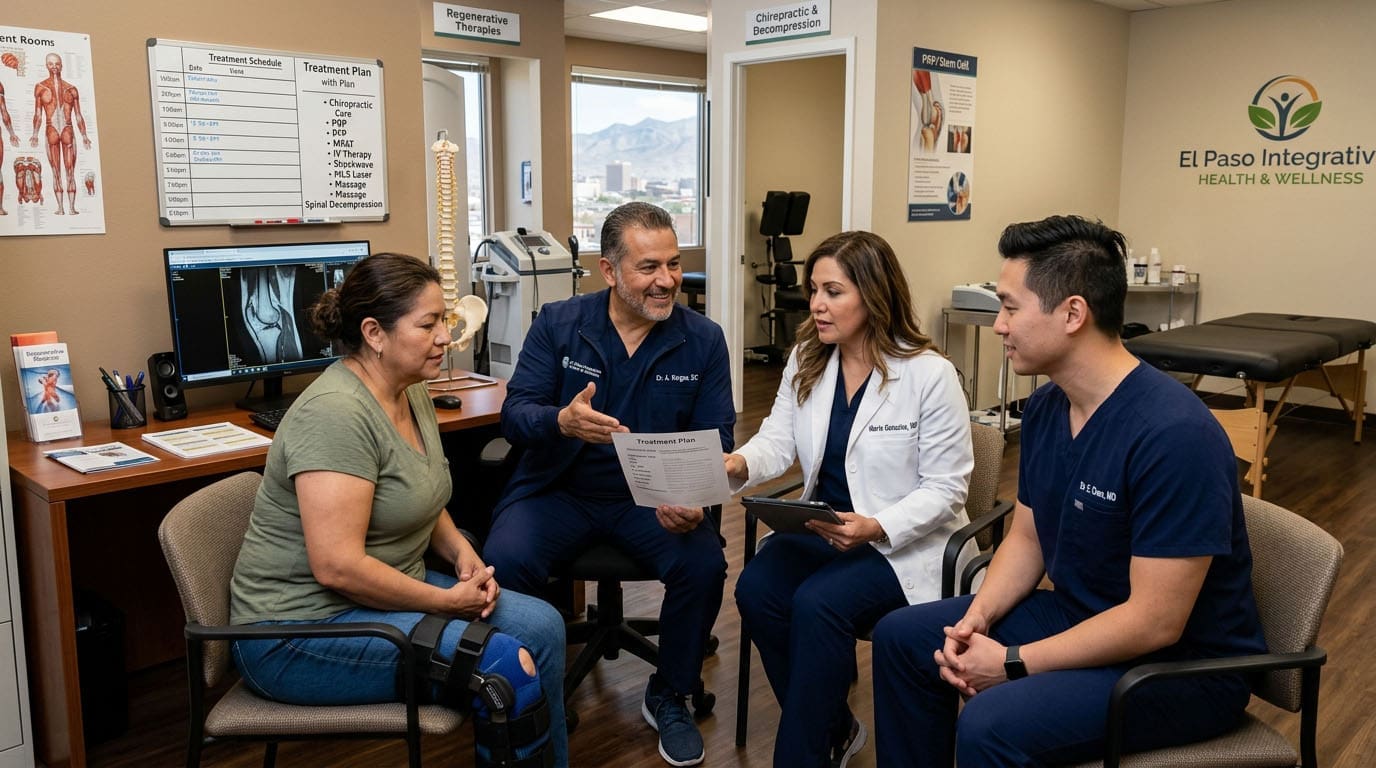
The Integrative Clinical Model: Bridging Chiropractic Care and Medical Oversight
At the core of our philosophy is a multidisciplinary, patient-centered approach. I am Dr. Alex Jimenez, DC, APRN, FNP-BC, CFMP, IFMCP, ATN, CCST, and my daily focus is restoring function, reducing pain, and optimizing health by uniting structural care, internal medicine insights, and functional medicine strategies. My diverse background allows me to view patient health through a wide-angle lens, constantly seeking to connect musculoskeletal integrity with systemic and metabolic function.
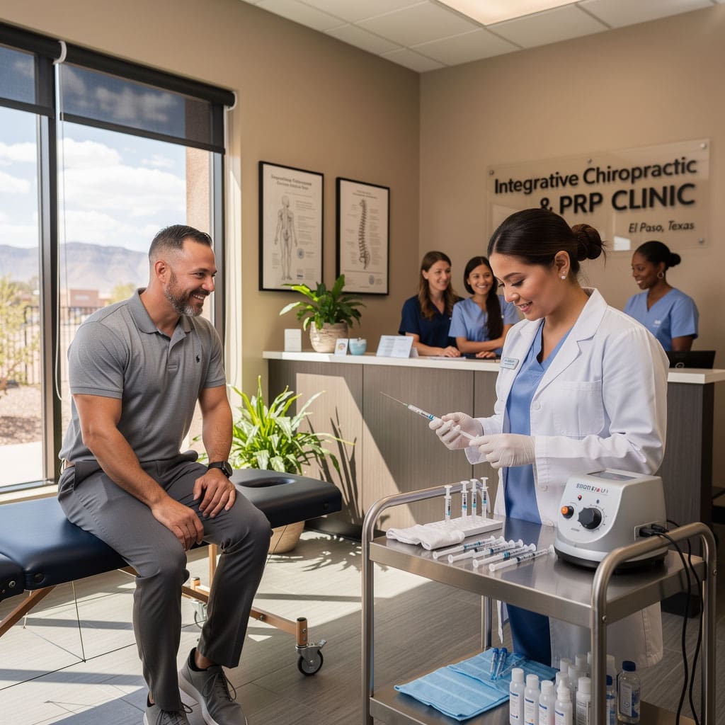
My work is seamlessly complemented and medically overseen by our esteemed Medical Director and Collaborative Physician, Dr. Maria Guadalupe Cardenas, MD. Dr. Cardenas is Board Certified in Internal Medicine and brings over 40 years of invaluable clinical experience to our team. Her NPI is #1164426749, and she is licensed in Texas under #J2933. Together, we practice at Injury Medical Clinic PA (also known as Mission Plaza Injury Medical Clinic) in El Paso, Texas.
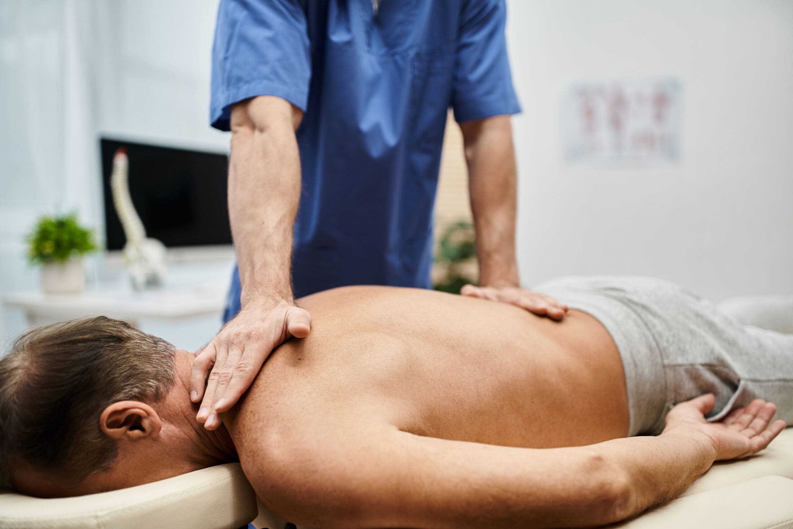
This multidisciplinary setup is a gold standard in integrative and personal injury care clinics. While I orchestrate the biomechanical and functional aspects of healing through chiropractic adjustments, neuromuscular rehabilitation, and functional medicine protocols, Dr. Cardenas provides the essential medical direction required for advanced modalities like peptide therapy. Her oversight ensures appropriate diagnostic workups—ranging from CBC and CMP to thyroid panels, lipid profiles, and inflammatory markers—and guarantees that every therapeutic pathway we select follows safe, evidence-informed protocols.
When a patient arrives at our clinic for a musculoskeletal injury, we do not just treat the local tissue damage. Through our functional medicine lens, we might identify underlying metabolic dysfunction or systemic inflammation that is stalling their recovery. With Dr. Cardenas’s medical guidance, we can safely incorporate advanced regenerative treatments alongside structural rehabilitation, creating a holistic ecosystem for true healing.
Understanding Biological Messengers: Peptides and Bioregulators
As a practitioner dedicated to regenerative medicine, I am perpetually amazed by the body’s inherent capacity to heal itself. This reverence for human physiology led me deep into the science of peptide therapy, a field that harnesses the body’s own signaling molecules to orchestrate repair. To fully grasp this, we must differentiate between peptides and bioregulators.
Peptides are short chains of amino acids. In the United States, we classify a chain of 2 to 40 amino acids as a peptide; beyond that, it is considered a protein (though in Europe, the threshold is often 50). These larger molecules typically function via a highly specific “lock and key” mechanism. They bind to designated receptors on a cell’s surface, sending instructions inward to trigger a specific cellular cascade, such as synthesizing collagen or reducing inflammation. Because they are highly targeted, they do not cause the unintended “off-target” side effects frequently seen with synthetic pharmaceuticals.
Bioregulators, conversely, are ultra-short peptide sequences, usually consisting of just two to four amino acids. Because of their microscopic size, they can pass directly through the cell membrane and into the nucleus. Here, they exert profound epigenetic effects by influencing DNA transcription—effectively turning specific gene expressions “on” or “off” without altering the underlying DNA sequence.
Many peptides we utilize are endogenous (naturally produced by the human body). A prime example is GHK-Cu (glycyl-L-histidyl-L-lysine bound to copper), known as the blue copper peptide. Found in our saliva and blood plasma, it possesses antibacterial properties and is critical for musculoskeletal repair by upregulating collagen types 1 and 3 following tissue injury.
Other peptides are exogenous, synthesized in a laboratory for therapeutic consistency. BPC-157 (Body Protection Compound) is derived from a protective protein found in human gastric juice. Interestingly, in the early 1900s, Ivan Pavlov, famous for his behavioral experiments, arguably became the first to commercialize BPC-157 by collecting gastric secretions from his dogs to sell as a medicinal elixir. While he did not know its precise molecular identity, he recognized its profound healing capacity.
A Brief History of Peptides in Medicine
Peptides are not a novel wellness trend; they are deeply woven into the history of modern medicine.
- 1901: German chemist Emil Fischer discovered peptides, a breakthrough that earned him the Nobel Prize in Chemistry.
- 1921: The first peptide drug, insulin, was administered to a 14-year-old Canadian named Leonard Thompson, waking him from a diabetic coma.
- 1922: Banting and Macleod were awarded the Nobel Prize for their work on insulin.
- 1953: The first lab-synthesized peptide, oxytocin (often dubbed the “love hormone”), was created, clarifying its role in childbirth and social bonding.
Today, there are hundreds of FDA-approved peptide drugs, with over 800 more in clinical pipelines, representing a rapid evolution in pharmacology alongside mRNA technology. Knowledge in medicine is doubling roughly every 25 years, and peptide science is riding the crest of this wave. Culturally, peptides have also entered the mainstream. High-profile figures like Jennifer Aniston and Gwyneth Paltrow praise them for skincare, while Joe Rogan has widely discussed the “Wolverine stack” (BPC-157 and TB-500) with actors like Matt Damon for injury recovery.
When the FDA notes “unknown risk factors” for compounds like BPC-157, it often means large-scale, formalized human clinical trials are pending, not that the compound is inherently dangerous. In decades of clinical application and research, there have been no documented adverse events in human trials for BPC-157. Under Dr. Cardenas’s medical direction, we prioritize rigorous third-party testing for purity, sterility, and heavy metals, ensuring every intervention is regulatory-compliant and meticulously safe.
The Science of Delivery: Enhancing Bioavailability and Efficacy
A significant challenge in peptide therapy is the delivery mechanism. Injectable delivery is highly effective but creates compliance barriers for needle-phobic patients and faces stringent regulatory scrutiny. For example, GHK-Cu is approved as a topical cosmetic and an oral supplement, but not for injection.
At our research lab, affiliated with the University of South Florida (USF) Research Park, we focused on non-invasive delivery. In November 2025, we were granted a provisional patent for a multi-stage oral delivery system that overcomes the destructive acidic environment of the stomach. Our system works in three stages:
- Enteric Coating: The capsule resists gastric acid, remaining intact until it reaches the alkaline environment of the small intestine, the optimal site for nutrient absorption.
- Trehalose Microcrystalline Pellet: Inside, the peptide is bound to trehalose, a specialized sugar utilized as a cryopreservant in stem cell science. This protective crystal shields the peptide, facilitating a slow, time-released dissolution.
- Bioavailability Enhancer: We integrate a drug solubility additive that temporarily increases the space between intestinal cells, enhancing cell wall permeability so the peptide can enter the bloodstream efficiently.
Similarly, we have achieved breakthroughs in transdermal delivery. Our transdermal serum system (such as ProCell Active) is formulated to cross the stratum corneum and epidermis in about 4 hours, reaching the dermis and subcutaneous layers within 24 hours. When applying Platelet-Rich Plasma (PRP) over skin pre-treated with our serum, we observe rapid, clean absorption compared to untreated skin. We also utilize botanical antioxidants and peptide stabilizers to extend the bioactivity and refrigerated shelf life of PRP, exosomes, and growth factors for up to 90 days, vastly improving clinical practicality.
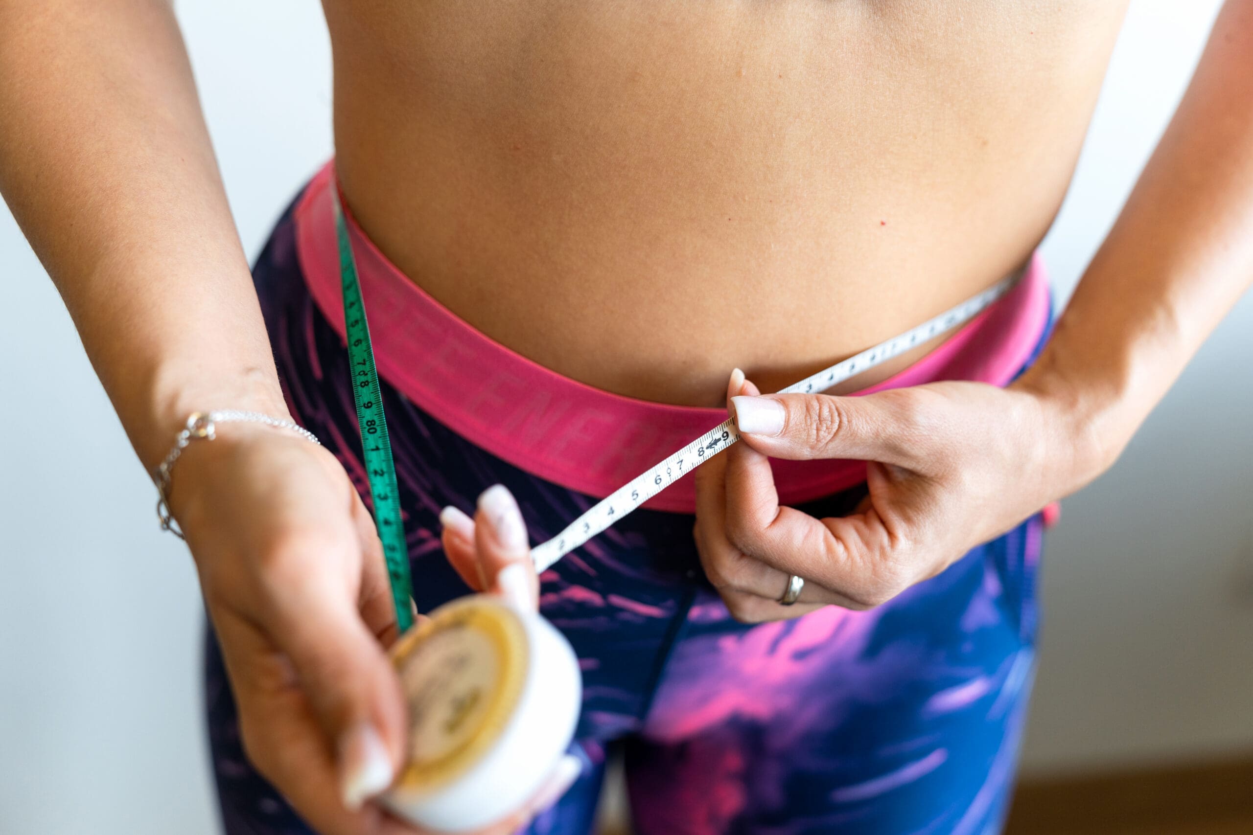
Strategic Weight Management: The GLP-1 Agonist Evolution
One of the most pressing health crises we face is metabolic disease. Drawing upon my clinical observations (available for review on my platforms at ChiroMed and LinkedIn), we have developed a highly effective, “good, better, best” approach to weight loss using advanced metabolic peptides.
- Good: Semaglutide. This GLP-1 (Glucagon-Like Peptide-1) receptor agonist mimics the natural GLP-1 hormone. It stimulates insulin release, suppresses glucagon (which raises blood sugar), drastically slows gastric emptying, and signals satiety to the brain. In 2025, it accounted for 12% of all US prescriptions.
- Better: Tirzepatide. This is a dual agonist acting on both GLP-1 and GIP (Glucose-dependent Insulinotropic Polypeptide) receptors. GIP enhances insulin secretion and favorably modulates fat metabolism. Targeting both pathways yields superior glucose control and lipid clearance.
- Best: Retatrutide. This groundbreaking triple-agonist targets GLP-1, GIP, and Glucagon receptors. While glucagon typically raises blood sugar, at this specific receptor level, it exponentially increases energy expenditure and fat oxidation.
I caution strongly against the “single lever approach”—simply pushing the dose higher when weight loss stalls. The TRIUMPH-1 study on retatrutide, completed in mid-2026, demonstrated that lower-dose groups often experienced fewer side effects and lower dropout rates than placebo groups. Rather than maxing out a dose, we pull “other levers.” We might add Cagrilintide, an amylin agonist that synergistically slows gastric emptying, or incorporate MitoQ or SS-31 (Elamipretide) if underlying mitochondrial dysfunction is impairing cellular energy production (ATP).
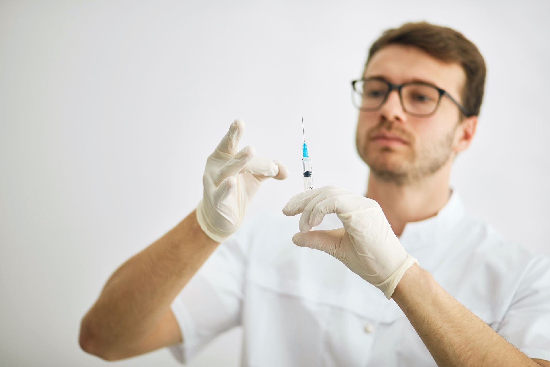
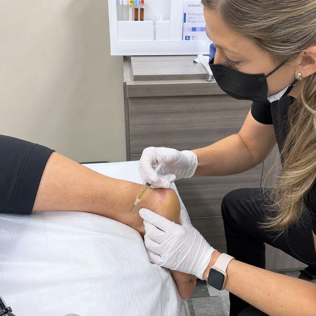
Patient safety dictates that we administer these injections in-office. We combine these peptides with lipotropic injections containing L-Carnitine (to shuttle long-chain fatty acids into the mitochondria for beta-oxidation) and B Vitamins (B6, B9, B12) for methylation and energy support. During these weekly visits, patients sit under a red-light therapy panel. This photobiomodulation stimulates dermal collagen production, proactively mitigating the skin laxity associated with rapid fat loss. For stubborn localized fat, we deploy a non-invasive 513-nanometer green laser, which creates temporary micropores in adipocytes, allowing lipids to leak out for lymphatic clearance.
Comprehensive Hair Restoration: Reawakening the Follicles
Hair loss requires a longer clinical window because we are attempting a biological shift: forcing follicles out of the resting (telogen) phase and back into the growth (anagen) phase. We approach this multidimensionally, steering clear of pharmaceuticals like finasteride or minoxidil that carry harsh rebound shedding upon cessation.
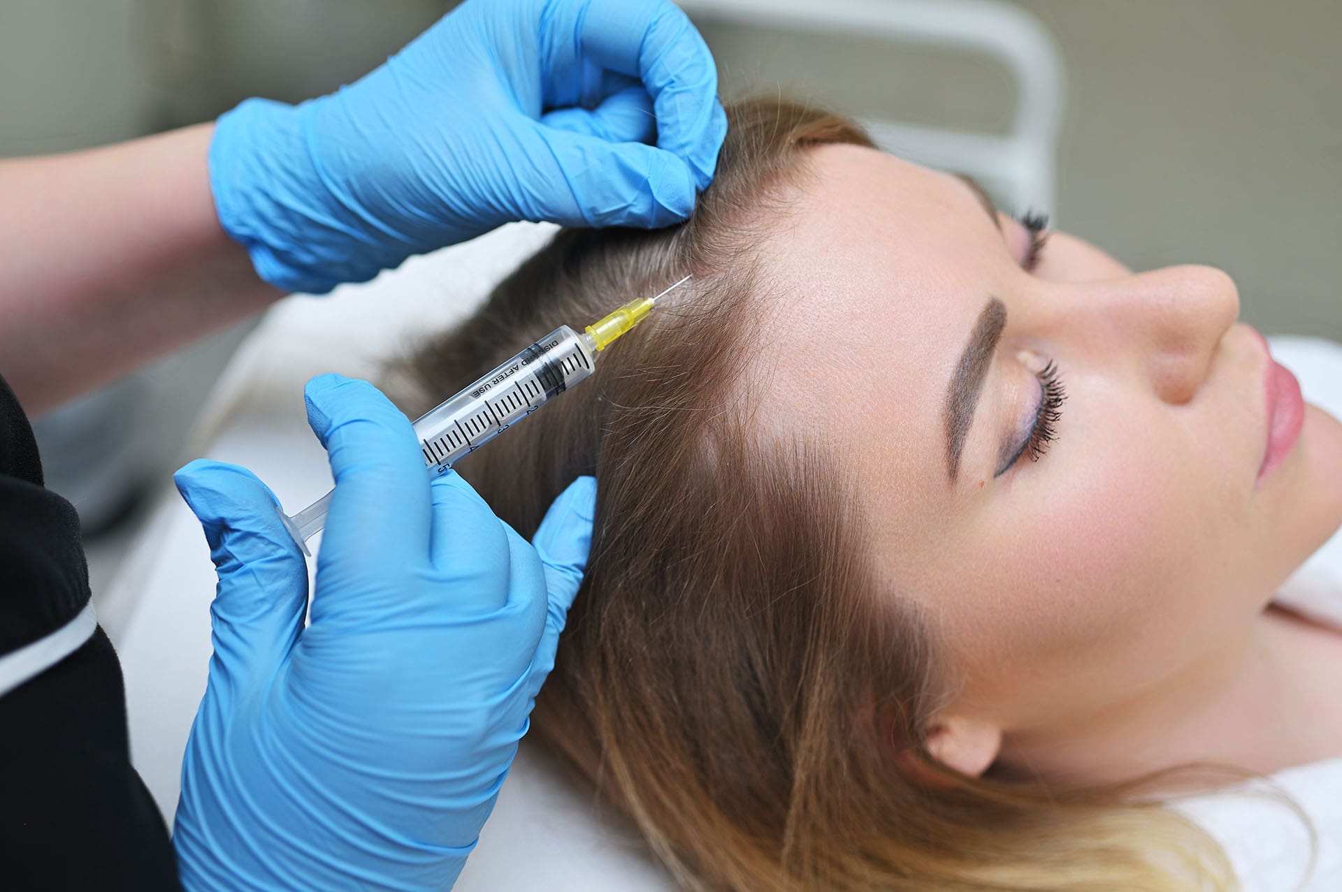
Our in-office procedures include PRP and exosome injections. Exosomes—extracellular vesicles carrying potent signaling molecules—are delivered directly into hair follicle stem cells, powerfully modulating anagen re-entry. We support this biochemically with topical GHK-Cu sprays to modulate dihydrotestosterone (DHT) pathways, effectively reducing the androgenic signaling that miniaturizes follicles.
Systemically, we utilize our oral, enteric-coated Recover capsules containing GHK-Cu, BPC-157, and carnosine. We use a microdosing strategy—five days on, two days off—to maintain cellular sensitivity and prevent receptor downregulation. My wife utilized this exact protocol; we visually documented the restoration of natural blonde pigmentation at the root of her graying hair, a testament to GHK-Cu’s melanin-supporting properties.
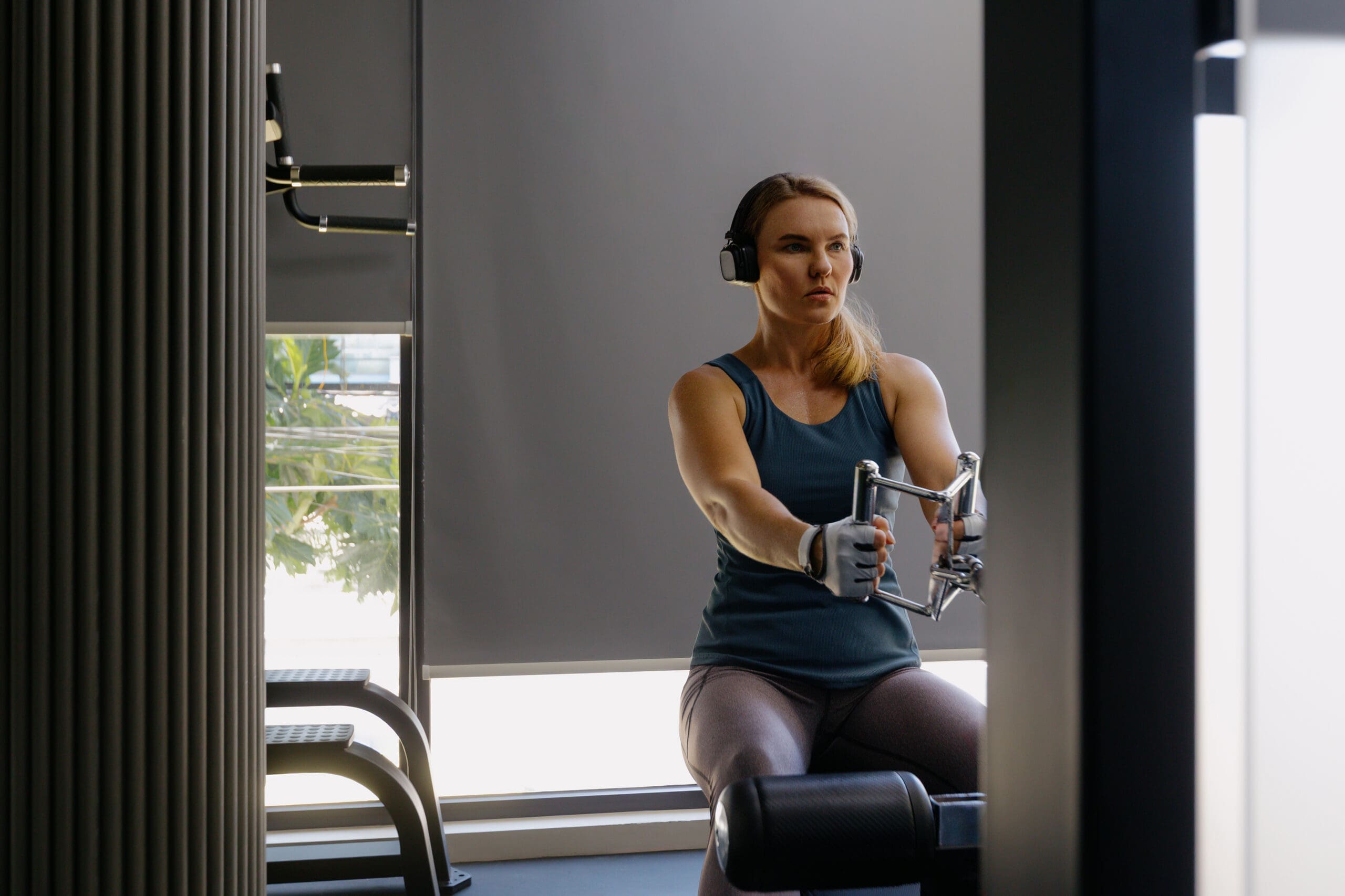
This is where integrative chiropractic care shines. Hair loss is influenced by autonomic imbalance and microvascular constriction. By restoring cervical-thoracic mobility and optimizing cranial-cervical mechanics through targeted chiropractic adjustments, we enhance scalp perfusion and vagal tone. Combined with functional nutrition (managing thyroid status, iron, and zinc), we create the ideal physiological terrain for the peptides to succeed. We also actively treat hair shedding triggered by GLP-1 weight loss using these restorative methods.
Regenerative Aesthetics: Beyond Topical Skincare
The demand for regenerative aesthetics is soaring, representing nearly 80% of our cash-based functional services. Patients want cellular repair, not just superficial masking.
For immediate wrinkle reduction, we utilize SNAP-8, an octapeptide that modulates the SNARE complex at the dermal-muscular interface. By gently attenuating muscular contraction, it provides a non-invasive, lower-risk alternative to botulinum toxin. During a “split-face” clinical demonstration, applying just 4 to 5 drops yields visible softening of fine lines within minutes.
For deep structural rejuvenation, we combine topical blue copper peptides (GHK-Cu) with PRP Microneedling (often called the “Vampire Facial”). The micro-injuries trigger acute inflammation, upregulating TGF-β and VEGF pathways for angiogenesis. By adding our transdermal peptide serums, we have seen tissue recovery times plummet from roughly 3 weeks to just 5 days. We also heavily utilize Polydeoxyribonucleotide (PDRN), a K-beauty innovation derived from salmon DNA extract. PDRN acts as a direct building block for cellular DNA repair, aggressively reducing inflammation and stimulating fibroblast activity.
Neurological Optimization and Longevity: Bioregulators in Practice
Beyond musculoskeletal and aesthetic health, we utilize neuroactive bioregulators for cognitive preservation and longevity.
- Semax and Selank are profoundly effective for cognitive optimization. Delivered via intranasal sprays to access the CNS directly through the olfactory and trigeminal nerve pathways, they bypass first-pass metabolism. I typically recommend Semax during the day to enhance attention and processing speed, while Selank is utilized at night for anxiolysis, mood stabilization, and sleep architecture support.
- Cerebrolysin, a polypeptide-rich formulation, is exceptional for executive function and neuroprotection, though I advise patients to administer it before 6:00 p.m. to avoid disrupting circadian rhythms.
- Epitalon directly influences telomere dynamics, reversing proxies of epigenetic aging.
- Ceruletide A5, derived from calf pineal glands, is the star of our oral Rebalance formulation. With over 60 years of research behind it, it promotes robust neurogenesis. Clinically, we have observed it rapidly resolve severe tinnitus (ringing in the ears) and alleviate early cognitive decline and peripheral neuropathy.
Oral Formulations and Targeted Healing Protocols
To help patients integrate these therapies effortlessly, we provide oral formulations using our patented delivery system:
- Recover (Musculoskeletal & Aesthetic Blend): Contains BPC-157, GHK-Cu, and carnosine. It drastically reduces inflammation, stimulates soft tissue angiogenesis, and visibly upregulates keratin and elastin for skin and nail health.
- Revive (Energy & Mitochondrial Health): An “exercise mimetic” featuring SLU-PP-332 to enhance mitochondrial efficiency, 5-Amino-1MQ to mobilize visceral fat, and NAD+ to fuel ATP synthesis.
- Rebalance (Neurocognitive & Sleep Support): Driven by the neurogenesis properties of Ceruletide A5.
- Oral GLP-1 (Metabolic Support): Features orforglipron, a newly approved oral GLP-1, paired with L-carnitine and L-pyroglutamate.
- GLP Support: Designed to combat injectable side effects with MitoQ, a synthetic bitter melon peptide for glucose regulation, and resveratrol to prevent the muscle wasting commonly termed “Ozempic face.”
In personal injury rehabilitation, merging these peptides with shockwave therapy, which promotes mechanotransduction, alongside my chiropractic joint mobilizations, drastically accelerates ligamentous and tendinous healing by minimizing nociceptive input and maximizing local blood flow.
The Peptide Protocol Portal and AI Integration
With the explosion of interest in regenerative medicine, clinical rigor is paramount. To that end, I spent over a year working 16-hour days to build the Peptide Protocol Portal, an exhaustive digital resource for practitioners.
This portal contains a searchable database detailing the mechanism of action, reconstitution calculators, titration schedules, and supporting human clinical data for every compound. We supply comprehensive Clinic Operator’s Manuals, WADA Disclosures for competitive athletes, and detailed Informed Consent forms. To mitigate malpractice risk, we provide Competency Checklists mandating that patients physically demonstrate safe self-administration techniques in-office before taking any injectable therapies home.
To synthesize this vast data, we trained an AI clinical assistant named Dr. Peppy over a 17-month period. Dr. Peppy can analyze a patient’s specific blood biomarkers, generate a customized, safety-assessed peptide protocol, and print an easy-to-read administration calendar for the patient.
My Personal Journey with Regenerative Healing
The obsessive dedication required to build these protocols took a severe toll on my own physical and mental health. My sleep fragmented, my systemic inflammation skyrocketed, and my metabolic health deteriorated. I was forced to become my own patient.
I utilized DSIP (Delta Sleep-Inducing Peptide) to salvage my circadian rhythms and anchor my REM sleep. I deployed NAD+ therapies to rescue my mitochondrial function, and I ultimately underwent intravenous infusions of mesenchymal signaling cells to quench the systemic fire I had ignited in my body. This grueling personal experience forged a profound, firsthand respect for these molecules. I sacrificed my own homeostasis to build these resources so that other providers and patients would not have to guess their way through the dark.
Conclusion
True integrative care is the elegant sequencing of the right interventions, under rigorous medical oversight, aligned with biomechanical structural freedom. Pathophysiology dictates that tissue heals only when mechanics, metabolism, and microcirculation harmonize. At Injury Medical Clinic PA, the convergence of my chiropractic-functional framework and Dr. Cardenas’s internal medicine leadership removes the friction that slows recovery. Peptides, bioregulators, and regenerative aesthetics are the master keys, but our collaborative, evidence-based model is the door through which lasting vitality is realized.
References
Anitua, E., Pino, A., Martinez, H., & Orive, G. (2013). Platelet-rich plasma for skin and hair: growth factor biology and clinical outcomes. Journal of Plastic, Reconstructive & Aesthetic Surgery, 66(5), 584-593.
Fink, B., & Runels, C. (2017). The “Vampire Facelift” and “Vampire Facial”: A review of the literature on platelet-rich plasma and microneedling in facial rejuvenation. Journal of Drugs in Dermatology, 16(11), 1074-1077.
Jeong, J., Lee, J., & Kim, S. (2020). Polydeoxyribonucleotide (PDRN) as a potential therapeutic agent in dermatology. Journal of Clinical Medicine, 9(6), 1785.
Khavinson, V. K., & Linkova, N. S. (2012). Epitalon and telomere biology: review of mechanisms and clinical observations. Bulletin of Experimental Biology and Medicine, 153(1), 104-108.
Lau, D. C. W., & Teoh, H. (2023). Cagrilintide, a long-acting amylin analogue for the treatment of obesity. The Lancet, 401(10373), 263-265.
Pickart, L., & Margolina, A. (2018). Regenerative and protective actions of the GHK-Cu peptide in the light of the new data. International Journal of Molecular Sciences, 19(7), 1987.
Wang, Y., Smith, J., & Lee, H. (2022). Topical peptide strategies for facial rejuvenation: SNAP-8 and neuromuscular modulation. Journal of Cosmetic Dermatology, 21(4), 1420-1428.
World Anti-Doping Agency (WADA). (2024). The World Anti-Doping Code International Standard Prohibited List. WADA.
Zaitsev, S. V., et al. (2019). Semax and Selank: intranasal neuroactive peptides for cognition and mood. Neurochemical Journal, 13(3), 205-212.
Clinical Observations and Professional Insights by Dr. Alex Jimenez, DC, APRN, FNP-BC, CFMP, IFMCP, ATN, CCST can be found at ChiroMed and LinkedIn.