Detoxing for More Energy: An Evidence-Based Approach

A Smarter Approach for ChiroMed Patients
Many people say they feel better after a detox. They report:
- More energy
- Less brain fog
- Better digestion
- Better focus
- Fewer sugar crashes
In many cases, that improvement is real. But it usually does not happen because a tea, cleanse, or supplement “flushes toxins” out of the body.
It usually happens because people start doing healthier things at the same time, such as:
- Drinking more water
- Eating fewer processed foods
- Cutting back on alcohol
- Sleeping better
- Moving more
- Eating more fiber and whole foods
That kind of reset can absolutely improve energy.
For ChiroMed readers, the best way to think about detoxing is this: support the body’s natural detox systems instead of chasing extreme cleanses. The body already has built-in detox organs—especially the liver, kidneys, digestive tract, and lungs. Your job is to support them with daily habits and, when needed, a personalized care plan (Alexander, 2020; BDA, 2025; Jones & Blackburn, 2025).
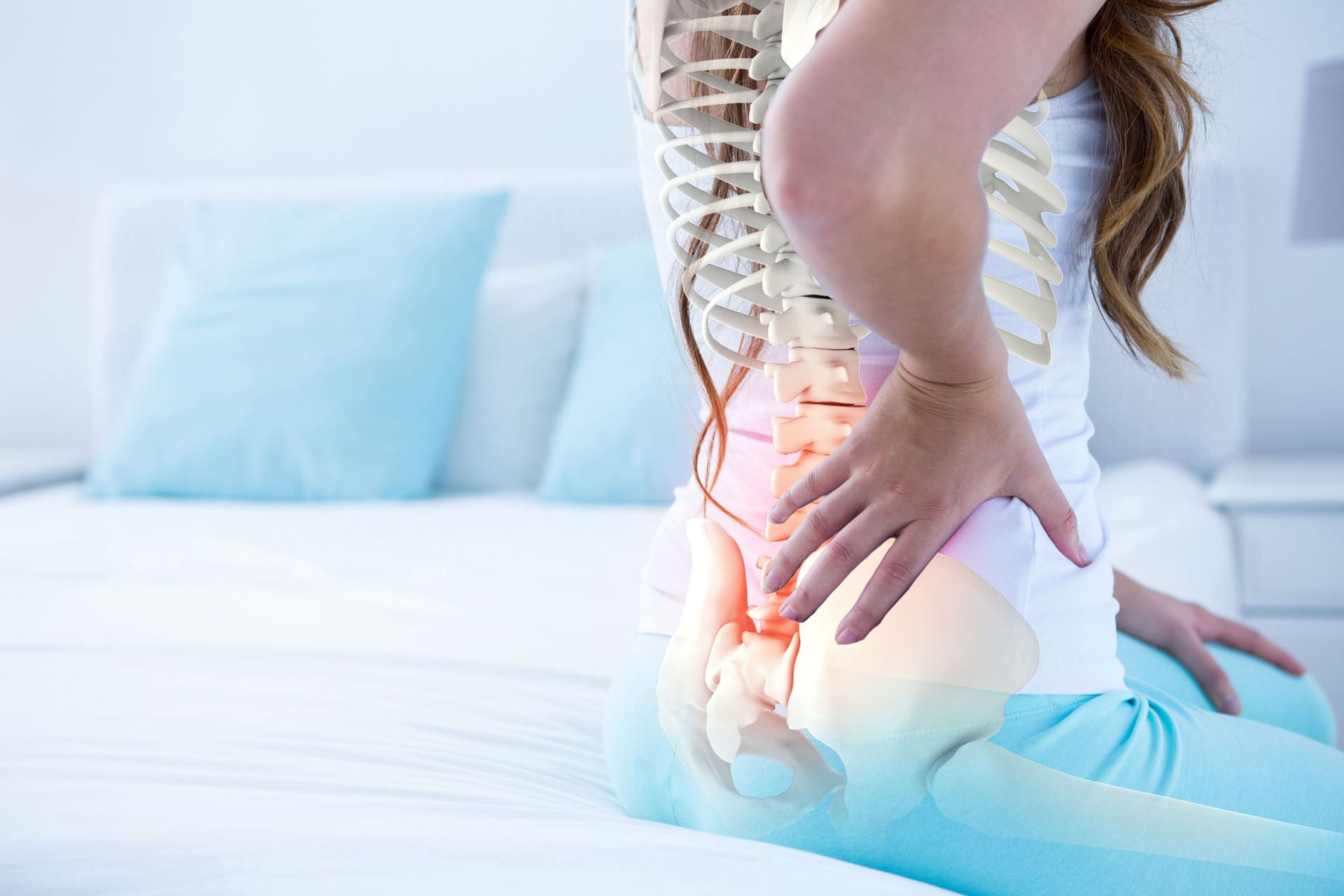
Your Body Already Has a Detox System
One of the biggest myths in wellness marketing is that the body needs a special product to detox itself.
MD Anderson directly addresses this and explains that your body is already designed to detoxify itself, with the liver playing a major role (Alexander, 2020). Their 2025 review also states that the body has its own detoxification system and usually clears harmful substances effectively (Jones & Blackburn, 2025).
The British Dietetic Association (BDA) says something similar. Their 2025 guidance explains that the idea of detox as a special cleanse is misleading, as the body already filters and removes waste through organs such as the skin, gut, liver, and kidneys (BDA, 2025).
Healthline also notes that there is little evidence that detox diets remove toxins, and it explains that the body clears waste through the liver, feces, urine, and sweat (Gunnars & Hultin, n.d.).
What this means for energy
If a person feels better after a “detox,” it is often because they have reduced the things that drain energy, not because they used a special cleansing product.
That is good news, because it means you can improve energy with sustainable habits.
Why Detoxing Can Improve Energy (When Done the Right Way)
The word “detox” is used in many confusing ways. A better phrase is “lifestyle reset.”
When people make better food and lifestyle choices, their energy can improve because their bodies experience less stress.
Common reasons energy improves during a detox-style reset
- Blood sugar becomes more stable
- Digestion improves
- Sleep improves
- Alcohol intake decreases
- Hydration improves
- Inflammation may decrease
- Nutrient intake improves
Healthline specifically notes that some people feel more focused and energetic during and after detox diets, but this may be because they removed processed foods and alcohol and improved vitamin and mineral intake (Gunnars & Hultin, n.d.).
BDA also points out that people may feel better during detox periods, but the likely reason is healthier habits like less alcohol, better sleep, fresh air, and exercise—not detox products (BDA, 2025).
That message is especially important for ChiroMed patients, because true energy recovery often comes from a whole-body plan, not a quick fix.
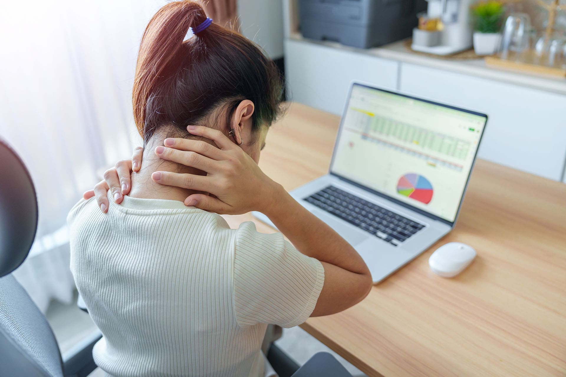
How Processed Foods and Sugar Can Drain Energy
A big reason many people feel tired is not “toxins” in a vague sense. It is often a daily food pattern.
Highly processed foods can lead to:
- Fast blood sugar spikes
- Fast crashes a few hours later
- Cravings
- Brain fog
- Mood swings
- Low energy
If a person starts a detox and removes sugary drinks, packaged snacks, and ultra-processed meals, they may feel better quickly simply because their blood sugar and appetite become more stable.
MD Anderson’s 2025 detox guidance recommends avoiding ultra-processed foods and building meals around vegetables, whole grains, fruits, and beans to support the body’s natural detox system (Jones & Blackburn, 2025).
BDA also recommends avoiding excess caffeine, alcohol, and high-fat, high-sugar, and high-salt foods, and instead choosing a balanced diet and adequate hydration (BDA, 2025).
Simple food swaps that support energy
- Soda → water or sparkling water
- Pastries → oats with nuts and fruit
- Chips → fruit + nuts or hummus + vegetables
- Fast food lunch → protein bowl with greens and beans
- Candy bars → yogurt, fruit, and seeds
These changes are not extreme. But they can make a major difference.
Fiber Helps Digestion, Gut Health, and Steadier Energy
Many people who feel “toxic” are actually dealing with digestive stress, low fiber intake, or poor meal quality.
Mass General’s nutrition article explains that fiber helps bind compounds in the gut and move them out through stool. It also notes that regular bowel movements reduce the time that harmful compounds remain in the intestinal tract, and that fiber-rich foods support healthy gut bacteria (Mass General, 2020).
This matters for energy because digestive health affects how you feel all day.
Signs that digestion may be affecting energy
- Bloating after meals
- Constipation
- Irregular bowel movements
- Feeling heavy or sluggish after eating
- Brain fog after processed meals
- Energy crashes
Fiber-rich foods that support a gentle detox lifestyle
- Beans and lentils
- Oats
- Whole grains
- Vegetables
- Fruits
- Nuts and seeds
Mass General specifically lists beans, whole grains, oats, vegetables, fruits, nuts, and seeds as fiber sources that support gut health and detox-related functions (Mass General, 2020).
For many people, simply increasing fiber and water leads to better energy within days.
Hydration Is One of the Fastest Ways to Feel Better
A lot of people start a detox and suddenly drink more water. That alone can improve how they feel.
Mild dehydration can contribute to:
- Fatigue
- Headaches
- Poor concentration
- Irritability
- Low exercise tolerance
MD Anderson’s 2025 article recommends drinking more water during the day as part of a healthy plan that supports the body (Jones & Blackburn, 2025). BDA also recommends staying hydrated with water and sugar-free drinks (BDA, 2025). Women’s Health Network also includes hydration as part of its gentle energy-supporting detox advice (Stills, 2025).
Easy hydration tips
- Start the day with a glass of water
- Drink water before coffee
- Keep a refillable bottle nearby
- Add lemon or cucumber if plain water is hard to drink
- Drink more on hot days or workout days
Hydration is not flashy, but it works.
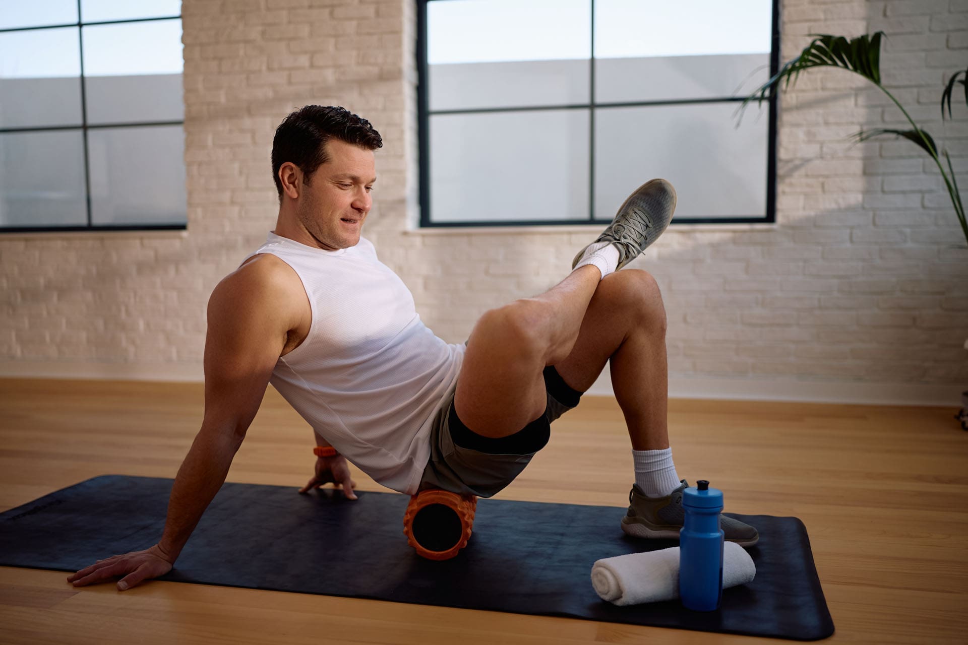
Movement Supports Energy and Natural Detox Pathways
A detox plan that does not include movement is missing a major piece.
Women’s Health Network explains that exercise supports detoxification by helping the lymph system and sweating, and it also supports mood and helps with fatigue (Stills, 2025).
MD Anderson’s 2025 guidance also highlights regular exercise as part of maintaining health and supporting a healthy weight, recommending weekly activity goals (Jones & Blackburn, 2025).
Why movement boosts energy
- Improves circulation
- Supports lymphatic flow
- Improves insulin sensitivity
- Helps mood and stress
- Improves sleep quality
- Reduces stiffness and pain
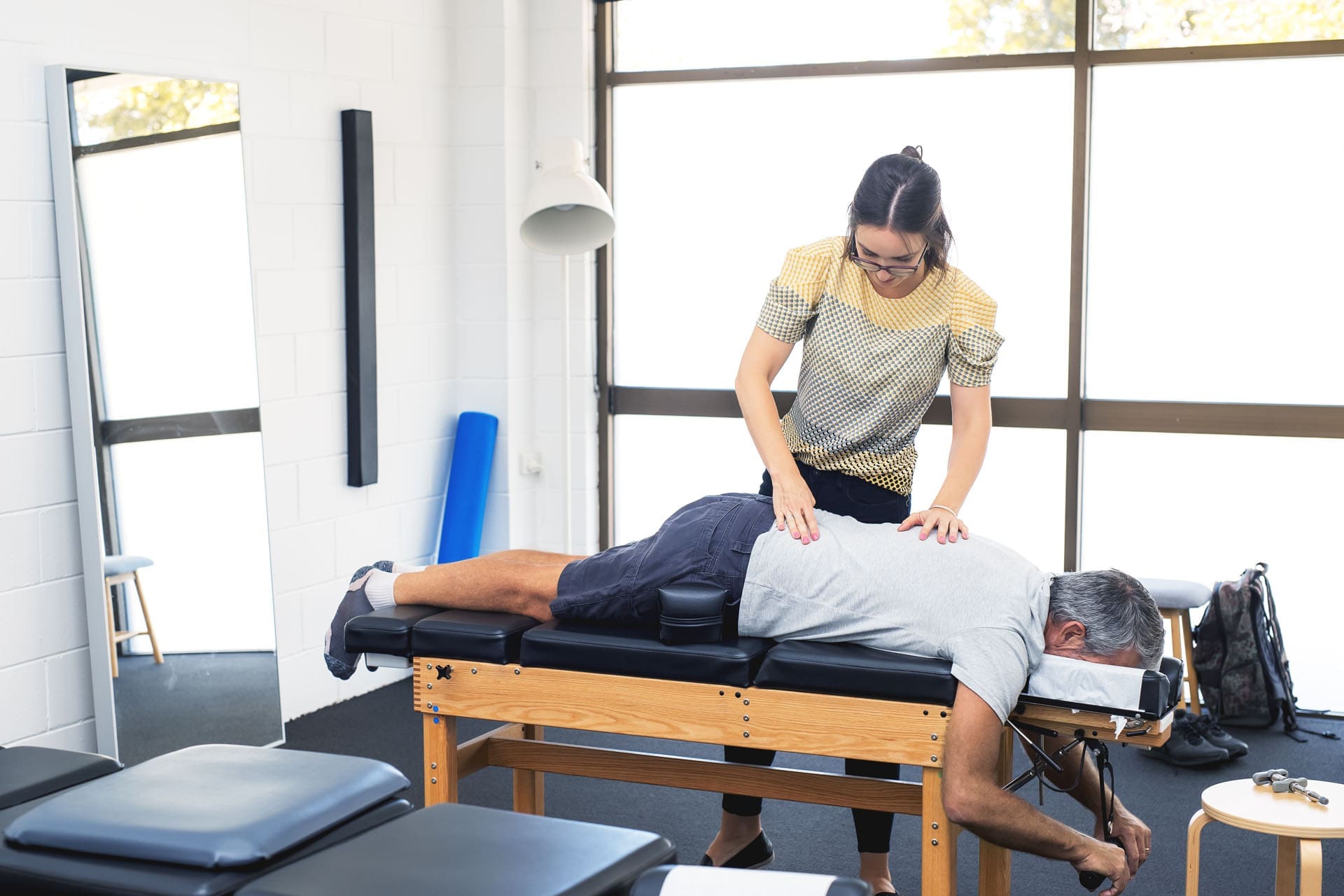
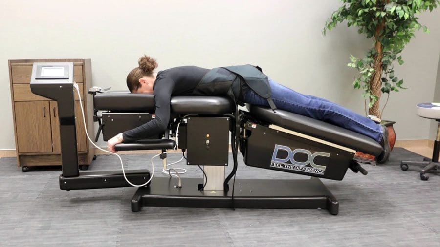
For ChiroMed patients, this is especially important. Pain, poor posture, and restricted movement can increase fatigue. A person who moves better usually feels better.
Good beginner options
- 20–30 minute walks
- Gentle mobility work
- Light stretching
- Bodyweight exercises
- Short exercise “snacks” during the day
You do not need a hard detox workout plan. You need consistency.
Cutting Back on Alcohol Often Improves Energy Fast
Many people notice a quick energy boost when they stop drinking alcohol for a few weeks.
Why?
Because alcohol can affect:
- Sleep quality
- Hydration
- Blood sugar
- Mood
- Recovery
- Appetite
BDA recommends avoiding excessive alcohol and focusing on balanced eating and hydration (BDA, 2025). MD Anderson also emphasizes that healthy habits are more useful than cleanses for supporting the body (Jones & Blackburn, 2025; Alexander, 2020).
If someone says, “I detoxed, and now I have more energy,” sometimes the biggest reason is simply that they took a break from alcohol and started sleeping better.
Why Some People Feel Worse Before They Feel Better
This is a common concern.
Some people begin a detox-style reset and feel tired, irritable, or headachy for a few days.
Healthline notes that many people report feeling unwell during detox periods (Gunnars & Hultin, n.d.). Cenikor also describes early symptoms such as fatigue, headaches, nausea, and mood swings during detox transitions (Cenikor Foundation, 2024).
Common reasons this happens
- Caffeine withdrawal
- Sugar withdrawal
- Eating too little
- Low protein intake
- Sudden diet changes
- Poor sleep
- Stress about changing habits
Cenikor also notes that sudden shifts in diet and stimulants can contribute to these symptoms (Cenikor Foundation, 2024).
Signs your detox plan is too aggressive
- Dizziness
- Shaking
- Severe weakness
- Trouble thinking clearly
- Constant headaches
- Nausea that does not improve
- Not enough calories or protein
A healthy reset should help your body, not punish it.
ChiroMed Approach: Whole-Body, Personalized Energy Support
For ChiroMed readers, a key part of this topic is the clinical approach.
ChiroMed describes Dr. Alex Jimenez as a dual-licensed provider (Doctor of Chiropractic and Advanced Practice Nurse Practitioner) who leads a multidisciplinary team in El Paso, with a patient-centered, integrative care model (ChiroMed, n.d.-a). The ChiroMed services page also describes evidence-based, integrative principles and lists Dr. Jimenez’s credentials and contact options (ChiroMed, n.d.-b).
ChiroMed also includes a dedicated article on integrative chiropractic detox support, describing Dr. Jimenez’s combined chiropractic and APRN perspective and how that model can support a broader detox and wellness strategy (ChiroMed, n.d.-c).
Why this matters for people with low energy
Low energy is often not caused by one issue.
A person may have a mix of problems, such as:
- Poor sleep
- Pain and inflammation
- Digestive stress
- Blood sugar swings
- Sedentary lifestyle
- Stress overload
- Poor recovery habits
A multidisciplinary clinic can help build a more complete plan.
Examples of what a personalized plan may include
- Nutrition changes (not crash cleanses)
- Hydration goals
- Activity and mobility support
- Chiropractic and rehab-based movement care
- Sleep and stress support
- Medical evaluation when fatigue persists
This is the difference between a generic detox trend and a real, personalized health plan.
A Safe, Practical Detox-for-Energy Plan (2–4 Weeks)
If your goal is better energy, here is a realistic plan that supports the body’s normal detox systems.
Eat more whole foods
Focus on:
- Vegetables
- Fruit
- Beans
- Whole grains
- Lean protein
- Nuts and seeds
This matches BDA and MD Anderson’s advice to prioritize balanced, nutrient-dense foods over restrictive cleanses (BDA, 2025; Jones & Blackburn, 2025).
Reduce common energy drains
Cut back on:
- Alcohol
- Sugary drinks
- Ultra-processed snacks
- Late-night heavy meals
- Excess caffeine
Hydrate daily
- Drink water consistently
- Use sugar-free drinks if needed
- Increase fluids with exercise or heat
Move every day
- Walk
- Stretch
- Do light strength work
- Keep it simple and repeatable
Sleep like it matters
- Set a regular sleep time
- Reduce screen time at night
- Avoid heavy meals late
- Cut late caffeine
Get help if fatigue stays
If fatigue is ongoing, severe, or getting worse, it may not be a detox issue. It may need medical evaluation.
That is where a clinic like ChiroMed can help guide the next steps with a broader view.
Important Safety Note About “Detox”
The word “detox” can also mean medical detox from alcohol or drugs, which is a different situation than a wellness reset.
Cenikor’s materials describe detox symptoms and management in the substance withdrawal setting, including fatigue, headaches, nausea, and mood changes (Cenikor Foundation, 2024). That type of detox may require medical supervision and should not be handled as a home wellness cleanse.
Also, MD Anderson’s colon cleansing guidance warns that colon cleanses lack proven health benefits and may cause harm, including dehydration and electrolyte disturbances (Bresalier, 2025).
Bottom line on safety
Avoid:
- Extreme fasting cleanses
- Colon cleanses
- Laxative detox teas
- Very low-calorie plans
- “Miracle” detox products
Choose:
- Balanced food
- Water
- Fiber
- Movement
- Sleep
- Personalized care
Final Takeaway for ChiroMed Readers
Yes, “detoxing” can improve energy—but the real benefit usually comes from a smarter reset, not a trendy cleanse.
The best energy-supporting detox plan is one that:
- Supports the liver, kidneys, gut, and lungs
- Uses whole foods instead of restrictive products
- Improves hydration and sleep
- Reduces alcohol and processed foods
- Includes movement
- Is personalized to the patient
That approach aligns well with ChiroMed’s integrative, patient-centered model. It is practical, evidence-based, and focused on root causes—not short-term hype.
If your low energy is persistent, the next best step is not another cleanse. It is a comprehensive evaluation and a plan tailored to your body.
References
- Alexander, H. (2020, October 26). 4 detox myths: Get the facts. MD Anderson Cancer Center.
- Bresalier, R. (2025, August 14). Colon cleanse: Health or hype? MD Anderson Cancer Center.
- British Dietetic Association. (2025). Detox diets.
- Cenikor Foundation. (2024, November 14). What to expect during detox.
- ChiroMed. (n.d.-a). ChiroMed – Integrated medicine holistic healthcare in El Paso, TX.
- ChiroMed. (n.d.-b). Integrated medicine services El Paso TX.
- ChiroMed. (n.d.-c). Nurse practitioners and integrative chiropractic detox.
- Gunnars, K., & Hultin, G. (n.d.). Do detox diets and cleanses really work? Healthline.
- Jones, V., & Blackburn, K. B. (2025, April 29). Detoxes, cleanses and fasts: What you should know. MD Anderson Cancer Center.
- Mass General. (2020, August 26). How to detox through diet. Massachusetts General Hospital Giving.
- Stills, D. (2025, November 3). 4 gentle energy-boosting detox tips. Women’s Health Network.