Care is the cornerstone of our practice

Give us a Call
+1 (915) 412-6680
Send us a Message
support@chiromed.com
Opening Hours
Mon-Thu: 7 AM - 7 PM
Fri - Sun: Closed

Rebuilding Strength After a Crash: How Sports Training and Physical Therapy Help Recover from Motor Vehicle Accidents


Introduction

Motor vehicle accidents (MVAs) can be life-changing. Even when injuries seem minor at first, their effects can linger for months or even years. From whiplash and nerve damage to gastrointestinal issues and chronic joint pain, the trauma goes far beyond broken bones. Fortunately, sports training and physical therapy offer a proven path to healing. These targeted methods restore mobility and improve long-term health. Integrative approaches—such as chiropractic care and functional medicine—further strengthen this recovery by treating the whole person, rather than just isolated injuries.


Understanding the Impact of Motor Vehicle Accidents

Hidden Damage: More Than Just Broken Bones

Motor vehicle accidents can cause injuries that aren’t immediately obvious. Even low-speed collisions can lead to:

  • Nerve compression or inflammation
  • Musculoskeletal joint pain
  • Back and neck misalignments
  • Gastrointestinal trauma
  • Balance and coordination issues

These injuries often develop over time, especially if left untreated. Whiplash, for example, might only seem like a sore neck at first—but it can lead to nerve irritation and chronic pain if not properly addressed. (IS Rehab, n.d.).


Chiropractic and Integrative Medicine: The Foundation of Holistic Recovery

Dr. Alexander Jimenez’s Dual-Scope Approach

As both a Doctor of Chiropractic and Family Nurse Practitioner, Dr. Alexander Jimenez specializes in diagnosing and treating complex MVA injuries in El Paso. His dual-scope expertise enables him to utilize advanced imaging and diagnostics—such as MRIs and musculoskeletal ultrasound—to identify underlying problems that general practitioners might overlook.

He also incorporates functional medicine, a method that views the body’s systems as interconnected. According to Dr. Jimenez, this approach helps pinpoint the root causes of chronic pain, nerve inflammation, or gut imbalances that can occur following a crash. (Jimenez, n.d.).


The Role of Physical Therapy in MVA Recovery

Why Movement Is Medicine

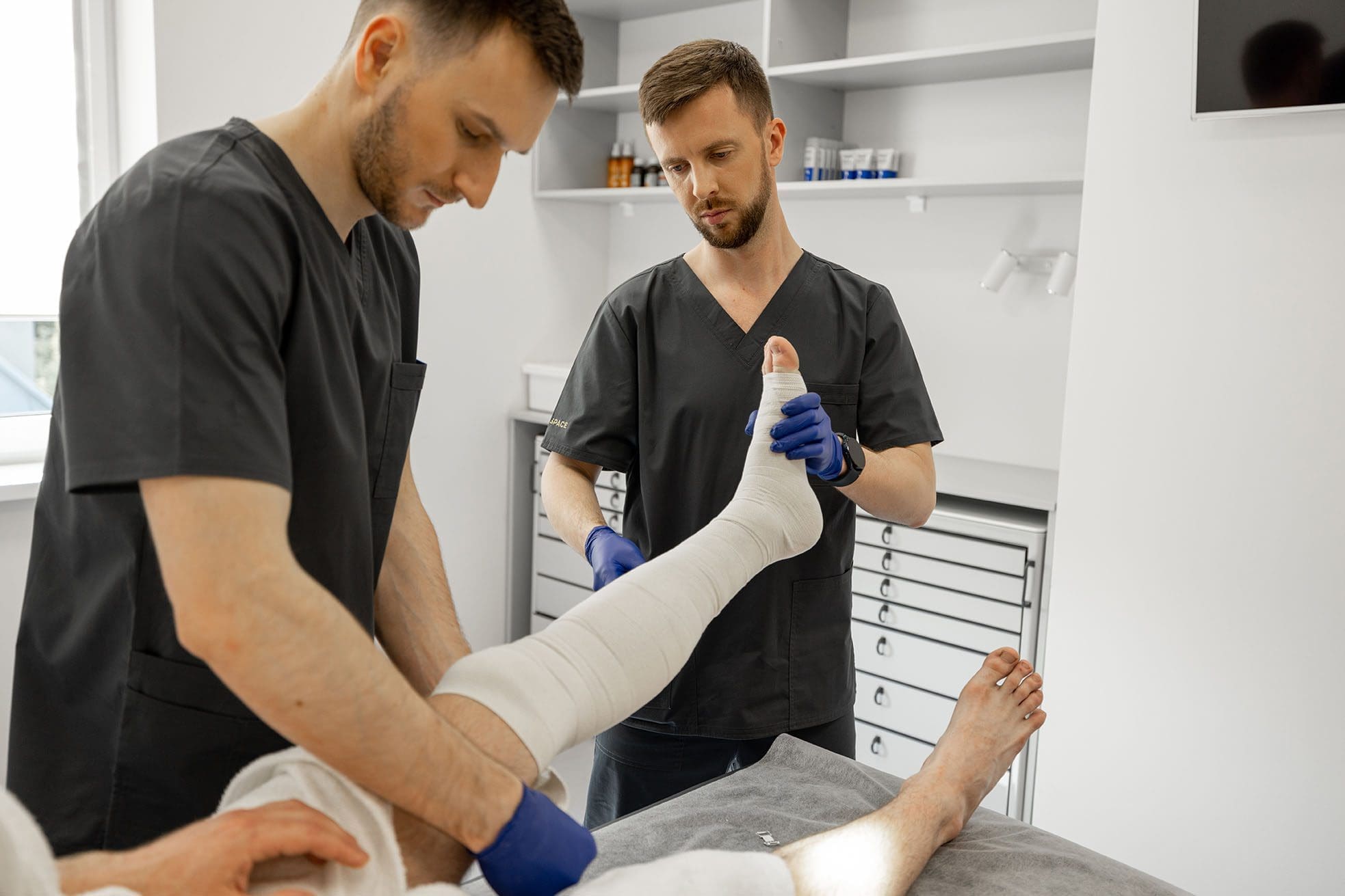
Physical therapy (PT) is one of the most effective tools for MVA recovery. PT doesn’t just treat symptoms; it corrects postural imbalances, reduces inflammation, and restores strength and flexibility. According to Advanced Orthopedic & Physical Therapy, physical therapy (PT) helps reduce nerve compression and improve joint mobility through targeted exercises, such as nerve glides and resistance training. (Advance Orthopt, n.d.).

Common PT treatments after MVAs include:

  • Manual therapy to realign joints
  • Stretching to reduce stiffness
  • Strength-building exercises
  • Balance and coordination training
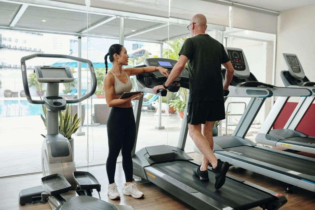
  • Gait retraining

Sports Training: Strengthening the Body Post-Injury

The Shift from Passive to Active Recovery

Sports training brings patients from passive recovery to active participation in their health. It includes:

  • Bodyweight exercises like push-ups or squats
  • Stretching and dynamic warm-ups
  • Balance and agility drills
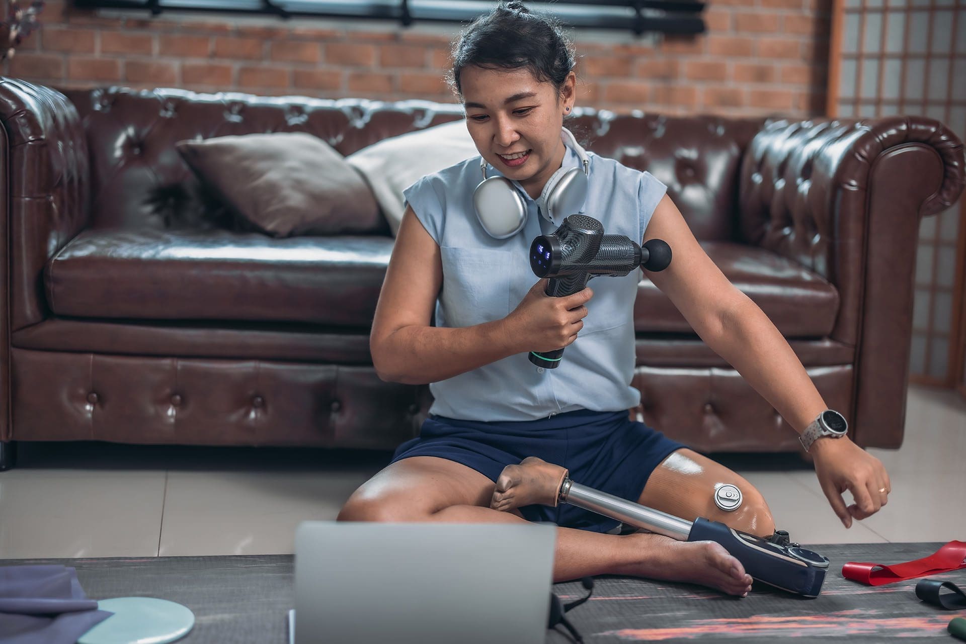
  • Resistance band workouts

As the body heals, structured physical activity becomes essential. It helps stabilize joints, build muscular endurance, and boost cardiovascular function—all while decreasing the likelihood of re-injury (Sports PT Centers, n.d.).


Managing Nerve Damage After an MVA

Understanding Neurological Trauma

Injuries to the neck and spine can pinch or inflame nerves, leading to symptoms like:

  • Tingling or numbness
  • Shooting pain in limbs
  • Muscle weakness

Chiropractic adjustments, nerve mobilization, and neuromuscular reeducation are all effective methods for managing these issues. Dr. Jimenez often uses digital motion X-rays and electromyography (EMG) to evaluate the extent of nerve damage before recommending treatments (Jimenez, n.d.).


Addressing Back and Neck Pain through Integrated Care

How Chiropractic and PT Work Together

Back and neck pain are among the most common complaints related to motor vehicle accidents (MVAs). Dr. Jimenez uses chiropractic care to realign the spine, reduce pressure on nerves, and support faster healing. He complements this with PT exercises aimed at restoring cervical and lumbar mobility.

Integrative care strategies may include:

  • Soft tissue massage
  • Spinal decompression
  • Heat/cold therapy
  • Core stabilization workouts

Together, these methods promote faster healing and improve long-term outcomes (Solutions Physical Therapy, n.d.).


The Gut-Brain Connection: Intestinal Injury After MVAs

A Lesser-Known but Serious Consequence

Not all injuries are external. Abdominal trauma from seatbelts or impact can cause intestinal bruising, bloating, or even motility issues. Stress hormones released after a crash may also disrupt the gut-brain axis, leading to chronic indigestion or constipation.

Dr. Jimenez uses functional lab testing and stool analysis to detect internal imbalances, often addressing them with:

  • Anti-inflammatory diets
  • Digestive enzymes
  • Probiotics
  • Manual visceral therapy

These approaches can help heal the gut from both physical and stress-related damage (PDF Source).


Rehabilitation Timeline: When Can You Start Moving?

From Rest to Regeneration

Patients often wonder when they can resume exercising. According to Maxwell Therapy, patients can begin gentle range-of-motion exercises within days of an accident, depending on the nature of the injury. Most rehab specialists recommend a progressive routine:

  1. Phase 1: Rest and Stabilization – Reduce swelling and protect injured areas.
  2. Phase 2: Light Movement – Gentle stretching and isometric exercises.
  3. Phase 3: Functional Exercises – Squats, lunges, light weights.
  4. Phase 4: Return to Sport or Activity – Full-body conditioning.

Delaying exercise can cause stiffness, muscle wasting, and postural dysfunction (Maxwell Therapy, n.d.).


How Dr. Jimenez Combines Legal and Medical Support

Injury Recovery Meets Documentation

One unique strength of Dr. Jimenez’s care in El Paso is his ability to merge medical treatment with legal documentation. Many personal injury cases require detailed reports, imaging, and prognosis notes for attorneys and insurance companies. Dr. Jimenez provides:

  • Functional range-of-motion assessments
  • Impairment ratings
  • SOAP notes for legal use
  • Narrative reports based on objective findings

His dual-scope licensure grants him the authority to order and interpret diagnostic tests, such as MRIs and CT scans, thereby strengthening both clinical care and legal claims. (Jimenez, n.d.).


Mental and Emotional Recovery: The Often-Forgotten Component

Addressing Psychological Trauma

After an MVA, it’s not just the body that suffers. Anxiety, PTSD, and depression are common. Exercise and chiropractic care naturally help regulate the nervous system by releasing endorphins and improving circulation. Functional medicine adds adaptogenic herbs and stress-reducing therapies like:

  • Acupuncture
  • Mind-body therapies (yoga, breathwork)
  • Lifestyle coaching

This full-spectrum approach makes long-term recovery more attainable and sustainable (PDF Source).


Conclusion: Movement Is the Best Medicine

Recovering from a motor vehicle accident takes more than rest. It requires a structured plan—one that includes physical therapy, sports training, chiropractic adjustments, and integrative medicine. Dr. Alexander Jimenez in El Paso serves as a model for this type of care. His ability to combine clinical diagnostics with rehabilitative therapies and legal documentation ensures that patients recover physically and secure the support they need to return to their lives with confidence.


References


Bunions Management: Relief and Care Strategies

What is the most effective method for managing the initial symptoms of bunions?

Bunions

A bunion, which is also known as hallux valgus, is a bony bump that is frequently excruciating and located on the side of the big toe. A misalignment of the big toe is the most prevalent cause of bunions. Redness, edema, tenderness, and thickened skin around the big toe joint are early indications of bunions. Although it is impossible to halt the progression of a bunion, it is possible to alleviate its symptoms as it progresses. Pain management strategies and various treatment options are among the early indications of bunions. (MedlinePlus, 2024)

Visual Signs of Early Symptoms

Bunions initially develop slowly and do not cause significant discomfort right away. When a bunion first begins to form, you can notice the following symptoms around your metatarsophalangeal (MTP) joint, which joins the base of your big toe to the remainder of your foot. (American Academy of Orthopaedic Surgeons, 2022)

  • Erythema
  • Edema Hyperpigmentation
  • Hyperkeratotic skin
  • A bony protrusion exists
  • Prominence where the hallux deviates towards the second digit or even beneath it
  • Calluses and corns

Initial Somatic Indicators

Although bunions may not be immediately apparent, they can still cause discomfort in the early stages. The following are some of the initial physical indicators of a developing bunion: (MedlinePlus, 2024)

  • Pain in the foot and big toe
  • This discomfort is especially evident during ambulation or when donning constrictive, pointed footwear.
  • Decreased movement of the big toe
  • Tenderness
  • Inflammation
  • Swelling
  • Stiffness
  • Heat

Stages

Bunions are typically progressive, meaning they tend to deteriorate over time. Failure to implement preventive measures for your bunions may result in consequences. Subsequent symptoms often accompany advanced-stage bunions. (American Academy of Orthopaedic Surgeons, 2022)

  • Persistent, intense pain in and around the metatarsophalangeal joint and the lateral and plantar aspects of the foot
  • Bursitis results in the formation of a fluid-filled cyst near the base of the big toe.
  • Hallux adducting and maybe overlapping the second toe
  • Excessive osseous proliferation along the lateral aspect of the hallux
  • Inability to accommodate your standard footwear
  • Impediment in ambulation
  • Hammertoe abnormalities occur when the second, third, or fourth toes exhibit an upward bend at the middle joint, like a hammer or claw.
  • Hallux rigidus, a kind of arthritis affecting the big toe

Halt the Advancement

Once bunions have begun to form, they will become irreversible. Nevertheless, some methods exist to prevent their exacerbation or the onset of additional issues. These encompass (American Academy of Orthopaedic Surgeons, 2022)

  • Refraining from wearing improperly fitting footwear and elevated heels
  • Utilizing orthopedic footwear and/or broad, comfy, soft-soled, and low-heeled shoes
  • Inserting spacers between the toes to avert friction and irritation
  • Applying over-the-counter (OTC) pads composed of felt, silicone, or foam to the bunion
  • Extending your calf muscles to enhance joint alignment

Analgesic Administration

Ice packs and non-steroidal anti-inflammatory medicines (NSAIDs), such as Advil or Motrin (ibuprofen), may alleviate bunion discomfort. Research indicates that Botox injections into the forefoot muscles may offer analgesic benefits. (Hurn, S. E., et al., 2022)

Non-invasive Therapy in Initial Phases

If your bunions continue to deteriorate despite using the aforementioned self-care measures, you may need to seek intervention from a podiatrist or another healthcare professional. A healthcare professional may advise

Foot orthoses, commonly referred to as orthotics

  • Orthoses, also known as foot orthoses, are specially designed implants that alleviate bunion-associated pain and prevent chafing.

Splints

Physical Therapy

  • A physical therapist can assist you by offering exercises to enhance the alignment of your feet and joints. They may further administer manual treatment to alleviate pain. (Hurn, S. E., et al., 2022)

Podiatrist

A podiatrist is a medical doctor (M.D.) specializing in the treatment of health issues affecting the foot, ankles, and lower legs. Request a referral to a podiatrist from your healthcare physician if you seek assistance in managing your bunion problems. (American Podiatric Medical Association, 2025)

Chiropractic Perspective

Chiropractors focus on biomechanical alignment and musculoskeletal health, viewing bunions as part of a broader kinetic chain dysfunction. Their approach emphasizes manual therapies and functional restoration.

  1. Biomechanical Assessment:
    • Evaluate foot, ankle, knee, hip, and spinal alignment to identify compensatory patterns contributing to bunion formation (e.g., overpronation, pelvic misalignment).
    • Assess gait and foot mechanics to identify areas of abnormal stress on the big toe joint.
  2. Chiropractic Interventions:
    • Adjustments: Perform gentle manipulations to the foot and ankle (e.g., metatarsal or tarsal adjustments) to improve joint mobility and reduce stress on the bunion. Spinal or pelvic adjustments may address upstream biomechanical issues.
    • Soft Tissue Therapy: Utilize techniques such as myofascial release or instrument-assisted soft tissue mobilization to reduce tension in the foot’s muscles and fascia, thereby improving circulation and flexibility.
    • Taping: Apply kinesiology tape to support the big toe and reduce strain during movement.
  3. Corrective Exercises:
    • Prescribe exercises to strengthen intrinsic foot muscles and improve toe alignment, such as:
      • Toe spreading: Actively spread toes apart to strengthen the abductor hallucis.
      • Arch strengthening: Perform short foot exercises to support the medial longitudinal arch.
      • Calf stretches: Address tight Achilles tendons that contribute to foot strain.
    • Recommend balance and proprioception exercises (e.g., standing on one leg) to improve overall foot stability.
  4. Orthotic and Footwear Guidance:
    • Fit patients with custom orthotics to correct overpronation or supination, which can exacerbate bunions.
    • Advise on minimalist or wide-toe-box shoes to promote natural foot mechanics, aligning with chiropractic principles of functional movement.
  5. Holistic Approach:
    • Address lifestyle factors, such as posture and ergonomics, that affect lower extremity alignment.
    • Educate on anti-inflammatory diets (e.g., rich in omega-3 fatty acids, low in processed sugars) to help reduce joint inflammation.

Nurse Practitioner Perspective

NPs focus on holistic patient care, symptom management, and coordinating multidisciplinary treatment. Their approach to bunions focuses on pain relief, slowing the progression, and providing patient education.

  1. Assessment and Diagnosis:
    • Conduct a thorough history and physical exam to evaluate bunion severity, pain level, and contributing factors (e.g., footwear, family history, arthritis).
    • Order imaging (X-rays) if needed to assess joint alignment and rule out complications like osteoarthritis or bone spurs.
    • Screen for systemic conditions (e.g., rheumatoid arthritis, gout) that may worsen bunions.
  2. Conservative Management:
    • Footwear Education: Recommend wide-toe-box shoes with low heels to reduce pressure on the bunion. Avoid high heels and narrow shoes.
    • Orthotics: Prescribe custom or over-the-counter orthotic inserts to support the arch, redistribute pressure, and correct foot mechanics.
    • Padding and Splinting: Suggest bunion pads or spacers to cushion the area and reduce friction. Night splints may help align the toe, though evidence for their effectiveness is mixed.
    • Pain Relief: Advise over-the-counter NSAIDs (e.g., ibuprofen) for pain and inflammation, ensuring proper dosing and monitoring for side effects. For severe cases, refer to a specialist for corticosteroid injections.
    • Physical Therapy: Refer to a physical therapist for exercises to strengthen foot muscles, improve flexibility, and enhance gait. Examples include toe stretches, towel curls, and marble pickups.
  3. Patient Education:
    • Educate on weight management to reduce stress on the feet.
    • Discuss lifestyle changes, such as avoiding prolonged standing or high-impact activities that worsen symptoms.
    • Highlight the progressive nature of bunions and the importance of early intervention.
  4. Referral and Coordination:
    • Refer to a podiatrist or orthopedic surgeon if conservative measures fail or if the bunion causes significant pain, deformity, or functional impairment.
    • Collaborate with chiropractors, physical therapists, or other specialists for a comprehensive care plan.

Both NPs and chiropractors emphasize:

  • Early Intervention: Addressing bunions early can prevent the worsening of deformity.
  • Footwear Modification: Wearing wide, supportive shoes is critical to reducing pressure.
  • Exercise and Mobility: Strengthening and stretching exercises improve foot function.
  • Pain Management: Non-invasive methods, such as padding, icing, or non-steroidal anti-inflammatory drugs (NSAIDs), are the first-line approach.
  • Referral for Surgery: If conservative measures prove ineffective, a specialist may be consulted for surgical options, such as bunionectomy, although this is typically considered a last resort.

Limitations and Considerations

  • Evidence Gaps: While orthotics and exercises are widely recommended, studies on their efficacy for bunions are limited. Splints may not correct severe deformities.
  • Individual Variation: Treatment must be tailored to the patient’s bunion severity, lifestyle, and comorbidities.
  • Surgical Indications: Severe pain, joint damage, or inability to walk may necessitate surgical consultation, which neither NPs nor chiropractors are qualified to perform.

Injury Medical Chiropractic and Functional Medicine Clinic

Dr. Jimenez, a nurse practitioner, integrates medical expertise with chiropractic care to address a wide range of conditions. The clinic provides individualized care programs that incorporate functional medicine, acupuncture, electroacupuncture, and sports medicine. The clinic addresses chronic pain syndromes and injuries by prioritizing strength, agility, and flexibility. Comprehensive care programs, combined with in-person and virtual health coaching, ensure personalized treatment and wellness outcomes for patients of all ages and abilities.


Enhance Your Performance with Functional Foot Orthotics


References

MedlinePlus (2024). Bunions. U.S. Department of Health and Human Services.

American Academy of Orthopaedic Surgeons. (2022). “Bunions.” OrthoInfo. from https://orthoinfo.aaos.org/en/diseases–conditions/bunions/.

Hurn, S. E., Matthews, B. G., Munteanu, S. E., & Menz, H. B. (2022). Effectiveness of Nonsurgical Interventions for Hallux Valgus: A Systematic Review and Meta-Analysis. Arthritis care & research, 74(10), 1676–1688. https://doi.org/10.1002/acr.24603

Aebischer, A. S., & Duff, S. (2020). Bunions: A review of management. Australian Journal of General Practice, 49(11), 720–723. https://doi.org/10.31128/AJGP-07-20-5541

American Podiatric Medical Association. (2025). “What is a podiatrist?” Advancing foot and ankle medicine and surgery. from https://www.apma.org/patients-and-the-public/what-is-a-podiatrist/.

The Throwing Motion and Its Impact on Performance

Can understanding the mechanics of the throwing motion help to understand why it may cause shoulder pain, the symptoms of a shoulder problem, the diagnosis, and the treatment options available?

Throwing a Ball and Shoulder Pain

The throwing motion is a complex shoulder movement that requires the mechanics of muscles, tendons, joints, ligaments, and bones. They all must move in a synchronized and stable pattern to move the shoulder joint. When these mechanics are interrupted or altered, inflammation can result in pain symptoms. (Wardell M., Creighton D., & Kovalcik C., 2022)

Throwing Motion

Distinct phases characterize the throwing motion:

  • The wind up
  • Cocking
  • Acceleration
  • Follow-through
  • Deceleration
  1. The rotator cuff and shoulder muscles sequentially guide the movements for effective throwing mechanics. (Wardell M., Creighton D., & Kovalcik C., 2022)
  2. The labrum stabilizes the ball in the socket of the shoulder.
  3. The shoulder blade rotation coordinates with the arm to ensure mobility. (Itoigawa Y. et al., 2023)

The throwing motion generates high torque and acceleration forces acting on the shoulder joint and the surrounding muscles, ligaments, and tendons.

Causes of Pain

Pain when throwing can come from the:

  • Shoulder blade
  • Shoulder joint – cartilage and labrum
  • Rotator cuff muscles and tendons
  • Nerves that control the muscles’ function 

The shoulder blade is attached to the upper back by ligaments, muscles, and tendons. The various muscles and tendons that control the movement of the shoulder blade impact movements. Abnormalities of any area can lead to shoulder dysfunction and pain when throwing. (Wardell M., Creighton D., & Kovalcik C., 2022) The most common is the tightness of the posterior shoulder capsule, causing a loss of normal internal rotation of the shoulder. If this is causing pain, individuals may notice that they can’t reach up as high on the side with the painful shoulder when reaching behind their back.

Symptoms

Whether an athlete or playing catch in the backyard, shoulder function abnormalities can cause significant pain. Some symptoms include.

Aching Pain

  • Often deep in the shoulder or extending down the upper arm.

Dead Arm

  • Lack of strength in the throwing motion.

Pain at Night

  • Pain can awaken you from sleep.

Diagnosis

Finding a healthcare provider familiar with sports injuries can be helpful. They can best determine if a structural abnormality needs to be addressed. (American Academy of Orthopaedic Surgeons, 2021)

Treatment

Most can improve with nonsurgical treatments. The earliest phase of treatment is resting the joint and reducing inflammation. Treatments can include:

  • Ice
  • Anti-inflammatory medications
  • Cortisone injection

Once the inflammation has subsided, the source of the discomfort can be addressed.

Physical Therapy

Therapy can include:

  • A structured shoulder stretching and strengthening program will help.
  • The physical therapist will focus on scapular mobility when managing shoulder joint problems.

Exercises may include: (American Academy of Orthopaedic Surgeons, 2024)

  • Stretching to improve internal rotation or any other lost motion can help allow a more normal throwing motion.
  • Strength exercises are often aimed at the rotator cuff, as these muscles initiate proper shoulder movements and stabilize the shoulder joint.
  • Maintaining flexibility and strength of the periscapular muscles (muscles that attach to the scapula bone) is important to ensure that the scapular movements are coordinated with the throwing motion.

Suppose improvements are not made within three months of therapy, or individuals can’t return to competitive sports within six months. In that case, the individual may need to return to their healthcare provider or see an orthopedic specialist who may recommend surgery. (American Academy of Orthopaedic Surgeons, 2024)

Injury Medical Chiropractic and Functional Medicine Clinic

As a Family Practice Nurse Practitioner, Dr. Jimenez combines advanced medical expertise with chiropractic care to address various conditions.

  • Wellness & Nutrition: Personalized plans to optimize health and prevent disease.
  • Chronic Pain Management: Non-invasive solutions for fibromyalgia, sciatica, and low back pain.
  • Personal Injury & Auto Accident Care: Tailored rehabilitation for whiplash, soft tissue injuries, and more.
  • Sports Injuries & Orthopedic Care: Treatment for sprains, strains, and complex injuries.
  • Functional Medicine: Root-cause analysis for chronic disorders, incorporating nutrition, lifestyle, and environmental factors.
  • Neuromusculoskeletal Health: Care for neck pain, migraines, herniated discs, and scoliosis.

Our clinic integrates Functional MedicineAcupunctureElectro-Acupuncture, and Sports Medicine to create customized care plans that promote natural healing, mobility, and long-term wellness. By focusing on flexibility, agility, and strength, we empower patients to thrive, regardless of age or health challenges.

At El Paso’s Chiropractic Rehabilitation Clinic & Integrated Medicine Center, we passionately focus on treating patients after frustrating injuries and chronic pain syndromes. We focus on improving your ability through flexibility, mobility, and agility programs tailored for all age groups and disabilities. We use in-person and virtual health coaching and comprehensive care plans to ensure every patient’s personalized care and wellness outcomes.


Lumbar Spine Injuries in Sports: Chiropractic Healing


References

Wardell, M., Creighton, D., & Kovalcik, C. (2022). Glenohumeral Instability and Arm Pain in Overhead Throwing Athletes: A Correlational Study. International journal of sports physical therapy, 17(7), 1351–1357. https://doi.org/10.26603/001c.39800

Itoigawa, Y., Koga, A., Morikawa, D., Kubota, A., Uehara, H., Maruyama, Y., Takazawa, Y., & Ishijima, M. (2023). Posterior shoulder stiffness was associated with shoulder pain during throwing in college baseball players: assessment of shear wave elastography. European journal of orthopaedic surgery & traumatology: orthopedie traumatologie, 33(4), 1237–1244. https://doi.org/10.1007/s00590-022-03286-z

American Academy of Orthopaedic Surgeons. (2021). Shoulder Injuries in the Throwing Athlete. https://orthoinfo.aaos.org/en/diseases–conditions/shoulder-injuries-in-the-throwing-athlete/

American Academy of Orthopaedic Surgeons. (2024). Shoulder Impingement/Rotator Cuff Tendinitis. https://orthoinfo.aaos.org/en/diseases–conditions/shoulder-impingementrotator-cuff-tendinitis

Running Fartlek Training for All Skill Levels

Can adding fartlek training improve speed and endurance for runners and running enthusiasts?

Running Fartlek Training

Fartlek training, which means speed play in Swedish, is a form of running training that involves alternating between bursts of fast running and slower recovery jogging.

  • It is a form of interval or speed conditioning.
  • It involves varying one’s pace throughout their run, alternating between fast treks and slow jogs.
  • Workouts are unstructured and allow runners to decide the duration and intensity of the fast and slow sections.
  • Unlike formal interval training, this flexibility offers a new way to improve speed, endurance, mental preparedness, and stamina.

Training Benefits

Traditional interval training uses specific timed or measured segments. Fartleks are more unstructured, with work-rest intervals based on how the body feels. With fartlek training, individuals can experiment with pace and endurance as they run, which helps them tune in to their body and adjust how it performs. Many runners enjoy the training because it involves speed work and is more flexible and less demanding.

  • A stopwatch is not needed for time intervals.
  • Training doesn’t have to be done on a track and can be performed on all terrain, such as roads, trails, or hills.
  • The training stresses the body’s systems, leading to faster speeds and improving anaerobic threshold. (Bacon, A. P. et al., 2013)
  • Improves the anaerobic threshold and increases the body’s ability to train longer at higher intensities. (Mazurek K. et al., 2016)
  • This is due to an increased VO2 max, which measures how much oxygen the body can take in and use.
  • A better VO2 Max generally leads to improved performance. (Scribbans T. D. et al., 2016)

Disadvantages

  • Training includes a higher risk of injury and strain.
  • Beginners are more prone to shin splints.
  • The training is demanding, so it should not be done daily.

Workout

The method is to vary brief periods of slightly higher pace into regular runs (Kumar P. 2015). Maintain a faster pace for a short distance or time interval, such as 200 meters or 30 seconds. Intervals can vary throughout the workout, and landmarks such as streetlights or telephone poles can be used to mark segments instead of measuring miles or meters. Once a fast segment is completed, slow to below-normal running cadence until the body fully recovers and breathing returns to normal. Return to running normally and incorporate slightly faster intervals later in the run.

  • Fartleks should be short because they are intense.
  • The actual higher-pace portion of the run should last up to 30 seconds.
  • Gradually add more time to the faster-paced portion as conditioning improves, up to 60 seconds.

Training Example

An example of a 40- to 45-minute fartlek workout suitable for beginners.

  • 10-minute warm-up at a light pace
  • 1 minute on (fast pace)
  • 2 minutes off (easy)
  • 2 minutes on
  • 1 minute off
  • Repeat the set 3 to 4 times
  • 10-minute cooldown at an easy pace

Remember that beginners should go slow when introducing fartlek training into their workouts. It is more intense and can increase the risk of injuries and strains, such as shin splints. Get help from a running coach or trainer if you are unsure how to incorporate the training into your routine.

Training on a Treadmill

Speed play can be done on a treadmill. The idea is to find ways to create speed variation intervals and help relieve treadmill boredom. Some examples include:

  • If watching television, use commercials to go into speed intervals.
  • At the gym, make a game out of the sprints and/or
  • Speed up during certain parts of songs where you can feel the energy moving you faster.

One precaution is learning to use the treadmill’s buttons to increase and decrease the pace. This can slow you down and disrupt form, so maybe do longer durations for each phase so there is less contact with the control panel.

Injury Medical Chiropractic & Functional Medicine Clinic

Fartlek training is an excellent way to add variety, fun, and interest to running. This speed work can also enhance performance, increase cardiovascular output, and allow individuals to run at higher intensities for longer periods. Injury Medical Chiropractic and Functional Medicine Clinic works with primary healthcare providers and specialists to develop an optimal health and wellness solution. We focus on what works for you to relieve pain, restore function, and prevent injury. We can also work with other medical professionals to integrate a treatment plan to resolve musculoskeletal issues.


Building a Stronger Body


References

Bacon, A. P., Carter, R. E., Ogle, E. A., & Joyner, M. J. (2013). VO2max trainability and high-intensity interval training in humans: a meta-analysis. PloS one, 8(9), e73182. https://doi.org/10.1371/journal.pone.0073182

Mazurek, K., Zmijewski, P., Krawczyk, K., Czajkowska, A., Kęska, A., Kapuściński, P., & Mazurek, T. (2016). High-intensity interval and moderate continuous cycle training in a physical education programme improves health-related fitness in young females. Biology of Sport, 33(2), 139–144. https://doi.org/10.5604/20831862.1198626

Scribbans, T. D., Vecsey, S., Hankinson, P. B., Foster, W. S., & Gurd, B. J. (2016). The Effect of Training Intensity on VO2max in Young Healthy Adults: A Meta-Regression and Meta-Analysis. International journal of exercise science, 9(2), 230–247. https://doi.org/10.70252/HHBR9374

Kumar, P. (2015). Effect of fartlek training for developing endurance ability among athletes. Int J Phys Ed Sports Health., 2(2), 291-293. https://www.kheljournal.com/archives/2015/vol2issue2/PartE/3-3-75-957.pdf

Post Surgery Leg Strengthening Tips for Patients

What are some leg-strengthening exercises that will expedite recovery for athletes and physically active individuals who have undergone leg surgery?

Post Surgery Leg Strengthening

Leg muscles may weaken after hip, knee, ankle, or foot surgery. This happens because those muscles are not used as much during recovery. Gaining back strength and muscle endurance after an injury or surgery is an important step in recovery. Leg exercises can help regain mobility and prevent complications like blood clots and pressure sores after surgery or an injury, which is why engaging in post-surgery leg strengthening is important once the doctor gives the ok.

Benefits

post-surgery leg strengthening exercises have several benefits, including

  • Rebuilds strength and confidence
  • Retraining for optimal mobility and flexibility
  • Prevents pressure sores
  • Prevents blood clots

When the leg muscles are contracted, they move blood like a pump, maintaining proper circulation. Moving in a bed after surgery also helps prevent pressure sores from forming and blood clots. A physical therapy team will determine the right leg exercises for each individual and their injury/condition. This is an important step when moving forward after surgery. (Hoogeboom T. J. et al., 2014)

Starting Out

The first exercises should target all the major muscles of the leg. (Madara K. C. et al., 2019)

Gluteal Sets

This isometric exercise means the muscles contract while no motion occurs at the hip joints. To perform:

  • Lie on your back and tighten your buttock muscles.
  • Hold the muscles tight for 5 seconds, then relax.
  • Repeat 10 or 15 times.
  • Gluteal sets can be done several times per day.

Heel Slide

Heel slides can help regain strength in the major muscles of the leg. To perform:

  • Lie on your back.
  • Bend the knee of the surgical leg and slowly slide the heel toward your butt.
  • Slide as far as possible and hold for 5 seconds.
  • Slowly return to the starting position and repeat.

Short Arc Quad

The short arc quad, or SAQ, is a simple way to get the quadricep muscles working. To perform:

  • Lie on your back with a towel roll, small ball, or something similar under the knee.
  • Slowly straighten the knee.
  • Tighten the quad muscle on the top of the thigh.
  • Hold for 3 seconds, then relax.
  • Repeat 10 to 15 times.

Quad Set

This exercise helps get the quad muscles working. It also helps control the position of the kneecap. To perform:

  • Lie on your back.
  • Place a small towel roll under the knee.
  • Try to press the back of the knee flat against the floor.
  • Hold for 10 seconds and release.
  • Repeat 10 to 15 times.

Individuals can complete quad sets bilaterally or with both knees simultaneously. This makes the stronger leg help strengthen the weaker side.

Straight Leg Raise

To perform:

  • Lie on your back.
  • Lift your leg straight off the floor until it is at the height of the opposite bent knee.
  • Hold for 10 seconds and slowly lower.
  • Repeat 10 to 15 times.

Be sure to keep the knee straight for the entire exercise. Keep the opposite knee bent for comfort. To ensure the knee is straight, individuals can complete a quad set first and then the straight leg raise. The exercise can be more challenging by increasing repetitions or adding a 2- to 3-pound ankle weight on the thigh. For even more challenge, add the ankle weight to the ankle.

Hamstring Strengthening

Working out the hamstrings after injury or surgery is important. The hamstring muscles bend the knee and extend the hip backward. To perform:

  • Lie on your stomach.
  • Bend one knee to raise the lower limb straight in the air.
  • Hold for 5 seconds and lower slowly.
  • Repeat 10 to 15 times.

Once the exercise is easy to do, increase the repetitions to 30. Individuals can also try adding a 2- to 3-pound ankle weight.

Physical therapy can help individuals regain mobility after injury or surgery. A therapist may prescribe exercises as part of an at-home exercise program. Over time progress will go from simple exercises to more challenging ones to improve balance and mobility. (Madara K. C. et al., 2019)

Injury Medical Chiropractic & Functional Medicine Clinic

Before starting this or any other exercise program, consult a doctor and a physical therapist to find the right exercises for your situation. Injury Medical Chiropractic and Functional Medicine Clinic works with primary healthcare providers and specialists to develop an optimal health and wellness solution. We focus on what works for you to relieve pain, restore function, and prevent injury. Regarding musculoskeletal pain, specialists like chiropractors, acupuncturists, and massage therapists can help mitigate the pain through spinal adjustments that help the body realign itself. They can also work with other medical professionals to integrate a treatment plan to resolve musculoskeletal issues.


Are You Recovering From Ankle Sprains?


References

Hoogeboom, T. J., Dronkers, J. J., Hulzebos, E. H., & van Meeteren, N. L. (2014). Merits of exercise therapy before and after major surgery. Current opinion in anaesthesiology, 27(2), 161–166. https://doi.org/10.1097/ACO.0000000000000062

Madara, K. C., Marmon, A., Aljehani, M., Hunter-Giordano, A., Zeni, J., Jr., & Raisis, L. (2019). PROGRESSIVE REHABILITATION AFTER TOTAL HIP ARTHROPLASTY: A PILOT AND FEASIBILITY STUDY. International Journal of Sports Physical Therapy, 14(4), 564–581.

Quadriceps Stretches: Essential for Leg Health

Can stretching quadriceps help relieve stiffness and pain and improve flexibility for individuals with consistently tight quadriceps?

Quadriceps Stretches

Walking, running, biking, and other daily activities can tighten the quadriceps muscles. The quadriceps are four muscles in the front of the thigh that extend the leg and strengthen the knee. Stretching the quadriceps may be a part of a home or gym exercise program or physical therapy treatment to maintain quadricep flexibility. Resting the quadriceps with an exercise program can greatly maximize mobility and prevent injury.

Tight quadriceps may sometimes result from injuries such as patellofemoral stress syndrome or iliotibial band friction syndrome. The quads may also become tight for individuals with spinal stenosis or other related problems with the lower back. (International Sports Sciences Association, 2023) Ely’s test, also known as the Duncan-Ely test, is one way to determine whether your quadriceps are tight. It is a physical examination used to assess the flexibility and potential spasticity of the rectus femoris muscle (a quadriceps muscle) by passively flexing the patient’s knee. Lie on your stomach and try to touch your foot to your buttocks. If you can’t, the rectus femoris, one of the main muscles, may be tight and benefit from quadricep stretches. (Olivencia, O. et al., 2020)

Safety and Precautions

Before trying this or any other exercise program, consult a healthcare provider to ensure exercise is safe for you and your conditions. A professional can help diagnose any overuse injury that might be causing tight quadriceps. Quadriceps stretches will be a little more comfortable after warming up. A few minutes of walking or biking will warm the quadriceps muscles to stretch more easily.

Stretches

To stretch the quadriceps, try the standing, side-lying, and prone quadriceps stretch once fully warmed up and after a workout. Individuals who frequently experience quadriceps tightness should stretch them daily. Incorporate all or some of these stretches into a cool-down or off-day flexibility routine.

Standing Stretch

The standing quadriceps stretch can be done anywhere in the office, gym, or outside. All you need is a place to stand. Here is how:

  • While standing, hold onto a countertop or back of a chair to help with balance.
  • Bend your knee by grasping your ankle.
  • Move your foot toward your buttocks.
  • Gently pull on your ankle to bend your knee as far as possible.
  • Maintain position for 30 seconds.
  • Return to the standing position.
  • Repeat the exercise 3 to 5 times with each leg.
  • Stop stretching if there are any sharp pains.

Side-Lying Stretch

The side-lying quad stretch lengthens the quadriceps. On the floor in a supported position can help focus on the stretch. Here’s how:

  • Lie on your side.
  • Bend the knee of your top leg as far as you can, gently pulling with your hand.
  • Maintain position for 30 seconds.
  • Return to the starting position.
  • Repeat the exercise 3 to 5 more times with each leg.

Prone Stretch

Stretch the quadriceps while lying on your stomach. In this position, the floor helps to stabilize the pelvis, minimizing rocking and maximizing the stretch. To do the stretch:

  • Lie on your stomach.
  • Bend your knee back as far as you are able.
  • Grab your ankle to pull your foot toward your buttocks.
  • Maintain position for 30 seconds.
  • Return to the starting position.
  • Repeat the exercise 3 to 5 more times with each leg.

If you have difficulty reaching your ankle, pull the leg up, wrap a towel or strap around the ankle, and use it to pull. This can help stretch the quadriceps effectively even if you cannot reach the ankle easily.

Injury Medical Chiropractic & Functional Medicine Clinic

Consult a healthcare provider or physical therapist to learn the recommended quadriceps stretches or other strengthening exercises. Keeping the quadriceps healthy will help keep the knees moving and maximize functional mobility. Injury Medical Chiropractic and Functional Medicine Clinic works with primary healthcare providers and specialists to develop an optimal health and wellness solution. We focus on what works for you to relieve pain, restore function, and prevent injury. Regarding musculoskeletal pain, specialists like chiropractors, acupuncturists, and massage therapists can help mitigate the pain through spinal adjustments that help the body realign itself. They can also work with other medical professionals to integrate a treatment plan to resolve musculoskeletal issues.


Chiropractic Care For Leg Instability


References

International Sports Sciences Association. (2023). How to Release Tight Quads in 2 Simple Steps. ISSA. https://www.issaonline.com/blog/post/how-to-release-tight-quads-in-2-simple-steps

Olivencia, O., Godinez, G. M., Dages, J., Duda, C., Kaplan, K., Kolber, M. J., Kaplan, & Kolber (2020). THE RELIABILITY AND MINIMAL DETECTABLE CHANGE OF THE ELY AND ACTIVE KNEE EXTENSION TESTS. International journal of sports physical therapy, 15(5), 776–782. https://doi.org/10.26603/ijspt20200776

Finger Pulley Anatomy: Understanding Your Fingers’ Structure

Finger pulley injuries are unique digital injuries distinct from sprains or dislocations. They occur specifically in rock climbers and occasionally in baseball pitchers. What are the symptoms, diagnoses, and treatments available?

Finger Pulley Injury

A finger pulley injury, common in activities like climbing, involves damage to the fibrous bands (pulleys) that hold tendons against bones. This causes pain, swelling, and potentially bowstringing of the tendons.

  • Finger pulleys are structures that hold tendons against the bones of the fingers.
  • Injury symptoms include pain, swelling, and a popping sound heard at the time of the injury.
  • Finger pulley injuries, or ruptures of the digital pulley, are seen almost exclusively in rock climbers. (Miro P. H. et al., 2021)

This activity stresses the digits when maneuvering along uneven surfaces while supporting the entire body’s weight. The injuries result from the mechanics of the finger tendons and joints and the position the fingers hold while rock climbing. Rock climbing has grown in popularity. The only other sport in which this injury has been described is baseball, in pitchers. The forces acting on the finger are very different in these activities, but both place high stress on the finger pulleys.

Digital Pulleys

Everyone has structures in their fingers called digital pulleys. These pulleys hold the tendons against the bones of the fingers. Each finger has eight pulleys, but only two are considered critical to prevent the finger tendons’ bowstringing (when one pulley gives out or ruptures). This can result in various injury outcomes, from a simple strain of the pulley to ruptures of multiple pulleys in a single digit. Pain, stiffness, and an inability to fully flex the finger can occur. (Carruthers K. H., Skie M., & Jain M. 2016) In severe situations, when the tendons are bowstringing, the tendon may lift away from the finger when making a fist.

Symptoms

Pain and Tenderness

  • Localized pain and tenderness at the finger’s base, particularly when gripping or bending. Pain on the palm side of finger and tenderness with pressure

Swelling

  • Swelling and bruising around the affected finger joint, especially on the palm side.

Popping Sound

Stiffness and Difficulty Bending

  • Stiffness and pain when bending the fingers or difficulty gripping. Difficulty forming a fist

Bowstringing

  • Visible displacement of the tendon from its normal position, causing a bulge at the finger’s base.

Most commonly, the middle or index digit is the injured finger. The two critical pulleys in the finger are designated the A2 and A4. (Carruthers K. H., Skie M., & Jain M. 2016) Individuals may see swelling, redness, and inflammation at the base of the finger (A2) and/or in the space between the two finger joints closest to the tip of the finger (A4). In rock climbers, either or both of those pulleys may be injured. In baseball pitchers, the injury is typically isolated to the A4 pulley.

Causes

  • Overuse and Repetitive Strain: Frequent or intense gripping or crimping, common in rock climbing and other activities, can cause pulley injuries.
  • Dynamic or Sudden Movements: Desperate or dynamic moves or poor technique can lead to injury.
  • Excessive Force: Pulleys can rupture when the force exerted on them is too great.
  • Mechanism of injury: The A2 pulley is the most commonly injured, followed by the A4 pulley.

Diagnosis

Emergency treatment is generally unnecessary. However, it is important to have suspected digital pulley injuries examined by a specialist within several days to a week after the injury. The most important aspect of an evaluation is determining whether the injury has caused the bowstringing of the tendons. Imaging tests may be performed to help with the diagnosis and plan treatment. An ultrasound is recommended as the initial imaging technique. (Miro P. H. et al., 2021)

If an ultrasound is inconclusive, an MRI may be advised. Sometimes, an MRI is performed with the finger held straight and then bent to see if the tendons are bowstringing. An X-ray can also help exclude other causes of finger pain, including sprains and fractures.

Treatment

Conservative Care

  • Immobilization, physical therapy, and pulley-protective measures, such as splints or taped fingers, are often used.

Surgery

  • Surgery may be necessary for severe grade IV injuries where conservative care fails.
  • Only in situations where there are multiple pulley ruptures or if there is delayed treatment should surgery be necessary.

Rehabilitation

  • Focuses on regaining flexibility, strength, and grip function through exercises and physical therapy.

If the tendons do not bowstring, treatment usually protects the injured finger until swelling and pain subside. If there is bowstringing of the tendons, more careful management of the injury is needed. Individuals who suspect a pulley injury rest or splint the finger and use nonsteroidal anti-inflammatory drugs as necessary for pain until they can get a medical evaluation. (Carruthers K. H., Skie M., & Jain M. 2016) Physical therapy is recommended for most injuries, along with immobilization, the H-tape method, and a protective pulley splint. (Miro P. H. et al., 2021) Specialized splints and therapy techniques can allow the pulleys to heal properly.

Returning to activity varies significantly with the severity of the injury. With mild pulley strains, full activity can be resumed as soon as swelling and pain have subsided. Treatment for full ruptures being treated non-surgically is typically between one and three months. For individuals requiring surgical reconstruction of a pulley injury, restrictions may apply up to a year after the surgery.

Injury Medical Chiropractic & Functional Medicine Clinic

To prevent complications, a healthcare provider should evaluate pulley injuries as soon as possible. Treatment most often consists of physical therapy, but surgery may be necessary. Injury Medical Chiropractic and Functional Medicine Clinic works with primary healthcare providers and specialists to develop an optimal health and wellness solution. We focus on what works for you to relieve pain, restore function, and prevent injury. Regarding musculoskeletal pain, specialists like chiropractors, acupuncturists, and massage therapists can help mitigate the pain through spinal adjustments that help the body realign itself. They can also work with other medical professionals to integrate a treatment plan to resolve musculoskeletal issues.


Sports Injury Treatments


References

Miro, P. H., vanSonnenberg, E., Sabb, D. M., & Schöffl, V. (2021). Finger Flexor Pulley Injuries in Rock Climbers. Wilderness & environmental medicine, 32(2), 247–258. https://doi.org/10.1016/j.wem.2021.01.011

Carruthers, K. H., Skie, M., & Jain, M. (2016). Jam Injuries of the Finger: Diagnosis and Management of Injuries to the Interphalangeal Joints Across Multiple Sports and Levels of Experience. Sports Health, 8(5), 469–478. https://doi.org/10.1177/1941738116658643

Hula Hoop Workouts and Their Health Benefits

Can hula hooping be an option for individuals and athletes needing an effective, light, fun workout?

Hula Hoop Benefits for Core and Full Body Fitness

Hula Hoop Workout

A hula hoop workout is a low-impact exercise that involves spinning a hula hoop around your body. It can help improve balance, core strength, and aerobic fitness. Hula hooping is a great aerobic exercise that raises the heart rate and engages the whole body. Exercises can be done with a weighted or non-weighted hula hoop. A standard hoop will increase heart rate after about three minutes. A weighted hula hoop can target and build important core and lower body muscles, including the hamstrings, calves, quadriceps, and glutes. Lifting the weighted hula hoop also works the upper body muscles, giving the all-over body workout.

Weighted Hula Hoop

A weighted hula hoop can offer several health benefits.

Burns Calories

According to a research study, hula hooping can burn an average of 200 calories during a 30-minute workout. Researchers found that hooping is comparable in calories burned to boot camp-style fitness classes, kickboxing, and step aerobics. The average heart rate of the study participants was 151 beats per minute, equal to 84% of the age-predicted heart rate maximum. This can result in improved cardiovascular health and muscle conditioning. (American Council on Exercise, 2011)

Helps Build Muscle Mass

A study found that hula hooping increased trunk muscle mass and decreased waist circumference more than walking alone. Participants hula hooped an average of 12.8 minutes daily and walked almost 10,000 steps daily. The results showed more benefits to the core with hula hooping. The body fat percentage in the core region decreased significantly with hula hooping compared to walking. (Lahelma M. et al., 2019)

Lowers LDL Cholesterol

  • In the same study, researchers found that hula hooping can reduce LDL cholesterol more than walking. The results demonstrated an LDL-lowering effect similar to what resistance training does for cholesterol levels. Hula hooping for 13 minutes daily could benefit anyone with elevated cholesterol levels. (Lahelma M. et al., 2019)

Fun Workout

  • Weighted hula hooping can help individuals get out of a workout rut if they’re bored with a routine. It is recommended as either a warmup or a full workout.

Allows for Multitasking

  • If time to work out is limited, you can multitask using a hula hoop, easily add it to an exercise routine, and get moving while speaking on the phone, during breaks, or watching TV.

Benefits

  • Core strength: The exercise requires core strength to keep the hoop spinning.
  • Balance: Helps improve balance.
  • Aerobic fitness: Hooping can be used as a primary cardio routine.
  • Weight loss: Hooping can help burn calories and contribute to weight loss.

Hula Hoop Workout

  • Stand with a straight spine and feet shoulder-width apart.
  • Draw your abdomen in to engage your core.
  • Place the hoop around your waist, just above your hips.
  • Hold the hoop with both hands and toss it to one side.
  • Keep your back straight and move forward and back as fast as you can.
  • Pulse your hips and feet in a rocking motion.
  • Keep your arms out to the side or above your head.
  • Be mindful of posture, which will help you hoop better.

Weighted Workout

A full-weighted hula hoop workout. Start with a warmup for 5 minutes with a light jog or running in place. Then, perform three sets of the following:

Minute 1

  • 50 seconds of a hula hoop halo with the right arm – swinging the hoop around your arm
  • 10 seconds of rest

Minute 2

  • 50 seconds of a hula hoop halo with the left arm
  • 10 seconds of rest

Minute 3

  • 50 seconds around the waist, hula hooping
  • 10 seconds of rest

Minute 4

  • 50 seconds of hula hoop squats, keeping the hoop around the legs and not dropping it
  • 10 seconds of rest

Minute 5

  • 50 seconds of hula hoop sit-ups, keeping the hoop around your legs and not dropping it
  • 10 seconds of rest

Minute 6

  • 0 seconds of hula jumping front to back, keeping the hoop around your legs and not dropping it
  • 10 seconds of rest

How long you hula hoop each day is a matter of personal preference. To gain cardiovascular benefits, it is recommended that adults engage in at least 150 minutes per week of moderate-intensity aerobic activity. Spreading out the exercise throughout the week is preferable. (American Heart Association, 2024)

Injury Medical Chiropractic and Functional Medicine Clinic

Injury Medical Chiropractic and Functional Medicine Clinic works with primary healthcare providers and specialists to build optimal health and wellness solutions. Regarding musculoskeletal pain, specialists like chiropractors, acupuncturists, and massage therapists can help mitigate the pain through spinal adjustments that help the body realign itself. The clinic can also work with other medical professionals to integrate a treatment plan to resolve musculoskeletal problems.


Can Core Exercises Help With Back Pain?


References

American Council on Exercise. (2011). ACE-sponsored research: Hooping—Effective workout or child’s play? https://www.acefitness.org/certifiednewsarticle/1094/ace-sponsored-research-hooping-effective-workout-or-child-s-play/

Lahelma, M., Sädevirta, S., Lallukka-Brück, S., Sevastianova, K., Mustelin, L., Gylling, H., Rockette-Wagner, B., Kriska, A. M., & Yki-Järvinen, H. (2019). Effects of Weighted Hula-Hooping Compared to Walking on Abdominal Fat, Trunk Muscularity, and Metabolic Parameters in Overweight Subjects: A Randomized Controlled Study. Obesity facts, 12(4), 385–396. https://doi.org/10.1159/000500572

American Heart Association. (2024). American Heart Association Recommendations for Physical Activity in Adults and Kids. https://www.heart.org/en/healthy-living/fitness/fitness-basics/aha-recs-for-physical-activity-in-adults

Bicycle Stationary Conversion for Home Workouts

Can converting a bicycle into a stationary bike be a way for individuals to exercise more while still enjoying cycling?

Bicycle Stationary Conversion

Is your bike collecting dust in the garage? Do you miss riding, but the weather or temperatures keep you indoors?  Why not quickly and painlessly turn your bike into a stationary bike? An indoor stationary bike is a great way to maintain physical activity while avoiding inclement weather and outdoor elements. Plus, bicycle stationary conversion in your home, office, or garage is quick and straightforward. A bike trainer or roller is needed to convert a bicycle into a stationary bike. The bike is mounted onto the trainer stand, and its rear axle is replaced with a special axle provided with the trainer kit to secure it and hold the wheel in place while allowing you to pedal, creating resistance for indoor cycling.

Cycling

Bicycle stationary conversion isn’t only for die-hard cyclists. Research shows indoor cycling is a great way to stay healthy and consistent with your workouts. One study showed that indoor cycling improves aerobic capacity, blood pressure, lipid profile, and body composition. (Chavarrias, M. et al., 2019)

Stationary Bikes Types

For those who don’t have the space or know that they won’t regularly use a stationary bike, indoor bike trainers and rollers can provide convenient, economical, and compact options. Whether individuals want to exercise more, maintain a riding schedule, or train for an event, trainers and rollers can be highly effective tools for any cyclist. However, choosing between a bike trainer and rollers varies depending on the pros and cons. Selecting the right equipment depends on several factors, including:

  • Personal needs
  • Space – indoor or garage.
  • Cycling experience

Most cyclists, even hobbyists, prefer indoor cycling bikes over traditional stationary bikes because they are riding a real bicycle, including the positioning and pedaling, compared to upright, air, or recumbent bikes.

Trainer

A bike trainer is a stand that lets an individual ride a regular bicycle while stationary. Cyclists often use it to warm up before races, and it comes in handy when weather conditions or time constraints prevent riding outdoors. Most trainer stands can accommodate bicycles. Trainers are suitable for beginners and professional riders. There are two types of indoor bike trainers.

Direct-drive

  • Direct-drive trainers attach to the bike’s rear dropouts, replacing the wheel and providing a direct connection to the resistance unit.
  • These can cost more, but they’re worth the investment because they’re more accurate and deliver the highest resistance levels.

Friction

  • A small roller is installed against the rear wheel with friction trainers, which use magnetic or fluid resistance.
  • These are typically lighter and easier to transport than direct-drive trainers but are less accurate and provide less resistance.

Rollers

Rollers are the most basic type of indoor stationary bike but may not be the best option for individuals new to indoor cycling. This is because it requires advanced cycling techniques, such as balancing and keeping the front tire straight while riding. The cycle is on rollers instead of being fixed, allowing the resistance to range from zero to the equivalent of a direct-drive trainer. Rollers help refine pedaling technique and improve cycling form because you balance and stabilize while riding.

Conversion – What Is Needed

The most important equipment needed to convert the cycle is a bike stand, which will be the foundation of the stationary bike setup. (Bike To Work Day, 2025) There are two-piece stands and one-piece stands. (Bike To Work Day, 2025) Tools like an Allen wrench may be needed. Various bike trainer kits contain all the parts needed for this conversion, but extra standard tools may be required to remove the back wheel and replace the axle.

  • Choose the right trainer: Consider your bike type, desired resistance levels, and budget.
  • Install the trainer: Follow the manufacturer’s instructions to position and secure the trainer properly.
  • Mount the bike: Remove the rear wheel quick release, insert the trainer’s axle, and tighten it securely.
  • Adjust resistance: Most trainers have a knob or lever to control the difficulty level.
  • If you are uncertain whether your bike stand is set up correctly or having issues with the installation, contact a professional bike mechanic. They can help get the stationary bike up and running properly.

Considerations

  • Tire wear: Using a dedicated trainer tire for the rear wheel can minimize wear and tear.
  • Noise level: Some trainers can be noisy, especially wheel-on types.
  • Stability: Ensure the bike is properly secured on the trainer and your training area is stable.

Injury Medical Chiropractic and Functional Medicine Clinic

Injury Medical Chiropractic and Functional Medicine Clinic works with primary healthcare providers and specialists to build optimal health and wellness solutions. Regarding musculoskeletal pain, specialists like chiropractors, acupuncturists, and massage therapists can help mitigate the pain through spinal adjustments that help the body realign itself. The clinic can also work with other medical professionals to integrate a treatment plan to resolve musculoskeletal problems.


Body In Balance: Chiropractic, Fitness, and Nutrition


References

Chavarrias, M., Carlos-Vivas, J., Collado-Mateo, D., & Pérez-Gómez, J. (2019). Health Benefits of Indoor Cycling: A Systematic Review. Medicina (Kaunas, Lithuania), 55(8), 452. https://doi.org/10.3390/medicina55080452

Bike To Work Day. (2025). How to turn a bike into a stationary bike: Things to know. https://biketoworkday.us/turn-a-bike-into-a-stationary-bike/#Setting_up_Your_Stationary_Bike

Female Hernia: Insights into Diagnosis and Care

In females, hernia symptoms are often smaller and deeper without a noticeable lump and can mimic gynecological issues, with misdiagnoses being common. Can knowing the risk factors and how female hernias are treated help women get relief?

Female Hernia

A hernia occurs when an internal structure pushes through a weak spot in the abdominal wall, the muscles, and the tissue covering the front of the torso. The more common include:

  • Groin hernia, known as an inguinal hernia.
  • Upper thigh or femoral hernia.

However, a hernia can develop anywhere from the ribcage to the upper thigh. Hernias are less common in women, have different symptoms than in men, and are often misdiagnosed. Lower abdominal and pelvic hernias present differently in women than men, who typically have a visible bulge. Instead, female hernias tend to be smaller, deeper, and less noticeable. They can also cause chronic pelvic pressure or pain that can be mistaken for gynecological problems.

Hernia Symptoms For a Woman

Hernias in women tend to be smaller and deeper than male hernias, with no lump showing. Instead, female hernias can cause chronic, deep pelvic pain and occasional sharp, stabbing pain that comes on fast and lingers. (Köckerling F., Koch A., & Lorenz R. 2019) Hernia pain worsens with exercise, laughing, coughing, or straining to evacuate the bowels. The pain is often described as:

  • Dull
  • Aching
  • Pinching
  • Sharp
  • Shooting
  • Burning

Inguinal hernia pain is usually felt at or above the groin and may radiate to the hip, lower back, vulva, or thigh. Many women find the pain increases during their menstrual cycle. The pain can also be exacerbated by any activity that generates extra pressure on the pelvic floor, including:

  • Prolonged sitting or standing.
  • Bending
  • Getting in or out of bed.
  • Getting in or out of a car.
  • Sexual intercourse

Emergency

Hernias in the pelvic area are at risk of becoming incarcerated hernias. An incarcerated hernia occurs when a portion of the intestine or other abdominal tissue becomes trapped in the hernial sac, making it impossible to push it back into place. If this gets trapped or strangulated, it can cause tissue death. Strangulated hernias are a medical emergency. Symptoms include:

  • Deep red or purple tissues.
  • The hernia bulge does not shrink when you lie down.

Other  symptoms that warrant immediate medical attention include: (Johns Hopkins Medicine, 2025)

  • Worsening pain
  • Bloating
  • Difficulty with bowel movements
  • Nausea
  • Fever
  • A fast, racing heartbeat.

Contact a healthcare provider or the emergency room if experiencing any of the above symptoms.

Types

Hernias can occur anywhere on the abdominal wall. They may be caused by:

  • Internal pressure, such as during pregnancy.
  • A sports injury
  • Tissue weakness

Hernias in the lower abdomen or groin are typically indirect inguinal hernias. The inguinal canal comprises multiple layers of muscles and fascia that the thin round ligament threads through. Other groin and pelvic hernias include:

  • A direct inguinal hernia
  • A femoral hernia at the top of the inner thigh.
  • An obturator hernia in the front upper thigh, although this type is rare.

Other common hernias in women are:

  • Incisional hernia – at the site of a surgical incision
  • Umbilical hernia – around the belly button
  • Ventral hernia – abdominal midline

Less common hernias include:

  • Hiatal hernia – diaphragm
  • Perineal hernia – pelvic floor

Risk Factors

Risk factors for developing a hernia include: (Johns Hopkins Medicine, 2025)

  • Obesity
  • Frequent constipation
  • Abdominal or pelvic surgery.
  • Allergies with chronic sneezing.
  • A chronic cough.
  • Collagen defects or connective tissue disorders.

Pregnancy and repeated pregnancies are linked to an increased risk of hernia. Types that are more common in pregnancy include:

  • Umbilical hernia
  • Ventral hernia
  • Inguinal hernia

Umbilical hernias are the most common. However, only a small percentage of pregnant individuals get them. (Kulacoglu H. 2018)

Diagnosis

A hernia diagnosis is made with a physical examination and, if needed, imaging studies. Patients are asked to describe their symptoms precisely, where the pain is located, and any activities that exacerbate it. To check for a hernia, the healthcare provider will palpate for a hernia while the patient sits, stands, or coughs. Imaging tests can include:

  • Ultrasound
  • CT scan
  • Endoscopy – a camera is used to see inside the esophagus and stomach.

Misdiagnoses

Female hernia symptoms can be vague, which often points healthcare providers in the wrong direction. Female hernias are commonly misdiagnosed as: (Köckerling F., Koch A., & Lorenz R. 2019)

  • Cysts in the reproductive organs
  • Endometriosis
  • Fibroid tumors

Treatment

A small hernia that does not cause problems or pain may be treated with a wait-and-evaluate protocol. A hernia often worsens over time and could eventually require surgery. (University of Michigan Health, 2024) Self-care treatments include:

Medical treatments usually start with conservative measures, including physical therapy, stretching, exercise, and rest. Physical therapists often use myofascial release techniques to relieve muscle spasms. Surgery may be needed to repair the weak area of the abdominal wall to relieve symptoms. (University of Michigan Health, 2024) Hernia repair surgery is typically performed as a laparoscopic surgery. (Köckerling F., Koch A., & Lorenz R. 2019) Most patients heal quickly from the surgery and can return to regular activities in a week or two.

Injury Medical Chiropractic and Functional Medicine Clinic

Injury Medical Chiropractic and Functional Medicine Clinic works with primary healthcare providers and specialists to develop an optimal health and wellness solution. We focus on what works for you to relieve pain, restore function, and prevent injury. Regarding musculoskeletal pain, specialists like chiropractors, acupuncturists, and massage therapists can help mitigate the pain through spinal adjustments that help the body realign itself. They can also work with other medical professionals to integrate a treatment plan to resolve musculoskeletal issues.


Lumbar Spine Injuries in Sports: Chiropractic Healing


References

Köckerling, F., Koch, A., & Lorenz, R. (2019). Groin Hernias in Women-A Review of the Literature. Frontiers in surgery, 6, 4. https://doi.org/10.3389/fsurg.2019.00004

Johns Hopkins Medicine. (2025). How to tell if you have a hernia. https://www.hopkinsmedicine.org/health/conditions-and-diseases/how-to-tell-if-you-have-a-hernia

Kulacoglu H. (2018). Umbilical Hernia Repair and Pregnancy: Before, during, after…. Frontiers in surgery, 5, 1. https://doi.org/10.3389/fsurg.2018.00001

University of Michigan Health. (2024). Inguinal hernia: Should I have surgery now, or should I wait? https://www.uofmhealth.org/health-library/za1162

American Academy of Orthopaedic Surgeons. (2022). Sports hernia. https://orthoinfo.aaos.org/en/diseases–conditions/sports-hernia-athletic-pubalgia/

Northeast Georgia Health System. (2022). Living with a hernia. Northeast Georgia Health System Improving the health of our community in all we do. https://www.nghs.com/2022/02/15/living-with-a-hernia