Chiropractic and ESWT Support Flexibility and Movement
Flexibility is a big part of feeling well and moving with ease. It helps you bend, twist, reach, walk, lift, and exercise with less strain. When the body becomes stiff, tight, or out of balance, even simple daily activities can become harder. Many people notice this in the neck, shoulders, lower back, hips, knees, calves, or feet. Over time, those restrictions can affect posture, comfort, and physical performance.
At ChiroMed, an integrative chiropractic approach focuses on more than quick symptom relief. The goal is to help the body move better by improving joint alignment, reducing muscle tension, supporting nervous system function, and strengthening movement patterns. When Extracorporeal Shockwave Therapy, or ESWT, is added to the treatment plan, it can further support flexibility by addressing soft tissue problems such as scar tissue, tendon strain, and chronic tightness. Together, these therapies may help restore range of motion, reduce stiffness, and support long-term mobility (Gentle Chiropractic, 2025; San Diego NUCCA, n.d.).
Why Flexibility Is Important
Flexibility is not just for athletes or people who exercise every day. It matters for anyone who wants to move comfortably and stay active. Healthy flexibility helps muscles and joints work together so the body can move smoothly and efficiently. It also supports better posture, balance, coordination, and comfort throughout the day.
When flexibility decreases, the body often begins to compensate. One area may tighten while another area becomes overworked. This can lead to poor movement habits and ongoing discomfort.
Common signs of reduced flexibility include:
- Stiffness when getting out of bed
- Tightness after sitting too long
- Trouble bending, reaching, or twisting
- Reduced range of motion in the shoulders, hips, or back
- Feeling sore or restricted during exercise
- Muscle tension that keeps coming back
These problems often develop slowly. Poor posture, long hours of sitting, repetitive movements, sports-related stress, and old injuries can all worsen flexibility over time (ThinkVida, n.d.; TXMAC, n.d.-a).
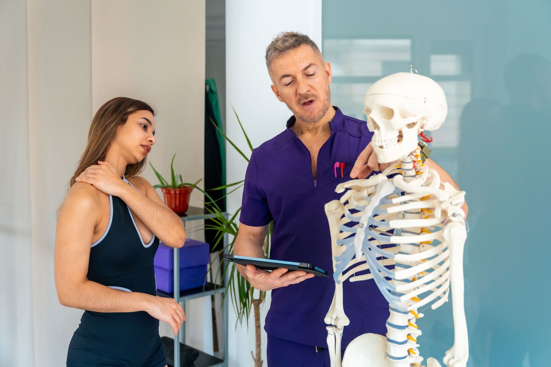
How Integrative Chiropractic Care Helps the Body Stay Flexible
Integrative chiropractic care is designed to address both structure and function. Instead of focusing only on where pain is felt, it looks at how the whole body moves. This can include chiropractic adjustments, stretching, soft tissue support, posture advice, and therapeutic exercises.
This type of care helps flexibility in several ways.
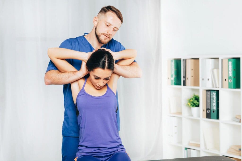
Restoring Better Joint Motion
When the spine or other joints are not moving well, the body often becomes stiff and guarded. Chiropractic adjustments are used to improve motion in restricted joints. Improved joint mobility can make everyday activities easier and may reduce stress on surrounding muscles and tissues (Dubuque Chiropractic, n.d.; Rodgers Stein Chiropractic, n.d.-a).
Many people describe this change as feeling looser or less stuck after treatment. That improved motion can be especially helpful in the neck, upper back, lower back, shoulders, hips, knees, and ankles.
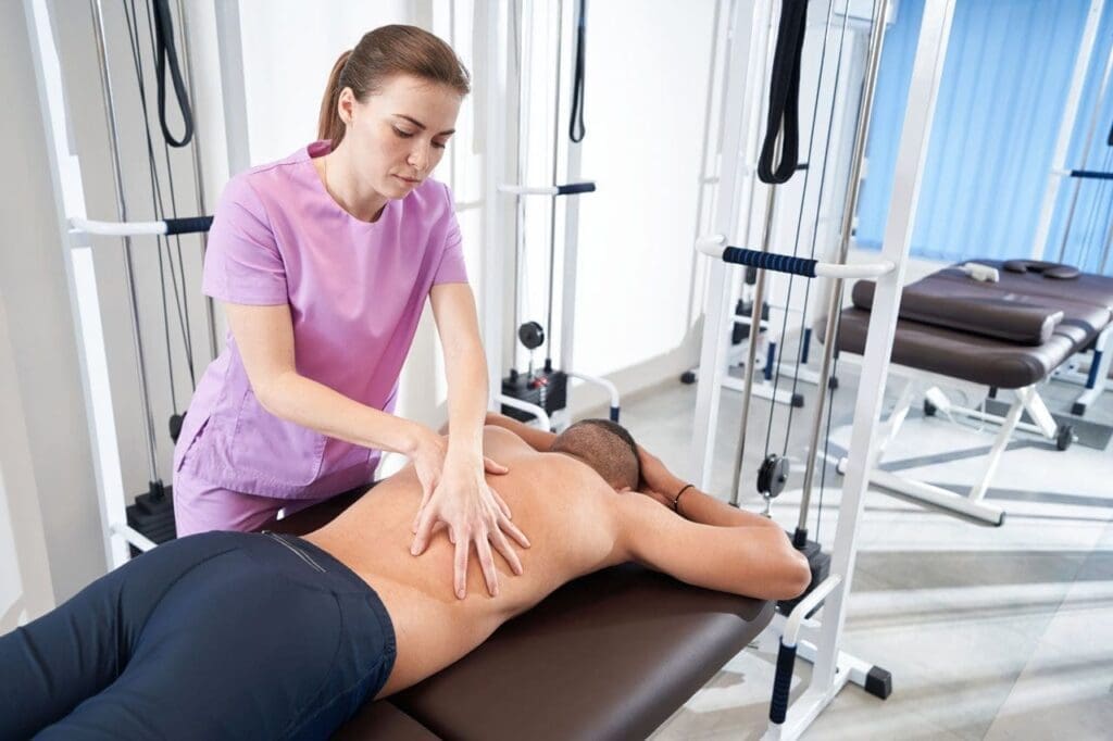
Reducing Muscle Tension
Tight muscles can limit flexibility even when the joints are not severely damaged. When muscles stay tense for long periods, they can pull the body out of balance and make movement feel restricted. Integrative chiropractic treatment often includes stretching and soft-tissue work to help muscles relax and function more effectively (Chiropractic Fitness, n.d.; Alter Chiropractic, n.d.).
When tension goes down, movement often becomes smoother and less painful.
Supporting the Nervous System
The nervous system helps control posture, muscle activity, balance, and coordination. Chiropractic care often focuses on improving how the spine and joints interact with the nervous system. When that system works more efficiently, muscles may respond better, and movement can become more natural (Gentle Chiropractic, 2025; Thrive Health Systems, n.d.).
This is important because flexibility is not only about tissue length. It is also about how the brain and body communicate during motion.
Improving Movement Patterns
Good flexibility is easier to maintain when the body learns better movement habits. That is why therapeutic exercises are such an important part of integrative care. Exercises help strengthen weak muscles, improve control, and support proper joint function. This makes it easier for the body to keep the benefits of treatment over time (OAA Orthopaedic Specialists, n.d.; Chiropractic Fitness, n.d.).
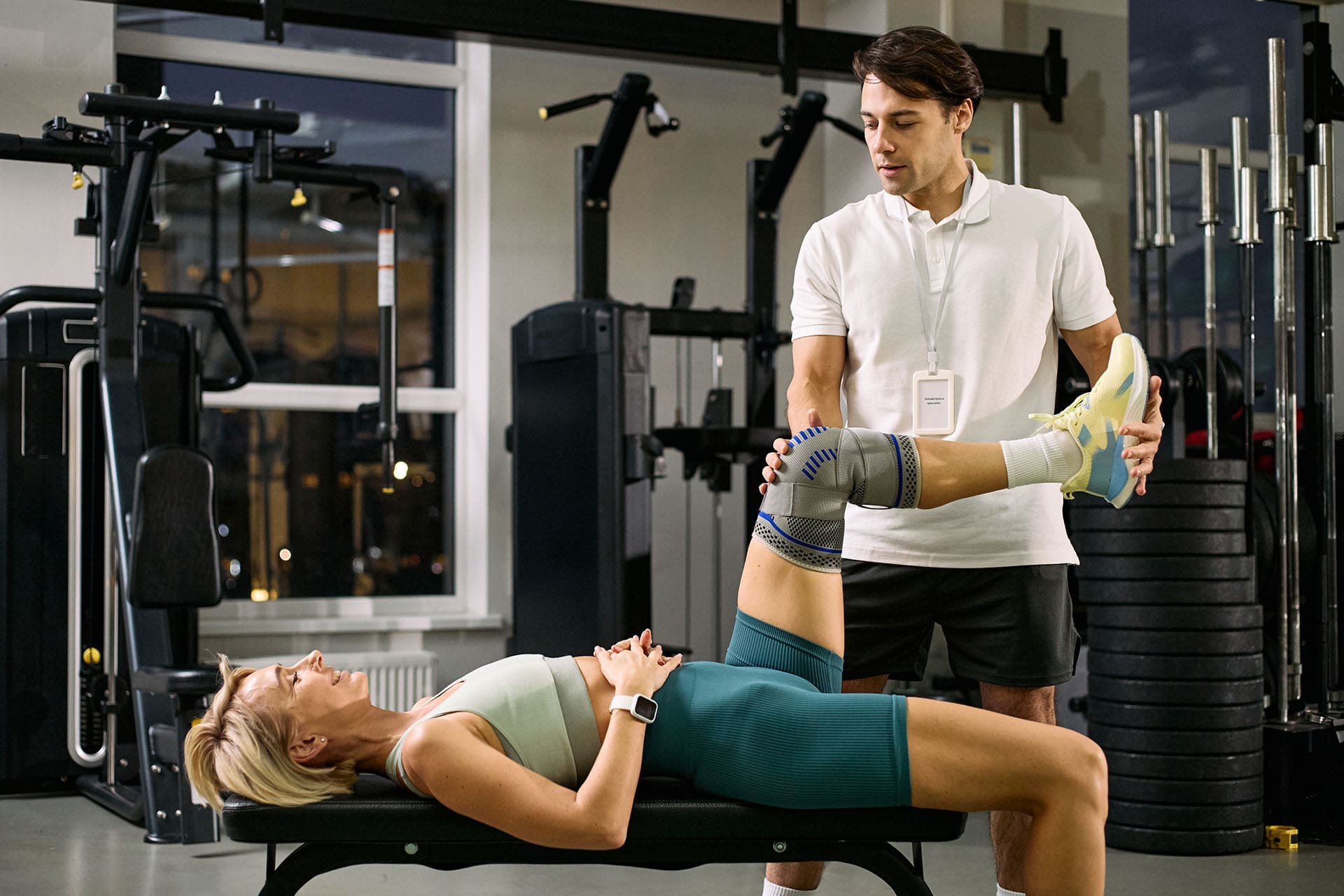
Why Stretching and Therapeutic Exercise Matter
Adjustments can help restore motion, but stretching and exercise help the body hold onto those gains. Stretching supports tissue length and mobility. Therapeutic exercise helps improve stability, coordination, and body control.
A flexibility-focused plan may include:
- Gentle stretching for tight muscle groups
- Mobility drills for stiff joints
- Core exercises for spinal support
- Postural exercises for daily alignment
- Strengthening work for weak stabilizing muscles
- Balance and coordination training
These methods work together so muscles and joints can support one another more effectively. That is one of the key ideas behind integrative chiropractic care. The body needs both mobility and stability to stay flexible and strong (Rodgers Stein Chiropractic, n.d.-b; TXMAC, n.d.-b).
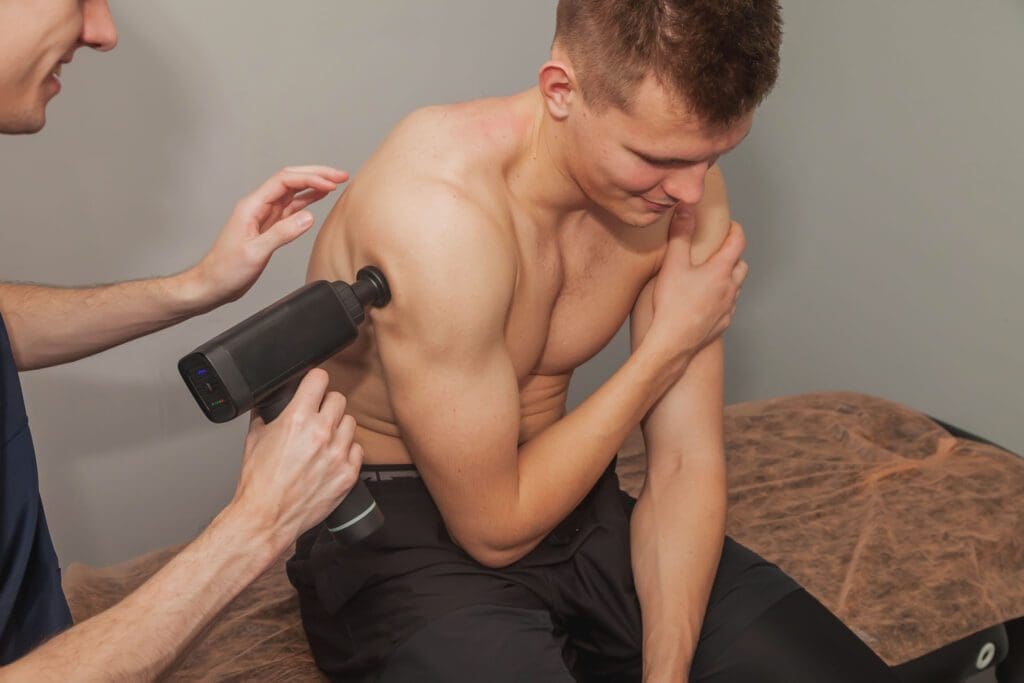
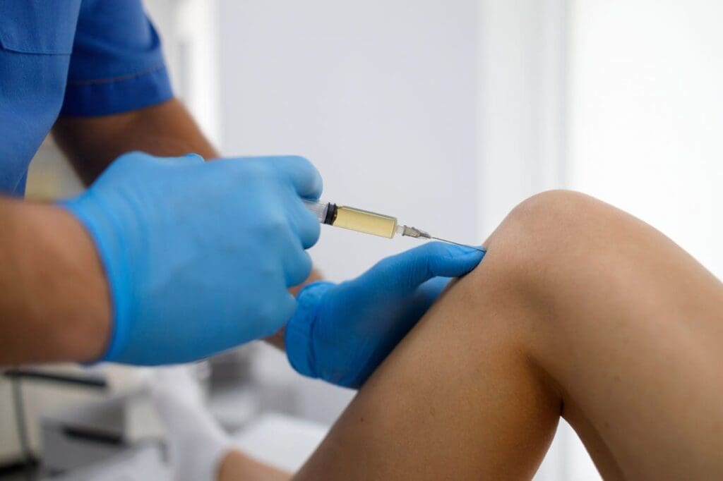
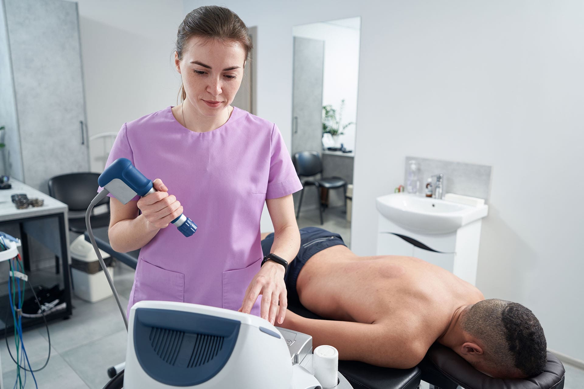
What ESWT Is and Why It Helps Flexibility
Extracorporeal Shockwave Therapy, or ESWT, is a noninvasive treatment that uses acoustic waves to stimulate tissue repair. It is commonly used for chronic soft tissue problems that can limit motion and cause long-term discomfort.
Many flexibility problems are not caused by joint restriction alone. In some cases, the main issue is in the muscles, tendons, or fascia. Scar tissue, chronic inflammation, tendon overload, and soft tissue adhesions can make movement feel tight and painful. ESWT is often used to address these issues by promoting blood flow and tissue healing (Bend Total Body Chiropractic, 2023; Corrective Chiropractic, n.d.).
ESWT may help by:
- Increasing circulation to the treated area
- Supporting tissue repair
- Reducing pain and inflammation
- Breaking down scar tissue and adhesions
- Improving tissue elasticity
- Helping muscles and tendons move more freely
This can be especially useful when a patient has chronic tightness that does not improve enough with stretching or rest alone (InSpine Chiropractic, n.d.; Chiropractic Experience, n.d.).
Why Chiropractic Care and ESWT Work Well Together
Chiropractic care and ESWT address different aspects of the same problem. Chiropractic adjustments help restore motion in the joints and spine. ESWT helps improve the condition of the soft tissues around those joints. When both are used together, the body may respond better than it would with only one treatment.
This two-part approach can help:
- Improve joint mechanics
- Reduce muscle guarding
- Break up scar tissue
- Improve blood flow
- Lower chronic inflammation
- Increase range of motion
- Support better long-term movement
This is one reason many integrative clinics combine chiropractic care and ESWT. The goal is to improve both how the body moves and the condition of the tissues that support that movement (San Diego NUCCA, n.d.; My Office Info, n.d.; Holistiq, n.d.).
Conditions That Can Limit Flexibility
A combined approach of chiropractic care and ESWT is often used for conditions involving both movement restriction and soft-tissue stress.
Frozen Shoulder
Frozen shoulder can cause severe stiffness, pain, and loss of motion. It often makes reaching overhead or behind the back very difficult. Adjustments, mobility work, and ESWT may help improve movement and reduce soft-tissue restrictions around the shoulder complex (Gentle Chiropractic, n.d.; Chiro Oklahoma City, 2025).
Achilles Tendinopathy
The Achilles tendon can become painful and tight, especially in active people or in those with faulty movement mechanics. ESWT is often used to support tendon healing, while chiropractic treatment may help improve the mechanics of the ankle, foot, knee, hip, and spine that affect how the tendon is loaded (Chiropractic First, n.d.; Dr. Alex Jimenez, 2026a).
Chronic Muscle Tightness
Long-term tightness in the neck, back, hips, or legs can come from stress, poor posture, repetitive work, or old injuries. In these cases, chiropractic care may restore joint motion while ESWT helps address stubborn tissue restrictions. This may make it easier for patients to stretch, exercise, and move without constant pulling or stiffness (Bend Total Body Chiropractic, 2023; TXMAC, n.d.-a).
Clinical Observations That Support an Integrative Approach
Dr. Alexander Jimenez, DC, APRN, FNP-BC, has published clinical material that supports a whole-body view of flexibility and recovery. His work describes a model that combines chiropractic care with rehabilitation, functional medicine, and advanced treatment strategies to improve mobility, strength, and overall function (Dr. Alex Jimenez, 2026b).
His published material on shockwave therapy also explains how ESWT can fit into a broader care plan addressing both joint mechanics and soft-tissue healing. That kind of combined strategy is useful because many movement problems involve more than one tissue type. A patient may have joint restriction, muscle tension, tendon overload, and scar tissue simultaneously. A well-rounded plan is often needed to improve function in a lasting way (Dr. Alex Jimenez, 2026a).
For a clinic like ChiroMed, that kind of integrative thinking fits naturally with patient-centered care. Instead of chasing only symptoms, the focus is on why movement is limited and how to improve it safely and effectively.
What Patients May Notice With Consistent Care
When chiropractic care, stretching, therapeutic exercise, and ESWT are used together in the right setting, patients may notice:
- Less stiffness in the morning
- Easier movement during daily tasks
- Better flexibility in the shoulders, hips, and back
- Reduced muscle tightness
- More comfort during walking, lifting, or exercise
- Better posture and body awareness
These improvements often build over time. Flexibility is not something that changes only from one visit. It usually improves best through consistent care, home exercises, better posture, and regular movement.
Conclusion
Integrative chiropractic care helps the body stay flexible by restoring joint alignment, easing muscle tension, and improving nervous system function. When regular adjustments are combined with stretching and therapeutic exercises, patients may experience improved range of motion, reduced stiffness, and more efficient movement in daily life.
When ESWT is added, the treatment plan can become even more effective for people dealing with scar tissue, chronic tendon problems, and long-term muscle tightness. By addressing both joint mechanics and soft-tissue limitations, chiropractic care and ESWT work together to improve mobility, support healing, and help the body remain flexible and strong.
For a practice like ChiroMed, this integrative model reflects a practical, modern approach to supporting long-term movement, recovery, and function (San Diego NUCCA, n.d.; Dr. Alex Jimenez, 2026a).
References
Alter Chiropractic. (n.d.). Why choose chiropractic for enhanced flexibility?
Bend Total Body Chiropractic. (2023, October 25). Exploring the uses, benefits, side effects of shockwave therapy
Chiro Oklahoma City. (2025, October 25). What is shockwave therapy?
Chiropractic Experience. (n.d.). Shockwave therapy – ESWT
Chiropractic First. (n.d.). How shockwave therapy complements chiropractic treatments
Chiropractic Fitness. (n.d.). Boost mobility and flexibility with chiropractic care
Corrective Chiropractic. (n.d.). Shockwave therapy
Dr. Alex Jimenez. (2026a). Shockwave therapy for healing: Understanding ESWT
Dr. Alex Jimenez. (2026b). Why choose our clinical team?
Dubuque Chiropractic. (n.d.). 5 ways chiropractic adjustments enhance flexibility
Gentle Chiropractic. (2025, March 14). Can chiropractic care improve joint flexibility and range of motion?
Gentle Chiropractic. (n.d.). Frozen shoulder relief and treatment
Holistiq. (n.d.). Chiropractic treatment and shockwave treatment
InSpine Chiropractic. (n.d.). Shockwave therapy in chiropractic care
My Office Info. (n.d.). Why you should integrate shockwave therapy into your chiropractic care plan
OAA Orthopaedic Specialists. (n.d.). How regular chiropractic visits boost mobility
Rodgers Stein Chiropractic. (n.d.-a). Why thousands trust chiropractors for greater flexibility
Rodgers Stein Chiropractic. (n.d.-b). Transform your flexibility with chiropractic care
San Diego NUCCA. (n.d.). Shockwave therapy and chiropractic adjustments
ThinkVida. (n.d.). Chiropractic and flexibility
TXMAC. (n.d.-a). Why choose chiropractic for enhanced flexibility?
TXMAC. (n.d.-b). Boost mobility and flexibility with chiropractic care
Thrive Health Systems. (n.d.). How chiropractic adjustments can improve mobility and flexibility