ChiroMed’s Adaptive Exercises for Senior Health
Best Exercises for Seniors at ChiroMed – Integrated Medicine
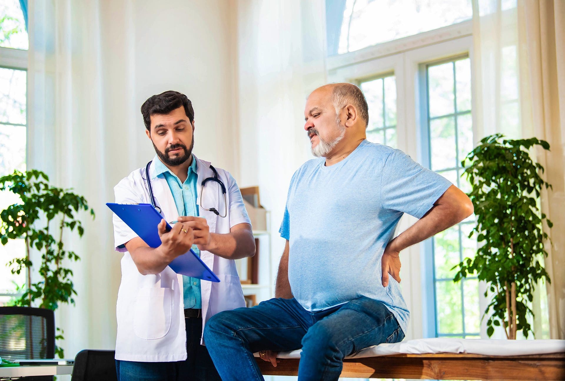
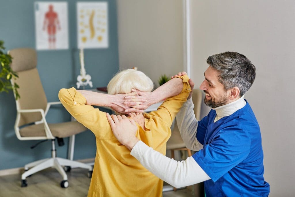
At ChiroMed – Integrated Medicine in El Paso, TX, we believe that staying active is key to a healthy, independent life, especially for seniors or those with limited mobility. Our integrative approach combines chiropractic care, nurse practitioner services, and complementary therapies like acupuncture and massage to create personalized, low-impact exercise plans. These exercises are designed to improve flexibility, balance, and strength while supporting spinal health, managing pain, and reducing fall risks. Led by Dr. Alexander Jimenez, DC, APRN, FNP-BC, our team uses advanced diagnostic tools and holistic methods to help patients recover from injuries and enhance their well-being. This article examines the most effective exercises for seniors, specifically designed to complement ChiroMed’s integrative care model.
Why Choose ChiroMed for Integrative Care?
Located in the heart of El Paso, ChiroMed – Integrated Medicine offers a unique blend of chiropractic adjustments, naturopathy, rehabilitation, nutrition counseling, and acupuncture. Our mission is to address the root causes of health issues, not just the symptoms, through patient-centered care (ChiroMed, 2025). For seniors or those with limited mobility, our team creates customized treatment plans that integrate gentle exercises with spinal adjustments to promote natural healing and long-term wellness.
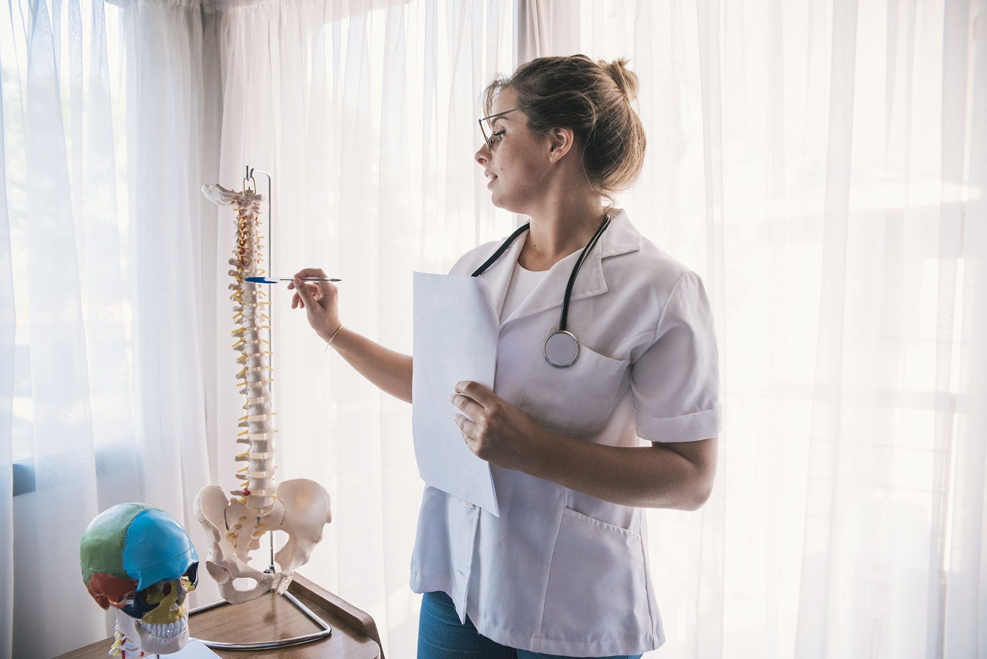
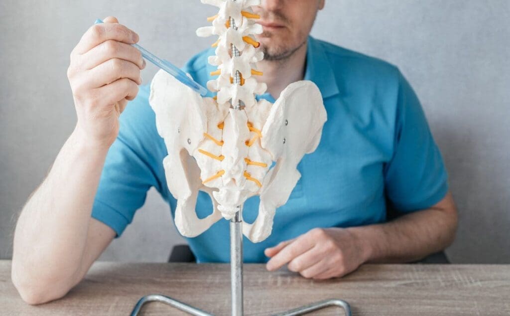
Dr. Alexander Jimenez, a board-certified chiropractor and family nurse practitioner, brings a dual-scope approach to care. With expertise in treating injuries from work, sports, personal incidents, and motor vehicle accidents (MVAs), he uses advanced neuromusculoskeletal imaging, such as X-rays and MRIs, to diagnose conditions accurately (Jimenez, 2025). This allows ChiroMed to design exercise programs that align with chiropractic adjustments, ensuring safe and effective outcomes for seniors.
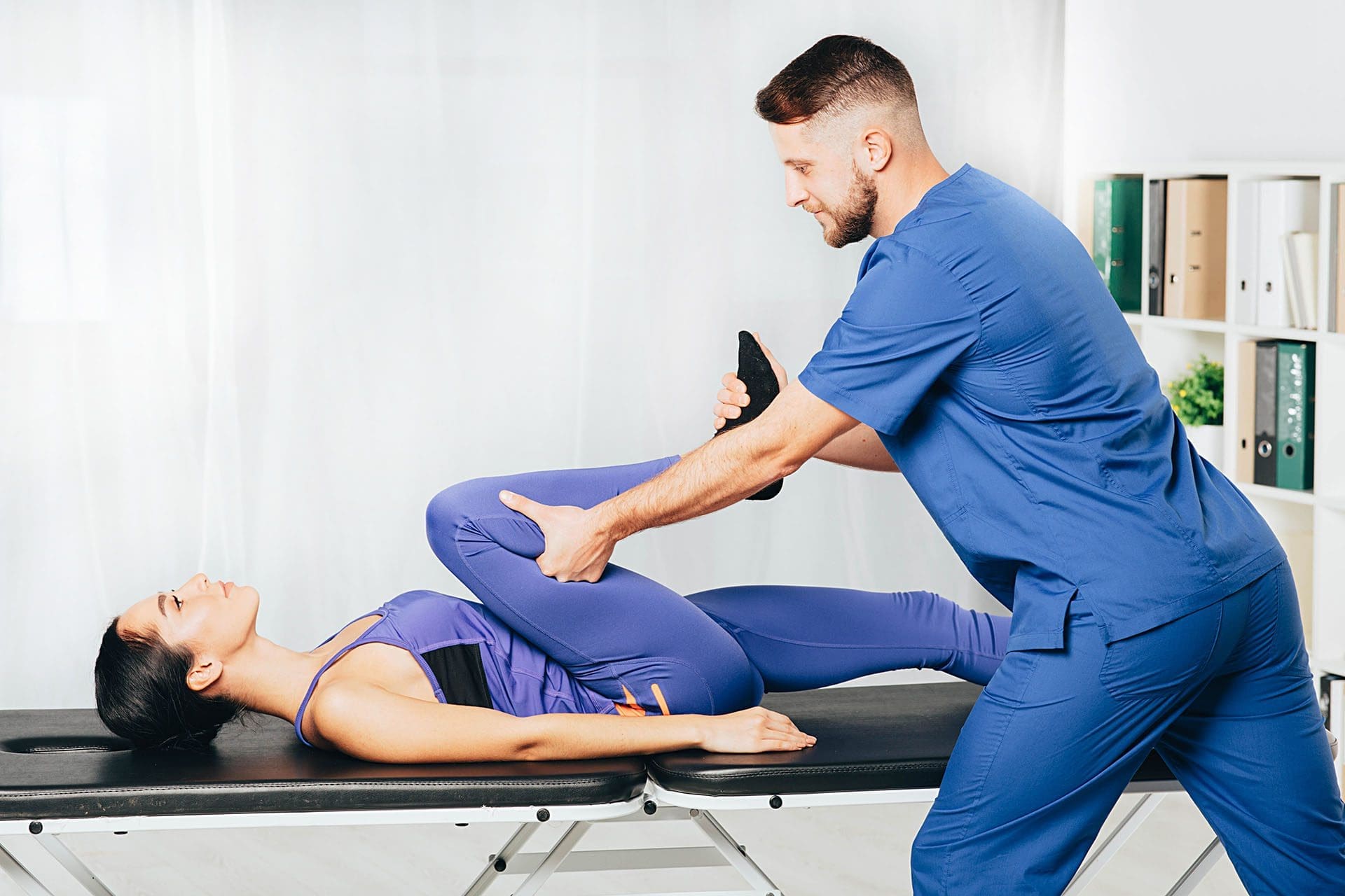
The Importance of Low-Impact, Adaptive Exercises
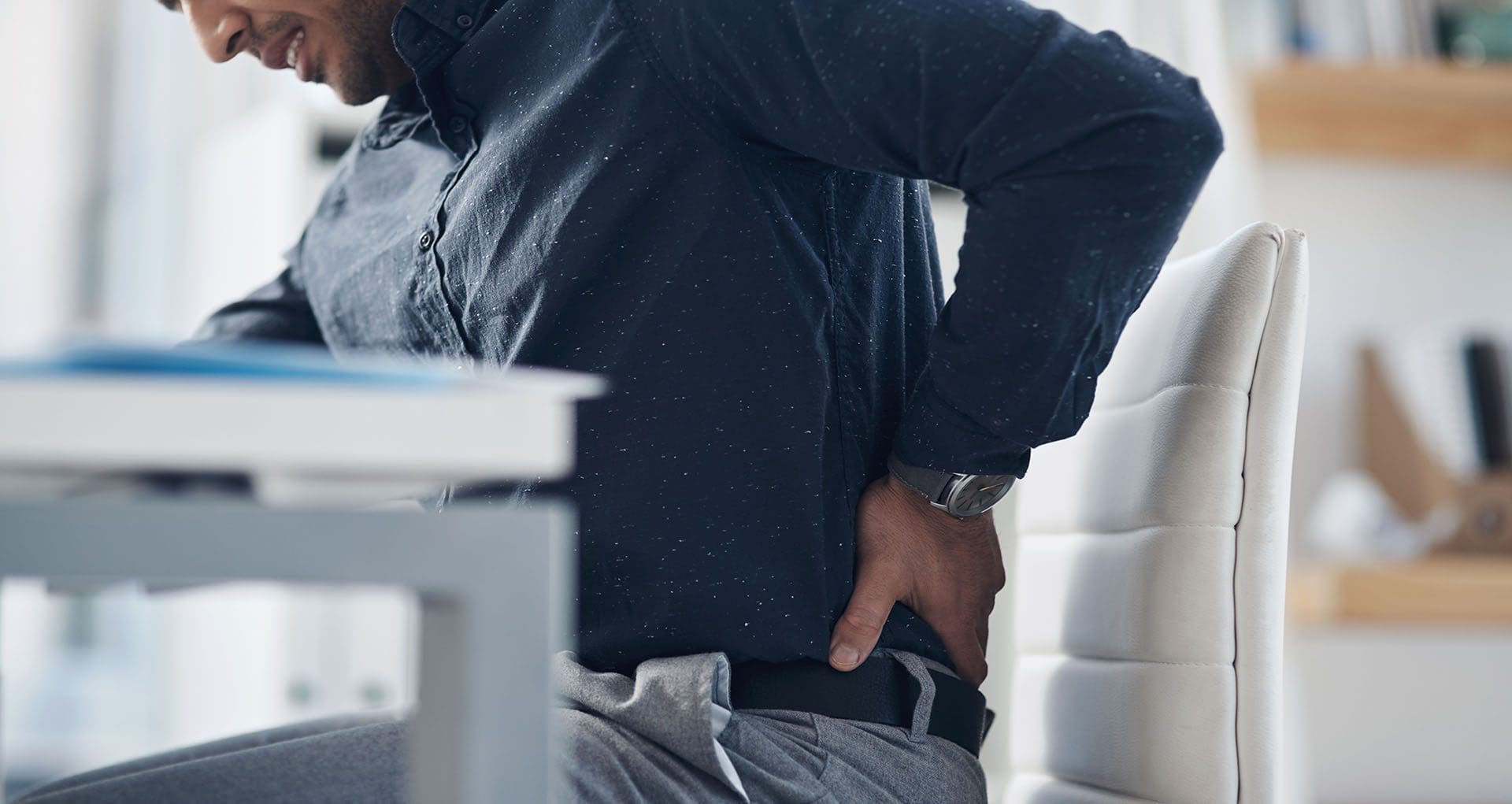
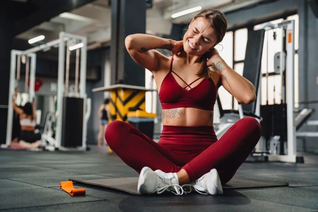
Low-impact exercises are ideal for seniors because they minimize stress on joints and muscles while improving strength, flexibility, and balance. These movements are especially helpful for those with arthritis, osteoporosis, or post-injury limitations, as they reduce the risk of further injury (Atlas Senior Living, 2024). At ChiroMed, our adaptive exercises are modified to suit individual abilities, making them accessible to patients who use walkers, wheelchairs, or other mobility aids (Live2BHealthy, 2024). These exercises support our chiropractic adjustments by enhancing spinal alignment, reducing pain, and improving mobility.
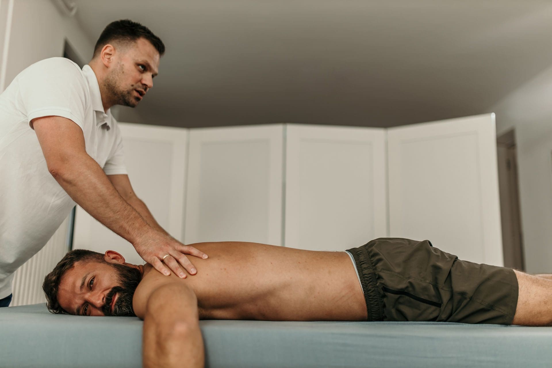
Dr. Jimenez’s clinical approach emphasizes personalized care. For example, patients recovering from MVAs may have soft tissue damage or spinal misalignments, which are assessed using diagnostic tools and treated with tailored exercises, adjustments, and therapies like massage or acupuncture (Jimenez, 2025). This holistic method ensures that seniors can stay active while addressing their specific health challenges.
Top Exercises at ChiroMed for Seniors
ChiroMed’s exercise programs are designed to complement our integrative care model. Below are some of the best low-impact, adaptive exercises we recommend for seniors or those with limited mobility, all tailored to support chiropractic treatment and overall health.
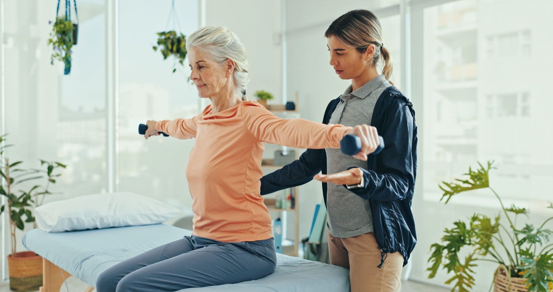
1. Chair-Based Exercises
Chair exercises are safe and effective for seniors with balance issues or mobility limitations. Performed seated, these movements improve strength, flexibility, and circulation while reducing fall risks (BLHC, 2023).
- Seated Marches: Sit in a sturdy chair with feet flat on the floor. Lift one knee toward your chest, then lower it, alternating legs like marching. Do 10–15 repetitions per leg. This strengthens leg muscles and supports hip alignment, complementing pelvic adjustments at ChiroMed (Comfort Keepers, 2024).
- Seated Leg Extensions: Extend one leg straight out, hold for 3–5 seconds, then lower slowly. Repeat 10 times per leg. This builds quadriceps strength, aiding knee stability and mobility (Lakehouse Three Rivers, 2024).
- Arm Raises: Hold light weights or water bottles, raise arms to shoulder level, and lower slowly. Perform 10–12 repetitions. This improves shoulder mobility and supports upper spine health, aligning with ChiroMed’s thoracic adjustments (Olive Elder Care, 2025).
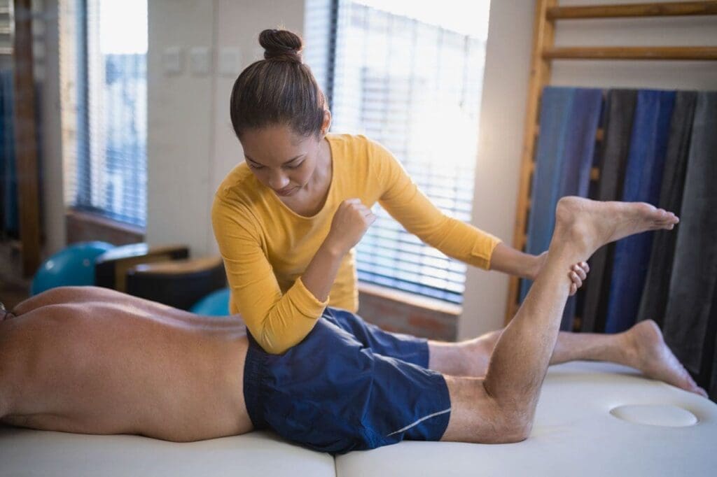
2. Stretching Exercises
Stretching keeps muscles and joints flexible, reduces tension, and enhances circulation, supporting ChiroMed’s chiropractic adjustments (Eaton Chiropractic, 2024).
- Neck Tilts: Gently tilt your head to one side, bringing your ear toward your shoulder, and hold for 10–15 seconds. Repeat on the other side. This relieves cervical spine tension, a focus of ChiroMed’s adjustments (Care Indeed, 2024).
- Shoulder Shrugs: Lift your shoulders toward your ears, hold for 3 seconds, then release. Repeat 10 times. This loosens the upper back, reducing strain on the thoracic spine (Village Green Retirement, 2024).
- Ankle Circles: Lift one foot slightly and rotate your ankle in circles, 10 times each direction. This improves ankle mobility, supporting lower body stability (McCarthy & Stone, 2024).
3. Core Strengthening Exercises
A strong core supports the spine, reduces misalignment risks, and enhances posture, all key goals of ChiroMed’s chiropractic care (Erie Chiropractic, 2024).
- Seated Cat-Cow Stretch: Sit upright, arch your back slightly while lifting your chest (cow), then round your back while tucking your chin (cat). Repeat 8–10 times. This enhances spinal flexibility, supporting lumbar adjustments (Elevate to Life, 2024).
- Pelvic Tilts: Sit or lie down, tighten your abdominal muscles, and tilt your pelvis upward. Hold for 5 seconds, repeat 10 times. This strengthens the lower back, aligning with ChiroMed’s spinal health focus (Best Grand Rapids Chiropractor, 2024).
- Seated Crunches: Cross your arms over your chest, engage your core, and lean forward slightly, then return upright. Do 10–15 repetitions. This builds core strength safely (Olive Elder Care, 2025).
4. Balance Exercises
Balance exercises are vital for preventing falls, a major concern for seniors. At ChiroMed, these movements are integrated with chiropractic care to improve coordination and stability (Rush Chiropractic, 2024).
- Heel-to-Toe Walking: Hold onto a wall or chair for support and walk by placing one foot directly in front of the other, heel touching toe. Take 10–15 steps. This strengthens leg muscles and improves balance (Village Green Retirement, 2024).
- Single-Leg Stand: Hold a chair, lift one foot slightly, and maintain the position for 10–20 seconds. Switch sides. This enhances ankle stability, supporting ChiroMed’s lower body adjustments (McCarthy & Stone, 2024).
- Chair Squats: Stand in front of a chair, lower your body as if sitting, then stand up without fully sitting. Repeat 8–10 times. This strengthens legs and core, aiding posture (Peregrine Crossgate, 2024).
5. Water-Based Exercises
Aquatic exercises, recommended by ChiroMed, use water’s buoyancy to reduce joint stress while building strength and mobility (Live2BHealthy, 2024).
- Water Walking: Walk in waist-deep water for 10–15 minutes, swinging arms naturally. This boosts cardiovascular health and leg strength, supporting overall mobility (Atlas Senior Living, 2024).
- Aquatic Arm Lifts: In chest-deep water, raise arms to shoulder level, then lower slowly. Repeat 10–12 times. This strengthens the upper body, complementing shoulder adjustments (Lakehouse Three Rivers, 2024).
- Leg Swings: Hold the pool edge and swing one leg forward and backward gently, 10 times per leg. This improves hip mobility, aligning with pelvic adjustments (Cordia Westmont, 2024).
6. Tai Chi and Yoga
ChiroMed incorporates mind-body exercises like Tai Chi and yoga to promote balance, flexibility, and relaxation, enhancing chiropractic outcomes (Baxter Senior Living, 2023).
- Chair Yoga: Sit in a chair, inhale while raising arms overhead, exhale while twisting gently to one side. Hold for 10 seconds, switch sides. This improves spinal flexibility (Health with Nargis, 2025).
- Tai Chi Flow: Perform slow movements like “wave hands like clouds” for 10 minutes. This enhances balance and coordination, supporting neuromusculoskeletal health (Be On The Move, 2025).
- Seated Sun Salutations: Modify sun salutations with seated arm and torso movements. Repeat 5–8 cycles. This boosts flexibility and circulation (Life in Lines, 2024).
Dr. Alexander Jimenez’s Expertise at ChiroMed
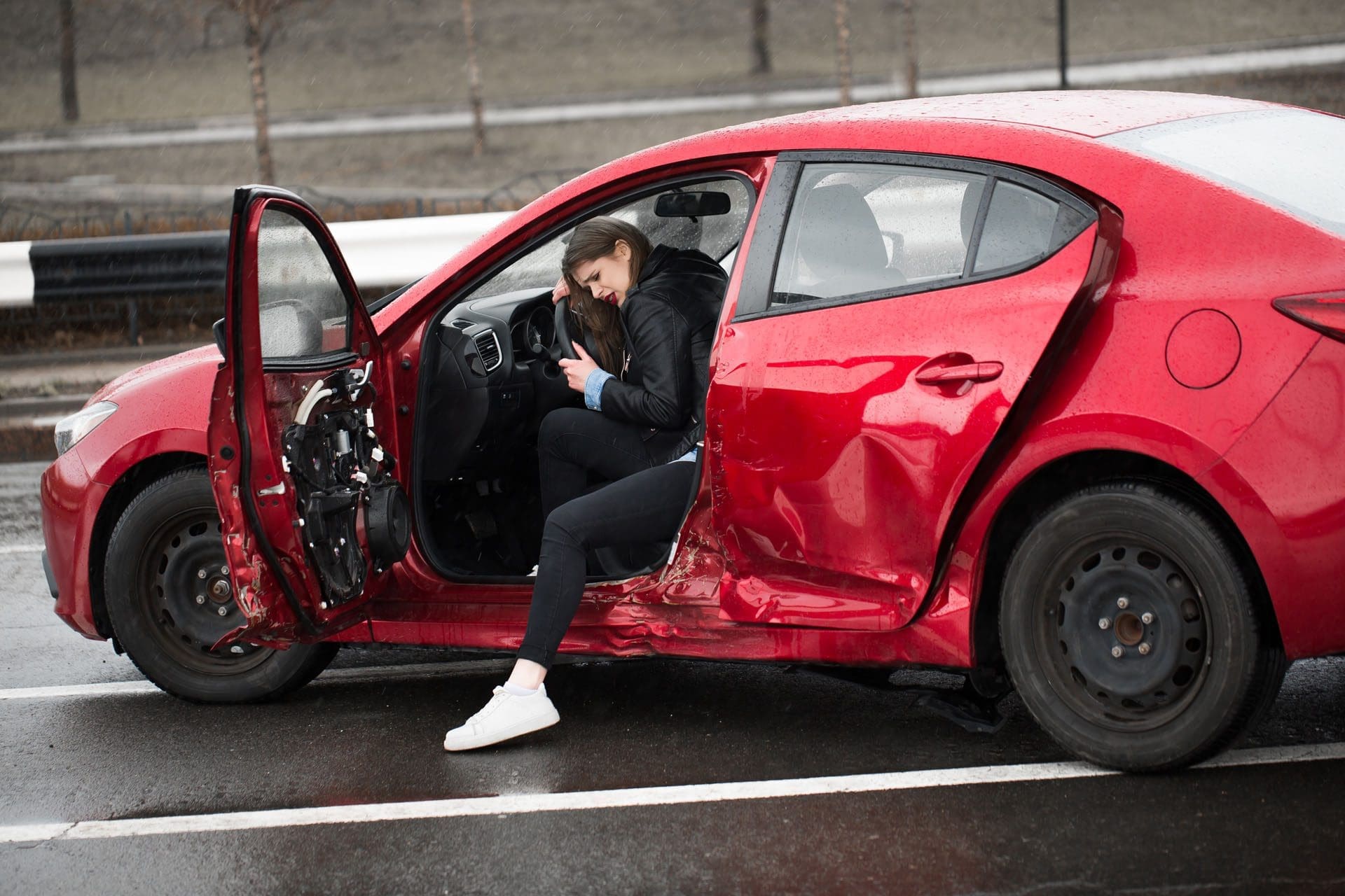
Dr. Alexander Jimenez leads ChiroMed with a dual-scope approach, combining chiropractic and nurse practitioner expertise. His clinic specializes in treating injuries from work, sports, personal incidents, and MVAs, using advanced imaging like MRIs and CT scans to diagnose conditions accurately (Jimenez, 2025). For seniors, this means tailored exercise plans that address specific limitations, such as arthritis or post-injury mobility issues, while promoting natural healing.
In MVA cases, Dr. Jimenez’s team provides comprehensive medical care and legal documentation, ensuring accurate records for insurance or legal needs. For example, they assess soft tissue injuries, spinal misalignments, and neurological impacts, then create plans with exercises, adjustments, and therapies like massage or acupuncture (Dallas Accident and Injury Rehab, 2024). At ChiroMed, massage therapy relaxes muscles before adjustments, while acupuncture reduces inflammation, enhancing exercise effectiveness (Integra Health, 2024).
Benefits of ChiroMed’s Exercise Programs
ChiroMed’s integrative exercise programs offer numerous benefits for seniors:
- Enhanced Spinal Health: Exercises like cat-cow stretches support spinal flexibility and alignment, reinforcing adjustments (Elevate to Life, 2024).
- Pain Relief: Low-impact movements reduce joint and muscle pain, especially for arthritis, by improving circulation (Chiro Health KC, 2024).
- Fall Prevention: Balance exercises like single-leg stands strengthen stabilizing muscles, reducing fall risks (Fall Prevention Foundation, 2024).
- Improved Strength and Flexibility: Stretching and strength exercises maintain muscle mass and joint mobility, supporting daily activities (Comfort Keepers, 2024).
- Better Mental Health: Mind-body exercises like Tai Chi release endorphins, reducing stress and boosting mood (Baxter Senior Living, 2023).
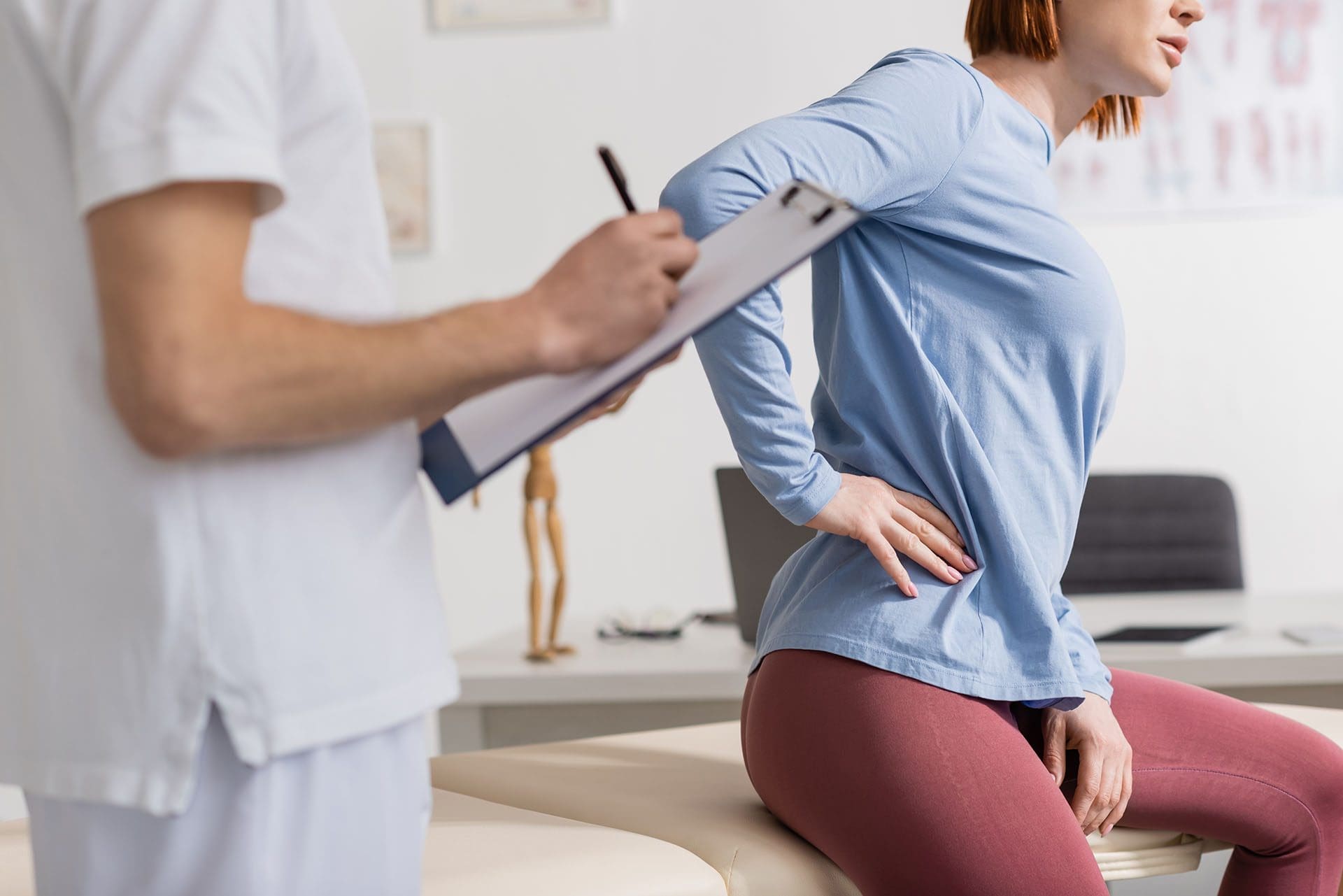
Safety Tips at ChiroMed
Before starting exercises, ChiroMed conducts thorough assessments to ensure safety, especially for seniors with chronic conditions or recent injuries. Dr. Jimenez’s diagnostic approach customizes plans to avoid overexertion (Jimenez, 2025). Safety tips include:
- Use supportive equipment like chairs or pool railings.
- Start with short sessions (5–10 minutes) and increase gradually.
- Stop if pain occurs and consult ChiroMed’s team.
- Exercise in a well-lit, stable environment with non-slip shoes (Baxter Senior Living, 2023).
Conclusion
ChiroMed – Integrated Medicine in El Paso, TX, offers seniors and those with limited mobility a holistic path to better health through low-impact, adaptive exercises. Under Dr. Alexander Jimenez’s leadership, our integrative approach combines chiropractic adjustments, personalized exercises, and therapies like massage and acupuncture to support spinal health, manage pain, and prevent falls. By addressing injury causes with advanced diagnostics and tailored care, ChiroMed helps patients stay active and independent. Visit us at ChiroMed to experience personalized, holistic care that prioritizes your well-being.
References
Best Grand Rapids Chiropractor. (2024). Corrective exercises for chiropractic patients.
BLHC. (2023). Home care: The best exercises for seniors who have limited mobility.
Chiro Health KC. (2024). Age 55 or over? See your chiropractor for exercise options.
Chirocare Fairlawn. (2024). Therapeutic exercises.
ChiroMed. (2025). Integrated Medicine Holistic Healthcare in El Paso, TX.
Comfort Keepers. (2024). Exercise for seniors with limited abilities.
Cordia Westmont. (2024). Beginner-friendly low-impact exercises for seniors.
Dallas Accident and Injury Rehab. (2024). The role of chiropractic care in older adults.
Eaton Chiropractic. (2024). 8 stretching & balancing exercises for older adults.
Elevate to Life. (2024). Top 7 exercises to support your chiropractic treatment.
Erie Chiropractic. (2024). The role of exercise in maintaining chiropractic adjustments.
Health with Nargis. (2025, February 2). 10 best low-impact exercises for seniors over 60.
Integra Health. (2024). Chiropractic for ankle pain.
Jimenez, A. (2025). Clinical observations and treatment approaches.
Lakehouse Three Rivers. (2024). Effective low-impact exercises for seniors to boost mobility.
Live2BHealthy. (2024). Adaptive fitness: Exercise modifications for seniors with mobility issues.
McCarthy & Stone. (2024). Balance exercises for seniors at home.
Peregrine Crossgate. (2024). The best exercises for seniors to stay fit & active.
Rush Chiropractic. (2024). Safe and effective exercise for seniors under chiropractic care.
Team Chiro. (2024). Chiropractic care for seniors.
The Joint Chiropractic. (2024, March 20). Chiropractic care for those with limited mobility.
UNC Health Talk. (2024). How to exercise with limited mobility.
Village Green Retirement. (2024). Low-impact exercises for seniors.